Embed Size (px)
Citation preview
Rev. Agr. Acad., v. 4, n. 4, Jul/Ago (2021)
Revista Agrária Acadêmica
Agrarian Academic Journal
Volume 4 – Número 4 – Jul/Ago (2021)
________________________________________________________________________________
doi: 10.32406/v4n4/2021/31-42/agrariacad
Myocardial bridges and coronary distribution in dairy cattle from Brazil Northeast. Pontes
miocárdicas e distribuição coronariana em bovinos leiteiros do Nordeste brasileiro.
Vinícius Lima Brito1, Kethelyn Freitas de Lima1, Anielly Mirelly de Assunção Ramalho1, Jadson Vieira de
Melo1, Júnior Mário Baltazar de Oliveira 2, Amara Maria de Sousa Barbosa1, Emanuela Polimeni de Mesquita1, Daniela Oliveira 1*
1- Laboratory of Animal Anatomy and Pathology, Federal University of Agreste of Pernambuco, Garanhuns, Brazil. Av.
Bom Pastor, s/n, Boa Vista, Garanhuns, PE, Brazil. 55292-270, (87) 37645500, ramal 5573. E-mail:
[email protected] 2- University Center Unifavip, Caruaru, Brazil.
________________________________________________________________________________
Abstract
This study evaluated the distribution and possible variations of the coronary arteries and the presence of myocardial
bridges in dairy cattle from Northeastern Brazil. Thus, healthy hearts were analyzed according to many variables. Left
coronary artery originate the rami paraconalis, circumflexus et subsinuosus. Ramus subsinuosus and right coronary artery
showed a short subepicardial path. Lengths of the main branches showed statistical differences only in relation to age
group and weight of the animal. Myocardial bridges had a prevalence of 55% and were more frequent over the ramus
paraconalis. Veterinary medicine is able to provide experimental models to expand the necessary study to understand
pathophysiology and clinical relevance of the bridges.
Keywords: Blood circulation. Myocardium. Cardiovascular system. Bovine.
Resumo
Este estudo avaliou a distribuição e possíveis variações das artérias coronárias e a presença de pontes miocárdicas em
bovinos leiteiros do Nordeste do Brasil. Assim, corações saudáveis foram analisados de acordo com muitas variáveis. A
artéria coronária esquerda origina os rami paraconalis, circumflexus et subsinuosus. O ramus subsinuosus e a artéria
coronária direita apresentavam trajeto subepicárdico curto. Os comprimentos dos ramos principais apresentaram
diferenças estatísticas apenas em relação à faixa etária e ao peso do animal. As pontes miocárdicas tiveram prevalência
de 55% e foram mais frequentes sobre o ramus paraconalis. A medicina veterinária é capaz de fornecer modelos
experimentais para expandir o estudo necessário para entender a fisiopatologia e a relevância clínica das pontes.
Palavras-chave: Circulação sanguínea. Miocárdio. Sistema cardiovascular. Bovino.
________________________________________________________________________________
31
Rev. Agr. Acad., v. 4, n. 4, Jul/Ago (2021)
Introduction
According to the OECD-FAO (Food and Agriculture Organization of the United Nations)
Brazil stands out in world livestock production and, together with the United States and the European
Union, will be responsible for approximately 60% of the world meat exports in 2029 (OECD-FAO,
2020). Substances necessary for the production of meat and milk (nutrients, hormones, etc.) circulate
through the blood to the muscles and the udder, respectively (GETTY, 1986; DYCE et al., 2010).
The circulatory system, therefore, is directly related to animal production.
The heart is compared to a large and powerful pump that drives blood to all tissues in the body.
Thus, the heart, similar to any other tissue, also needs to have its own blood supply, receiving the
equivalent of 15% of the cardiac output of the left ventricle (DYCE et al., 2010). The right and left
coronary arteries, which emerge right after the aortic bulb, are responsible for this distribution, occupy
the sulcus coronarius and present different paths and distributions according to each species
(GETTY, 1986; FRANDSON et al., 2017).
According to König et al. (2011), angiology is the branch of science dedicated to the study of
the shape, structure, topography and functioning of blood vessels. Thus, knowing and better
understanding the distribution of cardiac vessels, especially coronary arteries and their branches,
allows a better understanding of the blood supply in this organ, as well as possible pathologies that
can be established on these, such as myocardial infarction and atherosclerosis (SIERVULI et al.,
2014).
Myocardial bridge is the term used to describe the muscle bands that overlap the segments of
the epicardial coronary arteries (ZHAO et al., 2019). A potential relationship is reported between
myocardial bridges and some cardiac changes such as ischemia (TARANTINI et al., 2016),
myocardial fibrosis (BRODSKY et al., 2008) and coronary spasm (TERAGAWA et al., 2003).
Studies on the variation in the anatomical distribution of the coronary arteries and myocardial
bridges are still scarce in dairy cattle from northeastern Brazil (RODRIGUES et al., 1999; CORREIA-
OLIVEIRA et al., 2013, 2014). Therefore, given the importance of this study, this research evaluated
and described the path and possible variations in the distribution of coronary arteries and the presence
of myocardial bridges in cattle from a dairy region in northeastern Brazil.
Material and methods
All methodology adopted for the development of the present research was submitted and
approved by the Ethics Committee on the Use of Animals (CEUA) of the Federal Rural University
of Pernambuco (UFRPE), Recife-PE, under protocol number 028/2019.
Twenty-two hearts of 17 females and five bovine males aged between two days and nine years,
weighing between 30 and 620 kg were studied. The animals came from necropsies performed at the
Animal Pathology Sector of the Animal Anatomy and Pathology Laboratory (LAPA) at UFAPE and
sent to the Animal Anatomy Sector of the same laboratory. Age, gender, weight and breed were
recorded on the necropsied animal's record. The breeds recorded in the chart were Holstein (eight),
Gir x Holstein (five), other crossbred (seven), Brown-Swiss (one) and Guzerá (one). The main cause
of death was related to locomotor problems (fractures, traumas, muscle rupture, among others) and
reproductive problems (uterine rupture and torsion, dystocia).
32
Rev. Agr. Acad., v. 4, n. 4, Jul/Ago (2021)
The hearts were removed by sectioning the vessels at the base. Subsequently, the specimens
were washed, and the clots removed before being fixed in a 10% formaldehyde solution, where they
remained until dissected. Then, the coronary arteries and their branches were evidenced from the
origins in the aorta, with the use of appropriate dissection instruments, until their macroscopically
visible terminations.
The main branches of the coronary arteries and whenever myocardial bridges were observed,
the lengths were measured with the aid of a digital caliper and a flexible ruler, as well as the location
(topography and vasculotopy) of each of them in each studied heart was recorded and documented.
All information obtained was recorded and reported using the Veterinary Anatomical
Nomenclature (INTERNATIONAL, 2017).
Measurements of the length of the branches of the coronary arteries and the bridge of the bovine
ramus paraconalis were tabulated in an Excel spreadsheet. Initially, a logarithmic transformation was
carried out in order to homogenize the variances of the groups. After that, the normality of the data
was tested by the Shapiro-Wilk test. Then, a comparison was made of the data obtained when
measuring branches of the coronary arteries and the bridge of the ramus paraconalis, with dependent
variables: sex, breed, age and weight. For parametric analysis, Student's t test and Scheffe test were
used and for non-parametric analysis, Kruskal Wallis and Mann-Whitney tests were used.
Subsequently, a correlation analysis was performed between the length of the structures using
Pearson's or Spearman's Correlation (depending on the distribution of the variables) (SAMPAIO,
2015). The IBM SPSS Statistics 23.0 program was used to perform the aforementioned statistical
calculations. The level of significance was set to 5.0% for all analyses.
Results
Left coronary artery
It originated on the left side of the aorta and after a short course, in the sulcus coronarius, it
was divided into two branches, the ramus interventricularis paraconalis (ramus paraconalis) and the
left ramus circumflexus. The left ramus circumflexus continued in the sulcus interventricularis
subsinuosus, where it became known as the ramus interventricularis subsinuosus (ramus
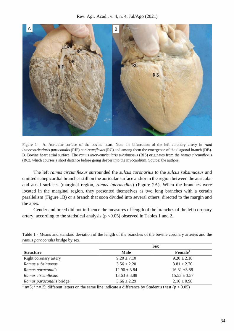
subsinuosus). An extra branch (diagonal branch) was found in four hearts, with the same origin as the
rami paraconalis and circumflexus, which runs above the left ventricle, on the auricular surface
(Figure 1A).
The ramus paraconalis coursed through the sulcus paraconalis and emitted one to two
subepicardial branches, mainly towards the left ventricle, although in a few cases it was also observed
over the right ventricle. The ramus paraconalis deepened in the myocardium close to the apex of the
heart. In only one case did this occur a few centimeters (3.7 cm) from the origin of the branch. On
the contrary, in most of the analyzed hearts, the ramus subsinuosus runned a short path and soon
entered the myocardium (Figure 1B), justifying subepicardial lengths much smaller than the ramus
paraconalis. Even so, the short ramus subsinuosus also emitted a subepicardial branch, found in four
specimens.
33
Rev. Agr. Acad., v. 4, n. 4, Jul/Ago (2021)
Figure 1 - A. Auricular surface of the bovine heart. Note the bifurcation of the left coronary artery in rami
interventricularis paraconalis (RIP) et circumflexus (RC) and among them the emergence of the diagonal branch (DB).
B. Bovine heart atrial surface. The ramus interventricularis subsinuosus (RIS) originates from the ramus circumflexus
(RC), which courses a short distance before going deeper into the myocardium. Source: the authors.
The left ramus circumflexus surrounded the sulcus coronarius to the sulcus subsinuosus and
emitted subepicardial branches still on the auricular surface and/or in the region between the auricular
and atrial surfaces (marginal region, ramus intermedius) (Figure 2A). When the branches were
located in the marginal region, they presented themselves as two long branches with a certain
parallelism (Figure 1B) or a branch that soon divided into several others, directed to the margin and
the apex.
Gender and breed did not influence the measures of length of the branches of the left coronary
artery, according to the statistical analysis (p <0.05) observed in Tables 1 and 2.
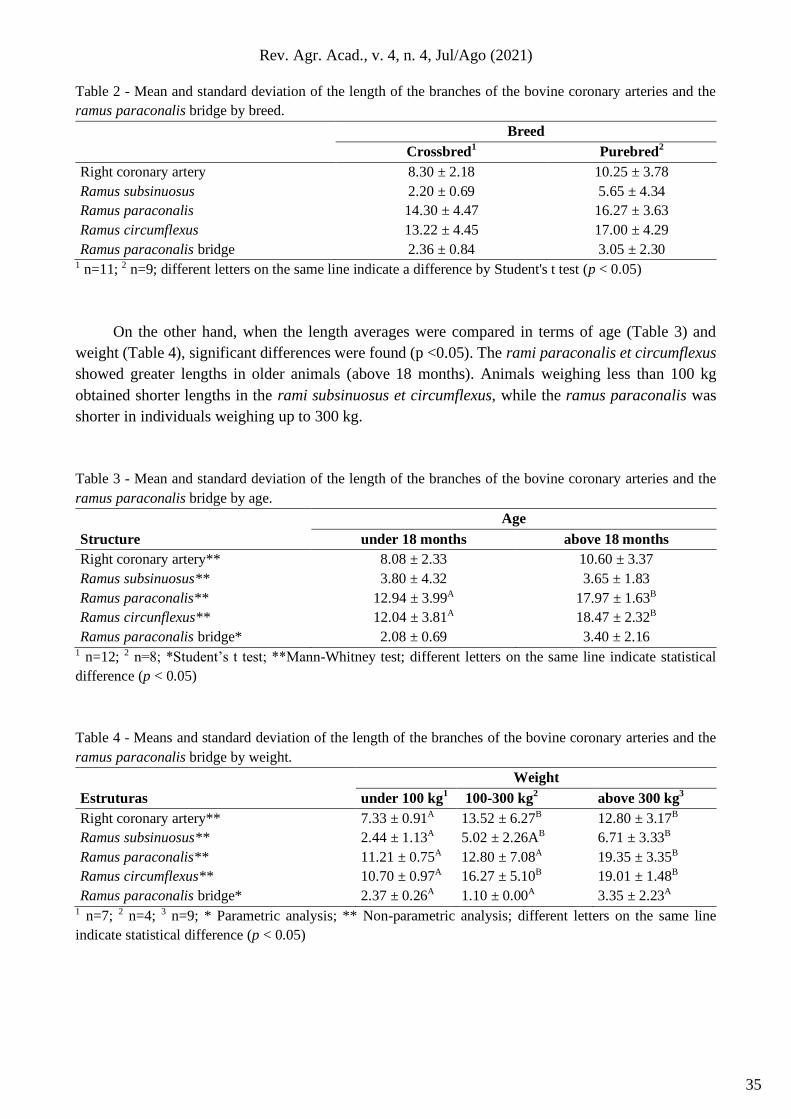
Table 1 - Means and standard deviation of the length of the branches of the bovine coronary arteries and the
ramus paraconalis bridge by sex.
Structure
Sex
Male Female2
Right coronary artery 9.20 ± 7.10 9.20 ± 2.18
Ramus subsinuosus 3.56 ± 2.20 3.81 ± 2.70
Ramus paraconalis 12.90 ± 3.84 16.31 ±3.88
Ramus circunflexus 13.63 ± 3.88 15.53 ± 3.57
Ramus paraconalis bridge 3.66 ± 2.29 2.16 ± 0.98 1 n=5; 2 n=15; different letters on the same line indicate a difference by Student's t test (p < 0.05)
34
Rev. Agr. Acad., v. 4, n. 4, Jul/Ago (2021)
Table 2 - Mean and standard deviation of the length of the branches of the bovine coronary arteries and the
ramus paraconalis bridge by breed.
Breed
Crossbred1 Purebred
2
Right coronary artery 8.30 ± 2.18 10.25 ± 3.78
Ramus subsinuosus 2.20 ± 0.69 5.65 ± 4.34
Ramus paraconalis 14.30 ± 4.47 16.27 ± 3.63
Ramus circumflexus 13.22 ± 4.45 17.00 ± 4.29
Ramus paraconalis bridge 2.36 ± 0.84 3.05 ± 2.30 1 n=11; 2 n=9; different letters on the same line indicate a difference by Student's t test (p < 0.05)
On the other hand, when the length averages were compared in terms of age (Table 3) and
weight (Table 4), significant differences were found (p <0.05). The rami paraconalis et circumflexus
showed greater lengths in older animals (above 18 months). Animals weighing less than 100 kg
obtained shorter lengths in the rami subsinuosus et circumflexus, while the ramus paraconalis was
shorter in individuals weighing up to 300 kg.
Table 3 - Mean and standard deviation of the length of the branches of the bovine coronary arteries and the
ramus paraconalis bridge by age.
Structure
Age
under 18 months above 18 months
Right coronary artery** 8.08 ± 2.33 10.60 ± 3.37
Ramus subsinuosus** 3.80 ± 4.32 3.65 ± 1.83
Ramus paraconalis** 12.94 ± 3.99A 17.97 ± 1.63B
Ramus circunflexus** 12.04 ± 3.81A 18.47 ± 2.32B
Ramus paraconalis bridge* 2.08 ± 0.69 3.40 ± 2.16 1 n=12; 2 n=8; *Student’s t test; **Mann-Whitney test; different letters on the same line indicate statistical
difference (p < 0.05)
Table 4 - Means and standard deviation of the length of the branches of the bovine coronary arteries and the
ramus paraconalis bridge by weight.
Estruturas
Weight
under 100 kg1 100-300 kg
2 above 300 kg
3
Right coronary artery** 7.33 ± 0.91A 13.52 ± 6.27B 12.80 ± 3.17B
Ramus subsinuosus** 2.44 ± 1.13A 5.02 ± 2.26AB 6.71 ± 3.33B
Ramus paraconalis** 11.21 ± 0.75A 12.80 ± 7.08A 19.35 ± 3.35B
Ramus circumflexus** 10.70 ± 0.97A 16.27 ± 5.10B 19.01 ± 1.48B
Ramus paraconalis bridge* 2.37 ± 0.26A 1.10 ± 0.00A 3.35 ± 2.23A 1 n=7; 2 n=4; 3 n=9; * Parametric analysis; ** Non-parametric analysis; different letters on the same line
indicate statistical difference (p < 0.05)
35
Rev. Agr. Acad., v. 4, n. 4, Jul/Ago (2021)
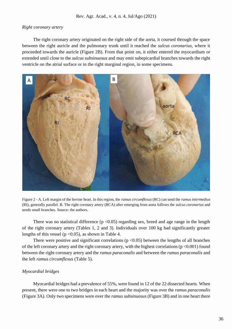
Right coronary artery
The right coronary artery originated on the right side of the aorta, it coursed through the space
between the right auricle and the pulmonary trunk until it reached the sulcus coronarius, where it
proceeded towards the auricle (Figure 2B). From that point on, it either entered the myocardium or
extended until close to the sulcus subsinuosus and may emit subepicardial branches towards the right
ventricle on the atrial surface or in the right marginal region, in some specimens.
Figure 2 - A. Left margin of the bovine heart. In this region, the ramus circumflexus (RC) can send the ramus intermedius
(RI), generally parallel. B. The right coronary artery (RCA) after emerging from aorta follows the sulcus coronarius and
sends small branches. Source: the authors.
There was no statistical difference (p <0.05) regarding sex, breed and age range in the length
of the right coronary artery (Tables 1, 2 and 3). Individuals over 100 kg had significantly greater
lengths of this vessel (p <0.05), as shown in Table 4.
There were positive and significant correlations (p <0.05) between the lengths of all branches
of the left coronary artery and the right coronary artery, with the highest correlations (p <0.001) found
between the right coronary artery and the ramus paraconalis and between the ramus paraconalis and
the left ramus circumflexus (Table 5).
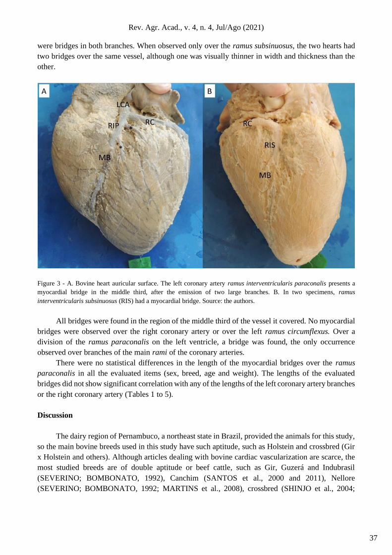
Myocardial bridges
Myocardial bridges had a prevalence of 55%, were found in 12 of the 22 dissected hearts. When
present, there were one to two bridges in each heart and the majority was over the ramus paraconalis
(Figure 3A). Only two specimens were over the ramus subsinuosus (Figure 3B) and in one heart there
36
Rev. Agr. Acad., v. 4, n. 4, Jul/Ago (2021)
were bridges in both branches. When observed only over the ramus subsinuosus, the two hearts had
two bridges over the same vessel, although one was visually thinner in width and thickness than the
other.
Figure 3 - A. Bovine heart auricular surface. The left coronary artery ramus interventricularis paraconalis presents a
myocardial bridge in the middle third, after the emission of two large branches. B. In two specimens, ramus
interventricularis subsinuosus (RIS) had a myocardial bridge. Source: the authors.
All bridges were found in the region of the middle third of the vessel it covered. No myocardial
bridges were observed over the right coronary artery or over the left ramus circumflexus. Over a
division of the ramus paraconalis on the left ventricle, a bridge was found, the only occurrence
observed over branches of the main rami of the coronary arteries.
There were no statistical differences in the length of the myocardial bridges over the ramus
paraconalis in all the evaluated items (sex, breed, age and weight). The lengths of the evaluated
bridges did not show significant correlation with any of the lengths of the left coronary artery branches
or the right coronary artery (Tables 1 to 5).
Discussion
The dairy region of Pernambuco, a northeast state in Brazil, provided the animals for this study,
so the main bovine breeds used in this study have such aptitude, such as Holstein and crossbred (Gir
x Holstein and others). Although articles dealing with bovine cardiac vascularization are scarce, the
most studied breeds are of double aptitude or beef cattle, such as Gir, Guzerá and Indubrasil
(SEVERINO; BOMBONATO, 1992), Canchim (SANTOS et al., 2000 and 2011), Nellore
(SEVERINO; BOMBONATO, 1992; MARTINS et al., 2008), crossbred (SHINJO et al., 2004;
37
Rev. Agr. Acad., v. 4, n. 4, Jul/Ago (2021)
CORREIA-OLIVEIRA, 2013 and 2014) and half-breed (SEVERINO et al., 1997), which reinforces
the importance of the contribution of the current research.
The nomenclature used to describe the studied bovine coronary arteries followed the most
recent Nomina Anatomica Veterinaria (INTERNATIONAL, 2017). However, it was noticed that
some branches found were not listed in the current official nomenclature, such as the diagonal branch
of the left coronary artery and the collateral branches of both coronary arteries. The diagonal branch
was previously found (CORREIA-OLIVEIRA et al., 2014) in seven (7/30) crossbred Gir x Holstein
cattle. In the studied hearts, four (4/22) had this branch. The literature does not name branches in the
right coronary artery in ruminants, citing them only as collateral (GETTY, 1986; CORREIA-
OLIVEIRA et al., 2013 and 2014). Martins et al. (2008) also noticed this scarcity of nomenclature to
describe the other branches of coronary arteries in cattle and had to rely on other articles to
complement the description.
In all hearts described, the left coronary artery gave rise to the rami paraconalis, circumflexus
et subsinuosus, while the right coronary artery was limited to the sulcus coronarius, as observed by
other authors (BORELLI; FERNANDES-FILHO, 1970; CORREIA-OLIVEIRA et al., 2013).
Coronary arterial dominance is determined according to the artery that gives rise to the posterior
interventricular artery (SANTOS et al., 2021), which corresponds to the animals' ramus subsinuosus
in terms of location. These authors described the dominance of human coronary arteries as
predominantly right (52.6%) (SANTOS et al., 2021), unlike the studied cattle, which had 100%
dominance of the left coronary artery. Of the domestic species, cats can have dominance in any of
the coronaries, but it is known that ruminants and canines have left dominance, and pigs and horses,
right (INTERNATIONAL, 2017). However, Borelli and Fernandes-Filho (1970) reported 6 cases
(4%) in which the right coronary artery gave rise to the right ramus circumflexus and the ramus
subsinuosus in five cattle of European origin (10%) and one zebu (1%).
No differences were found between the sexes regarding the lengths of the right coronary artery
or the branches of the left coronary artery, as well as Correia-Oliveira et al. (2013). Among the
measures taken by these authors (CORREIA-OLIVEIRA et al., 2013) in bovine coronary arteries, the
similarity in the lengths of the right coronary artery and rami paraconalis et circumflexus with the
current study stands out, but the authors describe significantly longer lengths for the ramus
subsinuosus. The cattle from northeastern Brazil showed a short subepicardial path of the ramus
subsinuosus and only in two hearts did this branch remain superficially beyond the middle third of
the sulcus interventricularis subsinuosus. It should be noted that in both studies, the animals evaluated
had the same aptitude (dairy), age groups consistent and allocated in mountainous geography, with
no external reasons at first glance for the difference in myocardial development in this region of the
evaluated hearts.
There are records of myocardial bridges in 100% of analyzed hearts (SEVERINO et al., 1997;
GULMEZ; SAH, 2021), however, in the current study the existence of this characteristic was
demonstrated in 55% of the 22 hearts. In pigs, from a total of 60 hearts, the presence of myocardial
bridges was reported in 36.36% of them (BOMBONATO et al., 1994). In humans, bridges were found
in 40.3% of the studied hearts, mainly in specimens with left dominance (22.8%) (SANTOS et al.,
2021). These authors emphasize that this frequency is not common in humans, and clinically may
suggest that bridges may be less frequent in populations with the usual distribution of arterial
dominance. The usual dominance in cattle is left, as observed in the current study and by Correia-
Oliveira (2014), however, no reports were found on the presence of myocardial bridges in cattle with
usual and unusual dominance.
38
Rev. Agr. Acad., v. 4, n. 4, Jul/Ago (2021)
Myocardial bridges have been reported in several species besides human (SANTOS et al.,
2021), such as ruminants (CORREIA-OLIVEIRA, 2013, 2014; GULMEZ; SAH, 2021), pigs
(BOMBONATO et al., 1994), mules (RIBEIRO et al., 2009), monkeys Cercopithecus aethiops
sabeus (NIKOLIĆ et al., 2009), among others.
The findings of the present study corroborate with the one that observed the vast majority
(94.3%) of bridges over the left coronary artery branches (SEVERINO et al., 1997). In other studies,
bridges were described in subdivisions of the main coronary branches in cattle in a significant amount
(29 of 106) (SEVERINO et al., 1997), in six pigs (BOMBONATO et al., 1994), and only one bovine
presented this characteristic in the current study.
In the evaluated hearts, all myocardial bridges were located predominantly in the middle portion
(middle third) of the vessel. Santos et al. (2021) observed the same location in most vessels that had
bridges, even more striking in hearts with left dominance. Bridges occur almost exclusively in the
middle region of the left anterior descending coronary artery (corresponding to the ramus paraconalis
of the animals) (ISHIKAWA et al., 2011).
The average length of the bovine myocardial bridges over the ramus paraconalis of this study
(24.6 ± 15.3 mm) was similar to that found in sheep (24.9 ± 16.1 mm) (GULMEZ; SAH, 2021). In
half-breed cattle, the bridges had an average length of 16.2 mm (SEVERINO et al., 1997) and in pigs,
7.5 mm (BOMBONATO et al., 1994)
Most of the myocardial bridges in pigs were found over the ramus subsinuosus
(BOMBONATO et al., 1994). It is pertinent to take into account the distinct origin of the ramus
subsinuosus between these two species (in ruminants it originates from the left coronary artery, and
in swine, from the right). Likewise, there was a difference regarding the presence of bridges in the
ramus circumflexus, which were present in pigs (3 on the right and 2 on the left), however, were
absent in the present study.
One of the importance of studying the vascularization of the heart is to understand how
functional structures are supplied, such as the conduction system. Both coronary arteries provide
branches for the sinoatrial node and the trunk of the atrioventricular fascicle, with the right ventricular
branch derived from the ramus subsinuosus of the left coronary artery and the rami septales derived
from the right coronary artery being the most frequent vessels of these structures, respectively
(MARTINS et al., 2008). Therefore, cardiac angiology research may contribute to the physiological
analysis, the interpretation of imaging tests and the clinical and surgical evaluation of the patient
(human or animal).
Several authors (ISHIKAWA et al., 2011; NASR, 2014; ZHAO et al., 2019) report that in
humans, myocardial bridges cause coronary heart disease by two distinct mechanisms, influenced by
the anatomical characteristics of the bridge, related to artery compression during cardiac systole and
increased coronary atherosclerosis due to stenosis of the artery near the myocardial bridge. They also
report that the study of myocardial bridges assists in the treatment decisions of heart disease and in
the clinical improvement of the patient. In Canchim cattle, the lesions found before the bridge, in the
bridge and in the post myocardial bridge are similar to the lesions that precede the formation of the
atherosclerotic plaque, although in the region of the bridge there are smaller lesions than in the other
regions (SANTOS et al., 2011). By analogy to the microscopic findings of the pre-bridge coronary
arteries, cattle have the same chance of developing atherosclerosis as humans (SHINJO et al., 2004).
However, coronary artery diseases resulting from myocardial bridges should not occur in sheep in
veterinary practice, as they observed bridges in 100% of the analyzed sheep hearts, with no detected
coronary disease (GULMEZ; SAH, 2021).
39
Rev. Agr. Acad., v. 4, n. 4, Jul/Ago (2021)
Myocardial bridges are an interesting area of study, as it is not clear whether they are a
pathology, a variation or even a physiological protection strategy for certain clinical conditions
(SANTOS et al., 2021). The literature is unanimous regarding the need to extend studies on the topic
to understand the pathophysiology and clinical relevance of these bridges. In this way, veterinary
medicine will be able to provide experimental models to expand knowledge on the subject.
Conclusion
Dairy cattle coronary arteries may present variation into their branches, especially the left one.
The ramus interventricularis paraconalis had a higher incidence of myocardial bridges, which
occurred in the middle third of the vessel. When observed in the subsinuous branch, they had thinner
characteristics in width and thickness.
Acknowledgements
To CBG (Bovine Clinic of Garanhuns) - UFRPE, which assisted patients subsequently
necropsied at LAPA and donated their hearts to this research.
Author’s Contributions
VLB, KFL, AMAR and JVM participated in its design, dissected the hearts, made the measurements,
collected the animal data, tabulated the data and measures for statistical analysis, JMBO participated
in the design of the study and performed the statistical analysis, AMSB participated in the collections
and applied the specimen conservation techniques, EPM participated in the design of the study, in the
writing and in the final revision of the manuscript, DO conceived of the study, and participated in its
design, analysis and coordination and helped to draft the manuscript. All authors read and approved
the final manuscript.
Competing Interests
The authors declared no potential conflicts of interest with respect to the research, authorship, and/or
publication of this article.
References
BOMBONATO, P. P.; DOMINGOS, C. de O; MARIANA, A. N. B; SILVA, F. O. C.; INTELIZANO, W.
Ocorrência de pontes do miocárdio em suínos. Brazilian Journal of Veterinary Research and Animal
Science, v. 31, n. 2, p. 107-111, 1994. https://pesquisa.bvsalud.org/portal/resource/pt/lil-240148
BORELLI, V.; FERNANDES FILHO, A. Sobre a origem do ramus descendens subsinuosus em bovinos.
Revista da Faculdade de Medicina Veterinária, São Paulo, v. 8, n. 2, p. 367-373, 1970.
http://docplayer.com.br/110147723-Sobre-a-origem-do-r-a-m-u-s-d-e-s-c-e-n-d-e-n-s-subsinuosus.html
BRODSKY, S. V.; ROH, L.; ASHAR, K.; BRAUN, A.; RAMASWAMY, G. Myocardial bridging of coronary
arteries: a risk factor for myocardial fibrosis? International Journal of Cardiology, v. 124, n. 3, p. 391-392,
2008. https://pubmed.ncbi.nlm.nih.gov/17399815/
40
Rev. Agr. Acad., v. 4, n. 4, Jul/Ago (2021)
CORREIA-OLIVEIRA, M.; MORAES, S. O. S.; GOMES, M. S.; PALHANO, H. B.; ABIDU-FIGUEIREDO,
M. Dominância entre as artérias coronárias em bovinos mestiços. Revista Brasileira de Ciência Veterinária,
v. 21, n. 2, p. 82-85, 2014. http://doi.editoracubo.com.br/10.4322/rbcv.2014.027
CORREIA-OLIVEIRA, M.; HERNANDEZ, J. M. F.; ABIDU-FIGUEIREDO, M. Morfometria cardíaca e
distribuição das artérias coronárias em bovinos mestiços. Biotemas, v. 26, n. 2, p. 199-207, 2013.
https://periodicos.ufsc.br/index.php/biotemas/article/view/2175-7925.2013v26n2p199
DYCE, K. M.; SACK, W. O.; WENSING, C. J. G. Tratado de Anatomia Veterinária. 4ª ed. Rio de Janeiro:
Elsevier, 2010.
FRANDSON, R. D.; WILKE, W. L.; FAILS, A. D. Anatomia e Fisiologia do Animais de Fazenda. 7ª ed.
Rio de Janeiro: Guanabara Koogan, 2017.
GETTY, R. Sisson/Grossman Anatomia dos Animais Domésticos. Rio de Janeiro: Guanabara Koogan,
1986.
GULMEZ, N.; SAH, H. The relationships between the myocardial bridge and ramus interventricularis
paraconalis characteristics in lamb and sheep. Anatomia, Histologia, Embryologia, v. 50, n. 2, p. 260-265,
2021. https://pubmed.ncbi.nlm.nih.gov/33009861/
INTERNATIONAL COMMITTEE ON VETERINARY GROSS ANATOMICAL NOMENCLATURE.
Nomina Anatomica Veterinaria. 6ª ed. World Association of Veterinary Anatomists: editorial Committee:
Rio de Janeiro, 2017.
ISHIKAWA, Y.; KAWAWA, Y.; KOHDA, E.; SHIMADA, K.; ISHII, T. Significance of the anatomical
properties of a myocardial bridge in coronary heart disease. Circulation Journal, v. 75, n. 7, p. 1559-1566,
2011. https://pubmed.ncbi.nlm.nih.gov/21467656/
KÖNIG, H. E.; LIEBICH, H. E. Anatomia dos Animais Domésticos. 4ª ed. Porto Alegre: Artmed, 2011.
MARTINS, A-K.; SEVERINO, R. S.; CARDOSO, J. R.; FIDELIS, A. A.; RODRIGUES, T. V.
Vascularização arterial do nó atrioventricular e tronco do fascículo atrioventricular em bovinos da raça Nelore.
Bioscience Journal, v. 24, n. 3, p. 102-107, 2008.
http://www.seer.ufu.br/index.php/biosciencejournal/article/view/6754
NASR, A. Y. Myocardial bridge and coronary arteries: morphological study and clinical significance. Folia
Morphologica (Warsz), v. 73, n. 2, p. 169-182, 2014. https://pubmed.ncbi.nlm.nih.gov/24902096/
NIKOLIĆ, V.; BLAGOJEVI, Z.; STIJAK, L.; RADONJI, V.; DORDEVI, M.; KOVACEVI, D.; FILIPOVI,
B. Myocardial bridges over the ramus interventricularis anterior and its branches in Cercopithecus aethiops
sabeus. Acta Veterinaria, v. 59, n. 2-3, p. 213-221, 2009. http://www.doiserbia.nb.rs/img/doi/0567-
8315/2009/0567-83150903213N.pdf
OECD/FAO. OECD/FAO Agricultural Outlook 2020-2029. OECD: Paris: FAO: Rome; 2020.
http://www.fao.org/publications/oecd-fao-agricultural-outlook/2020-2029/en/
RIBEIRO, A. L. C.; SEVERINO, R. S.; GUERRA, R. R.; FAVARON, P. O.; TOMMASI JUNIOR, H. L. P.;
RICCI, R. E. G.; FRANCIOLLI, P. R. F.; BOMBONATO, P. P. Biometria de pontes de miocárdio em muares
(Equus caballus x Equus asinus – Linnaeus 1758). Biotemas, v. 22, n. 3, p. 177-184, 2009.
https://periodicos.ufsc.br/index.php/biotemas/article/view/2175-7925.2009v22n3p177/0
RODRIGUES, T. M. A.; PALMEIRA, J. A. O.; MENDONÇA, J. T. DE; GOMES, O. M. Estudo evolutivo da
anatomia das artérias coronárias em espécies de vertebrados com técnica de moldagem em acetato de vinil
(vinilite). Revista Brasileira de Cirurgia Cardiovascular, v. 14, n. 4, p. 331-339, 1999.
https://www.rbccv.org.br/article/781/pt-BR/estudo-evolutivo-da-anatomia-das-arterias-coronarias-em-
especies-de-vertebrados-com-tecnica-de-moldagem-em-acetato-de-vinil--vinilite-
41
Rev. Agr. Acad., v. 4, n. 4, Jul/Ago (2021)
SAMPAIO, I. B. M. Estatística Aplicada à Experimentação Animal. 4ª ed. Belo Horizonte: Fundação de
Ensino e Pesquisa em Medicina Veterinária e Zootecnia, 2015.
SANTOS, J. C. C.; BARRETO, J. E. F.; RODRIGUES, C. F. de S.; LIMA JÚNIOR, F. A. S. de; OLIVEIRA,
A. de S. BRAGA. Morphological analysis of myocardial bridges and coronary arterial dominance in northeast
Brazil. Morphologie, 2021. https://pubmed.ncbi.nlm.nih.gov/33775545/
SANTOS, J. W.; BOMBONATO, P. P.; BELETTI, M. E.; SEVERINO, R. S.; CARNEIRO E SILVA, F. O.
Pontes de miocárdio em bovinos da raça Canchim, I - Aspectos microscópicos. Brazilian Journal of
Veterinary Research and Animal Science, v. 37, n. 2, p. 128-131, 2000.
https://www.scielo.br/j/bjvras/a/cNWz5Ctd93NXdvKCJg8Rqmv/?lang=pt
SANTOS, J. W.; WAFAE, N.; BELETTI, M. E. Ponte miocárdica associada a lesões cardiovasculares em
bovinos adultos da raça Canchim. Arquivos Brasileiros de Cardiologia, v. 98, n. 1, p. 22-28, 2011.
https://www.scielo.br/j/abc/a/YPJJwyvTBxKvmcjM54m3PSn/abstract/?lang=pt
SEVERINO, R. S.; CARNEIRO E SILVA, F. O; SANTOS, A. L. Q; DRUMMOND, S. S; BOMBONATO,
P. P; DURAN, F. P; MARÇAL, A. V. Pontes de miocárdio em bovinos azebuados. Brazilian Journal of
Veterinary Research and Animal Science, v. 34, n. 5, p. 288-291, 1997.
https://pesquisa.bvsalud.org/portal/resource/pt/lil-257117
SEVERINO, R. S.; BOMBONATO, P. P. Ocorrência de pontes de miocárdio em bovinos das raças Gir,
Guzerá, Indubrasil e Nelore. Brazilian Journal of Veterinary Research and Animal Science, v. 29, n. 1, p.
15-30, 1992. https://www.bvs-vet.org.br/vetindex/periodicos/brazilian-journal-veterinary-research-and-
animal-s/29-(1992)-1/ocorrencia-de-pontes-de-miocardio-em-bovinos-das-racas-gir-guzera-indu/
SHINJO, S. K.; OBA-SHINJO, S. M.; PRATES, N. E. V. B. Bovine myocardial bridge morphology and
association with coronary atherosclerosis, Brazilian Journal oh Morphological Sciences, v. 21, n. 2, p. 95-
98, 2004. https://pesquisa.bvsalud.org/portal/resource/pt/lil-406361
SIERVULI, M. T. F.; SILVA, A. de S.; SILVA, A. C. da; MUZZI, R. A. L.; SANTOS, G. A. B. Infarto do
miocárdio: alterações morfológicas e breve abordagem da influência do exercício físico. Revista Brasileira
de Cardiologia, v. 27, n. 5, p. 349-355, 2014. http://www.onlineijcs.org/english/sumario/27/pdf/v27n5a09.pdf
TARANTINI, G.; MIGLIORE, F.; CADEMARTIRI, F.; FRACCARO, C.; ILICETO, S. Left anterior
descending artery myocardial bridging: a clinical approach. Journal of the American College of Cardiology,
v. 68, n. 25, p. 2887-2899, 2016. https://pubmed.ncbi.nlm.nih.gov/28007148/
TERAGAWA, H.; FUKUDA, Y.; MATSUDA, K.; HIRAO, H.; HIGASHI, Y.; YAMAGATA, T.; OSHIMA,
T.; MATSUURA, H.; CHAYAMA, K. Myocardial bridging increases the risk of coronary spasm. Clinical
Cardiology, v. 26, n. 8, p. 377-83, 2003. https://pubmed.ncbi.nlm.nih.gov/12918640/
ZHAO, D. H.; FAN, Q.; NING, J. X.; WANG, X.; TIAN, J. Y. Myocardial bridge-related coronary heart
disease: Independent influencing factors and their predicting value. World Journal of Clinical Cases, v. 7, n.
15, p. 1986-1995, 2019. https://europepmc.org/article/MED/31423430
Recebido em 17 de junho de 2021
Retornado para ajustes em 13 de julho de 2021
Recebido com ajustes em 14 de julho de 2021
Aceito em 18 de julho de 2021
42