Embed Size (px)
Citation preview
R E U T I L I Z A T I O N OF L A B E L E D DNA BY R E G E N E R A T I N G A D R E N A L CELLS
(UDC 612.45:612.6.03].015.348)
M. Y a . N e k h l y u d o v a
Radiological Division of the Cantral Scient if ic Research Laboratory (Scient i f ic Supervisor, Professor M. F. Merkulov), Second Moscow Medical Institute (Presented by Member of the Academy of Medica l Sciences USSR A. F. Bilibin) Translated from Byulleten' ~ksperimental 'noi Biologii i Meditsiny, Vol. 61, No. 1, pp. 73-76, January, 1966 Original a r t ic ie submit ted June 8, 1964
Thymid ine -H s, which is a spec i f ic precursor of DNA, is widely used for the study of ce l lu lar kinetics. The conclusions drawn as a result of the first studies with thymid ine -H 3 were based upon the fact that after a single in - troduction of the labe led nucleoside, i t is u t i l ized for DNA synthesis only for 45-60 rain, since after this i t is ex- cre ted from the organism [7, 15, 16, 18, 20].
However, the results of a number of studies give evidence of the possibility of reut i l iza t ion of the isotope for
DNA synthesis by various cells of the organism. This process is detected in bone marrow cells [11-14], ep idermal cells of the skin [9], and cells of the l iver and smal l intest ine [5, 6]. Most authors consider the nuclear elements of
the blood, pr imari ly the lymphocytes, as the source of the isotope in its reut i l iza t ion [5, 6, 8, 9].
The purpose of this work was to determine the possibili ty of reut i l iza t ion of l abe led DNA by cells of the re- generat ing adrenal gland.
E X P E R I M E N T A L M E T H O D S
The experiments were conducted on males of the hybrid l ine of mace CsTB1 and CBA, weighing 28 g. Thy- m i d i n e - H 3, with specif ic ac t iv i ty 2.9 C/mM, was in jec tedin t rapet i toneal ly . Two animals received 3 injections each
of thymid ine -H z in doses of 30 microcuries at 12 h intervals. Two mice received a summary dose of 90 microcuries of thymid ine -H 3 in a single inject ion.
A day after the in jec t ion of thymid ine -H 3, the a n i m a h were operated upon - one adrenal was removed en- t i rely, and one third or one fourth of the other was resectioned. This method of operat ion induces the most pro- nounced regenerat ion of the organ [1, 3]. The operat ion may also be considered as a stressor. In stress reactions, the mass of the adrenals increases substantial ly in a short period of t ime. Moreover, numerous mitoses appear in
the organ [19].
All the mice were k i l led on the third day after the operation and on the fourth day after the inject ion of thymi din e - H s.
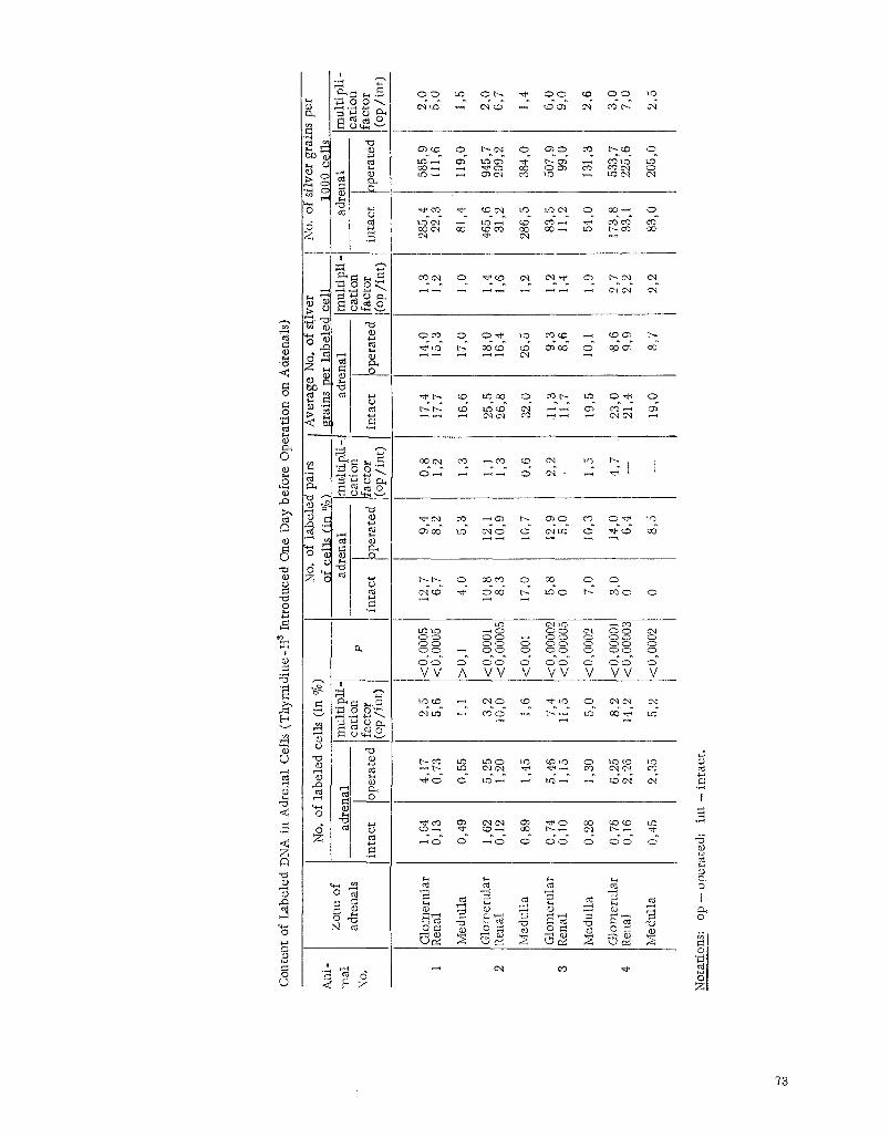
The adrenals removed during the operat ion and those in the ki l led animals were fixed in Camoy's Quid and imbedded in paraffin. Sections 5 p thick were stained with hematoxyl in-eos in and covered with l iquid nuclear emulsion type M (Scient if ic Research Institute of the Motion Picture Fi lm Industry) [2]. Part of the preparations was stained with methy tgreen-pyron ine through the emulsion after development. The preparations were exposed for a month. In the analysis of the radioautograph, the following indices were determined: 1) the percent content of l abe led cells, in this case from 5000 to 26,000 cells were counted in various zones of the adrenal cortex and adrenal cortex and adrenal medul la ; 2) the intensity of the labe l (number of grains of silver over each labe led cell), on the basis of these indices, the average number of grains of silver per l abe led ce l l and that per 1000 ceils were ca l cu - lated; 3) the percent content of l abe led pairs of cells with respect to a l l the labe led cells. A labeled pair of cells was considered to be those possessing two labe led nuclei with a distance between them not exceeding the d iameter
of one of the nuclei (see figure, a and b).
72
Con
tent
of
Lab
eled
DN
A i
n A
dren
al C
ells
(T
hym
idin
e-H
a In
trod
uced
One
Day
bef
ore
Ope
rati
on o
n A
dren
als)
Zon
e of
ad
rena
ls
Glo
mer
ular
R
enal
Med
ulla
Glo
mer
ular
R
enal
Med
ulla
Olo
mer
ular
R
enal
Med
ulla
Glo
mer
ular
R
enal
Med
ulla
No.
of
labe
led
cell
s (i
n %
)
adre
nal
mul
tipl
i i
cati
on
inta
ct
oper
ated
fa
ctor
(o
p/m
r)
I
4,17
0,
73
0,55
5,25
1,
20
1,45
5,46
1,
15
1,30
6,25
2,
26
2,35
2,5
5,6
1,1
3,2
0,0
1,6
7,4
1,5
5,0
8,2
4,2
5,2
1,64
0,
13
0,49
1,62
0,
12
0,89
0,74
0,
10
0,28
O, 7
6 0,
16
0,45
Ani
- m
at
No.
P
No.
of
labe
led
pair
s of
cel
ls (
in %
) ad
ren
al
mul
tipl
f-l
cati
on
inta
ct
oper
ated
fa
ctor
i(
op/i
nt)
Ave
rage
No.
of
silv
er
[ gr
ains
per
lab
eled
cel
l --
l-
adre
nal
mul
fipl
t-]
I ca
tion
I
inta
ct
Ope
rate
d fa
ctor
(o
p/in
t)
<0,
0005
2,
7 <
0,00
05
6,7
>0,
1 4,
0
<0,
0001
0,
8 <
0,00
005
8,3
<0,
001
7,0
<0,
0000
2 5,
8 <
0,00
005
0
<0,
0002
7,
0
<0,
0000
1 3,
0 <
0,00
003
0
<0,
0002
0
9,4
8,2
5,3
12,1
10
,9
10,7
12,9
5,
0
10,3
14,0
6,
4
8,5
0,8
1,2
1,3
1,1
1,3
0,6
2,2
1,5
4,7
17,4
17
,7
16,6
25,5
26
,8
32,0
11,3
I1
,7
19,5
23,0
21
,4
19,0
14,0
15
,3
17,0
18,0
16
,4
26,5
9,3
8,6
10,1
8,6
9,9
8,7
1,3
1,2
1,0
1,4
1,6
1,2
1,2
1,4
1,9
2,7
2,2
2,2
No.
of
silv
er g
rain
s pe
r 10
00 c
ells
ad
rena
l
inta
ct
oper
ated
285,
4 58
5,9
22,3
II
i ,6
81,4
11
9,0
465,
6 94
5,7
31,2
20
9,2
286,
5 38
4,0
83,5
50
7,9
1t ,2
99
,0
54,0
13
1,3
173,
8 53
3,7
33, 1
22
5,6
83,0
20
5,0
mul
ti p
li -
cati
on
fact
or
(op
/in
t)
2,0
5,0
1,5
2,0
6,7
1,4
6,0
9,0
2,6
3,0
7,0
2,5
Not
atio
ns:
op -
op
erat
ed;
int
- in
tact
,
g
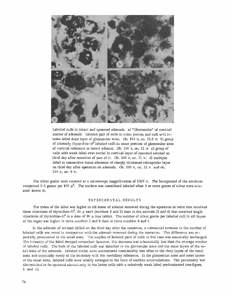
Labeled ceils in in tac t and operated adrenals, a) "Glomerulus" of cor t ica l mat te r of adrenals. Labeled pair of ceils in outer portion and ce l l with i n - tense label deep layer of g lomerular zone. Ob. 100 x, oc. 12.5 x; b) group of intensely thymid ine -H 3 labe led ceils in outer portions of g lomerular zone of cor t ical substance in in tac t adrenal. Ob. 100 x, oc. 11 x; c) group of
ceils with weak labe l over nuclei in cor t ical layer of operated adrenal on third day af ter resection of part of it. Ob. 100 • oc. 11 x; d) mul t ip le
l abe l in connect ive tissue elements of sharply thickened subcapsular layer on third day after operation on adrenals. Ob. 100 x, oc. 11 x and oh. 100 x , oc. 8 x .
The silver grains were counted at a microscope magnif icat ion of 1350 X. The background of the emulsion comprised 0-1 grains per 100 52. The nucleus was considered labe led when a or more grains of si lver were si tu- ated above it.
E X P E R I M E N T A L R E S U L T S
The index of the labe l was higher in a l l zones of adrenal removed during the operation in mice that received three injections of t hymid ine -H a, 30 ~ each (numbers 1 and 2) than in the animals (a and 4) that received single inject ions of t hymid ine -H 3 in a dose of 90 ~ (see table). The number of silver grains per labe led ce l l in a l l layers of the organ was higher in mice numbers 1 and 2 than in mice numbers ~ and 4.
In the adrenals of animals k i l led on the third day after the operation, a substantial increase in the number of l abe led ceils was noted in comparison with the adrenals removed during the operation. This difference was es- pec ia l ly pronounced in the renal zone. The number of l abe led pairs of ceils in this case was essential ly unchanged. The intensi ty of the labe l dropped somewhat; however, this decrease was substantially less than the average number of l abe led cells. The bulk of the labe led ceils was detected in the glomerular zone and the outer layers of the re - ha l zone of the adrenals. Labeled nuclei were encountered considerably less often in the deep layers of the renal zone and especia l ly rarely at the boundary with the medul lary substance. In the glomerular zone and outer layers of the renal zone, l abe led cells were usually arranged in the form of nest l ike accumulat ions. This pecul iar i ty was also retained in the operated adrenal ,only in the la t te r cells with a re la t ive ly weak label predominated (see figure, b and c).
74
The summary histograms of the labe led cel ls of the glomerular and renal zones in the operated and nonop-
cra ted adrenals represent single ver tex curves with a peak in the region of ceils containing 4 to 6 silver grains over
the nucleus. However, in the operated adrenals, this peak is higher in the region of weakly labeled nuclei.
Noteworthy is the content of the labe l in the connect ive tissue elements of the capsule on the third day after the operation. Usually the adrenal capsule consists of two to three rows of e longated dark-colored nuclei , among
which one to two labe led nuclei are detected on the entire section. The intensity of the label of these nuclei , as a rule, is equal to 18-20 grains of silver. In the operated adrenal , the capsule is substantially thickened, chiefly on account of connect ive tissue elements of the subcapsular layer. The la t ter forms a wedge, turned with its broad side toward the si te of the operat ion trauma, and consists of cel ls with very pa le cytoplasm; their nuclei are large, c i r cu-
lar or oval, weakly colored; almost every one of them contains a l abe l in 8 to 8 silver grains or more (see figure, d). At the base of the wedge-shaped expansion of the capsule, six to seven rows of the cells described are counted; they are most l ike ly fibroblasts and s imi lar cells.
At the leve l of the wedgel ike expansion of the capsule, the g lomerular zone of the cortex of the organ is dis- p laced inward. The structure of the zone cIose to the operated section is disturbed. In the parenchyma of the or- gan, along the l ine of the cut there is an outgrowth of strands of connective tissue. Outside them lies a structureless
mass of protein secretion, a fibrin clump, which is stained a pale rose color by eosin and methyl green-pyronine. On the inside of this c lump grow elements of connect ive tissue, while on the outside i t is adjoined by a mass of form- less detritus, stained by nuclear dyes. In this mass of nuclear detritus, a diffuse label is detected, which is 20-25 t imes as ~ e a t as the background act ivi ty.
As was ind ica ted above, most researchers be l i eve thymid ine -H a is rapidly u t i l ized for DNA synthesis under the conditions of the organism. Thus, a l l the processes in the operated adrenal took p lace against a background of the absence of free t hymid ine -H ~ in the blood plasma.
Under these conditions, we detected a substantial increase in the number of labe led ceils in various layers of the adrenal. This fact cannot be explained by division of the labeled ceils, since the increase in the number of
l abe led ceils was para l le led by an increase in the intensity. There is some basis for bel ieving that there is s t i l l some dilut ion of the labe l as a result of division of the ceils; however, i t cannot explain the increase in the number of labeled cells. The la t te r is due chief ly to secondary ut i l iza t ion of the isotope l iberated in other tissues of the or-
ganism for DNA synthesis in the adrenal. It cannot be stated on the basis of the results of our experiments what t i s - sues l ibera te the labe led DNA in this case. It is be l ieved [5, 6] that the donors of the label for reut i l iza t ion are the mononuclear blood cei ls . The degree of po lymer iza t ion of the labe led products of DNA that par t ic ipa te in the
process of reu t i l iza t ion is unknown. It may be assumed that the bulk of them are comprised of low-molecu la r com- pounds - nucleosides and nucleot ides [6, 7]. However, in this case the possibility of reut i l iza t ion of h igh-polymer compounds cannot be ent i rely excluded.
The operation may be considered as a strong stress factor. It is known that in the case of stress influences the processes of proliferation are most ac t iveIy expressed in the renal zone [4, 19]. Our data indicat ing that the i n - crease in the number of l abe led ceils after the operation on the adrenals is especial ly pronounced in the renal zone agree with this.
Of special interest are the changes in the ba lance of renal ac t iv i ty in the capsule of the operated adrenal. There is no doubt that the high content of labe led e lements in the thickened capsule cannot be due to division of
single labe led cells, encountered in the capsule of the in tac t organ. The labe led elements may also be " immigrants" with respect to the focus of inf lammat ion.
In the zone of operat ive intervention, there is nuclear detritus with an especia l ly high content of the label. This l abe l is not washed out by his tological t rea tment , which may be evidence of a high molecular state of the
labe led mater ia l . Whether the adrenal can direct ly u t i l i ze this l abe led mate r ia l for the synthesis of DNA of the proliferating e lements is unknown.
S U M M A R Y
Mice (Cs~B1 and CBA) weighing 28 g were given int raabdominat injections of thymid ine-H 3 (90 me) 24 h b e - fore an operation on the adrenals (excision of one gland and part of the other). On the third day after the operation a considerable increase in the index was noted, as wel l as of the total grain count in a l l the layers of the adrenal
75
operated upon and in the medullar layer. This increase was particularly notable in the fascicular zone of the cortex and in the connective-tissue elements of the subcapsular layer, which was markedly thickened after the operation. The accumulat ion of label in the organ operated upon took place in the absence of free H3-thymidine in the blood plasma. This fact may point to the process of reutilization of DNA metabolic products occurring in the mouse body.
L I T E R A T U R E CITED
I. M.A. Vorontsov and L D. Liozner, Physiological Regeneration [in Russian], Moscow (1955).
2. IVL F. Merkulov, Scientific Notes of the Second Moscow Medical Institute [in Russian], Vol. 6 (1957), p. 163.
3. G.V. Kharlova, In the book: Regeneration of Organs in Mammals, The Adrenals [in Russian], Moscow
(1960), p. 324. 4. R. 1Vs Brenner, Am. J. Anat., Vol. 112 (1963), p. 81. 5. B.J. Bryant, Exp. Cel l Res., Vol. 27 (1962), p. 70. 6. Idem, J. Cell. Biol., Vol. 18 (1963), po 515. 7. E.P. Cronkite, T. M. Fliedner, V. tL Bond et al., Arm., New York, VoL 77 (1959), p. 803. 8. H. Diderholm, Acta Pathol. Microbiol, Scan&, VoL 51, Suppl. 146 (1961), p. 5. 9. H. Diderholm, K. E. Fichtelius, and O. Linder, Exp. Cell. Res., Vol. 27 (1962), p. 43.
10. M. Hill, Nature, Vol. 183 (1959), p. 1059. 11. M. Hill and V. Drasil, Exp. Cell Res., Vol. 21 (1960), p. 569. 12. M. Hill, ]bid., VoL 24 (1961), p. 405. 13. M. Hill et al., ibid., Vol. 26 (1962), p. 541. 14. M. Hill, ]bid., Vol. 28 (1962), p. 21. 15. E. Koburg and W. Maurer, Biochem. Biophys. Acta, Vol. 61 (1962), p. 229. 16. H. Quastler and F. G. Sherman, Exp. Cell Res., Vol. 17 (1959), p. 420. 17. S .H. Robinson and G. Brecher, Science, Vol. 142 (1963), p. 392. 18. L tL Rubini, E. P. Cronkite, V. P. Bond et al., J. Clin. Invest., Vol. 39 (1960), p. 909. 19. H. Selye, Physiology and Pathology of Exposure to Stress, Montreal (1950). 20. 7. H. Taylor, Ann., New York Acad. Sci., Vol. 90 (1960), p. 409.
76