Embed Size (px)
Citation preview
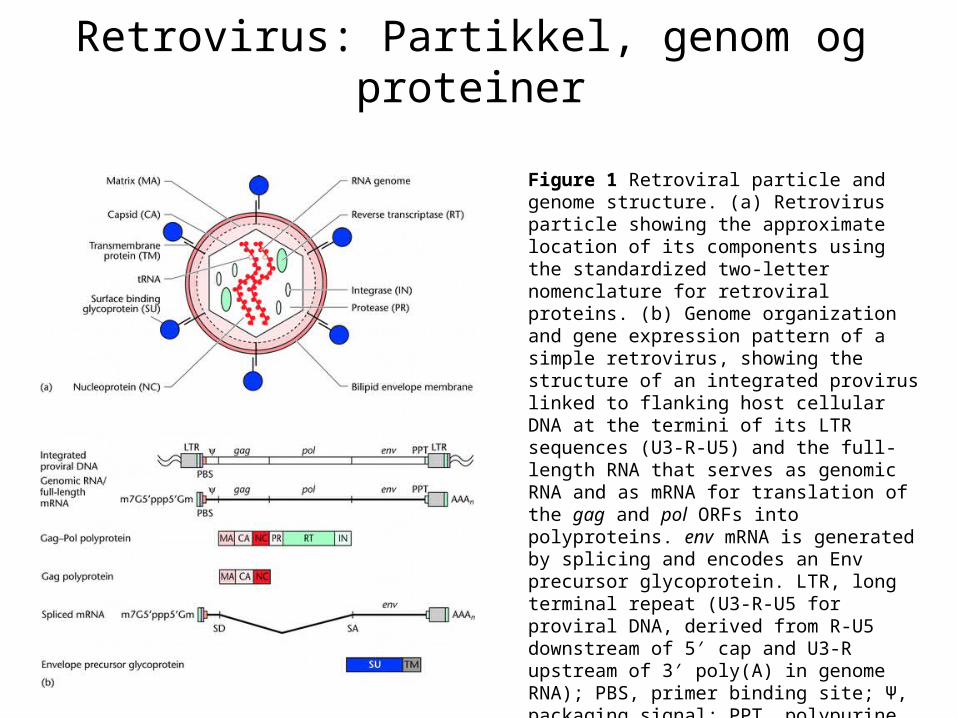
Retrovirus: Partikkel, genom og proteiner
Figure 1 Retroviral particle and genome structure. (a) Retrovirus particle showing the approximate location of its components using the standardized two-letter nomenclature for retroviral proteins. (b) Genome organization and gene expression pattern of a simple retrovirus, showing the structure of an integrated provirus linked to flanking host cellular DNA at the termini of its LTR sequences (U3-R-U5) and the full-length RNA that serves as genomic RNA and as mRNA for translation of the gag and pol ORFs into polyproteins. env mRNA is generated by splicing and encodes an Env precursor glycoprotein. LTR, long terminal repeat (U3-R-U5 for proviral DNA, derived from R-U5 downstream of 5′ cap and U3-R upstream of 3′ poly(A) in genome RNA); PBS, primer binding site; Ψ, packaging signal; PPT, polypurine tract; SD, splice donor site; SA, splice acceptor site.
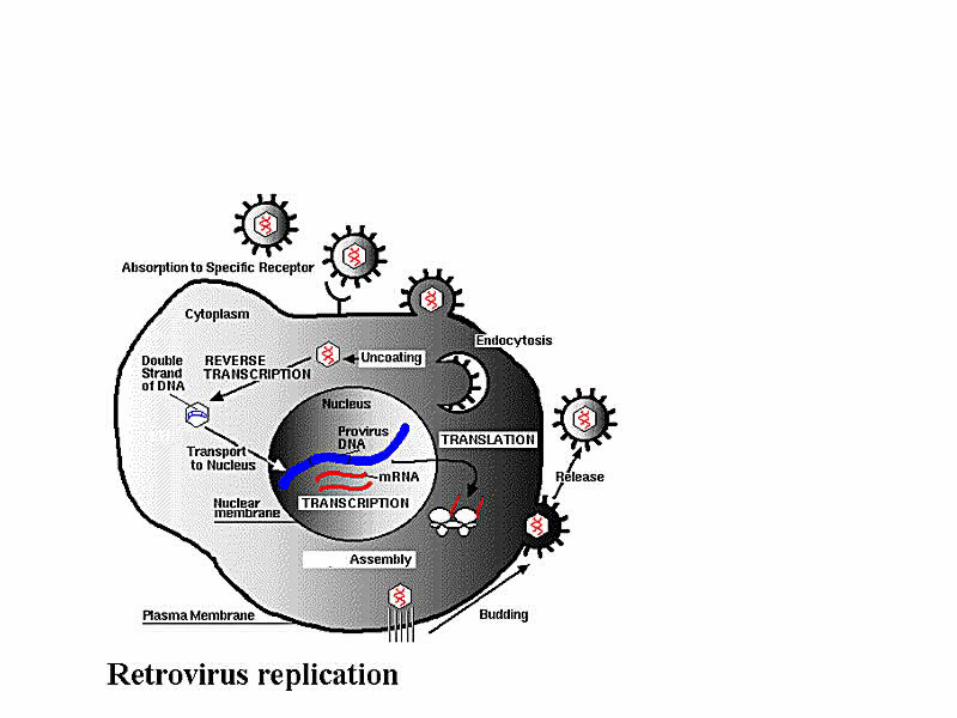
Retrovirus: Replikasjonssyklus
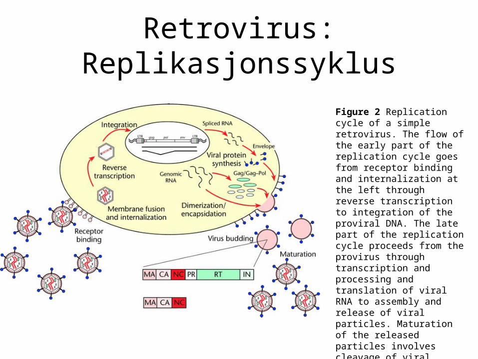
Figure 2 Replication cycle of a simple retrovirus. The flow of the early part of the replication cycle goes from receptor binding and internalization at the left through reverse transcription to integration of the proviral DNA. The late part of the replication cycle proceeds from the provirus through transcription and processing and translation of viral RNA to assembly and release of viral particles. Maturation of the released particles involves cleavage of viral polyproteins by PR (protease).
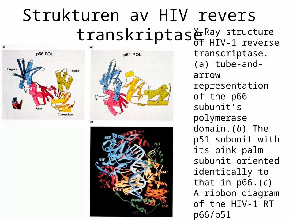
Strukturen av HIV revers transkriptase X-Ray structure of
HIV-1 reverse transcriptase. (a) tube-and-arrow representation of the p66 subunit’s polymerase domain.(b) The p51 subunit with its pink palm subunit oriented identically to that in p66.(c) A ribbon diagram of the HIV-1 RT p66/p51 heterodimer in complex with DNA.
Reaksjoner som katalyseres av revers transkriptase
Revers transkripsjon av genomisk RNA
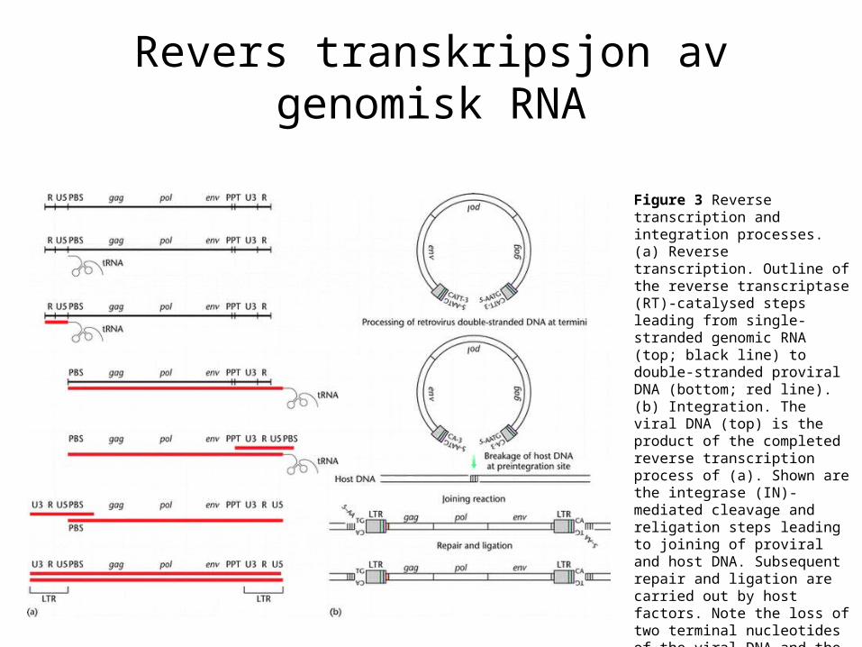
Figure 3 Reverse transcription and integration processes. (a) Reverse transcription. Outline of the reverse transcriptase (RT)-catalysed steps leading from single-stranded genomic RNA (top; black line) to double-stranded proviral DNA (bottom; red line). (b) Integration. The viral DNA (top) is the product of the completed reverse transcription process of (a). Shown are the integrase (IN)-mediated cleavage and religation steps leading to joining of proviral and host DNA. Subsequent repair and ligation are carried out by host factors. Note the loss of two terminal nucleotides of the viral DNA and the generation of a short repeat of host sequences of the integration site.
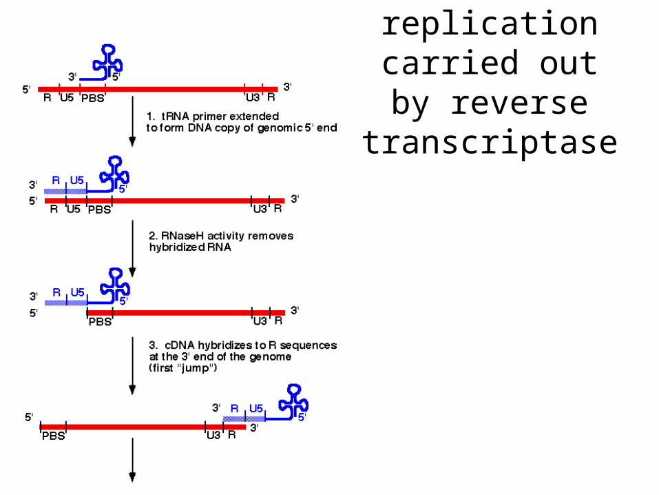
Retrovirus replication carried
out by reverse transcriptase
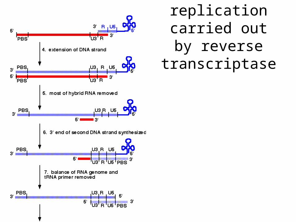
Retrovirus replication carried
out by reverse transcriptase
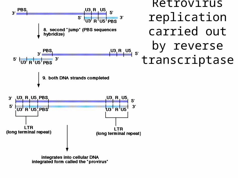
Retrovirus replication carried
out by reverse transcriptase
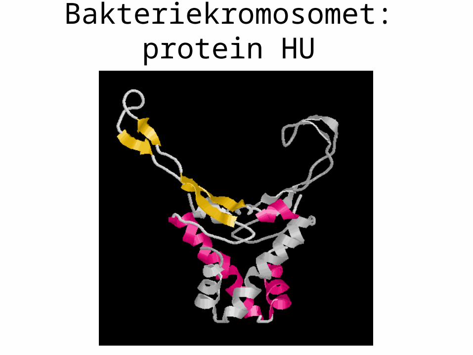
Bakteriekromosomet: protein HU
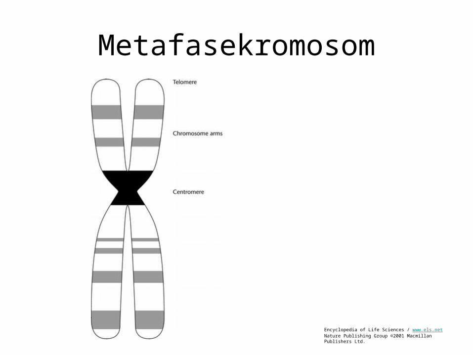
Metafasekromosom
Encyclopedia of Life Sciences / www.els.netNature Publishing Group ©2001 Macmillan Publishers Ltd.
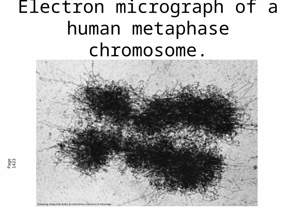
Electron micrograph of a human metaphase chromosome.
Pag
e 14
23
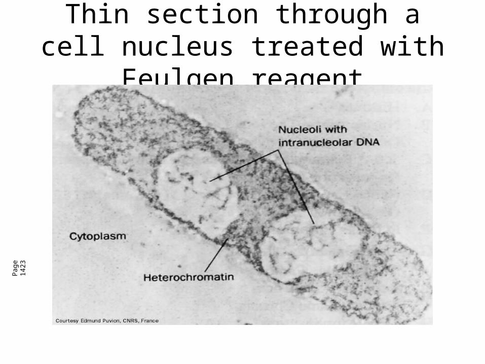
Thin section through a cell nucleus treated with Feulgen reagent
Pag
e 14
23

Calf Thymus HistonesP
age
1423
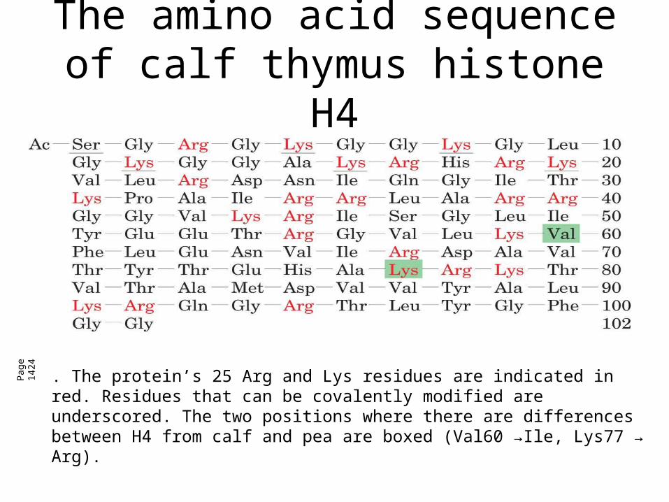
The amino acid sequence of calf thymus histone H4
Pag
e 14
24
. The protein’s 25 Arg and Lys residues are indicated in red. Residues that can be covalently modified are underscored. The two positions where there are differences between H4 from calf and pea are boxed (Val60 →Ile, Lys77 → Arg).
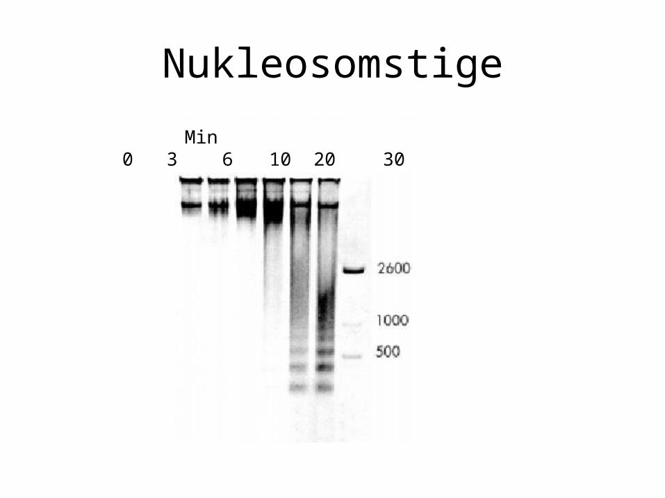
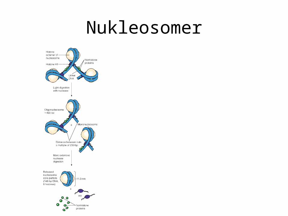
Nukleosomstige
Min0 3 6 10 20 30
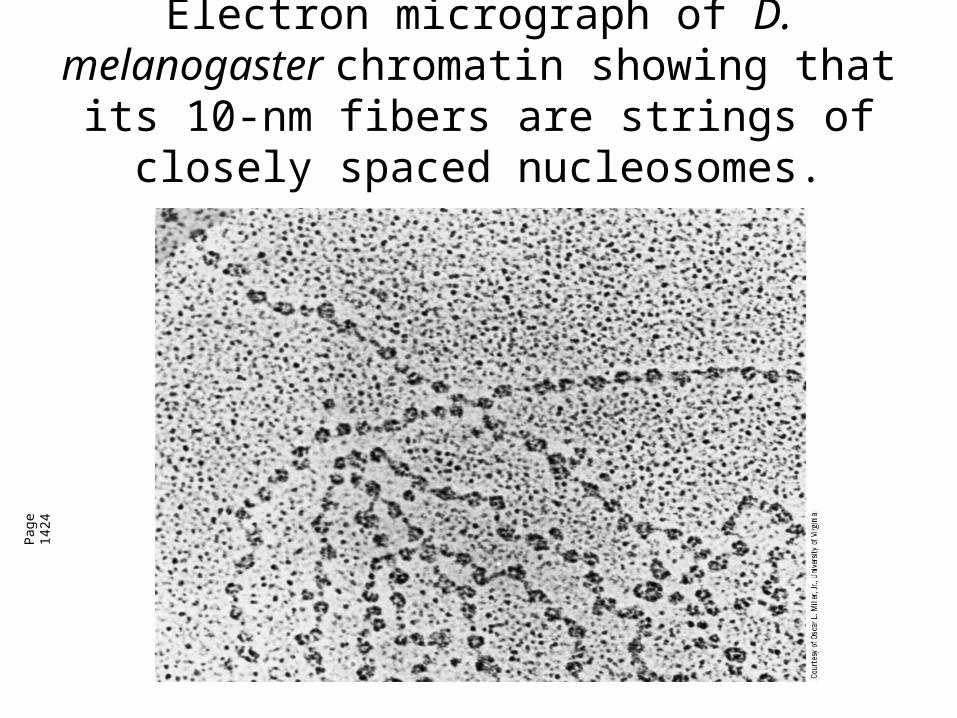
Electron micrograph of D. melanogaster chromatin showing that its 10-nm fibers are
strings of closely spaced nucleosomes.
Pag
e 14
24
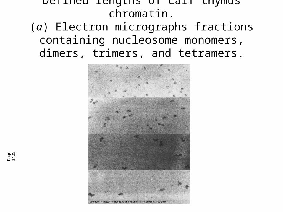
Defined lengths of calf thymus chromatin.(a) Electron micrographs fractions containing nucleosome monomers, dimers, trimers, and
tetramers.
Pag
e 14
25

Defined lengths of calf thymus chromatin.(b) Gel electrophoresis of DNA extracted from the nucleosome multimers are multiples of ~200 bp.
Pag
e 14
25
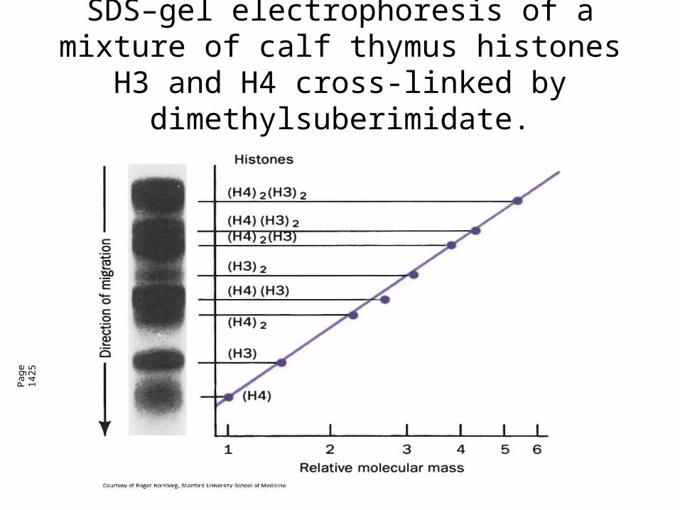
SDS–gel electrophoresis of a mixture of calf thymus histones H3 and H4 cross-linked by
dimethylsuberimidate.
Pag
e 14
25
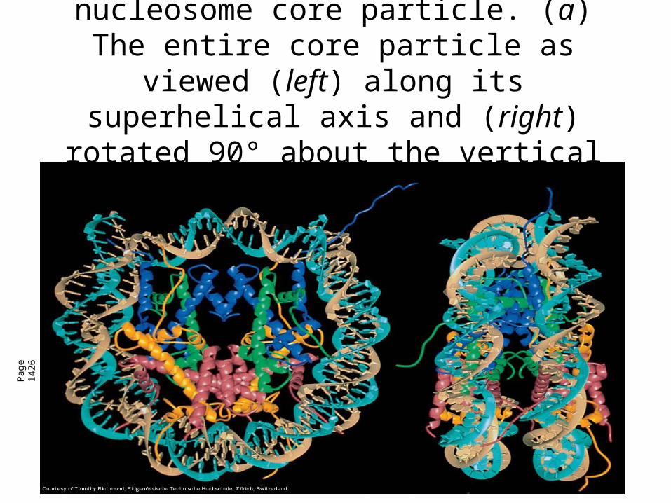
X-Ray structure of the nucleosome core particle. (a) The entire core particle as
viewed (left) along its superhelical axis and (right) rotated 90° about the vertical axis.
Pag
e 14
26
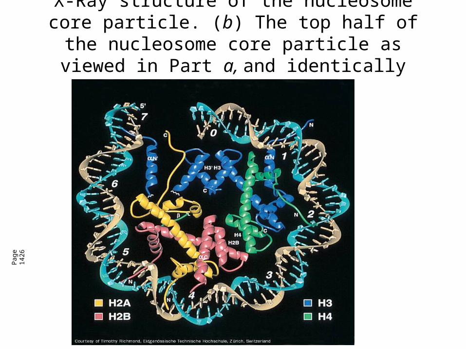
X-Ray structure of the nucleosome core particle. (b) The top half of the nucleosome core particle as
viewed in Part a, and identically colored.
Pag
e 14
26
Nukleosomer
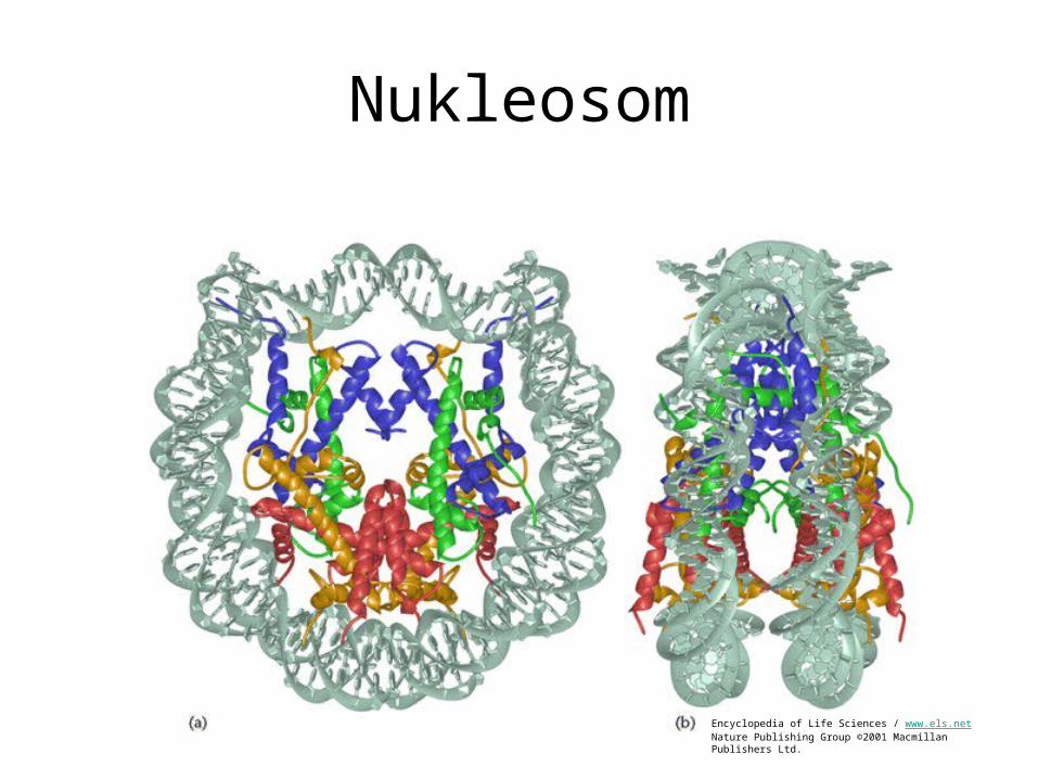
Nukleosom
Encyclopedia of Life Sciences / www.els.netNature Publishing Group ©2001 Macmillan Publishers Ltd.
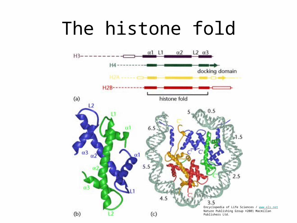
The histone fold
Encyclopedia of Life Sciences / www.els.netNature Publishing Group ©2001 Macmillan Publishers Ltd.
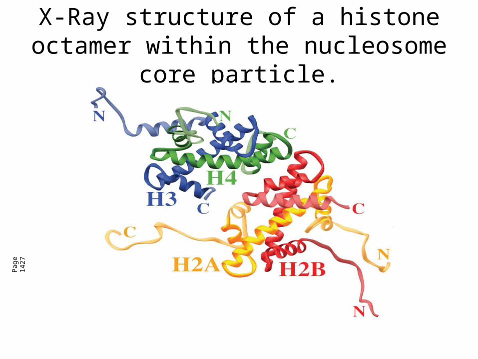
X-Ray structure of a histone octamer within the nucleosome core particle.
Pag
e 14
27
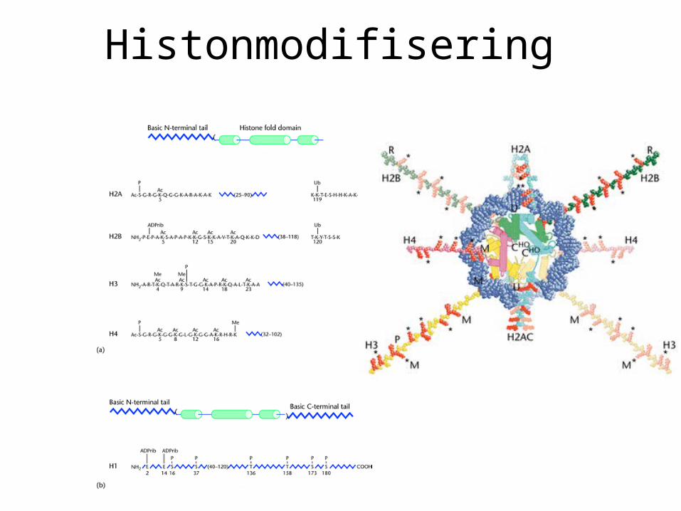
Histonmodifisering
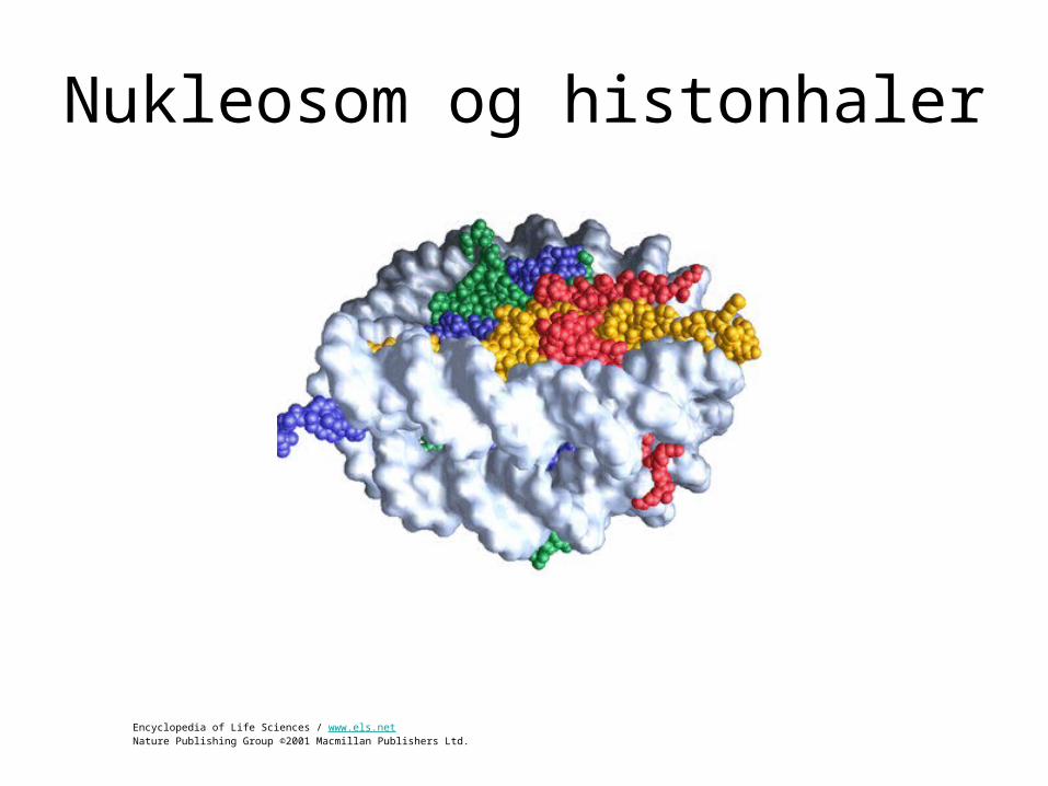
Nukleosom og histonhaler
Encyclopedia of Life Sciences / www.els.netNature Publishing Group ©2001 Macmillan Publishers Ltd.
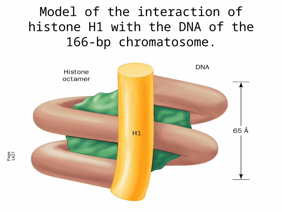
Model of the interaction of histone H1 with the DNA of the 166-bp chromatosome.
Pag
e 14
27
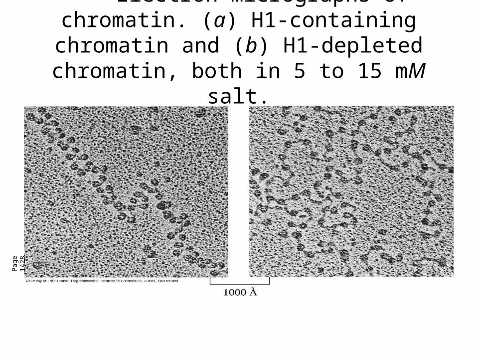
Electron micrographs of chromatin. (a) H1-containing chromatin and (b) H1-
depleted chromatin, both in 5 to 15 mM salt.
Pag
e 14
28
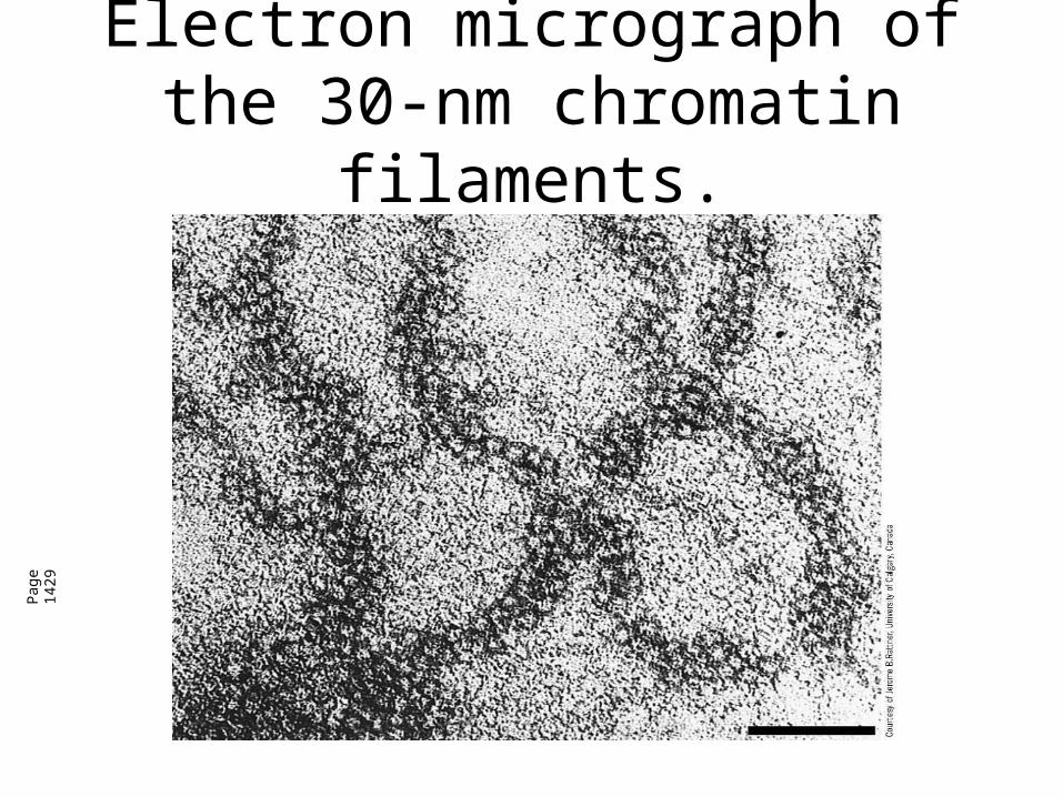
Electron micrograph of the 30-nm chromatin filaments.
Pag
e 14
29
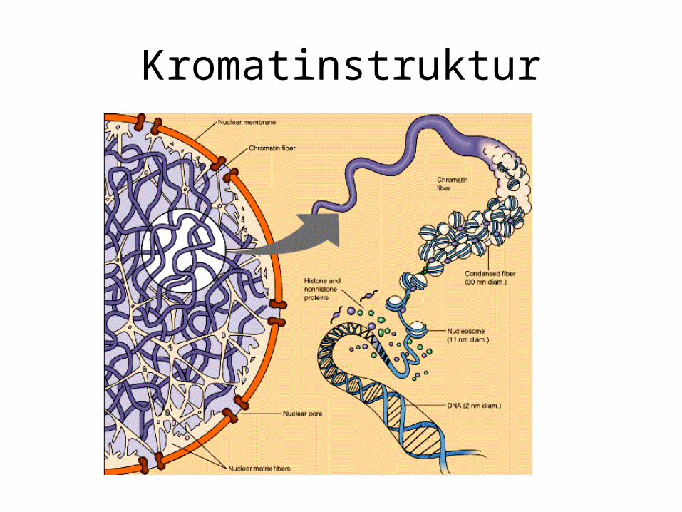
Kromatinstruktur
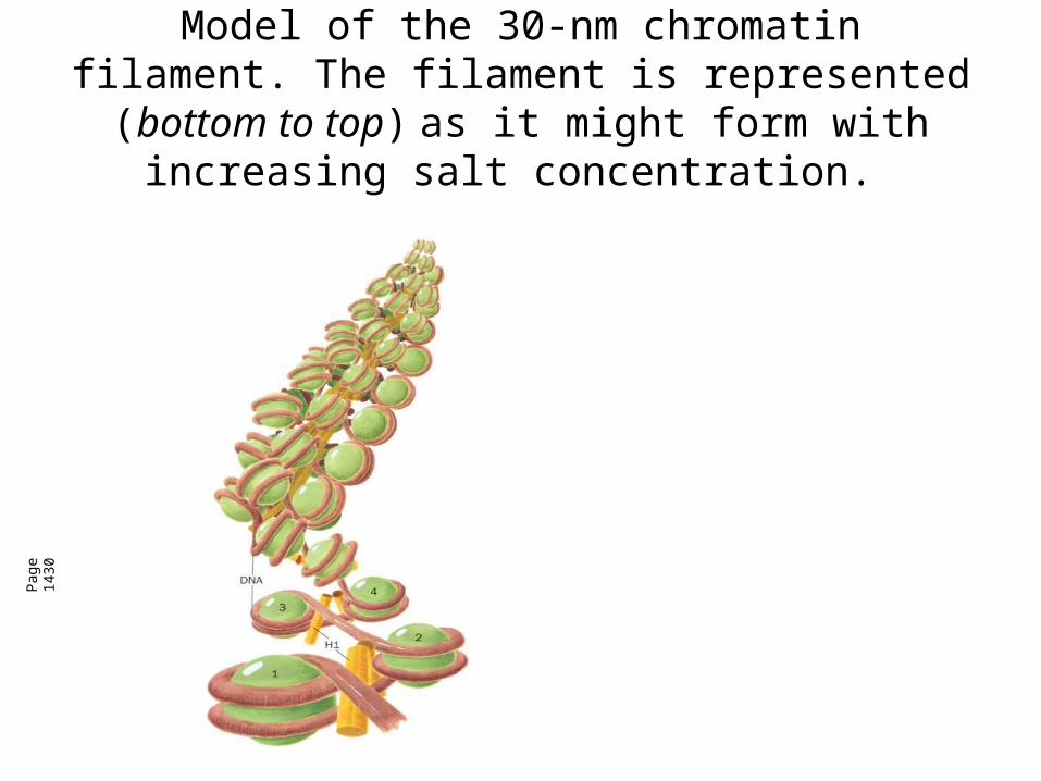
Model of the 30-nm chromatin filament. The filament is represented (bottom to top) as it might
form with increasing salt concentration.
Pag
e 14
30
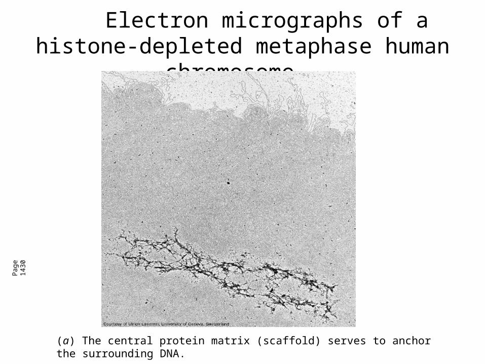
Electron micrographs of a histone-depleted metaphase human chromosome.
Pag
e 14
30
(a) The central protein matrix (scaffold) serves to anchor the surrounding DNA.
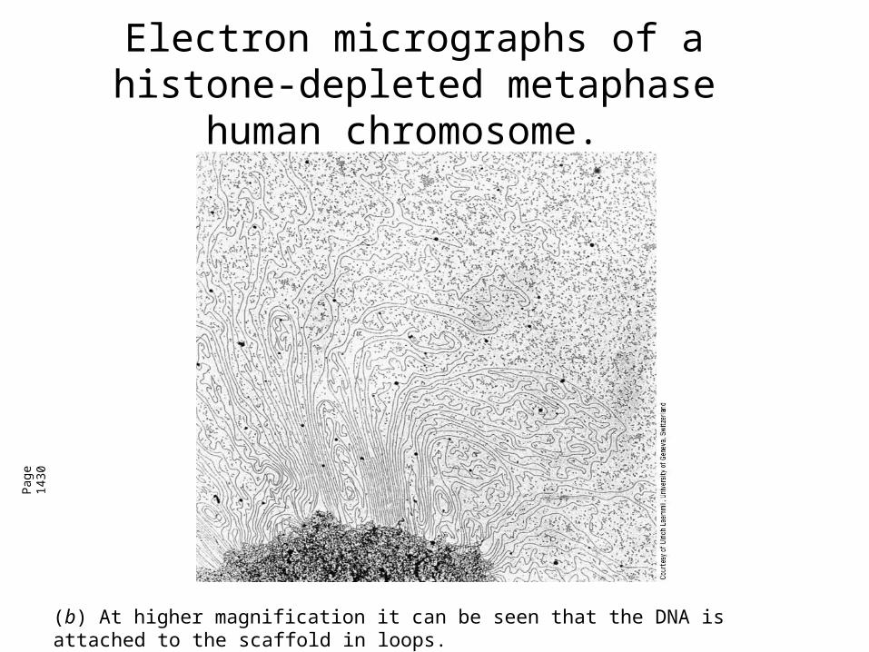
Electron micrographs of a histone-depleted metaphase human chromosome.
Pag
e 14
30
(b) At higher magnification it can be seen that the DNA is attached to the scaffold in loops.
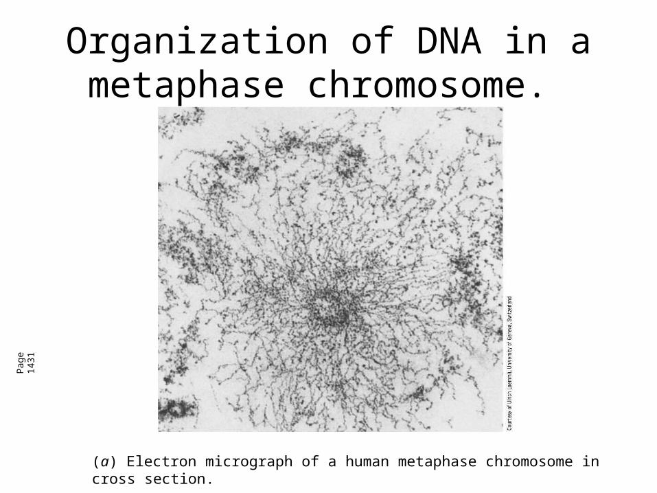
Organization of DNA in a metaphase chromosome.
Pag
e 14
31
(a) Electron micrograph of a human metaphase chromosome in cross section.
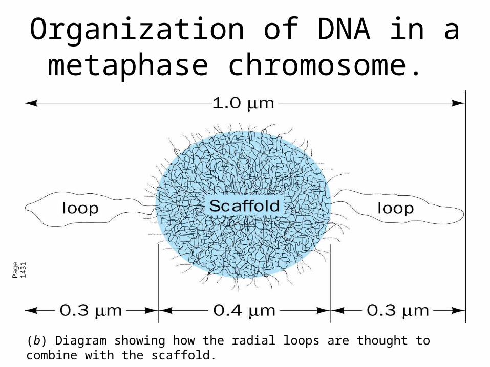
Organization of DNA in a metaphase chromosome.
Pag
e 14
31
(b) Diagram showing how the radial loops are thought to combine with the scaffold.
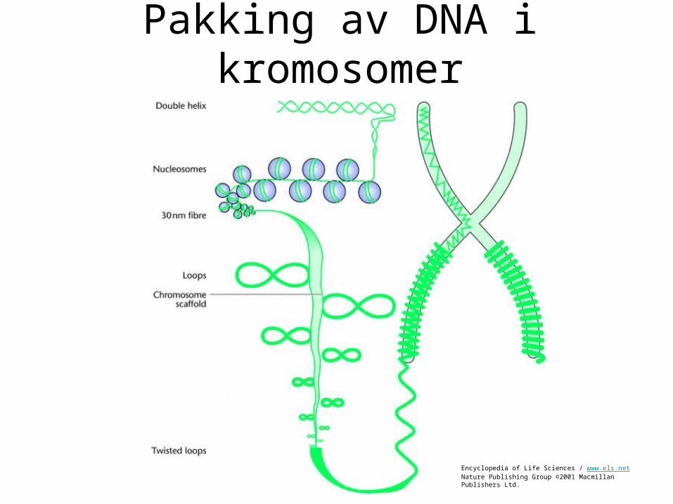
Pakking av DNA i kromosomer
Encyclopedia of Life Sciences / www.els.netNature Publishing Group ©2001 Macmillan Publishers Ltd.
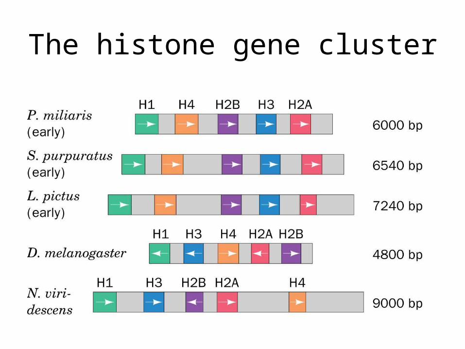
The histone gene cluster
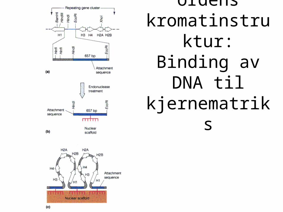
Høyere ordens kromatinstruktur: Binding av DNA til kjernematriks