Embed Size (px)
Citation preview
Retropupillary iris claw intraocular lens implantation in penetratingkeratoplasty patients.
Reshma Raj*, Vasanthi Kotian, Vijay Pai
Department of K.S. Hegde Charitable Hospital, Nithyanandanagar, Derlaketta, Mangalore, India.
Abstract
Objective-To evaluate the visual outcome and assess the complication rate after implantation of aretropupillary iris-claw Intraocular Lens (IOL) during Penetrating Keratoplasty (PKP).Method-A descriptive study on 15 eyes of 15 patients underwent penetrating keratoplasty along withretropupillary iris claw intraocular lens implantation. Reasons for penetrating keratoplasty includedpseudophakic bullous keratopathy, aphakic bullous keratopathy, and leukomatous corneal opacity.Mean follow up was 3 months for Visual Acuity (VA), Intraocular Pressure (IOP), graft clarity andany complications.RESULT-Of 15 eyes, visual acuity improvement was seen in 11 (74%) eyes and remained unchanged in4 (26%) eyes. Post operatively elevated IOP was seen in 8 (53%) eyes, which was managed medically.Improvement in graft clarity was noted in 7 (46%) eyes 1 week following the surgery.Conclusion-The results demonstrate that penetrating keratoplasty combined with retropupillary Irisclaw lens is an easy and effective method for the correction of aphakia in patients with no capsulesupport.
Keywords: Penetrating keratoplasty, Intraocular lens, Aphakia, Keratopathy.Accepted on October 03, 2018
IntroductionThe iris claw IOL attached to the anterior iris was developed byWorst in 1972 [1]. He has provided a technique to correctaphakia in the absence of capsular support and without directangle compromise. However, significant complication isdamage to endothelium [2], particularly in patients with narrowanterior chambers and in corneal transplantation. The techniquewas modifies by Brasse and Neuhann [3] by clipping the lensto the posterior iris, thereby protecting the endothelium, withthe A contant altered accordingly to 117.0 [4].
Intraocular lens implantation in eyes with pseudophakic oraphakic corneal edema and insufficient posterior capsularsupport is a surgical challenge [4]. The iris claw lens is fixatedto the iris without sutures which is a faster procedure whencompared to scleral fixating lens in combined penetratingkeratoplasty [4,5]. The iris claw lens has the advantage that itcan be fixated to the iris without sutures because the peripheraliris is incarcerated between the claws. Since an implantation ofiris claw lenses only takes a few minutes, the hypotonic opensky phase can be shortened compared to combined surgery withthe scleral fixation technique in cases of combined penetratingkeratoplasty [5,6].
The purpose of this study is to evaluate the visual outcome andassess the complication rate after implantation of aretropupillary iris-claw Intraocular Lens (IOL) duringPenetrating Keratoplasty (PKP).
MethodThis study was conducted during July 2016 to July 2017 at K.S. Hegde Hospital, Mangalore. Our study included 15 eyes of15 patients and all patients were operated by a single surgeon.Patients included were pseudophakic bullous keratopathy dueto Anterior Chamber Intraocular Lens (ACIOL), aphakicbullous keratopathy, leucomatous corneal opacity with lack ofposterior capsular support.
Patients underwent the following combined procedures:
1. Penetrating keratoplasty.
2. Anterior vitrectomy-if required.
3. Release of PAS- if needed.
4. PMMA iris claw lens fixation before graft suturing.
All patients underwent corneal trephination with trephine. Aftermaking side ports, the recipient corneal button was cut out withscissors (Figure 1). Removal of angle supported IOL implantedpreviously (Figure 2) was followed by anterior vitrectomy forfew cases. The retropupillary iris claw lens was then enclavedwith convex side down by an open sky approach on the midperipheral iris. The donor’s corneal button was then sutured torecipient bed with 10-0 nylon sutures. Post-operatively, patients
and then 3 months in the follow-up period (Figure 3). BestCorrected Visual Acuity (BCVA) and Intraocular Pressure(IOP) were noted and compared to preoperative data. The graftclarity was assessed by slit-lamp examination.
Research Article http://www.alliedacademies.org/clinical-ophthalmology-and-vision-science/
J Clin Ophthalmol 2018 Volume 2 Issue 292
were examined on the 1st , 7th , and 30th post-operative days,
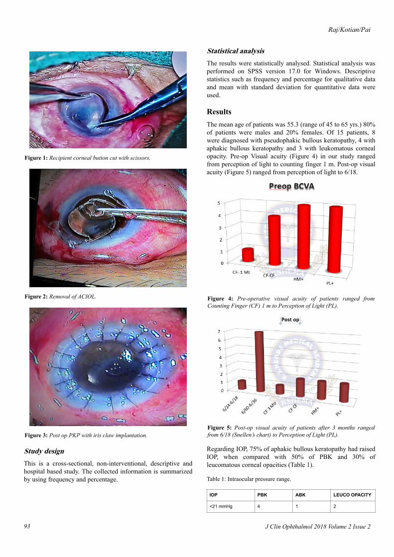
Figure 1: Recipient corneal button cut with scissors.
Figure 2: Removal of ACIOL.
Figure 3: Post op PKP with iris claw implantation.
Study designThis is a cross-sectional, non-interventional, descriptive andhospital based study. The collected information is summarizedby using frequency and percentage.
Statistical analysisThe results were statistically analysed. Statistical analysis wasperformed on SPSS version 17.0 for Windows. Descriptivestatistics such as frequency and percentage for qualitative dataand mean with standard deviation for quantitative data wereused.
ResultsThe mean age of patients was 55.3 (range of 45 to 65 yrs.) 80%of patients were males and 20% females. Of 15 patients, 8were diagnosed with pseudophakic bullous keratopathy, 4 withaphakic bullous keratopathy and 3 with leukomatous cornealopacity. Pre-op Visual acuity (Figure 4) in our study rangedfrom perception of light to counting finger 1 m. Post-op visualacuity (Figure 5) ranged from perception of light to 6/18.
Figure 4: Pre-operative visual acuity of patients ranged fromCounting Finger (CF) 1 m to Perception of Light (PL).
Figure 5: Post-op visual acuity of patients after 3 months rangedfrom 6/18 (Snellen’s chart) to Perception of Light (PL).
Regarding IOP, 75% of aphakic bullous keratopathy had raisedIOP, when compared with 50% of PBK and 30% ofleucomatous corneal opacities (Table 1).
Table 1: Intraocular pressure range.
IOP PBK ABK LEUCO OPACITY
<21 mmHg 4 1 2
Raj/Kotian/Pai
J Clin Ophthalmol 2018 Volume 2 Issue 293
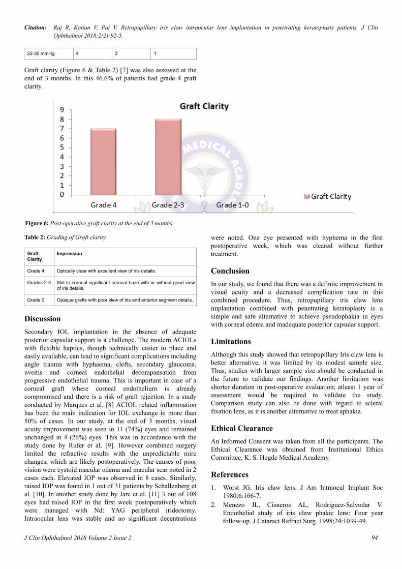
22-30 mmHg 4 3 1
Graft clarity (Figure 6 & Table 2) [7] was also assessed at theend of 3 months. In this 46.6% of patients had grade 4 graftclarity.
Figure 6: Post-operative graft clarity at the end of 3 months.
Table 2: Grading of Graft clarity.
GraftClarity
Impression
Grade 4 Optically clear with excellent view of iris details.
Grades 2-3 Mid to corneal significant corneal haze with or without good viewof iris details.
Grade 0 Opaque grafts with poor view of iris and anterior segment details.
DiscussionSecondary IOL implantation in the absence of adequateposterior capsular support is a challenge. The modern ACIOLswith flexible haptics, though technically easier to place andeasily available, can lead to significant complications includingangle trauma with hyphaema, clefts, secondary glaucoma,uveitis and corneal endothelial decompansation fromprogressive endothelial trauma. This is important in case of acorneal graft where corneal endothelium is alreadycompromised and there is a risk of graft rejection. In a studyconducted by Marques et al. [8] ACIOL related inflammationhas been the main indication for IOL exchange in more than50% of cases. In our study, at the end of 3 months, visualacuity improvement was seen in 11 (74%) eyes and remainedunchanged in 4 (26%) eyes. This was in accordance with thestudy done by Rufer et al. [9]. However combined surgerylimited the refractive results with the unpredictable mirechanges, which are likely postoperatively. The causes of poorvision were cystoid macular odema and macular scar noted in 2cases each. Elevated IOP was observed in 8 cases. Similarly,raised IOP was found in 1 out of 31 patients by Schallenberg etal. [10]. In another study done by Jare et al. [11] 3 out of 108eyes had raised IOP in the first week postoperatively whichwere managed with Nd: YAG peripheral iridectomy.Intraocular lens was stable and no significant decentrations
were noted. One eye presented with hyphema in the firstpostoperative week, which was cleared without furthertreatment.
ConclusionIn our study, we found that there was a definite improvement invisual acuity and a decreased complication rate in thiscombined procedure. Thus, retropupillary iris claw lensimplantation combined with penetrating keratoplasty is asimple and safe alternative to achieve pseudophakia in eyeswith corneal edema and inadequate posterior capsular support.
LimitationsAlthough this study showed that retropupillary Iris claw lens isbetter alternative, it was limited by its modest sample size.Thus, studies with larger sample size should be conducted inthe future to validate our findings. Another limitation wasshorter duration in post-operative evaluation; atleast 1 year ofassessment would be required to validate the study.Comparison study can also be done with regard to scleralfixation lens, as it is another alternative to treat aphakia.
Ethical ClearanceAn Informed Consent was taken from all the participants. TheEthical Clearance was obtained from Institutional EthicsCommittee, K. S. Hegde Medical Academy.
References1. Worst JG. Iris claw lens. J Am Intraocul Implant Soc
1980;6:166-7.2. Menezo JL, Cisneros AL, Rodriguez-Salvodar V.
Endothelial study of iris claw phakic lens: Four yearfollow-up. J Cataract Refract Surg. 1998;24:1039-49.
Citation: Raj R, Kotian V, Pai V. Retropupillary iris claw intraocular lens implantation in penetrating keratoplasty patients. J ClinOphthalmol 2018;2(2):92-5.
94J Clin Ophthalmol 2018 Volume 2 Issue 2

3. Brasse K, Neuhann TH. Posterior chamber verisyse lensimplantation to correct aphakia without capsular support.Video J Cataract Refract Surg. 2004;20.
4. Rijneveld WJ, Beekhuis WH, Hassman EF, et al. Iris clawlens: Anterior and posterior iris surface fixation in theabsence of capsular support during penetratingkeratoplasty. J Refract Corneal Surg. 1994;10:14-9.
5. Rüfer F, Saeger M, Nölle B, et al. Implantation ofretropupillar iris claw lenses with and without combinedpenetrating keratoplasty. Graefes Arch Clin ExpOphthalmol. 2009;247:457-62.
6. Kanellopoulos AJ. Penetrating keratoplasty and artisaniris-fixated intraocular lens implantation in themanagement of aphakic bullous keratopathy. Cornea.2004;23:220-4.
7. Shanbhag N, Cholera PP, Sahana N, et al. Evaluation ofgraft clarity post-penetrating keratoplasty. Int J Sci Study.2017;5:90-6.
8. Faria M, Pinto FN, Medeiros PJ, et al. Retropupillary irisclaw intraocular lens implantation in aphakia fordislocated intraocular lens (Corrigendum). Int Med CaseRep J. 2016;9:261-5.
9. Rüfer F, Saeger M, Nölle B, et al. Implantation ofretropupillar iris claw lenses with and without combinedpenetrating keratoplasty. Graefes Arch Clin ExpOphthalmol. 2008;247:457-62.
10. Schallenberg M, Dekowski D, Hahn A, et al. Aphakiacorrection with retropupillary fixated iris-claw lens(Artisan) long-term results. Clin Ophthalmol.2014;8:137-41.
11. Jare NM, Kesari AG, Gadkari SS, et al. The posterior iris-claw lens outcome study: 6-month follow-up. Indian JOphthalmol 2016;64:878-8.
Correspondence to:Reshma Raj
KS Hegde Medical Academy
Thaliparamba, Kannur
Kerala, India
E-mail: [email protected]
Tel: 00919995622881
Raj/Kotian/Pai
J Clin Ophthalmol 2018 Volume 2 Issue 295