Embed Size (px)
Citation preview
Retinopathy of Prematurity
ROP
PRESENTED BY
Maurice O. AdamsAlicia Lugo
Carolyn MetzgerAleida Valdez
ROP Retinopathy Of Prematurity is a
disorder of retinal blood vessel development in the premature infant.
The severe form is characterized by retinal vascular proliferation, scarring, retinal detachment, and blindness.
First diagnosed in 1942.
Major Causes Prematurity (younger than
32 weeks gestation) Low Birth Weight (about 2 ¾
lbs)
Need for oxygen in first week after birth
Unstable health immediately after birth

Other Contributing
Factors Respiratory distress Heart disease Infection Anemia Multiple Blood Transfusions Multiple births
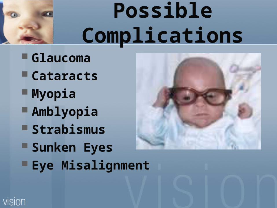
Possible Complications
Glaucoma Cataracts Myopia Amblyopia Strabismus Sunken Eyes Eye Misalignment
Signals of underlying problems
Holding things very close Difficulty seeing distant objects Favoring or winking one eye Reluctance to use one eye Poor vision (previously
undetected by the physician) Sudden decrease of vision Crossed or turned eye
ROP: Symptoms
White pupils (Leukocoria) Abnormal eye movements
(Nystagmus) Crossed eyes (Strabismus) Severe nearsightedness
(Myopia)
Effects of the condition on the
visual system Abnormal blood vessels may
develop which can lead to bleeding and scar tissue formation
This can stretch the retina and pull it out of position or cause it to detach completely
Blindness can occur
Development of ROP
In premature infants, the normal growth of blood vessels stops.
The area without adequate blood supply emits a chemical trigger to stimulate growth of the abnormal vessels.
These vessels lead to a formation of a ring of scar tissue attached to both the retina and the vitreous humor
As the scar contracts, it may pull on the retina creating a retinal detachment.
Stages of ROP
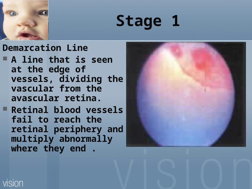
Stage 1
Demarcation Line A line that is seen at
the edge of vessels, dividing the vascular from the avascular retina.
Retinal blood vessels fail to reach the retinal periphery and multiply abnormally where they end .
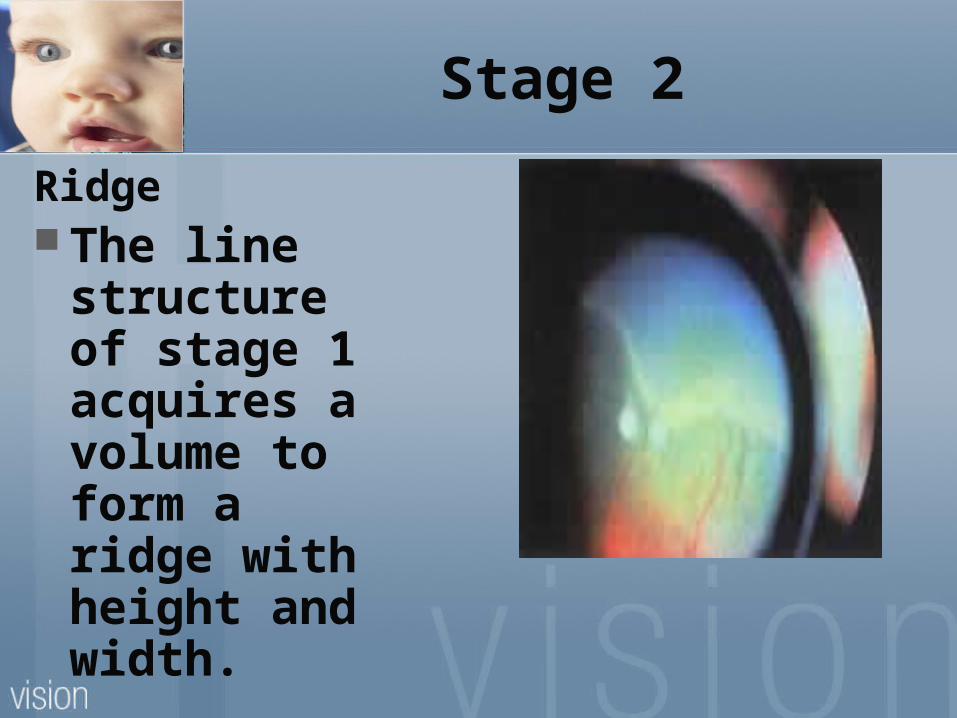
Stage 2
Ridge The line
structure of stage 1 acquires a volume to form a ridge with height and width.
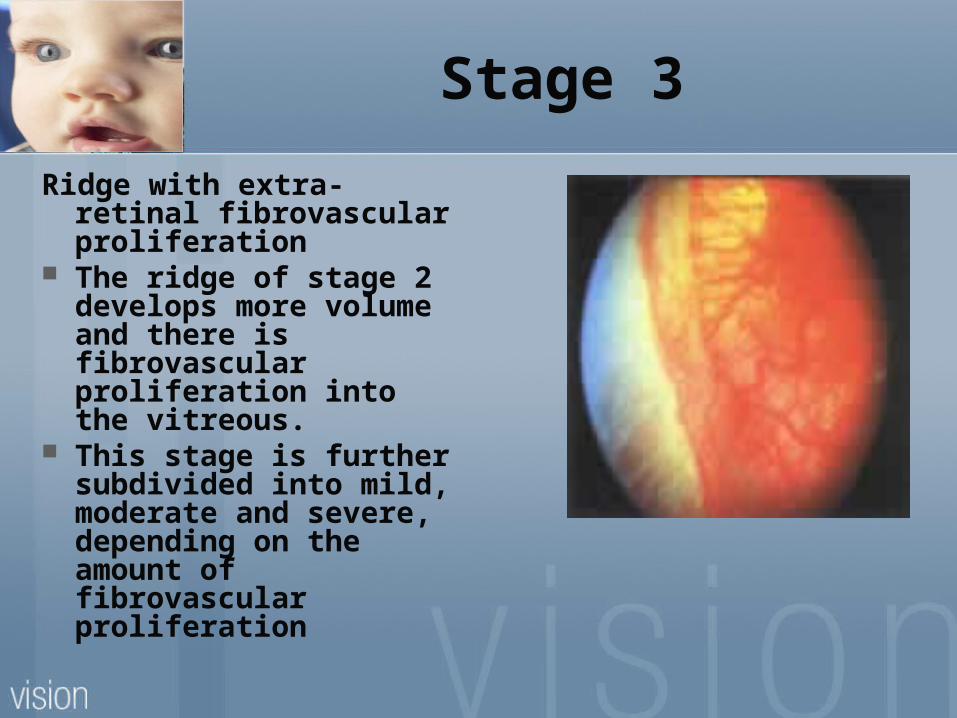
Stage 3
Ridge with extra-retinal fibrovascular proliferation
The ridge of stage 2 develops more volume and there is fibrovascular proliferation into the vitreous.
This stage is further subdivided into mild, moderate and severe, depending on the amount of fibrovascular proliferation
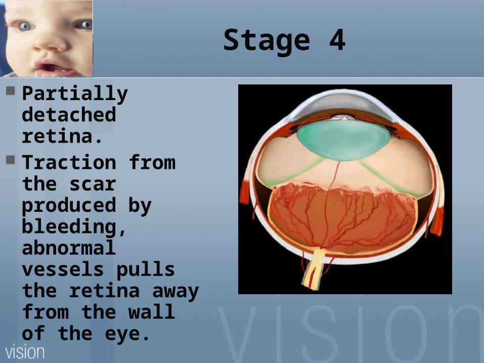
Stage 4
Partially detached retina.
Traction from the scar produced by bleeding, abnormal vessels pulls the retina away from the wall of the eye.
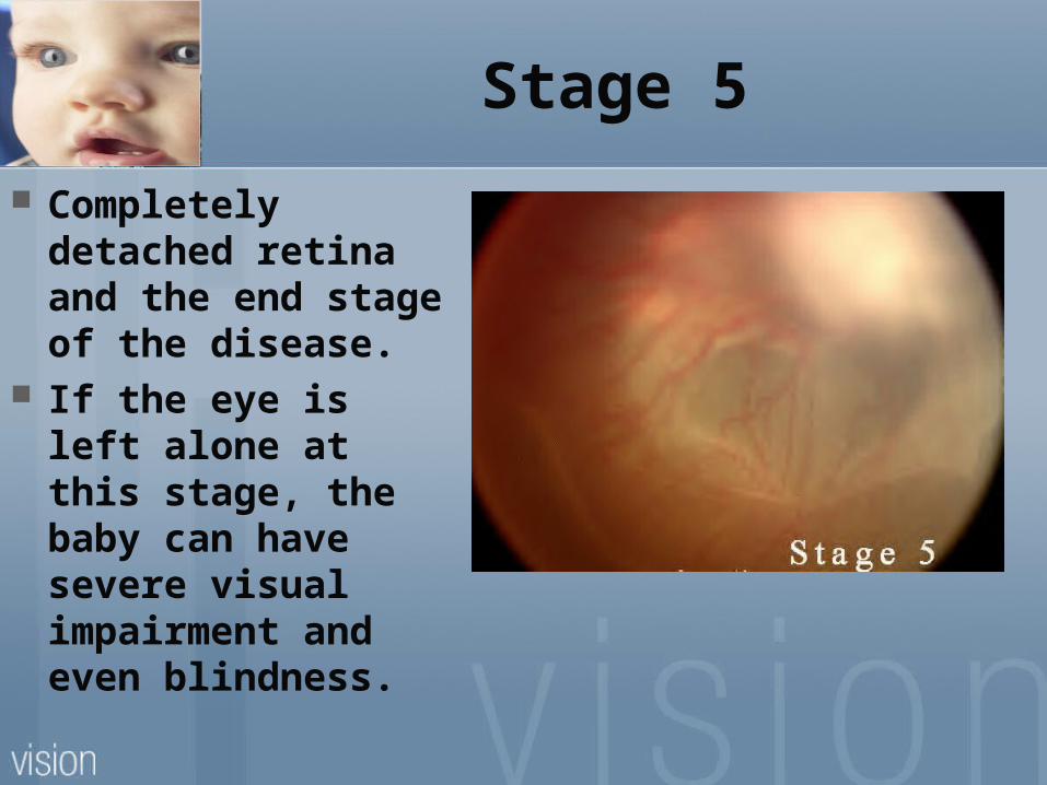
Stage 5
Completely detached retina and the end stage of the disease.
If the eye is left alone at this stage, the baby can have severe visual impairment and even blindness.
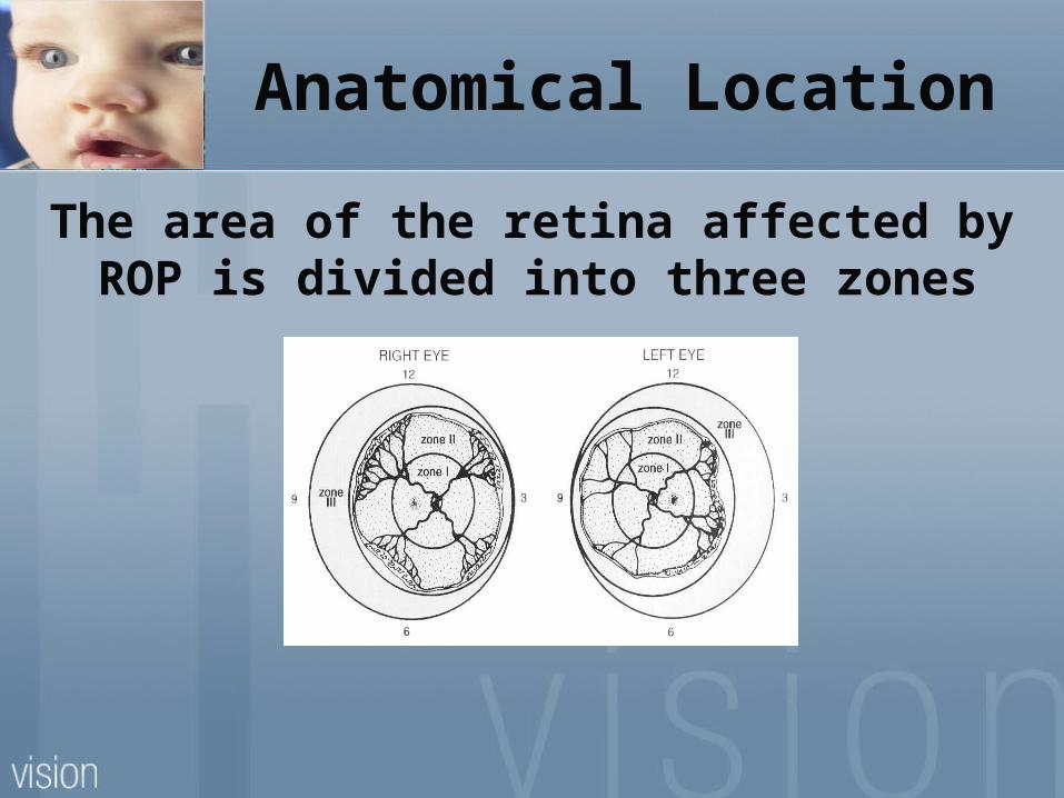
Anatomical Location
The area of the retina affected by ROP is divided into three zones
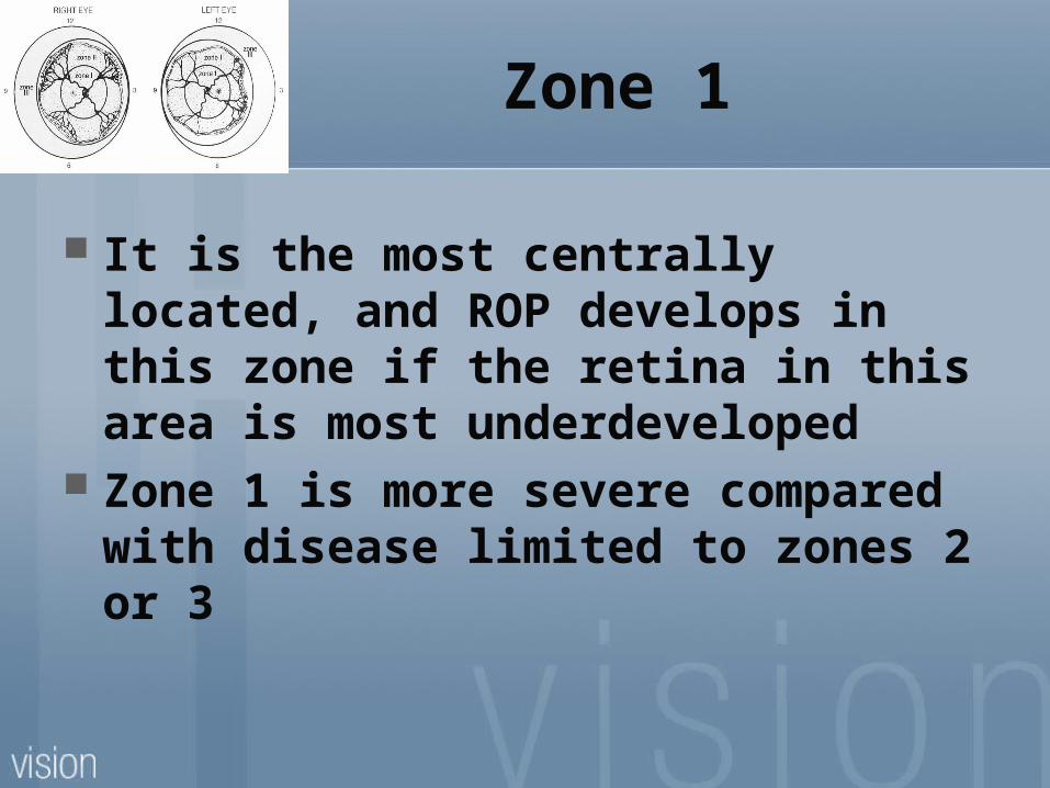
Zone 1
It is the most centrally located, and ROP develops in this zone if the retina in this area is most underdeveloped
Zone 1 is more severe compared with disease limited to zones 2 or 3
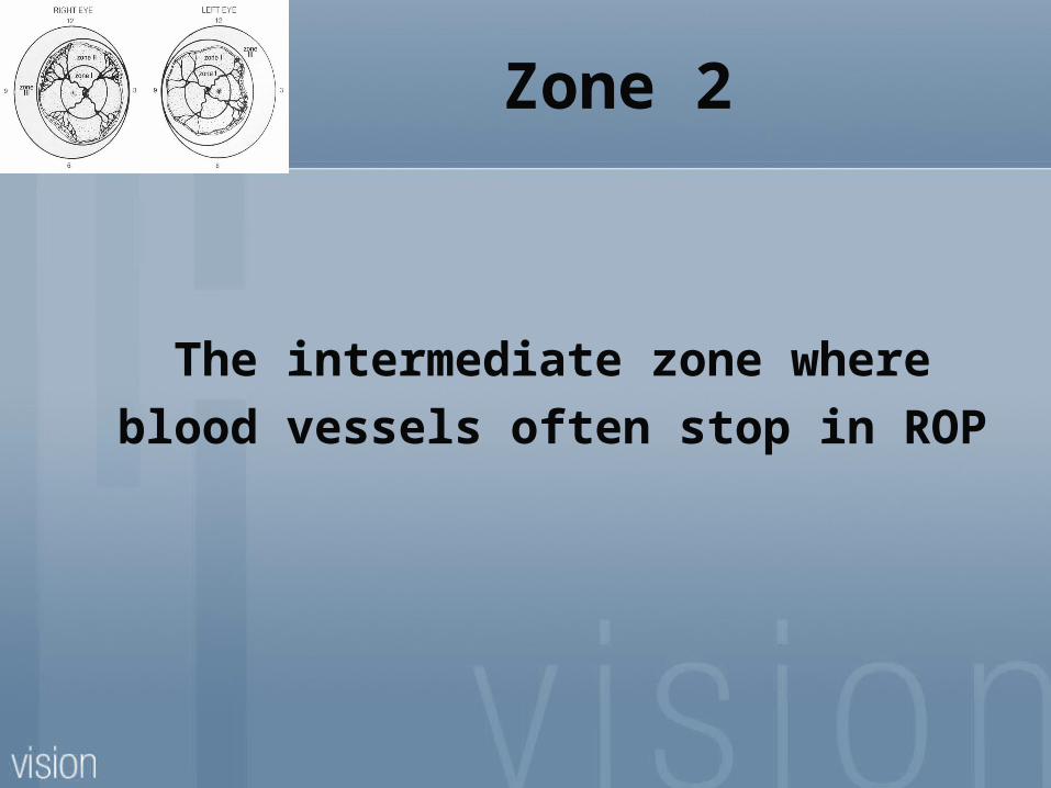
Zone 2
The intermediate zone where blood vessels often stop in ROP
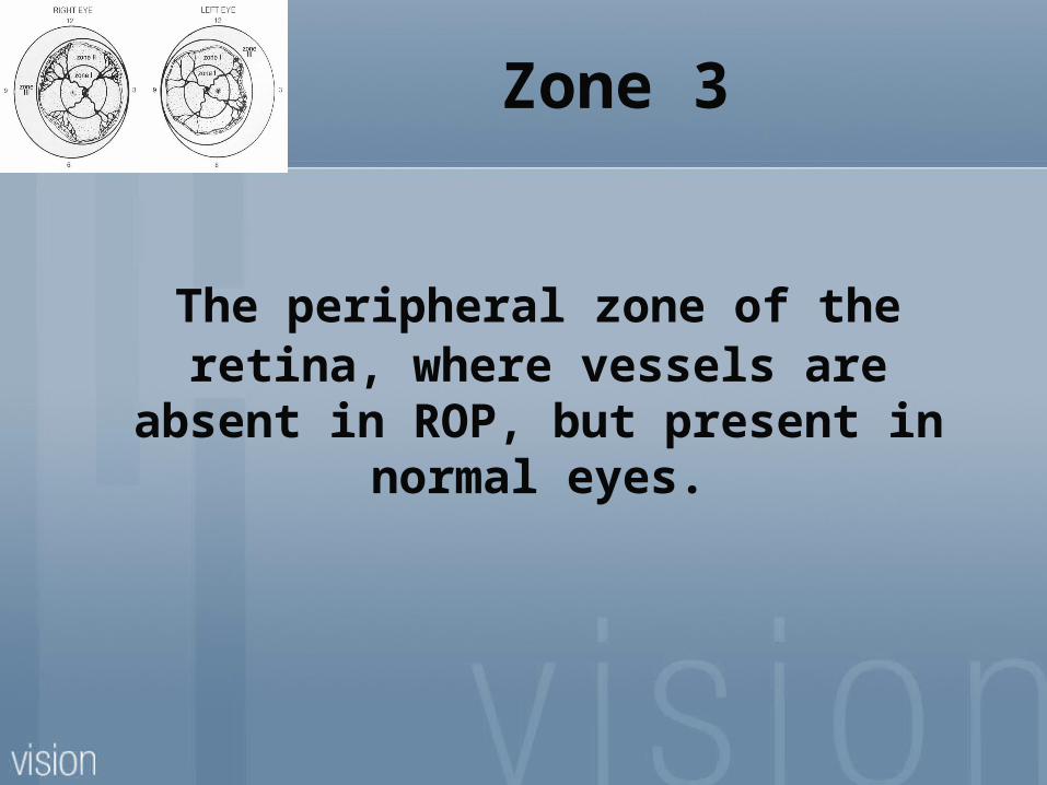
Zone 3
The peripheral zone of the retina, where vessels are absent in ROP,
but present in normal eyes.
ROP: Diagnosis
The only way to diagnose that baby has ROP is an eye exam by an ophthalmologist at 4 weeks of age.
The timing of examination and follow-up are important factors in diagnosis and treatment .
Common Treatments
Cryotherapy of the retina can reduce more severe complications by removing the stimulus for the growth of the abnormal blood vessels.
Laser surgery
ROP: Treatment
Treatment for ROP depends on the stage and severity of the condition.
The milder stages of the disease typically resolve themselves on their own, and do not require treatment.
If the disease has progressed to a point where the baby's vision is at risk, treatment is required
Treatment Goals
The treatment’s goal is to destroy the retina that is deprived of retinal vessels.
This helps to shrink the new vessels and prevents the formation of dense scars that usually follow
Laser Photocoagulation
Laser photocoagulation is the most common treatment.
A laser is directed to a designated spot to destroy abnormal vessels and seal leaks.
Laser photocoagulation is the preferred method of treatment by surgeons, because there is little postoperative pain and swelling
Cryotherapy Cryotherapy can be used to treat
threshold ROP but is not preferred It involves destroying abnormal
tissue by freezing and is often used to treat Stage III ROP
Cryotherapy reduces the risk for retinal detachment from 43% to 21%.
Drawback Cryotherapy also causes significant
swelling of the eye and eyelid, which makes postoperative assessment difficult.
Other treatments Scleral buckle is placing a silicon
band used to prevent pulling on the scar tissue and the retina. It is used for severe stage 4 and stage 5 retinopathies.
Vitrectomy is a complex procedure, involving the use of microscopic instruments to remove the vitreous from the eye and replacing it with a saline (salt) solution. Used only in Stage 5.
Discussion The incidence of ROP in moderately
premature infants has decreased dramatically with better care in the neonatal intensive care unit and better prenatal care.
However, this has led to high rates of survival of very premature infants who would have had little chance of survival in the past.
Since these very premature infants are at the highest risk of developing ROP, the condition may actually be becoming more common again.
Prevention
The most effective prevention of retinopathy of prematurity is prevention of premature birth.
Case Study Zachary is a 16 year old boy with ROP. He was born at 24 weeks of gestation and
had a birth weight of 1 lb. 5oz. He was diagnosed with ROP and
secondary glaucoma shortly after birth and is in stage 5 of the disease with no vision.
Prognosis: Permanent Vision Loss He is a very talented musician. He sings
the National Anthem at sporting events and is a Disc Jockey.

Zachary

Zachary
Cassin, Barbara . & Solomon, S. (1990). Dictionary of Eye Terminology 5th edition. Gainesville, FL: Triad Publishing Co.
Goldberg, S. (1991). Ophthalmology Made Ridiculously Simple. Miami: MedMaster, Inc.
Region IV ESC Resource. (2004). Program in Low Vision Therapy. Houston, TX: Education Service Center Region IV.
Resources
Emedicine from WebMD. “Retinopathy of Prematurity”. May 4, 2006. http://emedicine.com/ped/topic1998.htm.
Moss, Kate. “Retinopathy of Prematurity”. TSBVI Deafblind Outreach. Sept. 4,
2003. http://www.tsbvi.edu/Outreach/seehear/winter98/rop.htm
National Eye Institute. “The Early Treatment for Retinopathy of Prematurity Study (ETROP)”. Oct 2004. http://
www.nei.nih.gov/rop/photos.asp. Windsor, Richard L. & Windsor L. Vision
Enhancement Journal. “Understanding Retinopathy of Prematurity”. 7/25/07. http://lowvision.org/retinopathy_of_prematurityxx.htm.
Websites