Embed Size (px)
Citation preview
Retina Mosaicing Using Local Features
Philippe C. Cattin, Herbert Bay, Luc Van Gool, and Gabor Szekely
ETH Zurich - Computer Vision LaboratorySternwartstrasse 7, CH - 8092 Zurich
{cattin,bay,vangool,szekely}@vision.ee.ethz.ch
Abstract. Laser photocoagulation is a proven procedure to treat vari-ous pathologies of the retina. Challenges such as motion compensation,correct energy dosage, and avoiding incidental damage are responsiblefor the still low success rate. They can be overcome with improved in-strumentation, such as a fully automatic laser photocoagulation system.In this paper, we present a core image processing element of such a sys-tem, namely a novel approach for retina mosaicing. Our method relieson recent developments in region detection and feature description toautomatically fuse retina images. In contrast to the state-of-the-art theproposed approach works even for retina images with no discernable vas-cularity. Moreover, an efficient scheme to determine the blending masksof arbitrarily overlapping images for multi-band blending is presented.
1 Introduction
With the dramatic demographic shift towards an older population, age-relatedeye diseases, such as Glaucoma and Macular Degeneration, is about to becomeincreasingly prevalent and to develop into a serious public health issue. However,age-related eye diseases are not the only emerging causes of visual loss. Diabeticretinopathy that can be directly linked to the increase of diabetes in the de-veloped world has already surpassed age-related diseases as the main cause ofblindness.
All the aforementioned potential causes of blindness can be treated with laserphotocoagulation. The small bursts from the laser cause controlled damage thatcan be used to either seal leaky blood vessels, destroy abnormal blood vessels,reattach retinal tears or destroy abnormal tumorous tissue on the retina. Photo-coagulation laser surgery is an outpatient procedure. The treatment is performedwhile the patient is seated in a chair and eye drops will be given to dilate thepupil, immobilise and anesthetise the eye. The laser treatment itself is currentlyperformed manually and lasts from a few minutes up to half an hour, dependingon the type of treatment and number of laser spots to apply. Although photoco-agulation is the most widely used treatment for the aforementioned conditions,the success rate is still below 50% for both the first treatment and the possiblere-treatments [1]. Many of these failures can be attributed to the manual natureof the procedure, in particular to the difficulty to quickly respond to rapid eyeand head movements of the patient that can not be completely stopped, and
2
the problems to quantitatively measure and control the applied laser energy.As it takes several weeks before knowing if laser surgery has been successful orre-treatment is required to prevent further vision loss, the success rate of theprocedure has to be maximised. Additionally, the visual recovery declines witheach re-treatment.
A fully automatic surgery under controllable and reproducible conditionswould therefore be desirable. Computer vision methods for mosaicing retinaimages would be a useful tool to achieve these goals. Such mosaics can be usedon the one hand for diagnosis and interventional planning, and on the otherhand as a spatial map for the laser guidance system during treatment.
Many retinal image registration methods have been proposed in the litera-ture [2–4]. Can et al. proposed in [5] a benchmarking algorithm for the auto-matic stitching of retinal images. They used bifurcations of the vascular treeon the retinal surface to establish image correspondences. These bifurcationsare easy to detect but sometimes lack distinctiveness, and their localisation isoften quite inaccurate. To improve the landmark extraction Tsai proposed in[6] a model-based approach that greatly increased the accuracy and repeatabil-ity of estimating the locations where vascular structures branch or cross over.Stewart proposed in [7] a different approach that uses one or more initial cor-respondences defining the mapping only in a small area around these bootstrapregions. In each of these regions the transformations are iteratively refined usingonly information from this same area. The region is then expanded and testedto see if a higher order transformation model can be used. The expansion stopswhen the entire overlapping region of the images is covered. Finally, in [8] Tsaievaluated the performance of the two methods proposed by Can and Stewart ina clinically-oriented framework.
All the previously mentioned methods are limited to cases with clearly visiblevascular structures. Quite often, however, bleedings or tumorous tissue limits thenumber of detectable bifurcations or even prevents their proper segmentation,see Fig. 3(a) for an example.
The work described below is part of a project aiming towards a fully au-tomatic laser photocoagulation system. As will be argued, a key component ofsuch a system is the automatic mosaicing method capable to stitch and blend thecaptured retina images. The method should be robust against all morphologiesseen in the various pathologies subjected to laser photocoagulation.
2 Methods
Recent interest point detection/description schemes, such as SIFT [9] have broughtabout important progress in image mosaicing [10, 11]. Yet, in our experiments,these state-of-the-art methods failed to identify sufficient number of reliable, dis-tinct landmarks allowing to build retinal mosaics. The use of our very recentlydeveloped framework [12] proved more successful.
The method for obtaining a mosaic image from multiple images of the retina,using region detectors/descriptors, can be summarised with the following steps.
3
(1) Interest points are detected at different scales and their feature descriptorsare calculated in each image individually. (2) The region descriptors are robustlymatched for each image pair to find the overlapping images using the second-nearest-neighbour ratio matching strategy [13]. (3) A graph theoretical algorithmis used to find the anchor image that is connected to all the other images throughthe shortest path. (4) The mappings from all images into the coordinate systemof the anchor image are linearly estimated and then globally optimised. (5) Allimages are warped into the coordinate frame of the anchor image and multi-bandblending is used to seamlessly fuse the individual retina images. The subsequentparagraphs describe the aforementioned steps in more detail.
(1) Interest point detection and feature descriptors: The first task for image mo-saicing is the identification of a possibly high number of interest points in allimages of the same scene. As described in the introduction, the previously pre-sented retina mosaicing methods used the branches and crossovers of the vasculartree as interest points. Although this approach works very well it is bound to failfor pathological retinas, where the vascular tree is seriously occluded by bleedingor tumorous tissue, see Fig. 3(a) for an example. Only our recent developmentsin interest point detection and characterisation [12] made the application of thistechnology possible for the highly self-similar regions generally seen in retina im-ages. Our algorithm proved to be superior to the state-of-the-art methods withregard to repeatability and stability. It uses a Hessian-matrix based detector inorder to identify blob-like interest points and a 128-dimensional distribution-based vector to describe the neighbourhood of every interest point.
Given a set of N input images I1, ..., IN for the construction of the mosaic,let each image Ii have a number Ni of interest points. As a first step the im-age coordinates (u, v) of these interest points are extracted yielding pi,1, ...pi,Ni .Their respective feature descriptors are defined by ci,1, ..., ci,Ni . Once the interestpoints have been found, the overlapping images can be identified by establishingthe correspondences between all image pairs.
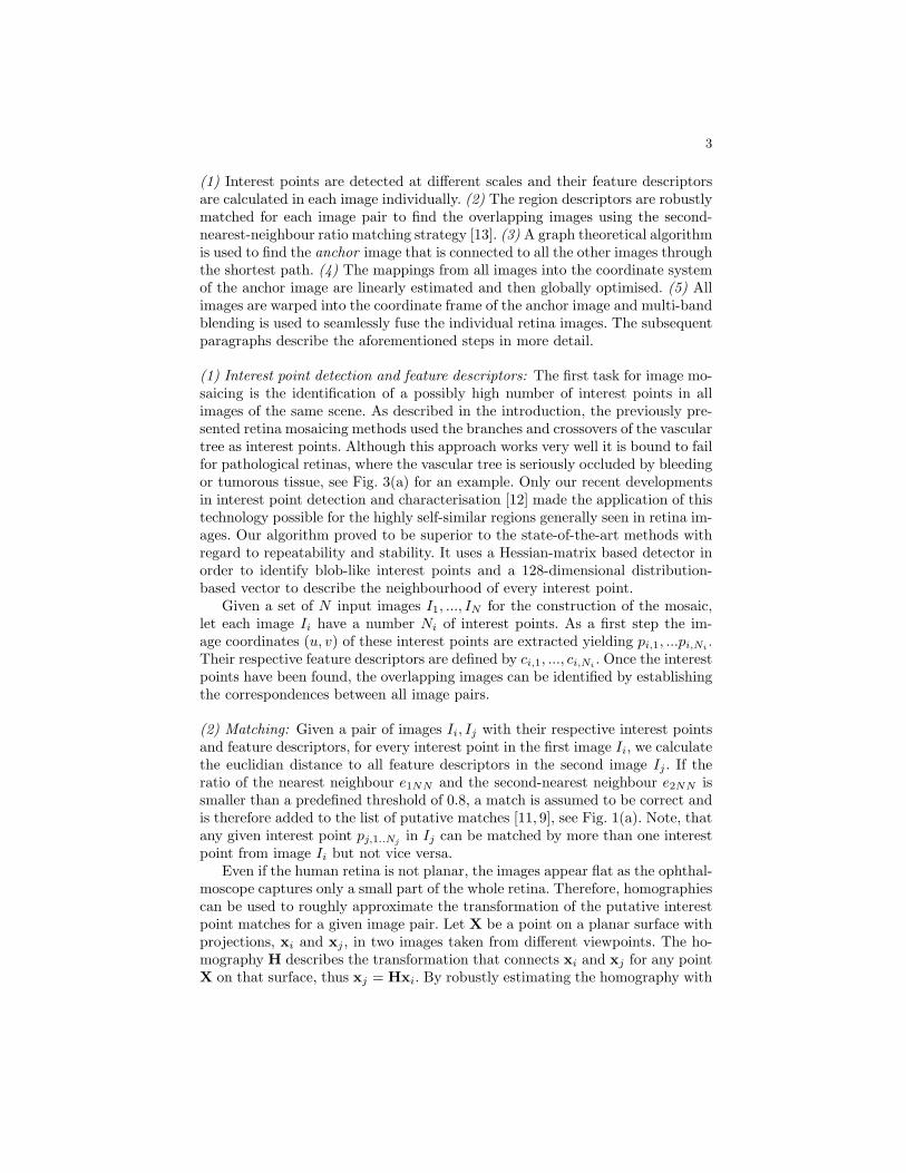
(2) Matching: Given a pair of images Ii, Ij with their respective interest pointsand feature descriptors, for every interest point in the first image Ii, we calculatethe euclidian distance to all feature descriptors in the second image Ij . If theratio of the nearest neighbour e1NN and the second-nearest neighbour e2NN issmaller than a predefined threshold of 0.8, a match is assumed to be correct andis therefore added to the list of putative matches [11, 9], see Fig. 1(a). Note, thatany given interest point pj,1..Nj in Ij can be matched by more than one interestpoint from image Ii but not vice versa.
Even if the human retina is not planar, the images appear flat as the ophthal-moscope captures only a small part of the whole retina. Therefore, homographiescan be used to roughly approximate the transformation of the putative interestpoint matches for a given image pair. Let X be a point on a planar surface withprojections, xi and xj , in two images taken from different viewpoints. The ho-mography H describes the transformation that connects xi and xj for any pointX on that surface, thus xj = Hxi. By robustly estimating the homography with
4
the classical RANSAC scheme [14], the transfer error d(xj , Hxi) can be used tofilter out possible mismatches from the list of putative matches.
As the initial matches are paired using a very conservative feature vectorcomparison, many pairs are not matched in the first step. The advantage ofthis initial conservative selection is that the ratio of inliers vs. outliers is betterand thus a correct homography can be found using RANSAC. Once the initialhomography is established, more correspondences can be found with a guidedmatching method. Knowing the homography, the final correspondences are es-tablished by ignoring the feature descriptors and just matching interest pointsif they are spatially close enough, see Fig. 1(b) for an example.
(3) Anchor Image Selection: Once the correspondences between all image pairshave been determined, the anchor image IA is identified. The selection of theanchor image among the available images IA ∈ I1, ...IN , plays a crucial role inimage mosaicing as the anchor defines the base coordinate system towards whichall other images are warped. A central image having direct correspondenceswith all other ones in the data set would be an ideal candidate. However, suchan image usually does not exist. The next logical candidate would thus be animage that connects to all other images through the least number of connections,see Fig. 1(c). Graph theoretical methods [15] allow to calculate the minimumdistances between nodes given the connectivity matrix. If more than one nodeis connected to the rest with the minimal number of connections, the node withthe highest number of correspondences with its neighbouring images is selected.
(a) (b)
I5
I2 I1
I6I3
IA
(c)
Fig. 1. (a) Correspondences after matching SURF features (b) after the guided match-ing. (c) Example graph of the connected images after pairwise matching.
(4) Mapping Estimation: Even if overlapping regions of image pairs can berelated with homographies, a planar transformation model for the mosaic wouldresult in a false representation of distances and angles. The curved nature of theretina can best be taken into account by using a quadric transformation modelΘ as proposed by Can et al. in [4]. It transforms a point x = (x, y)> of an inputimage to a point x′ = (x′, y′)> in the anchor image IA coordinate system.
5
(x′
y′
)=
(θ11 θ12 θ13 θ14 θ15 θ16
θ21 θ22 θ23 θ24 θ25 θ26
)
x2
xyy2
xy1
(1)
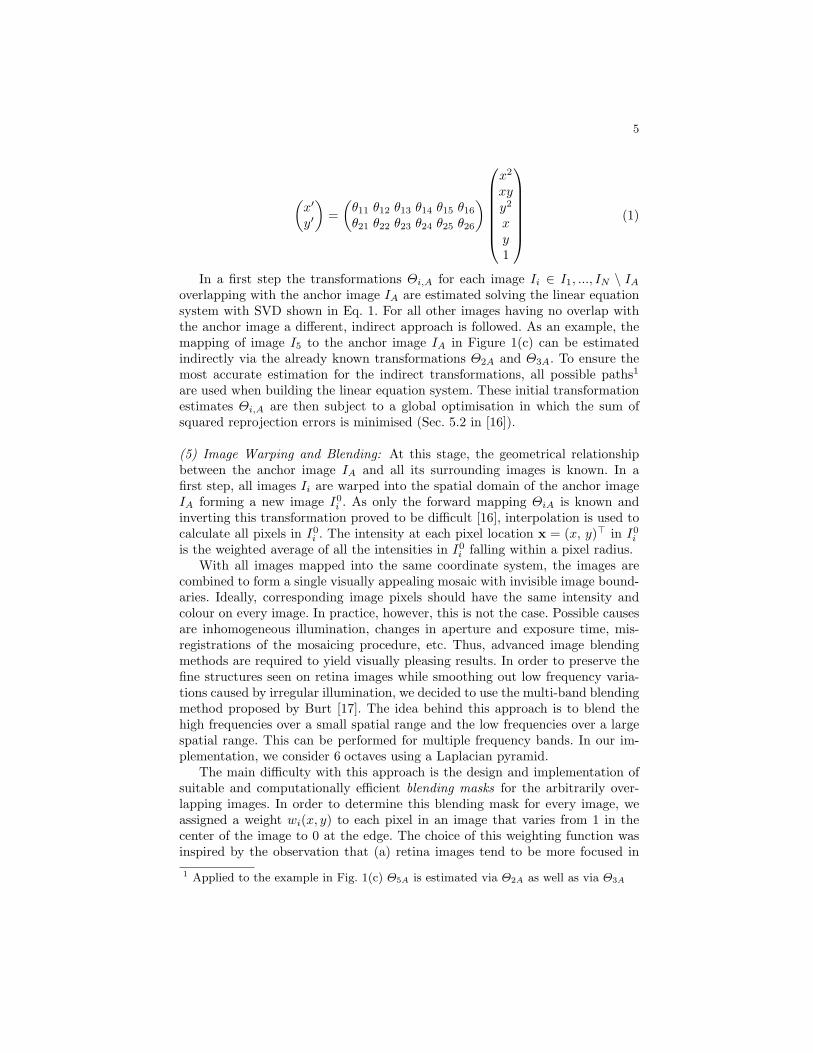
In a first step the transformations Θi,A for each image Ii ∈ I1, ..., IN \ IA
overlapping with the anchor image IA are estimated solving the linear equationsystem with SVD shown in Eq. 1. For all other images having no overlap withthe anchor image a different, indirect approach is followed. As an example, themapping of image I5 to the anchor image IA in Figure 1(c) can be estimatedindirectly via the already known transformations Θ2A and Θ3A. To ensure themost accurate estimation for the indirect transformations, all possible paths1
are used when building the linear equation system. These initial transformationestimates Θi,A are then subject to a global optimisation in which the sum ofsquared reprojection errors is minimised (Sec. 5.2 in [16]).
(5) Image Warping and Blending: At this stage, the geometrical relationshipbetween the anchor image IA and all its surrounding images is known. In afirst step, all images Ii are warped into the spatial domain of the anchor imageIA forming a new image I0
i . As only the forward mapping ΘiA is known andinverting this transformation proved to be difficult [16], interpolation is used tocalculate all pixels in I0
i . The intensity at each pixel location x = (x, y)> in I0i
is the weighted average of all the intensities in I0i falling within a pixel radius.
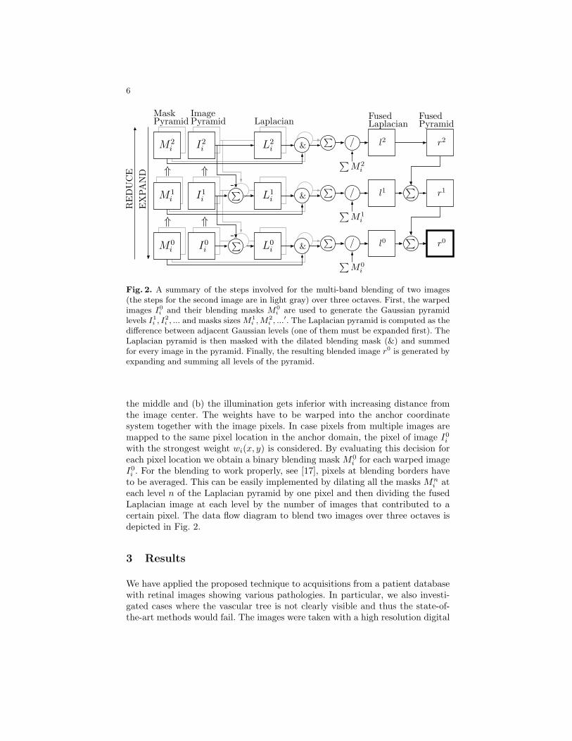
With all images mapped into the same coordinate system, the images arecombined to form a single visually appealing mosaic with invisible image bound-aries. Ideally, corresponding image pixels should have the same intensity andcolour on every image. In practice, however, this is not the case. Possible causesare inhomogeneous illumination, changes in aperture and exposure time, mis-registrations of the mosaicing procedure, etc. Thus, advanced image blendingmethods are required to yield visually pleasing results. In order to preserve thefine structures seen on retina images while smoothing out low frequency varia-tions caused by irregular illumination, we decided to use the multi-band blendingmethod proposed by Burt [17]. The idea behind this approach is to blend thehigh frequencies over a small spatial range and the low frequencies over a largespatial range. This can be performed for multiple frequency bands. In our im-plementation, we consider 6 octaves using a Laplacian pyramid.
The main difficulty with this approach is the design and implementation ofsuitable and computationally efficient blending masks for the arbitrarily over-lapping images. In order to determine this blending mask for every image, weassigned a weight wi(x, y) to each pixel in an image that varies from 1 in thecenter of the image to 0 at the edge. The choice of this weighting function wasinspired by the observation that (a) retina images tend to be more focused in
1 Applied to the example in Fig. 1(c) Θ5A is estimated via Θ2A as well as via Θ3A
6
& /P
& /P
& /P
PyramidMask
M2i
M1i
I0iM0
i
I1i
I2i
∑
∑
LaplacianFused
r1
r0
r2l2
l1
l0L0i
L1i
L2i ∑
M2i
∑M0
i
∑M1
i
LaplacianImagePyramid
P
P⇑ ⇑
⇑ ⇑
RE
DU
CE
EX
PAN
D
PyramidFused
-
-
Fig. 2. A summary of the steps involved for the multi-band blending of two images(the steps for the second image are in light gray) over three octaves. First, the warpedimages I0
i and their blending masks M0i are used to generate the Gaussian pyramid
levels I1i , I2
i , ... and masks sizes M1i , M2
i , ...′. The Laplacian pyramid is computed as thedifference between adjacent Gaussian levels (one of them must be expanded first). TheLaplacian pyramid is then masked with the dilated blending mask (&) and summedfor every image in the pyramid. Finally, the resulting blended image r0 is generated byexpanding and summing all levels of the pyramid.
the middle and (b) the illumination gets inferior with increasing distance fromthe image center. The weights have to be warped into the anchor coordinatesystem together with the image pixels. In case pixels from multiple images aremapped to the same pixel location in the anchor domain, the pixel of image I0
i
with the strongest weight wi(x, y) is considered. By evaluating this decision foreach pixel location we obtain a binary blending mask M0
i for each warped imageI0i . For the blending to work properly, see [17], pixels at blending borders have
to be averaged. This can be easily implemented by dilating all the masks Mni at
each level n of the Laplacian pyramid by one pixel and then dividing the fusedLaplacian image at each level by the number of images that contributed to acertain pixel. The data flow diagram to blend two images over three octaves isdepicted in Fig. 2.
3 Results
We have applied the proposed technique to acquisitions from a patient databasewith retinal images showing various pathologies. In particular, we also investi-gated cases where the vascular tree is not clearly visible and thus the state-of-the-art methods would fail. The images were taken with a high resolution digital
7
camera 2256× 2032 pixels, but scaled to half their size (i.e. 1128× 1016 pixels)for all the tests reported in this publication.
We evaluated the robustness of the matching step that automatically findsoverlapping image pairs using a set of 100 pairs. The matching method failed in6 cases. All of these cases had an overlap of less than 20.2% (average 11.4%). Onthe other hand it successfully managed to match 12 image pairs with an overlapof 20% or less (average 14.3%). Per image pair an average of 311 correspondences(maximum 1763, minimum 9) were found.
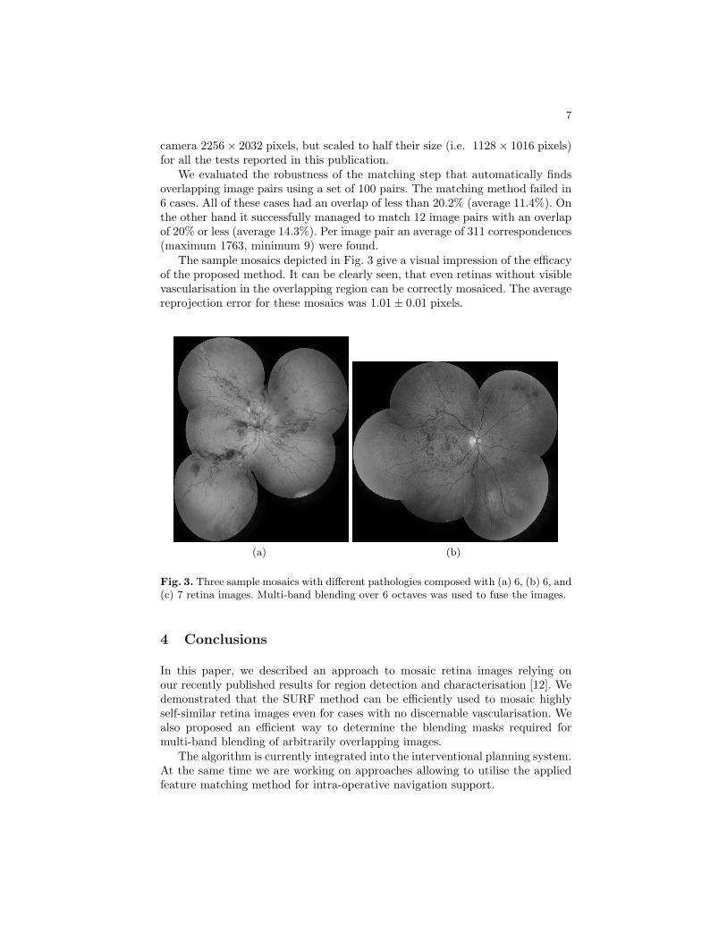
The sample mosaics depicted in Fig. 3 give a visual impression of the efficacyof the proposed method. It can be clearly seen, that even retinas without visiblevascularisation in the overlapping region can be correctly mosaiced. The averagereprojection error for these mosaics was 1.01± 0.01 pixels.
(a) (b)
Fig. 3. Three sample mosaics with different pathologies composed with (a) 6, (b) 6, and(c) 7 retina images. Multi-band blending over 6 octaves was used to fuse the images.
4 Conclusions
In this paper, we described an approach to mosaic retina images relying onour recently published results for region detection and characterisation [12]. Wedemonstrated that the SURF method can be efficiently used to mosaic highlyself-similar retina images even for cases with no discernable vascularisation. Wealso proposed an efficient way to determine the blending masks required formulti-band blending of arbitrarily overlapping images.
The algorithm is currently integrated into the interventional planning system.At the same time we are working on approaches allowing to utilise the appliedfeature matching method for intra-operative navigation support.
8
Acknowledgments
This work has been supported by the CO-ME/NCCR research network of theSwiss National Science Foundation (http://co-me.ch). We thank the UniversityHospital in Berne, Switzerland for providing the retina images.
References
1. Zimmer-Galler, I., Bressler, N., Bressler, S.: Treatment of choroidal neovascular-ization: updated information from recent macular photocoagulation study groupreports. Int. ophthalmology clinics 35 (1995) 37–57
2. Hart, W.E., Goldbaum, M.H.: Registering retinal images using automatically se-lected control point pairs. In: Proc. IEEE International Conference on ImageProcessing (ICIP). Volume 3. (1994) 576–81
3. Becker, D.E., Can, A., Turner, J.N., Tanenbaum, H.L., Roysam, B.: Image pro-cessing algorithms for retinal montage synthesis, mapping, and real-time locationdetermination. IEEE Trans. on Biomedical Engineering 45 (1998) 105–18
4. Can, A., Stewart, C.V., Roysam, B.: Robust hierarchical algorithm for construct-ing a mosaic from images of the curved human retina. In: IEEE Conference onComputer Vision and Pattern Recognition. Volume 2. (1999) 286–92
5. Can, A., Stewart, C.V., Roysam, B., Tanenbaum, H.L.: A feature-based techniquefor joint, linear estimation of highorder image-to-mosaic transformations: Mosaic-ing the curved human retina. IEEE Trans. Pattern Anal. Mach. Intell. 24 (2002)412–419
6. Tsai, C.L., Stewart, C.V., Tanenbaum, H.L., Roysam, B.: Model-based method forimproving the accuracy and repeatability of estimating vascular bifurcations andcrossover from retinal fundus images. IEEE Transactions on Information Technol-ogy in Biomedicine 8 (2004) 122–30
7. Stewart, C.V., Tsai, C.L., Roysam, B.: The dual-bootstrap iterative closest pointalgorithm with application to retinal image registration. IEEE Trans. Med. Imag-ing 22 (2003) 1379–1394
8. Tsai, C.L., Majerovics, A., Stewart, C.V., Roysam, B.: Disease-oriented evaluationof dual-bootstrap retinal image registration. Lect Note Comput Sci 2878 (2003)754–761
9. Lowe, D.G.: Distinctive image features from scale-invariant keypoints. Interna-tional Journal of Computer Vision (2004)
10. Brown, M., Lowe, D.G.: Recognising panoramas. In: 10th International Conferenceon Computer Vision. (2003) 1218–25
11. Brown, M., Szeliski, R., Winder, S.: Multi-image matching using multi-scale ori-ented patches. In: International Conference on Computer Vision and PatternRecognition (CVPR). (2005) 510–517
12. Bay, H., Tuytelaars, T., Gool, L.V.: SURF: Speeded up robust features. In: Eu-ropean Conference on Computer Vision (ECCV). Volume 3951 of LNCS. (2006)404–17
13. Mikolajczyk, K., Schmid, C.: A performance evaluation of local descriptors. Trans-actions on Pattern Analysis and Machine Vision (2005) Accepted to PAMI.
14. Hartley, R., Zisserman, A.: Multiple View Geometry in Computer Vision. Cam-bridge University Press (2000)
15. Diestel, R.: Graph Theory. New York: Springer-Verlag (1997)
9
16. Can, A., Stewart, C., Roysam, B., Tanenbaum, H.: A feature-based technique forjoint, linear estimation of high-order image-to-mosaic transformations: Applicationto mosaicing the curved human retina. In: IEEE Conf. on Computer Vision andPattern Recognition. Volume 2. (2000) 585–91
17. Burt, P.J., Adelson, E.H.: A multiresolution spline with application to imagemosaics. ACM Transactions on Graphics 2 (1983) 217–36