Embed Size (px)
Citation preview

3/9/12
1
Rethinking Risk and Benefit in Dental and Maxillofacial Imaging –
Dose matters
John B Ludlow, DDS, MS, FDS RCSEd University of North Carolina, Chapel Hill, NC
Background • Radiation risk is frequently front page news
recent example - Japan’s Fukushima Nuclear Plant – following an Earthquake & Tsunami - 3/11/2011
• Our patients are concerned and often confused about risk associated with different exposures
• We can help by being well informed

3/9/12
2
Learning objectives: • Identify the risks from ionizing radiation • Describe options in CBCT units which
affect dose • Discuss importance of matching options
to objectives of imaging • Explore ways to reduce patient risk • Explain how to talk about risks with
patients
What are the risks?
Gamma bomb exposure
Radioactive spider bite

3/9/12
3
Stochastic vs Deterministic Effects
Stochastic effects
• A linear-no-threshold hypothesis of x ray risk fits most data for cancer development but is extrapolated to doses below • 100 mGy (adult exposure) • 10-20 mG (fetal exposure)
• No expressions of germ cell mutations have been observed in human populations
Deterministic effects (Non stochastic effects)
• Threshold for – in-utero birth defects
100-250 mSv – Cataracts
2-5 Gy – radiation burns
3 Gy (reddening) – radiation mucositis
30+ Gy (therapy typically 60-80 Gy)
Source contribution to total effective dose (6.2 mSv) per capita in the US - 2006
6
Source % µSv Ubiquitous background 50 3100 Medical 48 2976 Consumer 2 124
From NCRP REPORT No. 160, 2009
3/9/12
4
CT procedures per year - US
From NCRP REPORT No. 160, 2009
Annual growth of >10% per year
Background NEJM 11-29-2007 Computed Tomography – An Increasing Source of Radiation Exposure David Brenner, et al.
From 1.5% to 2% of all cancers in the United States may be attributable to the radiation from CT studies

3/9/12
5
Background: CT & CBCT Effective Doses (2007 ICRP)
• Large FOV CBCT scans 68 – 1073 µSv
• Medium FOV CBCT scans 69 – 560 µSv
• Small FOV CBCT scans 189 – 652 µSv
• Medium FOV MDCT scans 534 – 860 µSv Ludlow JB, Ivanovic M. Comparative Dosimetry of Dental CBCT Devices and 64 row CT for Oral and
Maxillofacial Radiology Oral Surg Oral Med Oral Pathol Oral Radiol Endodont 2008;106:930-938
Dose and Risk Estimation • 1990 Recommendations of the
International Commission on Radiological Protection – Effective dose calculation (Sv) – Summed doses to weighted organs &
tissues known to be most susceptible to radiation damage
– Mathematical expression: E = ∑ wT x HT

3/9/12
6
ICRP 2007 Recommendations
• 2007 Recommendations of the ICRP – Reassessment of Risk based on cancer
incidence data from the Life Span Study of Japanese atomic bomb survivors
– Revision of list of tissues
– Adjustment of weights
Effective dose: E = ∑ wT x HT
* Adrenals, brain, upper large intestine, small intestine, kidney, muscle, pancreas, spleen, thymus, uterus † Adrenals, Extrathoracic region, Gall bladder, Heart, Kidneys, Lymphatic nodes, Muscle, Oral Mucosa, Pancreas, Prostate, Small Intestine, Spleen, Thymus, and Uterus/cervix.
Tissue weighting factors for calculation of Effective Dose – Comparison of 1990 and 2007 ICRP Recommendations
Tissue
19901 wT
20072
wT
Bone marrow 0.12 0.12
Breast 0.05 0.12
Colon 0.12 0.12
Lung 0.12 0.12
Stomach 0.12 0.12
Bladder 0.05 0.04
Esophagus 0.05 0.04
Gonads 0.20 0.08
Liver 0.05 0.04
Thyroid 0.05 0.04
Bone surface 0.01 0.01
Brain remainder 0.01
Salivary glands - 0.01
Skin 0.01 0.01
Remainder Tissues 0.05* 0.12† 1. 1990 Recommendations of the ICRP. Publication 60. Ann ICRP 1991; 21: 1-201 2. 2007 Recommendations of the ICRP.
Publication 103. Ann ICRP 2007; 37: 1-332

3/9/12
7
Summary of changes ICRP 1990 – 2007
• 4 additional weighted tissues • 10% increase in weight of tissues located in
maxillofacial area • 28% increase in weight adjusted for
distribution of tissues in maxillofacial area – 3 of the newly weighted tissues are entirely
within the maxillofacial area: oral mucosa, extrathoracic region, and salivary glands
Effective Dose and Detriment (Risk) Calculation
• Detriment includes the weighted probabilities – fatal and non-fatal cancer – relative length of life lost – hereditary effects
• Cancer risk alone may be used for the 2007 ICRP risk estimates for dental radiography
Cancer Risk = E(Sv) x 0.055 Cancer risks for children are 2 or more times greater than for adults

3/9/12
8
How do we measure dose? • Effective Dose calculation preferred • Human phantom studies
– Expensive • Simple acrylic phantoms – CTDIVOL
– Easy but inaccurate • Monte Carlo modeling
– Promising but model and software dependent • Dose area Product x coefficient for head/neck
exposure – Easy but inaccurate
RANDO
2345679
LevelsLevel 6
3/9/12
9
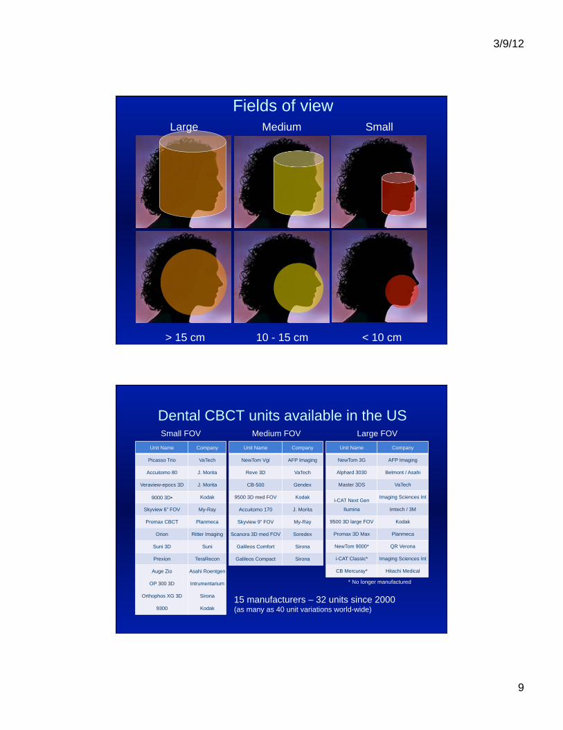
Fields of view Large Medium Small
> 15 cm 10 - 15 cm < 10 cm
Dental CBCT units available in the US
Unit Name Company
Picasso Trio VaTech
Accuitomo 80 J. Morita
Veraview-epocs 3D J. Morita
9000 3D• Kodak
Skyview 6” FOV My-Ray
Promax CBCT Planmeca
Orion Ritter Imaging
Suni 3D Suni
Prexion TeraRecon
Auge Zio Asahi Roentgen
OP 300 3D Intrumentarium
Orthophos XG 3D Sirona
9300 Kodak
Unit Name Company
NewTom Vgi AFP Imaging
Reve 3D VaTech
CB-500 Gendex
9500 3D med FOV Kodak
Accuitomo 170 J. Morita
Skyview 9” FOV My-Ray
Scanora 3D med FOV Soredex
Galileos Comfort Sirona
Galileos Compact Sirona
Unit Name Company
NewTom 3G AFP Imaging
Alphard 3030 Belmont / Asahi
Master 3DS VaTech
i-CAT Next Gen Imaging Sciences Int
Ilumina Imtech / 3M
9500 3D large FOV Kodak
Promax 3D Max Planmeca
NewTom 9000* QR Verona
i-CAT Classic* Imaging Sciences Int
CB Mercuray* Hitachi Medical
Small FOV Medium FOV Large FOV
15 manufacturers – 32 units since 2000 (as many as 40 unit variations world-wide)
* No longer manufactured

3/9/12
10
Field of view
• Note that detector sizes need to be larger than the FOV due to image magnification
• Image Intensifiers produce spherical FOVs
• Flat panels produce cylendrical FOVs
• Cylinders typically provide larger useful FOVs than spheres. Cylinder height ≠ Sphere diameter
Large FOV Units ex: Kodak 9500
• 60 - 90 kV
• 2 - 15 mA
• Pulsed
• Flat Panel large FOV version (18 cm x 20 cm)

3/9/12
11
Large FOV CBCT Dose Calculations (Based on ICRP 2007 Recommendations)
Large FOV Techniques Effective Dose in
µSv
Dose as multiple of average†
Panoramic Dose
Days of per capita back-
ground*
Probability of x in a million fatal cancer‡
NewTom3G – Large FOV 68 4 8 4
CB Mercuray – Facial FOV (maximum quality) 1073 67 131 59
CB Mercuray – Facial FOV (standard quality) 569 35 69 31
i-CAT Classic Extended Field 70 4 8 4
Next Generation i-CAT Portrait mode 74 5 9 4
Kodak 9500 21 cm x 18 cm (medium adult) 163 10 20 9
Iluma – (standard) 98 6 12 5
Iluma – (ultra) 498 31 61 27
SCANORA 3D dual scan 125 8 15 7
*3,000 µSv, NCRP Report No. 145, 2003 †Average of 5 units ‡dose in µSv x 5.5x10⁻²
7.7X
Medium FOV units ex: Kodak 9500
• 60 - 90 kV
• 2 - 15 mA
• Pulsed
• Flat Panel medium FOV version (9 cm x 15 cm)

3/9/12
12
Medium FOV CBCT Dose Calculations (Based on ICRP 2007 Recommendations)
Medium FOV Techniques Effective
Dose in µSv
Dose as multiple of average†
Panoramic Dose
Days of per capita back-
ground*
Probability of x in a
million fatal cancer‡
CB Mercuray – Panoramic FOV 560 35 68 30.8
Classic i-CAT – Standard scan 69 4 8 3.8
Next Generation i-CAT Landscape mode 87 5 11 4.8
Galileos – (default exposure) 70 4 9 3.9
SCANORA 3D – large FOV 76 5 9 4.2
Newtom VG 109 7 13 6.0
CB-500 – extended diameter scan 89 6 11 4.9
Kodak 9500 9 cm x 15 cm (medium adult) 98 6 12 5
Somaton 64 MDCT 860 53 105 47.3
Somaton 64 MDCT w/ CARE Dose 4D 534 33 65 29.4
≈
12X
*3,000 µSv, NCRP Report No. 145, 2003 †Average of 5 units ‡dose in µSv x 5.5x10⁻²
Small FOV Units ex: Kodak 9000 3D
• Panoramic unit • Sensor switches from
pan to 3D electronically • Volume size:
3.7 cm x 5 cm • Voxel size 67 µm • CMOS w/ optical fiber • 60 - 90 kV • 2 - 15 mA • Pulsed • 16 bit

3/9/12
13
Small FOV CBCT Dose Calculations (Based on ICRP 2007 Recommendations)
*3,000 µSv, NCRP Report No. 145, 2003; †Average of 5 units; ‡dose in µSv x 5.5x10⁻²
Small FOV Techniques Effective Dose in
µSv
Dose as multiple of average†
Panoramic Dose
Days of per capita back-
ground*
Probability of x in a
million fatal cancer‡
CB Mercuray – I FOV (maxillary) 407 25 50 22
CB-500 8 cm x 8 cm Standard 0.3 or 0.4 mm 115 7 14 6
Orthophos XG 3D – 8 cm x 8 cm (medium adult) 64 4 7 4
Promax 3D – 8 cm x 8 cm (medium adult) 216 30 59 27
PreXion 3D – 8 cm x 8 cm (standard exposure) 189 12 23 10
OP300 – 8 cm x 6 cm FOV Standard dose & res
66 4 8 4
SCANORA 3D – 6 cm x 6 cm (avg sextant) 38 3 6 3
Kodak 9000 – 4 cm x 5 cm (avg sextant) 21 1 2 1
6.4X
Kodak 9000 effective dose*
* ICRP 2007 calculation † Average of 5 units: Sirona - Orthophos XG, Planmeca - ProMax, Kodak - 9000, SCANORA 3D, Instrumentarium - OP 200 VT

3/9/12
14
Variable FOV units ex: Kodak 9300
• 7 FOVs + pan • Voxel size 90-500 µ • Scan time 12-28 sec
Effective Dose Comparison of Carestream CS 93001 Cone Beam CT (CBCT) and Multi-Slice CT (MSCT)2,3,4 Systems
Standard MSCT
Low-Dose MSCT
CS 9300 CBCT
1 Ludlow JB. Effective doses of CS 9300 cone beam CT system conducted in June 2011. ICRP 2007 tissue weights used. Absorbed dose calculated for bone marrow, thyroid, esophagus, skin, bone surface, salivary glands, brain, lymphatic nodes, extrathoracic airway, muscle, oral mucosa. 2 Faccioli et al. Radiation dose saving through the use of cone beam CT in hearing impaired patients. Radiol Med. 114: 1308-1319, 2009. 3 Niu et al. Radiation dose to the lens using different temporal bone CT scanning protocols. AJNR 31: 226-229, 2010. 4 Ludlow JB, Ivanovic M. Comparative dosimetry of dental CBCT devices and 64-slice CT for oral and maxillofacial radiology. Oral Surg Oral Med Oral Pathol Oral Radiol Endod. 106: 106-114, 2008.
Carestream CS 9300 (CBCT)1
and Multi-Slice CT (MSCT)2,3,4
Effective Dose Comparison
0.0 0.1 0.2 0.3 0.4 0.5 0.6 0.7 0.8 0.9
Full panoramic
TMJ 8x8 cm
Temporal 5x5 cm
Posterior maxillary 5x5 cm
Temporal 17x6 cm
Jaw 10x10 cm
Sinus 17x11 cm
Head 17x13.5 cm
Sinus 17x13.5 cm
Temporal 25x8 cm
Sinus 25x12 cm
Temporal 25x8 cm
Sinus 25x12 cm
mSv

3/9/12
15
Panoramic Replacement? • CBCT is inferior to conventional
panoramic imaging for – Caries interpretation
• False positive diagnoses • Radiation dose
– Periodontal assessment • Metal artifact effects
• Some CBCT units have a conventional panoramic option
Panoramic detector
CBCT detector
Kodak 9000 3D
20 mm
Conventional pan vs. CT panoramic layer
conventional
10 mm

3/9/12
16
20 mm
5 mm
Affect of layer thickness on CT panoramic image
Effective dose distribution in maxillofacial imaging
40% of total E dose is from bone marrow & thyroid exposure
55% of total E dose is from salivary gland and remainder exposure

3/9/12
17
Effect of Field of View on dose distribution
Dose proportion decreases with decreasing FOV
Dose proportion increases with decreasing FOV
Example of use of field restriction to reduce dose
Orthophos XG 3D

3/9/12
18
Other dose associated technical factors
• Pulsed x-ray source • Scintillator coating
– Cesium Iodide – Gadolinium Oxy-bromide
• Detector design – Image intensifier / Sphere – Flat panel / Cylinder
• Resolution?
Spatial Resolution and Dose
0 50 100 150 200 250
8 cm x 12 cm
8 cm x 8 cm
2007 ICRP Effective Dose in µSv
NewTom VGi 0.3 vs 0.15 voxel size
Standard Resolution
High Resolution
Average of 70% reduction in dose from use of the standard resolution option

3/9/12
19
Additional dose associated technical factors
• kVp • Added filtration
Kodak 9500 3D Effect of added filtration and increased kV Effective dose ICRP 2007
µSv pre-production configuration
added filtration configuration
% reduction in dose
Large FOV (18 x 21 cm)
Small adult 93
Medium adult 282 163 42%
Large adult 339 260 23% Medium FOV (9 x 15 cm)
Small adult 171 76 56%
Medium adult 200 98 51%
Large adult 166
avg reduction 43%
3/9/12
20
The most significant dose associated factor
patient selection criteria
ADA/FDA Selection Criteria
• THE SELECTION OF PATIENTS FOR DENTAL RADIOGRAPHIC EXAMINATIONS – Originally developed 1987 – Most recently revised 2004
http://www.ada.org/prof/resources/topics/topics_radiography_examinations.pdf

3/9/12
21
USE OF CONE-BEAM COMPUTED TOMOGRAPHY IN ENDODONTICS Joint Position Statement of the American Association of Endodontists and
the American Academy of Oral and Maxillofacial Radiology
• What patients are most likely to benefit? – Difficult diagnosis
• Equivocal signs / symptoms • Superimposed structure • Internal / external resorbtion
– Unusual morphology • Root or canal numbers • Root curvature
– Intraoperative complication – Refractory to conventional treatment – Pre-surgical
• Proximity and relationship to nerve canal or sinus – Pathology of non-endodontic origin suspected
Joint Position Statement of the American Association of Orthodontists and the
American Academy of Oral and Maxillofacial Radiology
THE USE OF RADIOLOGY RADIOGRAPHIC EXAMINATIONS IN ORTHODONTICS
currently under development
children may be two to ten times or more sensitive to radiation carcinogenesis than mature adults*
*Smith-Bindman R, et al. Radiation dose associated with common computed tomography examinations and the associated lifetime attributable risk of cancer. Arch Intern Med. 2009

3/9/12
22
Risk differences due to age
Brenner D. 2007 NEJM
Children may be 2 to 10 times more sensitive to radiation than adults
RISK
Risk vs Benefit
Risks • Dollars • Dose • ALARA Benefits?

3/9/12
23
Explaining Risk to patients
Do
• Provide an estimate of cancer risk (this should be adjusted for children)
• Compare with Ubiquitous Background Dose
• Compare with alternative exam equivalence (pan or FMX)
• Compare with Commonly encountered risks of life
Don’t
• Say it’s nothing, it’s unimportant, or similar dismissive statements.
• Use analogy of a day at the beach
Risk Quantity Nature
LifeLiving in stone building 2 Months Natural RadioactivityLiving in Denver, CO 2 weeks Cosmic Radiation
TravelCanoe 6 minutes AccidentBicycle 10 miles AccidentCar 300 miles AccidentAirplane 1000 miles AccidentAirplane 6000 miles Cosmic Radiation
WorkTypical Factory 10 days Accident
MiscellaneousSmoking 1.4 cigarettes Cardiovascular Disease, CancerWine 500 cc Cirrhosis
Comparable Risk Table Situation of a one in million risk of dying

3/9/12
24
Explaining Benefit to the patients and parents
• Accurate diagnosis = – Reduced cost – reduced time – reduced discomfort – Better outcomes – Fewer complications
Risk Example
• The effective dose from a Kodak 9000 medium adult panoramic scan is about 15 µSv
• The effective dose from average Kodak 9000 4 x 5 cm jaw sextant is about 21 µSv
• This dose from these combined examinations is equivalent to about 4 days of average naturally occurring background dose
• The added risk of cancer from this dose is about 2 in 1,000,000 exposures. Keep in mind the population risk of lifetime fatal cancer is 1 in 5.

3/9/12
25
In accordance with the AGD, I declare that I have received expense reimbursement and honoraria from Carestream Dental for dosimetry studies performed on Carestream CBCT units discussed in this presentation and have received expense reimbursement and an honorarium for this talk.
Course Code 4440-031115-58
1 - CE unit
Approved PACE Program Provider FAGD/MAGD Credit 5/31/2010 to 5/31/2012
Presentation slides available at: http://www.unc.edu/~jbl