Embed Size (px)
Citation preview
Research ArticleResveratrol-Enriched Rice Attenuates UVB-ROS-InducedSkin Aging via Downregulation of Inflammatory Cascades
Lalita Subedi,1 Taek Hwan Lee,2 Hussain Mustatab Wahedi,1,3 So-Hyeon Baek,4 andSun Yeou Kim1
1Laboratory of Pharmacognosy, College of Pharmacy and Gachon Institute of Pharmaceutical Sciences, Gachon University,Incheon 21936, Republic of Korea2College of Pharmacy, Yonsei University, No. 162-1, Songdo-dong, Yeonsu-gu, Incheon 406-840, Republic of Korea3Department of Biochemistry, Faculty of Biological Sciences, Quaid-i-Azam University, Islamabad, Pakistan4National Institute of Crop Science, Rural Development Administration, Iksan 570-080, Republic of Korea
Correspondence should be addressed to Sun Yeou Kim; [email protected]
Received 4 May 2017; Accepted 12 July 2017; Published 16 August 2017
Academic Editor: Sergio Di Meo
Copyright © 2017 Lalita Subedi et al. This is an open access article distributed under the Creative Commons Attribution License,which permits unrestricted use, distribution, and reproduction in any medium, provided the original work is properly cited.
The skin is the outermost protective barrier between the internal and external environments in humans. Chronic exposure toultraviolet (UV) radiation is a major cause of skin aging. UVB radiation penetrates the skin and induces ROS production thatactivates three major skin aging cascades: matrix metalloproteinase- (MMP-) 1-mediated aging; MAPK-AP-1/NF-κB-TNF-α/IL-6, iNOS, and COX-2-mediated inflammation-induced aging; and p53-Bax-cleaved caspase-3-cytochrome C-mediated apoptosis-induced aging. These mechanisms are collectively responsible for the wrinkling and photoaging characteristic of UVB-inducedskin aging. There is an urgent requirement for a treatment that not only controls these pathways to prevent skin aging but alsoavoids the adverse effects often encountered when applying bioactive compounds in concentrated doses. In this study, weinvestigated the efficacy of genetically modified normal edible rice (NR) that produces the antiaging compound resveratrol (R)as a treatment for skin aging. This resveratrol-enriched rice (RR) overcomes the drawbacks of R and enhances its antiagingpotential by controlling the abovementioned three major pathways of skin aging. RR does not exhibit the toxicity of R alone andpromisingly downregulates the pathways underlying UVB-ROS-induced skin aging. These findings advocate the use of RR as anutraceutical for antiaging purposes.
1. Introduction
In humans, the skin is the outermost barrier between theinternal and external environments [1]. Internal factors, suchas genetic changes, can cause intrinsic aging while externalfactors, such as UVB and environmental toxins, can resultin extrinsic aging [2]. Long-term exposure to ultraviolet(UV) radiation is a major cause of skin aging [3]. Histologi-cally, wrinkled skin is characterized by the accumulation ofaltered elastic fibers and degradation or degeneration of col-lagen bundles in the dermis [4, 5]. UVB-induced ROS pro-duction activates mitogen-activated protein kinase (MAPK)signaling and the transcription factors activator protein-1(AP-1) and nuclear factor-κB (NF-κB), which further inducethe inflammaging and apoptosis in cells and cause skin aging.
UVB can induce an imbalance in mitochondrial fusionand fission that itself causes mitochondrial dysfunction,oxidative stress, prolonged inflammation, and increased apo-ptosis, which are the major hallmarks of skin aging [6].Hence, the mechanisms underlying skin photoaging andwrinkling are closely associated with the inflammaging, apo-ptosis, and ROS-induced damage that occurs as part of thenormal homeostatic processes in the skin [7, 8–11]. Aging-associated inflammation, otherwise known as inflammaging,is a major consequence of immunosenescence. Most aging-associated diseases share inflammation-related characteris-tics, such as upregulated tumor necrosis factor-α (TNF-α)and interleukin-6 (IL-6) levels, which further complicatesuch conditions [12]. Inflammaging is responsible for theactivation of transcription factors such as NF-κB and
HindawiOxidative Medicine and Cellular LongevityVolume 2017, Article ID 8379539, 15 pageshttps://doi.org/10.1155/2017/8379539
sirtuins, which propagate inflammation-induced signals andaggravate skin aging by inducing apoptosis and increasedROS production [13]. The production of ROS and matrixmetalloproteinases is common in both intrinsic and extrinsicaging. It has been reported that the accumulation of ROSinduces the activation of MAPK pathways. The activationof extracellular signal-regulated kinases (ERKs), c-Jun N-terminal kinases (JNKs), and p38 MAPKs induces the activa-tion of the transcription factors AP-1 and NF-κB. Addition-ally, there is upregulated transcription of inflammatorymediators such as NO, iNOS, COX-2, and proinflammatorycytokines (including TNF-α and IL-6) [14]. Such inflamma-tory mediators will further induce collagen degradation bypromoting apoptosis in dermal fibroblasts, enhancing theexpression of the matrix metalloproteinases MMP-1, MMP-3, and MMP-9, and preventing the expression of procollagen[15]. In particular, UVB- or ROS-induced MMP-1, known asinterstitial collagenase, initiates the degradation of TGF-β,elastin, and collagen types I, II, and III, especially procollagentype I (PIP-1) [16, 17]. These cascades have also been shownto induce inflammation and apoptosis in cultured cells,further hastening the skin aging process [18]. Apoptosiscan result from direct DNA damage (intrinsic), the clusteringof death receptors on the cell surface (extrinsic), and thegeneration of ROS and activation of tumor suppressor genep53-mediated modulation of Bcl2 family proteins [19].
Natural products are often used in the cosmeticsindustry because of the consumers’ growing preferencefor environment-friendly items [20]. Resveratrol (R) is atrihydroxy derivative of stilbene (3,5,4′-trihydroxystilbene)that is present in grapes, berries, peanuts, and red wine[21]. It has been widely used in the cosmetics andpharmaceutical industries for its antitumor, anti-inflamma-tory, antiaging, and antimelanogenic effects [21–23]. R isparticularly well suited to addressing inflammatory pro-cesses in the skin, because its antioxidant properties workwell against the high levels of oxidative stress frequentlyencountered by skin cells. However, there are severalimpediments to applying this promising agent as a treat-ment, such as its poor bioavailability and fast metabolism[24]. Most of the adverse effects associated with R occurat higher doses and relate primarily to nephrotoxicity.Such limitations have attracted attention from researchersseeking to design a derivative, nanoparticle, or geneticallyengineered vehicle for R, so that its therapeutic effect canbe elicited in a safer, more effective, and more promisingway. In the case of designing a genetically engineered vehi-cle for R, it is necessary to select a foodstuff that can beeasily consumed or applied. These conditions led us toconsider using rice (Oryza sativa L. var. japonica) as thevehicle for a genetically engineered R product. Rice hasbeen used in folk medicine and the cosmetics industry inKorea, China, and Japan for many years [25]. It has beenused for the treatment of various allergic disorders, suchas dermatitis and bronchitis, as well as skin aging andother conditions [26–28]. Because of the demonstratedbiological efficacy of R and normal rice (NR) in variousskin disorders, we hypothesized that resveratrol-enrichedrice (RR) may exhibit synergistic or additive effects. The
transgenic cereal crop, called RR, was designed to overex-press the stilbene synthase gene isolated from the peanut(Arachis hypogaea var. Palkwang) and therefore containshigh levels of R. The excellent antiobesity and antimelano-genic effects of RR have previously been demonstrated[29]. We have previously reported that the effects of NRand R combine synergistically in RR, when used to treatobesity in mice fed on a high-fat diet or to control meta-bolic syndrome and its related disorders [29, 30]. How-ever, the efficacy of RR in a UVB-induced skin agingmodel has not been reported thus far. Hence, we investi-gated the antiwrinkle properties of RR relative to thoseof R or NR. In order to evaluate the antiwrinkle propertiesof RR, we used a cellular model of photoaging (UVB-induced damage to dermal fibroblasts) and determinedthe effects of RR, R, and NR on aging-related parameters.
The search for improved cosmetic products hasprompted the development of multifunctional cosmetic for-mulations. Those formulations that harness the synergisticeffects of different active substances and maintain integrityagainst UVB-induced toxicity could be better candidates forthe prevention and treatment of UVB-induced skin agingand skin disorders. Three of the major molecular pathwaysthat can be downregulated to reduce skin aging (character-ized by skin wrinkles and photoaging) include MMP-mediated aging, inflammaging, and apoptosis-induced aging.This study demonstrated that NR, R, and RR have goodpotential for protecting the skin against UVB-induced toxic-ity. The additive effect elicited by RR renders it a potentialcandidate for the preparation of safe and effective cosmeceu-ticals in the future. This study found that geneticallyengineered natural products can not only be better for skinprotection but also safer and of greater potential utility as acosmetic preparation.
2. Methods
2.1. Materials and Chemicals. Dulbecco’s modified eagle’smedium (DMEM), fetal bovine serum (FBS), and penicillinstreptomycin were purchased from Gibco BRL (GrandIsland, NY, USA). Dimethyl sulfoxide (DMSO) and 3-(4,5-dimethylthiazol-2-yl)-2,5-diphenyltetrazolium bromide(MTT) were purchased from Sigma-Aldrich (St. Louis,MO, USA). An enzyme-linked immunosorbent assay(ELISA) kit for PIP-1 was obtained from Takara (Procolla-gen Type I C-Peptide enzyme immunoassay (EIA) Kit;Takara, Shiga, Japan). The ELISA kit for MMP-1, TNF-α, and IL-6 was purchased from R&D Systems (HumanTotal MMP-1, TNF-α, and IL-6 kit R&D Systems Inc.,Minneapolis, MN, USA). Transfer membrane was pur-chased from the Millipore Corporation (Bedford, MA,USA). Materials for the enhanced chemiluminescence(ECL) detection and lysis buffer for skin cells and tissueswere purchased from Intron (Sungnam, Korea). Theantibodies against α-tubulin, MMP-1, type I procollagen,iNOS, COX-2, ERK, JNK, p38, Bax, Bcl2, cleaved cas-pase-3, p53, TGF-β, and elastin were purchased fromSanta Cruz (Dallas, TX, USA), Cell Science (Canton, MA,USA), and Cell Signaling (Beverly, MA, USA). Secondary
2 Oxidative Medicine and Cellular Longevity
antibodies conjugated to horseradish peroxidase were pur-chased from Santa Cruz. Resveratrol was purchased fromSigma-Aldrich (St. Louis, Missouri, USA). Rice (NR) (Oryzasativa var. japonica) and resveratrol-enriched rice (RR) weresupplied by the Rural Development Administration (RDA)of South Korea.
2.2. Extract Preparation. NR and RR obtained from RDAwere undergone for extraction in methanol. Firstly, eachsample weighed 10 g. 100mL MeOH was added in bothcrude drugs and then placed in an ultrasonic bath for60min with sonication. After 60min incubation for extrac-tion, the mixture was filtered and evaporated using rotaryevaporator followed by freeze drying for complete evapora-tion. The obtained yield was dissolved in MeOH in order tomake a stock of 10mg/mL concentration. This stock wasdiluted and used for the treatment of cells as well as recon-structed skin tissue during experiment.
2.3. Cell Culturing. Normal human dermal fibroblast cells(NHDFs) were obtained by skin biopsy from a healthy youngmale donor (MCTT Core Inc., Seoul, Korea). The cells wereplated in 100mm tissue culture dishes and cultured inDMEM supplemented with 10% heat-inactivated FBS and1% penicillin-streptomycin at 37°C in a humidified atmo-sphere with 5% CO2. Cells were cultured in 100mm culturedishes and seeded in 60mm culture dishes (1.2× 10⁵ cells/well) when they reached more than 80% confluence. Allexperiments were performed using cells between passages 6and 10.
2.4. UVB Irradiation and Sample Treatments. UVB irradia-tion and treatment with the samples were performed accord-ing to a method previously reported by Hwang et al. [5].When NHDFs seeded in 60mm culture dishes covered morethan 80% of the dish, the cells were washed twice withphosphate-buffered saline (PBS). The cells were suspendedin a small amount of PBS and exposed to UVB (144mJ/cm2) using a UVB irradiation machine (Bio-Link BLX-312;Vilber Lourmat GmbH, Marne-la-Vallée, France). AfterUVB irradiation, the cells were washed with warm PBS threetimes. The cells were immediately treated with the samplesNR, RR, and R (10 and 100 μg/mL) under serum-freemediumconditions. Nonirradiated control cells were maintainedunder the same culture conditions without UVB exposure.
2.5. Measurement of Cell Viability (MTT Assay). The MTTassay measures cell viability by monitoring color change dur-ing the reduction of MTT to formazan dye, which is purple incolor. MTT assay was performed as described previously [31]with slight modification. NHDF cells were treated with UVBfollowed by sample treatment and incubated for total 72 h.After 72h of incubation, the volume of the medium wasreduced to 1mL, and 100μL of 1mg/mL MTT was addedto each well. Next, the cells were incubated in the presenceof 5% CO2 at 37
°C for 2 h. The substrate-containing mediumwas removed, and 800μL of DMSO was added to each well todissolve the formazan crystals. The plates were shaken on anorbital shaker for 10min at room temperature. The absor-bance of 100μL aliquots of formazan dissolved in DMSO
was quantified by measuring the optical density (OD) at570 nm using an ELISA reader (Molecular Devices E09090;San Francisco, CA, USA).
2.6. Measurement of ROS Production. After 24 h of UVBirradiation (144mJ/cm2) and sample treatment, NHDFswere stained with 30μM 2′,7′-dichlorofluorescein diacetate(DCFH-DA; Sigma-Aldrich) for 30min at 37°C in a CO2incubator. The cells were then analyzed using flow cytometry(FACSCalibur™; Becton-Dickinson, San Jose, CA, USA).
2.7. Measurement of MMP-1, Type I Procollagen, TNF-α, andIL-6. After 72h of incubation, cell medium was collectedfrom each well. The concentrations of MMP-1, type I procol-lagen, TNF-α, and IL-6 were analyzed from conditionedmedium using commercially available ELISA kits (HumanTotal MMP-1, TNF-α, and IL-6 kit; R&D Systems Inc.; Pro-collagen Type I C-Peptide EIA Kit, Takara) in accordancewith the manufacturers’ instructions. Each sample was ana-lyzed in triplicate.
2.8. Western Blot Analysis. For theWestern blot analysis, cellswere lysed with lysis buffer (50mM Tris-Cl, pH8.0, 0.1%sodium dodecyl sulfate (SDS), 150mM NaCl, 1% NP-40,0.02% sodium azide, 0.5% sodium deoxycholate, 100μg/mLphenylmethylsulfonyl fluoride, 1μg/mL aprotinin, and phos-phatase inhibitor) and centrifuged at 12,000×g for 20min at4°C temperature. Cell and skin lysates were then homoge-nized to yield equivalent amounts of protein based on proteinconcentration measurements carried out with Bradfordreagent (Bio-Rad, Hercules, CA, USA). Homogenized pro-teins were resolved using 6% or 10% SDS polyacrylamidegel electrophoresis (SDS-PAGE) and transferred to nitrocel-lulose membranes (Amersham Pharmacia Biotech, Bucking-hamshire, UK). The membranes were then blocked with 5%nonfat milk in Tris-buffered saline with tween (TBST)(50mmol/L Tris-HCl, pH7.5, 150mmol/L NaCl, and 0.1%Tween 20) for 1 h at room temperature to block nonspecificinteractions. The membranes were incubated in primaryantibodies overnight at 4°C, washed with TBST three times,and incubated with secondary antibody (Santa Cruz Biotech-nology Inc.) for 1 h at room temperature. Protein levels weredetermined using ECL reagents (Fujifilm, LAS-4000, Tokyo,Japan) and Image Master TM 17 2D Elite software, version3.1 (Amersham Pharmacia Biotech, NJ, USA).
2.9. Reconstructed Human Skin Tissue Model. Reconstructedhuman skin (Keraskin™ FT) was purchased from ModernCell & Tissue Technologies Inc. (Seoul, Korea). The recon-structed skin model is composed of multilayered keratino-cytes and fibroblasts. To evaluate the effects of NR, RR, andR on UVB-induced photoaging, the reconstructed skin wastopically treated with the samples. The samples were dis-solved at a concentration of 1% (w/v) in 10% propylene gly-col with phosphate-buffered saline (PBS) to form thetreatment solution, 20μL of which was applied to the recon-structed skin. After 24h, the skin tissue was exposed to100mJ/cm2 UVB radiation. The UV source, which generatedradiation at a wavelength of 310nm, was supplied by SankyoDenki sunlamps (Kanagawa, Japan). After 24h, the skin
3Oxidative Medicine and Cellular Longevity
tissue was collected and fixed in 10% formalin and processedfor histological analysis. Paraffin sections (4μm) were stainedwith hematoxylin-eosin (H&E) and Masson’s trichrome(MT) and immunohistochemically analyzed. To carry outthe immunohistochemical analysis, the sections were incu-bated in 0.1% protease in PBS for antigen retrieval and werethen incubated in 3% H2O2 in PBS for 10–15min. The sec-tions were incubated with 2% normal horse serum in PBS.After 1 h, the sections were incubated with primary antibodyprocollagen type I (Santa Cruz Biotechnology Inc.) andMMP-1 (Abcam, Cambridge, MA, USA). After washing withPBS, the slides were incubated in Vectastain ABC reagent(Vector Laboratory, Piscataway, NJ, USA) for 1 h. The colorwas developed with 3,3′-diaminobenzidine (DAB).
2.10. Statistical Analysis. The results were evaluated usingthe Statistical Analysis System (GraphPad Prism 5, La Jolla,CA, USA). The results are presented as mean± standarderror of the mean (SEM), and all results are the mean of atleast three independent experiments. A statistical comparisonof different treatment groups was determined by one-wayanalysis of variance (ANOVA) followed by Newman-Keulsmultiple comparison test. A value of p < 0 05 was consideredstatistically significant.
3. Results
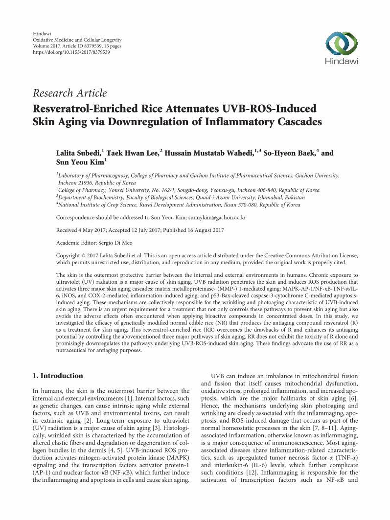
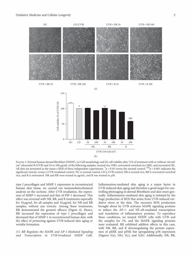
3.1. RR Protects against UVB-Induced Toxicity in NHDFCells. UVB exposure induces cell death in dermal fibroblasts,as well as various inflammatory cascades, resulting in skinaging, skin wrinkling, and skin pigmentation. In order toevaluate the cytotoxicity of the samples, after 72 h of UVB(144mJ/cm2) and sample treatment, the viability of NHDFcells was measured using an MTT assay (as described inthe Methods). We photographed the cells to show theirmorphology with or without UVB and sample treatment.In the UVB-exposed cells, NR and RR were not toxic anddid not affect the normal morphology of NHDF cells untilthe concentration reached 100μg/mL. However, R aloneshowed significant toxicity, causing cell death at the higherconcentration of 100μg/mL and a completely altered,shrunken cell morphology. Using a lower concentration(10μg/mL) of R induced the level of cell death, but its signif-icant toxicity was still revealed at this dose by comparing themorphology of the treated cells with that of the cells in theUVB-treated control group (Figure 1).
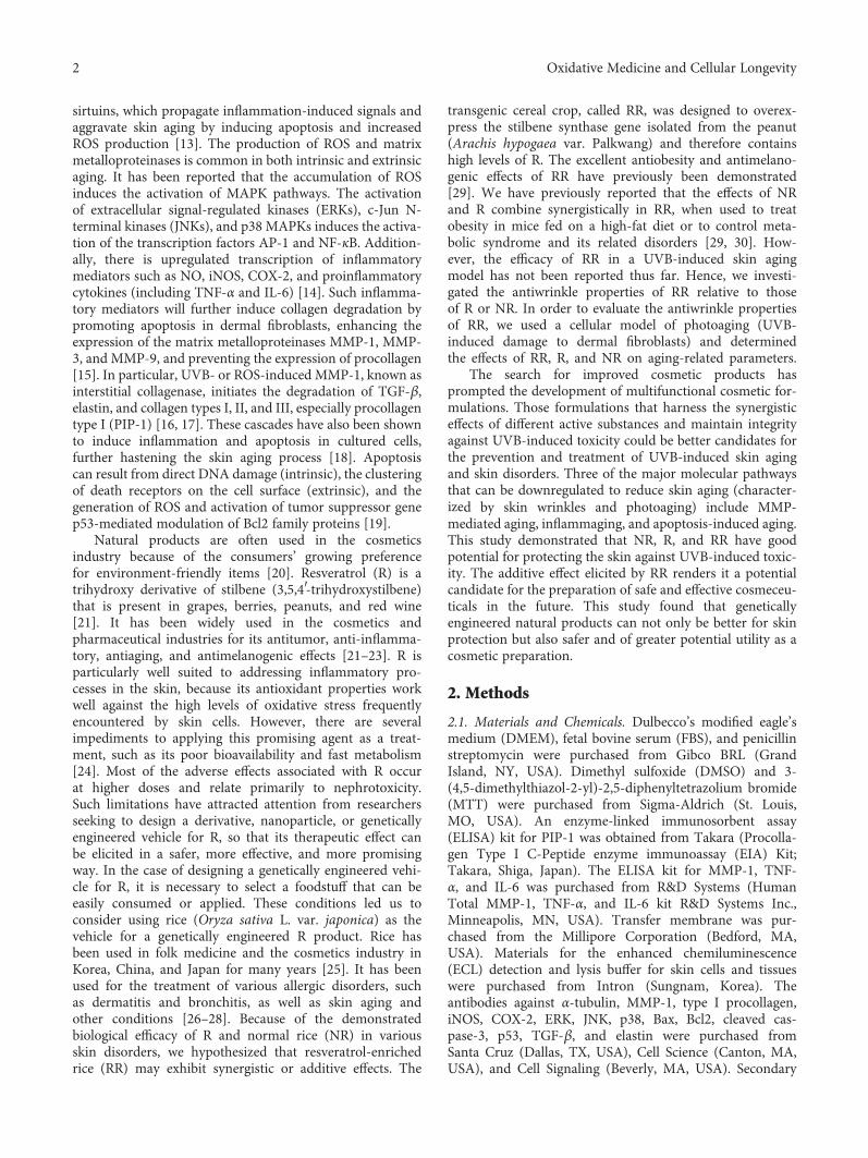
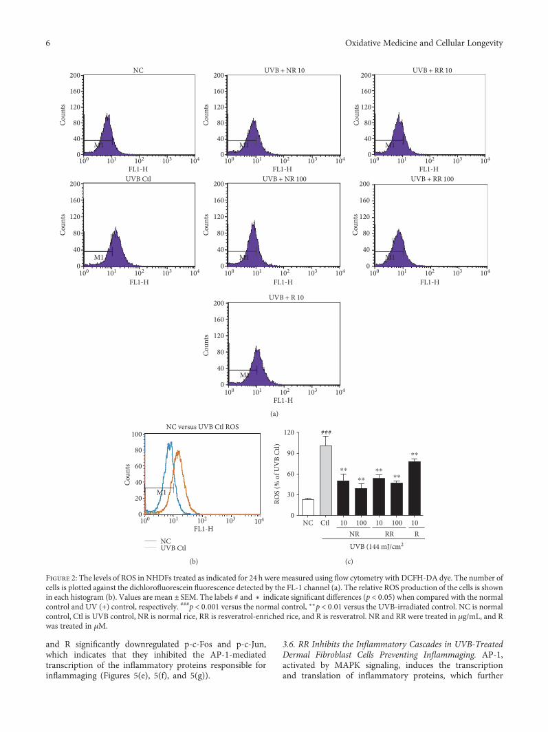
3.2. RR Downregulated UVB-Induced ROS Production inNHDF Cells. ROS are the major toxic substances generatedby UVB exposure in the skin and dermal fibroblast cells. Tomeasure ROS production in NHDFs, we treated cells withUVB and the samples for 24 h. The change in intracellularROS compared with the nonirradiated controls was deter-mined using 2′,7′-dichlorofluorescein diacetate (DCF-DA),which is oxidized by ROS in cells to DCF. The cells werestained with 30μM of DCF-DA and incubated for 30min,after which the fluorescence level was measured. TheUVB-induced ROS production in dermal fibroblast cellswas significantly reduced following treatment with NR,
RR, and R. RR demonstrated a greater reduction in ROSproduction at concentrations of 10 and 100μg/mL thanNR or R alone. While all of the samples were capable ofreducing UVB-induced ROS production, RR was foundto be the most effective one (Figure 2).
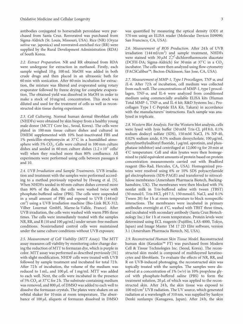
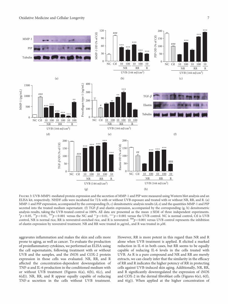
3.3. NR, RR, and R Control the Level of MMP-1, TGF-β, andPIP-1 in NHDF Cells. To evaluate the effects of the sampleson MMP-1 and PIP production in UVB-exposed NHDFcells, the levels of protein expression and MMP-1 and PIP-1 secretion were measured by using Western blotting andan ELISA, respectively (Figures 3(a), 3(b), 3(c), 3(d), and3(e)). According to both the protein expression measure-ments and secreted protein assay, NR, RR, and R all reducedUVB-induced MMP-1 production and increased PIP pro-duction. The RR-mediated downregulation of MMP-1 andupregulation of PIP appear to have been caused by the addi-tive effect of NR and R. This is because, despite R showing themost potent activity in reducing MMP-1 and increasing PIPlevels (even at only 10μg/mL), the activity of RR seems to bebetter than that of NR and R alone in terms of the levels ofsecreted MMP-1 and PIP when tested using the ELISA kit.NR, RR, and R play significant roles in reducing MMP-1,but RR exhibits significantly greater (and concentration-dependent) activity against UVB-induced MMP-1 produc-tion. Only RR demonstrated the ability to increase PIP pro-duction almost two- and threefold at concentrations of 10and 100μg/mL, respectively, in comparison with the UVB-treated control group, whereas NR and R were unable toaffect a significant increase. This result stimulated our inter-est in elucidating the mechanism by which RR increasesPIP levels to this extent. We therefore evaluated the proteinexpression of TGF-β, as the TGF-β/Smad pathway is a majorpathway controlling PIP production. We found that RR sig-nificantly induced TGF-β protein expression in UVB-irradiated NHDF cells and thereby stimulated PIP produc-tion. This has the concomitant effect of preventing UVB-induced skin wrinkle formation, as upregulating TGF-βand PIP levels also results in increased elastin production.Similar result was obtained here in the RR and UVB-treated group. RR increased elastin production to a greaterextent than NR and R alone (Figures 3(f), 3(g), and 3(h)).This result suggests that RR can protect NHDF cells againstUVB-ROS-MMP-1-induced skin aging, particularly skinwrinkle formation.
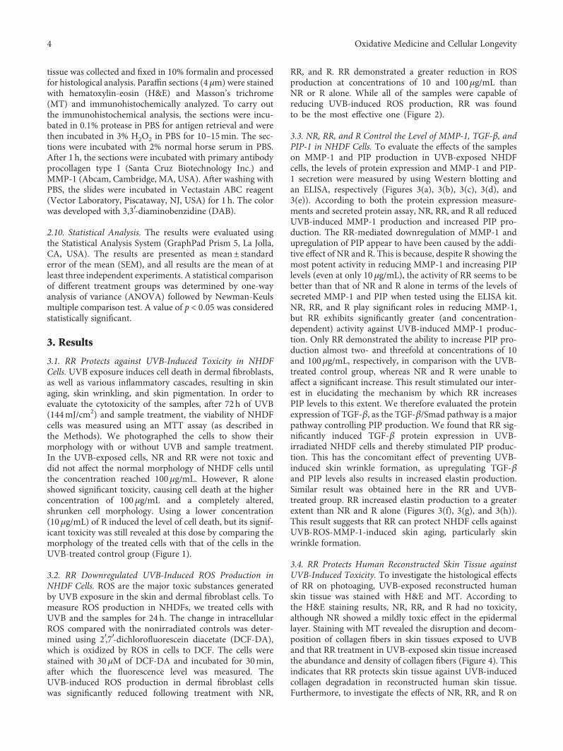
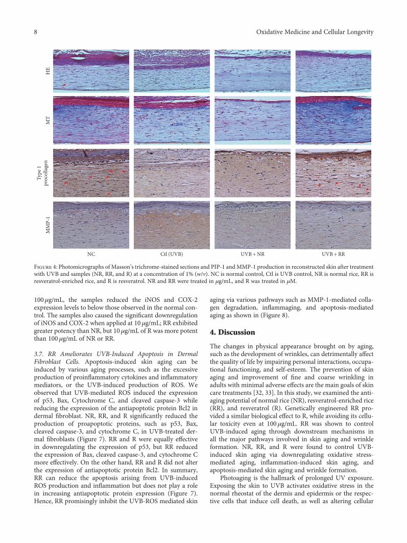
3.4. RR Protects Human Reconstructed Skin Tissue againstUVB-Induced Toxicity. To investigate the histological effectsof RR on photoaging, UVB-exposed reconstructed humanskin tissue was stained with H&E and MT. According tothe H&E staining results, NR, RR, and R had no toxicity,although NR showed a mildly toxic effect in the epidermallayer. Staining with MT revealed the disruption and decom-position of collagen fibers in skin tissues exposed to UVBand that RR treatment in UVB-exposed skin tissue increasedthe abundance and density of collagen fibers (Figure 4). Thisindicates that RR protects skin tissue against UVB-inducedcollagen degradation in reconstructed human skin tissue.Furthermore, to investigate the effects of NR, RR, and R on
4 Oxidative Medicine and Cellular Longevity
type I procollagen and MMP-1 expression in reconstructedhuman skin tissue, we carried out immunohistochemicalanalysis on the sections. After UVB irradiation, the expres-sion of MMP-1 increased and that of PIP-1 decreased. Thiseffect was reversed with NR, RR, and R treatments especiallyfor 10μg/mL for all samples and 10μg/mL for NR and RRsamples, without any toxicity. Among these treatments,RR demonstrated the greatest efficacy (Figure 4). Hence,RR increased the expression of type I procollagen anddecreased that of MMP-1 in reconstructed human skin, withthe effect of protecting against UVB-induced skin aging orwrinkle formation.
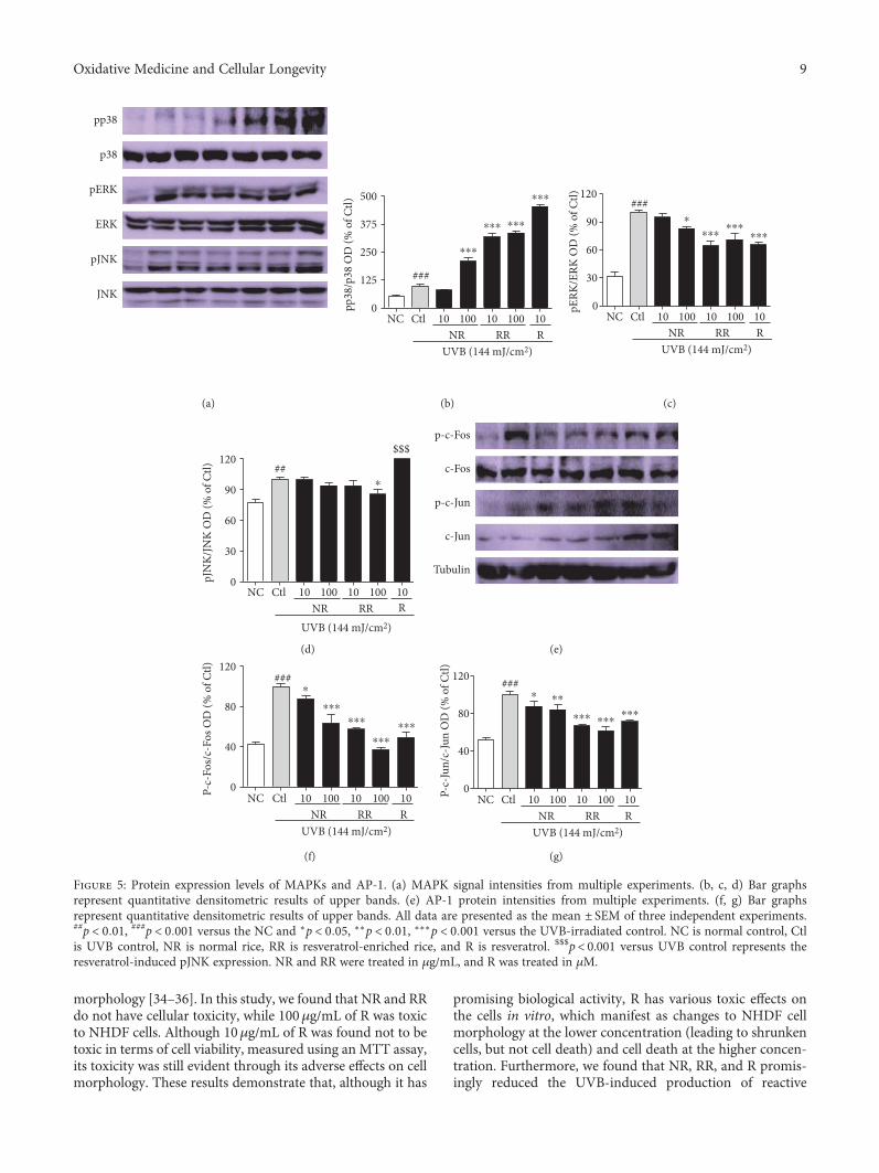
3.5. RR Regulates the MAPK and AP-1-Mediated Signalingand Transcription in UVB-Irradiated NHDF Cells.
Inflammation-mediated skin aging is a major factor inUVB-induced skin aging and therefore a good target for con-trolling photoaging in dermal fibroblasts and skin more gen-erally. Inflammation-mediated skin aging is initiated by thehuge production of ROS that arises from UVB-induced oxi-dative stress in the skin. The excessive ROS productionbrought about by UVB activates MAPK signaling proteinsto induce the AP-1- and NF-κB-mediated transcriptionand translation of inflammatory proteins. To reproducethese conditions, we treated NHDF cells with UVB andthe samples for 3 h, and the MAPK signaling proteinswere evaluated. RR exhibited additive effects in this case,with NR, RR, and R downregulating the protein expres-sion of pERK and pJNK but upregulating p38 expression(Figures 5(a), 5(b), 5(c), and 5(d)). Additionally, NR, RR,
NC Ctl (UVB) UVB + NR 10 UVB + NR 100
UVB + RR 10 UVB + RR 100 UVB + R 10 UVB + R 100
(a)
Cell
viab
ility
(% o
f UV
B Ct
l)
0
50
100
150
NC Ctl 10 100 10 100 10
NR RR R
UVB (144 mJ/cm2)
100
#
$$$
(b)
Figure 1: Normal human dermal fibroblast (NHDF). (a) Cell morphology and (b) cell viability after 72 h of treatment with or without 144mJ/cm2 ultraviolet B (UVB) and 10 or 100 μg/mL of the following samples: normal rice (NR), resveratrol-enriched rice (RR), and resveratrol (R).All data are presented as the mean± SEM of three independent experiments. #p < 0 05 versus the normal control. $$$p < 0 001 indicates thesignificant toxicity versus a UVB-irradiated control. NC is normal control, Ctl is UVB control, NR is normal rice, RR is resveratrol-enrichedrice, and R is resveratrol. NR and RR were treated in μg/mL, and R was treated in μM.
5Oxidative Medicine and Cellular Longevity
and R significantly downregulated p-c-Fos and p-c-Jun,which indicates that they inhibited the AP-1-mediatedtranscription of the inflammatory proteins responsible forinflammaging (Figures 5(e), 5(f), and 5(g)).
3.6. RR Inhibits the Inflammatory Cascades in UVB-TreatedDermal Fibroblast Cells Preventing Inflammaging. AP-1,activated by MAPK signaling, induces the transcriptionand translation of inflammatory proteins, which further
160
200
120
80
40
0M1 M1 M1
M1
M1
M1 M1
Coun
ts
160
200
120
80
40
0
Coun
ts
Coun
ts
160
200
120
80
40
0
160
200
120
80
40
0
Coun
ts160
200
120
80
40
0
Coun
ts
160
200
120
80
40
0
Coun
ts
160
200
120
80
40
0
Coun
ts
100 101 102
FL1-H103 104 100 101 102
FL1-H103 104 100 101 102
FL1-H103 104
100 101 102
FL1-H103 104
100 101 102
FL1-H103 104
100 101 102
FL1-H103 104 100 101 102
FL1-H103 104
UVB + R 10
UVB + RR 100
UVB + RR 10UVB + NR 10NC
UVB Ctl UVB + NR 100
(a)
60
80
100
20M1
40
0
Coun
ts
100 101 102
FL1-H103 104
NCUVB Ctl
NC versus UVB Ctl ROS
(b)
0
30
60
90
120
ROS
(% o
f UV
B Ct
l)
NC Ctl 10 100 10 100 10NR RR R
UVB (144 mJ/cm2
###
⁎⁎
⁎⁎
⁎⁎⁎⁎
⁎⁎
(c)
Figure 2: The levels of ROS in NHDFs treated as indicated for 24 h were measured using flow cytometry with DCFH-DA dye. The number ofcells is plotted against the dichlorofluorescein fluorescence detected by the FL-1 channel (a). The relative ROS production of the cells is shownin each histogram (b). Values are mean ± SEM. The labels # and ∗ indicate significant differences (p < 0 05) when compared with the normalcontrol and UV (+) control, respectively. ###p < 0 001 versus the normal control, ∗∗p < 0 01 versus the UVB-irradiated control. NC is normalcontrol, Ctl is UVB control, NR is normal rice, RR is resveratrol-enriched rice, and R is resveratrol. NR and RR were treated in μg/mL, and Rwas treated in μM.
6 Oxidative Medicine and Cellular Longevity
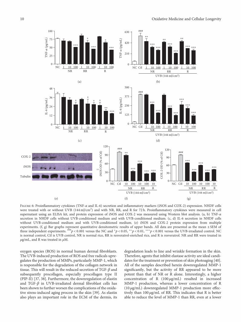
aggravates inflammation and makes the skin and cells moreprone to aging, as well as cancer. To evaluate the productionof proinflammatory cytokines, we performed an ELISA usingthe cell supernatants, following treatment with or withoutUVB and the samples, and the iNOS and COX-2 proteinexpression in those cells was evaluated. NR, RR, and Raffected the concentration-dependent downregulation ofTNF-α and IL-6 production in the conditioned medium withor without UVB treatment (Figures 6(a), 6(b), 6(c), and6(d)). NR, RR, and R appear equally capable of reducingTNF-α secretion in the cells without UVB treatment.
However, RR is more potent in this regard than NR and Ralone when UVB treatment is applied. R elicited a markedreduction in IL-6 in both cases, but RR seems to be equallycapable of reducing IL-6 levels in the cells treated withUVB. As R is a pure compound and NR and RR are merelyextracts, we can clearly infer that the similarity in the efficacyof RR and R indicates the higher potency of RR in protectingcells against UVB-induced skin aging. Additionally, NR, RR,and R significantly downregulated the expression of iNOSand COX-2 in the dermal fibroblast cells (Figures 6(e), 6(f),and 6(g)). When applied at the higher concentration of
PIP
MMP-1
Tubulin
(a)
0
30
60
90
120
MM
P-1
OD
(% o
f Ctl)
NC Ctl 10 100 10 100 10NR RR R
⁎⁎⁎⁎⁎⁎
⁎⁎⁎
⁎⁎⁎ ⁎⁎⁎
###
UVB (144 mJ/cm2)
(b)
⁎⁎⁎
⁎⁎⁎
⁎⁎⁎
0
50
100
150
200
PIP
OD
(% o
f Ctl)
NC Ctl 10 100 10 100 10NR RR R
###
UVB (144 mJ/cm2)
(c)
⁎⁎⁎⁎⁎⁎
⁎⁎⁎
⁎⁎
⁎⁎⁎ ⁎⁎⁎
###
NC
1500
1000
MM
P-1
(pg/
mL)
500
0Ctl 10 100 10 100 10
NR RR R UVB (144 mJ/cm2)
100
(d)
⁎⁎⁎
⁎⁎
#
NC Ctl 10 100 10 100 10NR RR R
100
Proc
olla
gen
type
-1 (n
g/m
L)
UVB (144 mJ/cm2)
400
300
200
100
0
(e)
Elastin
TGF-�훽
Tubulin
(f)
⁎⁎⁎⁎⁎⁎ ⁎⁎⁎
⁎⁎⁎
⁎⁎⁎
0
50
100
150
200
NC Ctl 10 100 10 100 10NR RR R
###
TGF-�훽
OD
(% o
f Ctl)
UVB (144 mJ/cm2)
(g)
⁎⁎⁎ ⁎⁎⁎ ⁎⁎⁎
0
50
100
150
200
NC Ctl 10 100 10 100 10NR RR R
$$$
##
Elas
tin O
D (%
of C
tl)
UVB (144 mJ/cm2)
(h)
Figure 3: UVB-MMP1-mediated protein expression and the secretion of MMP-1 and PIP were measured using Western blot analysis and anELISA kit, respectively. NHDF cells were incubated for 72 h with or without UVB exposure and treated with or without NR, RR, and R. (a)MMP-1 and PIP expression, accompanied by the corresponding (b, c) densitometric analysis results (d, e) and the quantities MMP-1 and PIPsecreted into the treated medium supernatant. (f) TGF-β and elastin expression, accompanied by the corresponding (g, h) densitometricanalysis results, taking the UVB-treated control as 100%. All data are presented as the mean ± SEM of three independent experiments.#p < 0 05, ##p < 0 01, ###p < 0 001 versus the NC and ∗∗p < 0 01, ∗∗∗p < 0 001 versus the UVB control. NC is normal control, Ctl is UVBcontrol, NR is normal rice, RR is resveratrol-enriched rice, and R is resveratrol. $$$p< 0.001 versus UVB control represents the inhibitionof elastin expression by resveratrol treatment. NR and RR were treated in μg/mL, and R was treated in μM.
7Oxidative Medicine and Cellular Longevity
100μg/mL, the samples reduced the iNOS and COX-2expression levels to below those observed in the normal con-trol. The samples also caused the significant downregulationof iNOS and COX-2 when applied at 10μg/mL; RR exhibitedgreater potency than NR, but 10μg/mL of R was more potentthan 100μg/mL of NR or RR.
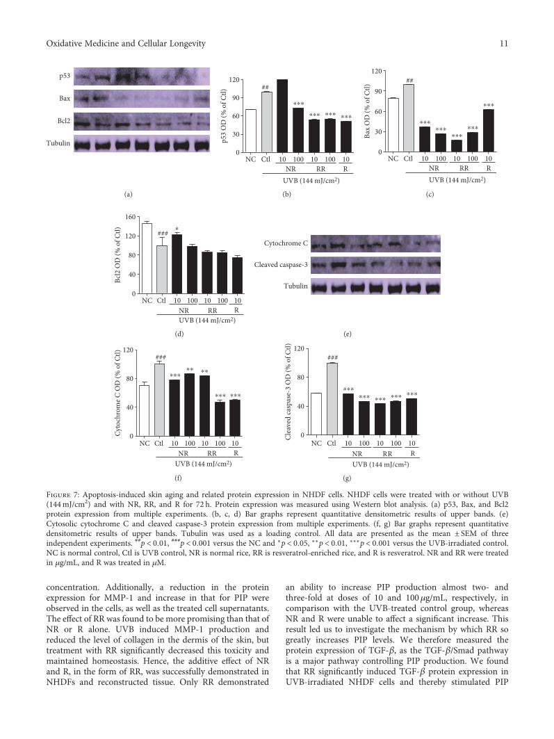
3.7. RR Ameliorates UVB-Induced Apoptosis in DermalFibroblast Cells. Apoptosis-induced skin aging can beinduced by various aging processes, such as the excessiveproduction of proinflammatory cytokines and inflammatorymediators, or the UVB-induced production of ROS. Weobserved that UVB-mediated ROS induced the expressionof p53, Bax, Cytochrome C, and cleaved caspase-3 whilereducing the expression of the antiapoptotic protein Bcl2 indermal fibroblast. NR, RR, and R significantly reduced theproduction of proapoptotic proteins, such as p53, Bax,cleaved caspase-3, and cytochrome C, in UVB-treated der-mal fibroblasts (Figure 7). RR and R were equally effectivein downregulating the expression of p53, but RR reducedthe expression of Bax, cleaved caspase-3, and cytochrome Cmore effectively. On the other hand, RR and R did not alterthe expression of antiapoptotic protein Bcl2. In summary,RR can reduce the apoptosis arising from UVB-inducedROS production and inflammation but does not play a rolein increasing antiapoptotic protein expression (Figure 7).Hence, RR promisingly inhibit the UVB-ROS mediated skin
aging via various pathways such as MMP-1-mediated colla-gen degradation, inflammaging, and apoptosis-mediatedaging as shown in (Figure 8).
4. Discussion
The changes in physical appearance brought on by aging,such as the development of wrinkles, can detrimentally affectthe quality of life by impairing personal interactions, occupa-tional functioning, and self-esteem. The prevention of skinaging and improvement of fine and coarse wrinkling inadults with minimal adverse effects are the main goals of skincare treatments [32, 33]. In this study, we examined the anti-aging potential of normal rice (NR), resveratrol-enriched rice(RR), and resveratrol (R). Genetically engineered RR pro-vided a similar biological effect to R, while avoiding its cellu-lar toxicity even at 100μg/mL. RR was shown to controlUVB-induced aging through downstream mechanisms inall the major pathways involved in skin aging and wrinkleformation. NR, RR, and R were found to control UVB-induced skin aging via downregulating oxidative stress-mediated aging, inflammation-induced skin aging, andapoptosis-mediated skin aging and wrinkle formation.
Photoaging is the hallmark of prolonged UV exposure.Exposing the skin to UVB activates oxidative stress in thenormal rheostat of the dermis and epidermis or the respec-tive cells that induce cell death, as well as altering cellular
NC Ctl (UVB) UVB + NR UVB + RR
HE
MT
Type
1pr
ocol
lage
nM
MP‑
1
Figure 4: Photomicrographs of Masson’s trichrome-stained sections and PIP-1 andMMP-1 production in reconstructed skin after treatmentwith UVB and samples (NR, RR, and R) at a concentration of 1% (w/v). NC is normal control, Ctl is UVB control, NR is normal rice, RR isresveratrol-enriched rice, and R is resveratrol. NR and RR were treated in μg/mL, and R was treated in μM.
8 Oxidative Medicine and Cellular Longevity
morphology [34–36]. In this study, we found that NR and RRdo not have cellular toxicity, while 100μg/mL of R was toxicto NHDF cells. Although 10μg/mL of R was found not to betoxic in terms of cell viability, measured using an MTT assay,its toxicity was still evident through its adverse effects on cellmorphology. These results demonstrate that, although it has
promising biological activity, R has various toxic effects onthe cells in vitro, which manifest as changes to NHDF cellmorphology at the lower concentration (leading to shrunkencells, but not cell death) and cell death at the higher concen-tration. Furthermore, we found that NR, RR, and R promis-ingly reduced the UVB-induced production of reactive
pp38
p38
pERK
ERK
pJNK
JNK
(a)
0
125
250
375
500
pp38
/p38
OD
(% o
f Ctl)
NC Ctl 10 100 10 100 10NR RR R
UVB (144 mJ/cm2)
⁎⁎⁎
⁎⁎⁎ ⁎⁎⁎
⁎⁎⁎
###
(b)
UVB (144 mJ/cm2)
⁎⁎⁎
⁎⁎⁎⁎
⁎⁎⁎
0
30
60
90
120
pERK
/ERK
OD
(% o
f Ctl)
NC Ctl 10 100 10 100 10NR RR R
###
(c)
UVB (144 mJ/cm2)
$$$
0
30
60
90
120
NC Ctl 10 100 10 100 10NR RR R
pJN
K/JN
K O
D (%
of C
tl) ##⁎
(d)
Tubulin
p-c-Fos
c-Fos
p-c-Jun
c-Jun
(e)
UVB (144 mJ/cm2)
⁎⁎⁎⁎⁎⁎
⁎⁎⁎
⁎⁎⁎
0
40
80
120
NC Ctl 10 100 10 100 10NR RR R
P-c-
Fos/
c-Fo
s OD
(% o
f Ctl)
###⁎
(f)
UVB (144 mJ/cm2)
⁎⁎⁎ ⁎⁎⁎⁎⁎⁎
0
40
80
120
NC Ctl 10 100 10 100 10NR RR R
P-c-
Jun/
c-Ju
n O
D (%
of C
tl)
###⁎ ⁎⁎
(g)
Figure 5: Protein expression levels of MAPKs and AP-1. (a) MAPK signal intensities from multiple experiments. (b, c, d) Bar graphsrepresent quantitative densitometric results of upper bands. (e) AP-1 protein intensities from multiple experiments. (f, g) Bar graphsrepresent quantitative densitometric results of upper bands. All data are presented as the mean ± SEM of three independent experiments.##p < 0 01, ###p < 0 001 versus the NC and ∗p < 0 05, ∗∗p < 0 01, ∗∗∗p < 0 001 versus the UVB-irradiated control. NC is normal control, Ctlis UVB control, NR is normal rice, RR is resveratrol-enriched rice, and R is resveratrol. $$$p< 0.001 versus UVB control represents theresveratrol-induced pJNK expression. NR and RR were treated in μg/mL, and R was treated in μM.
9Oxidative Medicine and Cellular Longevity
oxygen species (ROS) in normal human dermal fibroblasts.The UVB-induced production of ROS and free radicals upre-gulates the production of MMPs, particularly MMP-1, whichis responsible for the degradation of the collagen network intissue. This will result in the reduced secretion of TGF-β andsubsequently procollagen, especially procollagen type II(PIP-II) [37, 38]. Furthermore, the downregulation of elastinand TGF-β in UVB-irradiated dermal fibroblast cells hasbeen shown to further worsen the complications of the oxida-tive stress-induced aging process in the skin [39]. As elastinalso plays an important role in the ECM of the dermis, its
degradation leads to line and wrinkle formation in the skin.Therefore, agents that inhibit elastase activity are ideal candi-dates for the treatment or prevention of skin photoaging [40].All of the samples described herein downregulated MMP-1significantly, but the activity of RR appeared to be morepotent than that of NR or R alone. Interestingly, a higherconcentration of R (100μg/mL) resulted in increasedMMP-1 production, whereas a lower concentration of R(10μg/mL) downregulated MMP-1 production more effec-tively than 100μg/mL of RR. This indicates that R is betterable to reduce the level of MMP-1 than RR, even at a lower
0
25
50
75
100
TNF-�훼
(pg/
mL)
100NR RR R
101100101100101NC
⁎⁎⁎⁎⁎⁎ ⁎⁎⁎
⁎⁎
(a)
⁎⁎⁎
⁎⁎
⁎⁎⁎
⁎⁎
⁎⁎⁎
⁎⁎⁎
⁎⁎
⁎⁎⁎ ⁎⁎⁎
0
210
420
630
TNF-�훼
(pg/
mL)
100NR RR R
UVB (144 mJ/cm2)
101100101100101NC Ctl
###
(b)
0
12
24
36
48
IL-6
(pg/
mL)
100NR RR R
101100101100101NC
⁎⁎
⁎ ⁎
⁎⁎⁎⁎⁎
(c)
UVB (144 mJ/cm2)
0
125
250
375
500
IL-6
(pg/
mL)
100NR RR R
101100101100101NC Ctl
###
⁎⁎⁎
⁎⁎⁎
⁎⁎⁎
⁎⁎⁎
⁎⁎⁎
⁎⁎⁎
⁎⁎⁎
(d)
COX-2
iNOS
Tubulin
(e)
UVB (144 mJ/cm2)
0
30
60
90
120
iNO
S O
D (%
of C
tl)
NC Ctl 10 100 10 100 10NR RR R
###⁎⁎
⁎⁎⁎
⁎⁎⁎
⁎⁎⁎ ⁎⁎⁎
(f)
UVB (144 mJ/cm2)
0
30
60
90
120
COX-
2 O
D (%
of C
tl)
NC Ctl 10 100 10 100 10NR RR R
###
⁎⁎⁎
⁎⁎⁎
⁎⁎⁎
⁎⁎⁎⁎⁎⁎
(g)
Figure 6: Proinflammatory cytokines (TNF-α and IL-6) secretion and inflammatory markers (iNOS and COX-2) expression. NHDF cellswere treated with or without UVB (144mJ/cm2) and with NR, RR, and R for 72 h. Proinflammatory cytokines were measured in cellsupernatant using an ELISA kit, and protein expression of iNOS and COX-2 was measured using Western blot analysis. (a, b) TNF-αsecretion in NHDF cells without UVB-conditioned medium and with UVB-conditioned medium. (c, d) IL-6 secretion in NHDF cellswithout UVB-conditioned medium and with UVB-conditioned medium. (e) iNOS and COX-2 protein expression from multipleexperiments. (f, g) Bar graphs represent quantitative densitometric results of upper bands. All data are presented as the mean ± SEM ofthree independent experiments. ###p < 0 001 versus the NC and ∗p < 0 05, ∗∗p < 0 01, ∗∗∗p < 0 001 versus the UVB-irradiated control. NCis normal control, Ctl is UVB control, NR is normal rice, RR is resveratrol-enriched rice, and R is resveratrol. NR and RR were treated inμg/mL, and R was treated in μM.
10 Oxidative Medicine and Cellular Longevity
concentration. Additionally, a reduction in the proteinexpression for MMP-1 and increase in that for PIP wereobserved in the cells, as well as the treated cell supernatants.The effect of RR was found to be more promising than that ofNR or R alone. UVB induced MMP-1 production andreduced the level of collagen in the dermis of the skin, buttreatment with RR significantly decreased this toxicity andmaintained homeostasis. Hence, the additive effect of NRand R, in the form of RR, was successfully demonstrated inNHDFs and reconstructed tissue. Only RR demonstrated
an ability to increase PIP production almost two- andthree-fold at doses of 10 and 100μg/mL, respectively, incomparison with the UVB-treated control group, whereasNR and R were unable to affect a significant increase. Thisresult led us to investigate the mechanism by which RR sogreatly increases PIP levels. We therefore measured theprotein expression of TGF-β, as the TGF-β/Smad pathwayis a major pathway controlling PIP production. We foundthat RR significantly induced TGF-β protein expression inUVB-irradiated NHDF cells and thereby stimulated PIP
Bax
Bcl2
Tubulin
p53
(a)
0
30
60
90
120
p53
OD
(% o
f Ctl)
NC Ctl 10 100 10 100 10NR RR R
UVB (144 mJ/cm2)
##
⁎⁎⁎
⁎⁎⁎ ⁎⁎⁎ ⁎⁎⁎
(b)
0
30
60
90
120
Bax
OD
(% o
f Ctl)
UVB (144 mJ/cm2)
NC Ctl 10 100 10 100 10NR RR R
##
⁎⁎⁎⁎⁎⁎
⁎⁎⁎⁎⁎⁎
⁎⁎⁎
(c)
UVB (144 mJ/cm2)
0
40
80
120
160
Bcl2
OD
(% o
f Ctl)
NC Ctl 10 100 10 100 10NR RR R
###⁎
(d)
Cleaved caspase-3
Tubulin
Cytochrome C
(e)
0
40
80
120
Cyto
chro
me C
OD
(% o
f Ctl)
UVB (144 mJ/cm2)
NC Ctl 10 100 10 100 10NR RR R
###
⁎⁎⁎⁎⁎ ⁎⁎
⁎⁎⁎ ⁎⁎⁎
(f)
0
40
80
120
Clea
ved
casp
ase-
3 O
D (%
of C
tl)
UVB (144 mJ/cm2)
NC Ctl 10 100 10 100 10NR RR R
###
⁎⁎⁎⁎⁎⁎ ⁎⁎⁎ ⁎⁎⁎ ⁎⁎⁎
(g)
Figure 7: Apoptosis-induced skin aging and related protein expression in NHDF cells. NHDF cells were treated with or without UVB(144mJ/cm2) and with NR, RR, and R for 72 h. Protein expression was measured using Western blot analysis. (a) p53, Bax, and Bcl2protein expression from multiple experiments. (b, c, d) Bar graphs represent quantitative densitometric results of upper bands. (e)Cytosolic cytochrome C and cleaved caspase-3 protein expression from multiple experiments. (f, g) Bar graphs represent quantitativedensitometric results of upper bands. Tubulin was used as a loading control. All data are presented as the mean ± SEM of threeindependent experiments. ##p < 0 01, ###p < 0 001 versus the NC and ∗p < 0 05, ∗∗p < 0 01, ∗∗∗p < 0 001 versus the UVB-irradiated control.NC is normal control, Ctl is UVB control, NR is normal rice, RR is resveratrol-enriched rice, and R is resveratrol. NR and RR were treatedin μg/mL, and R was treated in μM.
11Oxidative Medicine and Cellular Longevity

production. This can simultaneously prevent UVB-inducedskin wrinkle formation, as upregulating TGF-β and PIPlevels also results in increased elastin production. RR wasshown to increase elastin production to a greater extent thanNR and R alone. To confirm this in vitro finding, we con-ducted the same experiment in reconstructed tissue andobtained very similar results. Even in the reconstructed tis-sue, NR, RR, and R had no toxicity, although NR showed amildly toxic effect in the epidermal layer. The disruptionand decomposition of collagen fibers were observed in skintissues exposed to UVB using Masson’s trichrome (MT)staining, hematoxylin and eosin (H&E) staining, and stainingfor the determination of PIP and MMP-1. RR treatmentincreased the abundance and density of collagen fibers inUVB-exposed skin tissue. NR and RR treatment reducedthe level of MMP-1 and increased that of type I procollagenin cells exposed to UVB. Of all the tested samples, RR mosteffectively increased the expression of type I procollagenand decreased that of MMP-1 in reconstructed humanskin, thereby protecting against UVB-induced skin agingor wrinkle formation. These data collectively indicate thatRR downregulates UVB-induced oxidative stress morepotently than NR or R alone and therefore reduces itscontribution to skin aging.
UVB irradiation induces ROS production. ROS-mediated oxidative stress activates MAPK signaling byincreasing the phosphorylation of p38, JNK, and ERK(pp38, pJNK, and pERK) to induce inflammaging. Inflam-maging is closely associated with many aging-associated dis-eases, such as Alzheimer’s disease, as well as atherosclerosis,heart disease, type II diabetes, and cancer. One factor that
exacerbates UVB-induced ROS-mediated inflammaging isimmunosenescence [41]. Inflammaging is initiated afterthe activation of MAPK signaling, AP-1 (c-Fos and c-Jun) activation and the increased transcription of inflam-matory mediators. Through the MAPK signaling path-ways, AP-1 controls the expression of MMPs, especiallyMMP-1, MMP-2, and MMP-9, in inflammation-inducedskin aging. Heterodimer complexes made between c-Junand c-Fos, with various growth factors, cytokines, andUV exposure, can cause aggressive inflammation and skinaging [42]. In this study, we found that irradiating NHDFcells with UVB activates MAPK signaling, which activatesthe AP-1- and NF-κB-mediated transcription of MMPs,proinflammatory cytokines, inflammatory mediators, andso forth, while simultaneously downregulating PIP, TGF-β, and elastin production [43]. Inflammatory mediators,such as iNOS, COX-2, and cytokines, as well as IL-6, IL-1β,and TNF-α produced by innate immune cells, will causechronic inflammation and thus initiate inflammaging [44].In this study, we demonstrated that treatment with NR, RR,and R can significantly modulate MAPK and AP-1 signaling,by inhibiting NF-κB-mediated transcription. This was evi-denced by the activation of PIP and the inhibition of TNF-α, IL-6, and MMP-1 in the treated dermal fibroblast cells.RR most effectively reduced TNF-α and IL-6 productionin UVB-irradiated NHDF cells, but its ability to reduceiNOS and COX-2 levels, while still superior to that ofNR, was inferior to that of R alone. This result furtherdemonstrates the anti-inflammatory properties of thesetreatments and their great potential for protecting againstUVB-induced inflammaging.
p38 JNK ERK
In�ammaging
TNF-�훼,IL-6
iNOS COX-2
ROS
UVB
MMP1 TFG-�훽
PIPElastin
Skin wrinklePhotoaging
p53 Bax , Bcl2
Cleaved caspase-3Cytochrome C
Apoptosis-mediatedskin aging
NR, RR, RNR, RR, R
NR, RR, R
NR, RR, R
NR, RR, R
NR, RR, R
NR, RR, R NR, RR, R
Figure 8: Scheme of the UVB-ROS-mediated skin aging and the protective role of resveratrol rice against its toxicity to prevent skin aging.
12 Oxidative Medicine and Cellular Longevity
Oxidative stress and inflammaging together induceapoptosis, which is another key factor in skin aging,photoaging, wrinkling, and related disorders [45]. UVB-induced ROS cause further oxidative stress in the cellularenvironment, with the MAPK- and NF-κB-mediated tran-scription of proinflammatory cytokines, particularly TNF-α, being the major cause of cell apoptosis and aging byapoptosis in the skin [46]. UVB, ROS, proinflammatorycytokines, and other toxins produced by UVB will inducethe expression of apoptotic protein p53. This subsequentlyactivates the expression of Bax, Bad, PUMA, and cleavedcaspase-3, while simultaneously downregulating theexpression of antiapoptotic proteins such as Bcl2 [47].Activated cleaved caspase-3 translocates mitochondrialcytochrome C to the cytosol, which further induces apo-ptosis and contributes to skin aging by further activatingfactors that aggravate skin aging and wrinkle formation[19]. In this study, we confirmed that NR, RR, and Rpromisingly downregulate the levels of p53, Bax, cleavedcaspase-3, and cytochrome C and that RR and R did notsimultaneously alter the expression of the antiapoptoticprotein Bcl2. RR downregulated the expression of Baxand cleaved caspase-3 more effectively than the othertreatments and was as potent as R for the inhibition ofp53 in the UVB-treated fibroblast cells. The RR-mediatedreduction in p53 lowered the transcription of Bax andcleaved caspase-3. Bax and cleaved caspase-3 were conse-quently unable to translocate mitochondrial cytochromeC to the cytosol, thereby protecting fibroblasts from mito-chondrial apoptosis and apoptosis-induced skin aging. Inthis way, and considering that RR (an extract) is generallyat least as effective as R (a pure compound) or NR alone,we conclude that the pure compounds or standard com-pound in RR might have better synergistic biological activ-ity than R alone for every considered aging pathway.
In conclusion, NR, R, and particularly RR have beenshown to control MMP-1-mediated UVB-induced skinaging, apoptosis-induced skin aging, and inflammation-mediated complications called inflammaging in dermalfibroblasts. A schematic explanation for the UVB-ROS-mediated aging and the role of NR, RR, and R has beenshown in Figure 8. In this way, our study has demon-strated the potential of RR as an antiaging product forthe prevention of UVB-induced complications in vitroand ex vivo.
Conflicts of Interest
The authors declare that there is no conflict of interestsregarding the publication of this paper.
Acknowledgments
This work was supported by a grant from the Next-Generation Bio-Green 21 Program (no. PJ01118803), RuralDevelopment Administration, Republic of Korea and NationalResearch Foundation of Korea (NRF) grant funded by theKorean government (MSIP) (no. NRF-2017R1A4A1015594)and also supported by the High Value-added Food Technology
Development Program, the Ministry of Agriculture, Foodand Rural Affairs (114006041HD020).
References
[1] C. J. Pendegrass, A. E. Goodship, J. S. Price, and G. W. Blunn,“Nature’s answer to breaching the skin barrier: an innovativedevelopment for amputees,” Journal of Anatomy, vol. 209,no. 1, pp. 59–67, 2006.
[2] M. Wlaschek, I. Tantcheva-Poor, L. Naderi et al., “Solar UVirradiation and dermal photoaging,” Journal of Photochemistryand Photobiology B: Biology, vol. 63, no. 1–3, pp. 41–51, 2001.
[3] L. Rittie and G. J. Fisher, “Natural and sun-induced aging ofhuman skin,” Cold Spring Harbor Perspectives in Medicine,vol. 5, no. 1, article a015370, 2015.
[4] M. J. Sherratt, “Tissue elasticity and the ageing elastic fibre,”Age (Dordrecht, Netherlands), vol. 31, no. 4, pp. 305–325,2009.
[5] K. A. Hwang, B. R. Yi, and K. C. Choi, “Molecular mechanismsand in vivo mouse models of skin aging associated with dermalmatrix alterations,” Laboratory Animal Research, vol. 27, no. 1,pp. 1–8, 2011.
[6] M. R. Rippo, F. Olivieri, V. Monsurro, F. Prattichizzo, M. C.Albertini, and A. D. Procopio, “MitomiRs in humaninflamm-aging: a hypothesis involving miR-181a, miR-34aand miR-146a,” Experimental Gerontology, vol. 56, pp. 154–163, 2014.
[7] T. G. Lim, J. E. Kim, S. Y. Lee et al., “The daidzein metabolite,6,7,4′-trihydroxyisoflavone, is a novel inhibitor of PKC alphain suppressing solar UV-induced matrix metalloproteinase1,” International Journal of Molecular Sciences, vol. 15,no. 11, pp. 21419–21432, 2014.
[8] E. Hwang, S. Y. Park, H. J. Lee et al., “Vigna angulariswater extracts protect against ultraviolet b-exposed skin agingin vitro and in vivo,” Journal of Medicinal Food, vol. 17, no. 12,pp. 1339–1349, 2014.
[9] Q. Yu, H. M. Zou, S. Wang, Y. M. Xu, J. M. Li, and N. Zhang,“Regulative effect of bakuchiol on ESF-1 cells anti-aging gene,”Zhong Yao Cai, vol. 37, no. 4, pp. 632–635, 2014.
[10] E. Hwang, T. H. Lee, S. Y. Park, T. H. Yi, and S. Y. Kim,“Enzyme-modified Panax ginseng inhibits UVB-induced skinaging through the regulation of procollagen type I andMMP-1 expression,” Food & Function, vol. 5, no. 2,pp. 265–274, 2014.
[11] J. E. Oh, M. S. Kim, W. K. Jeon et al., “A nuclear factor kappaB-derived inhibitor tripeptide inhibits UVB-induced photoag-ing process,” Journal of Dermatological Science, vol. 76, no. 3,pp. 196–205, 2014.
[12] S. Salvioli, D. Monti, C. Lanzarini et al., “Immune system,cell senescence, aging and longevity—inflamm-aging reap-praised,” Current Pharmaceutical Design, vol. 19, no. 9,pp. 1675–1679, 2013.
[13] A. P. Navarrete-Reyes and M. Montana-Alvarez, “Inflamma-ging. Aging inflammatory origin,” Revista de InvestigaciónClínica, vol. 61, no. 4, pp. 327–336, 2009.
[14] S. J. Cooper and G. T. Bowden, “Ultraviolet B regulationof transcription factor families: roles of nuclear factor-kappaB (NF-kappaB) and activator protein-1 (AP-1) in UVB-induced skin carcinogenesis,” Current Cancer Drug Targets,vol. 7, no. 4, pp. 325–334, 2007.
13Oxidative Medicine and Cellular Longevity
[15] F. Debacq-Chainiaux, C. Leduc, A. Verbeke, and O. Toussaint,“UV, stress and aging,” Dermato-endocrinology, vol. 4, no. 3,pp. 236–240, 2012.
[16] E. Hwang, S. Y. Park, H. J. Lee, T. Y. Lee, Z. W. Sun, and T. H.Yi, “Gallic acid regulates skin photoaging in UVB-exposedfibroblast and hairless mice,” Phytotherapy Research, vol. 28,no. 12, pp. 1778–1788, 2014.
[17] E. Hwang, D. G. Lee, S. H. Park, M. S. Oh, and S. Y. Kim,“Coriander leaf extract exerts antioxidant activity and protectsagainst UVB-induced photoaging of skin by regulation of pro-collagen type I and MMP-1 expression,” Journal of MedicinalFood, vol. 17, no. 9, pp. 985–995, 2014.
[18] D. Kulms and T. Schwarz, “Molecular mechanisms of UV-induced apoptosis,” Photodermatology, Photoimmunology &Photomedicine, vol. 16, no. 5, pp. 195–201, 2000.
[19] C. H. Lee, S. B. Wu, C. H. Hong, H. S. Yu, and Y. H. Wei,“Molecular mechanisms of UV-induced apoptosis and itseffects on skin residential cells: the implication in UV-basedphototherapy,” International Journal of Molecular Sciences,vol. 14, no. 3, pp. 6414–6435, 2013.
[20] F. Rodrigues, A. Palmeira-de-Oliveira, J. das Neves, B.Sarmento, M. H. Amaral, and M. B. Oliveira, “Coffee silver-skin: a possible valuable cosmetic ingredient,” PharmaceuticalBiology, vol. 53, no. 3, pp. 386–394, 2015.
[21] M. Ndiaye, C. Philippe, H. Mukhtar, and N. Ahmad, “Thegrape antioxidant resveratrol for skin disorders: promise,prospects, and challenges,” Archives of Biochemistry andBiophysics, vol. 508, no. 2, pp. 164–170, 2011.
[22] J. Dun, X. Chen, H. Gao, Y. Zhang, H. Zhang, and Y.Zhang, “Resveratrol synergistically augments anti-tumoreffect of 5-FU in vitro and in vivo by increasing S-phasearrest and tumor apoptosis,” Experimental Biology and Med-icine (Maywood, New Jersey), vol. 240, no. 12, pp. 1672–1681, 2015.
[23] J. Wittenauer, S. Mackle, D. Sussmann, U. Schweiggert-Weisz,and R. Carle, “Inhibitory effects of polyphenols from grapepomace extract on collagenase and elastase activity,” Fitotera-pia, vol. 101, pp. 179–187, 2015.
[24] C. H. Cottart, V. Nivet-Antoine, C. Laguillier-Morizot, andJ. L. Beaudeux, “Resveratrol bioavailability and toxicity inhumans,” Molecular Nutrition & Food Research, vol. 54,no. 1, pp. 7–16, 2010.
[25] G. S. Sim, D. H. Lee, J. H. Kim et al., “Black rice (Oryza sativaL. var. japonica) hydrolyzed peptides induce expression ofhyaluronan synthase 2 gene in HaCaT keratinocytes,” Journalof Microbiology and Biotechnology, vol. 17, no. 2, pp. 271–279,2007.
[26] T. H. Lee, J. O. Seo, M. H. Do, E. Ji, S. H. Baek, and S. Y.Kim, “Resveratrol-enriched rice down-regulates melaninsynthesis in UVB-induced Guinea pigs epidermal skin tis-sue,” Biomolecules & Therapeutics (Seoul), vol. 22, no. 5,pp. 431–437, 2014.
[27] H. Utsunomiya, A. Takaguri, A. M. Bourne et al., “An extractfrom brown rice inhibits signal transduction of angiotensinII in vascular smooth muscle cells,” American Journal ofHypertension, vol. 24, no. 5, pp. 530–533, 2011.
[28] S. H. Lee, Y. S. Sohn, K. K. Kang, J. W. Kwon, and M. Yoo,“Inhibitory effect of DA-9201, an extract of Oryza sativa L.,on airway inflammation and bronchial hyperresponsivenessin mouse asthma model,” Biological & Pharmaceutical Bulle-tin, vol. 29, no. 6, pp. 1148–1153, 2006.
[29] S. H. Baek, H. J. Chung, H. K. Lee et al., “Treatment of obesitywith the resveratrol-enriched rice DJ-526,” Scientific Reports,vol. 4, p. 3879, 2014.
[30] S. H. Baek, W. C. Shin, H. S. Ryu et al., “Creation ofresveratrol-enriched rice for the treatment of metabolicsyndrome and related diseases,” PLoS One, vol. 8, no. 3,article e57930, 2013.
[31] E. Hwang, S. H. Kim, S. Lee et al., “A comparative study ofbaby immature and adult shoots of Aloe vera on UVB-induced skin photoaging in vitro,” Phytotherapy Research,vol. 27, no. 12, pp. 1874–1882, 2013.
[32] L. Hsin-Ti, L. Wen-Sheng, W. Yi-Chia et al., “The effect intopical use of Lycogen via sonophoresis for anti-aging onfacial skin,” Current Pharmaceutical Biotechnology, vol. 16,no. 12, pp. 1063–1069, 2015.
[33] Y. Tamatsu, K. Tsukahara, Y. Sugawara, and K. Shimada,“New finding that might explain why the skin wrinkles moreon various parts of the face,” Clinical Anatomy, vol. 28, no. 6,pp. 745–752, 2015.
[34] J. Y. Lim, O. K. Kim, J. Lee, M. J. Lee, N. Kang, and J. K. Hwang,“Protective effect of the standardized green tea seed extract onUVB-induced skin photoaging in hairless mice,” NutritionResearch and Practice, vol. 8, no. 4, pp. 398–403, 2014.
[35] M. Rinnerthaler, J. Bischof, M. K. Streubel, A. Trost, and K.Richter, “Oxidative stress in aging human skin,” Biomolecules,vol. 5, no. 2, pp. 545–589, 2015.
[36] J. D'Orazio, S. Jarrett, A. Amaro-Ortiz, and T. Scott, “UV radi-ation and the skin,” International Journal of MolecularSciences, vol. 14, no. 6, pp. 12222–12248, 2013.
[37] Y. F. Hong, H. Lee, B. J. Jung, S. Jang, D. K. Chung, andH. Kim, “Lipoteichoic acid isolated from Lactobacillus plan-tarum down-regulates UV-induced MMP-1 expression andup-regulates type I procollagen through the inhibition of reac-tive oxygen species generation,” Molecular Immunology,vol. 67, no. 2, Part B, pp. 248–255, 2015.
[38] T. G. Lim, S. K. Jung, J. E. Kim et al., “NADPH oxidase is anovel target of delphinidin for the inhibition of UVB-induced MMP-1 expression in human dermal fibroblasts,”Experimental Dermatology, vol. 22, no. 6, pp. 428–430, 2013.
[39] Z. W. Sun, E. Hwang, H. J. Lee et al., “Effects of Galla chinensisextracts on UVB-irradiated MMP-1 production in hairlessmice,” Journal of Natural Medicines, vol. 69, no. 1, pp. 22–34,2015.
[40] R.Ganceviciene, A. I. Liakou,A. Theodoridis, E.Makrantonaki,and C. C. Zouboulis, “Skin anti-aging strategies,” Dermato-endocrinology, vol. 4, no. 3, pp. 308–319, 2012.
[41] S. Xia, X. Zhang, S. Zheng et al., “An update on inflamm-aging: mechanisms, prevention, and treatment,” Journal ofImmunology Research, vol. 2016, Article ID 8426874, 12pages, 2016.
[42] D. R. Bickers and M. Athar, “Oxidative stress in the pathogen-esis of skin disease,” The Journal of Investigative Dermatology,vol. 126, no. 12, pp. 2565–2575, 2006.
[43] Y. Jeon, Y. Jung, J. K. Youm, K. S. Kang, Y. K. Kim, and S. N.Kim, “Abietic acid inhibits UVB-induced MMP-1 expressionin human dermal fibroblast cells through PPARalpha/gammadual activation,” Experimental Dermatology, vol. 24, no. 2,pp. 140–145, 2015.
[44] S. Ponnappan and U. Ponnappan, “Aging and immune func-tion: molecular mechanisms to interventions,” Antioxidants& Redox Signaling, vol. 14, no. 8, pp. 1551–1585, 2011.
14 Oxidative Medicine and Cellular Longevity

[45] Y. Zhuang and J. Lyga, “Inflammaging in skin and othertissues - the roles of complement system and macrophage,”Inflammation & Allergy Drug Targets, vol. 13, no. 3,pp. 153–161, 2014.
[46] S. Reuter, S. C. Gupta, M. M. Chaturvedi, and B. B. Aggarwal,“Oxidative stress, inflammation, and cancer: how are theylinked?,” Free Radical Biology & Medicine, vol. 49, no. 11,pp. 1603–1616, 2010.
[47] S. Elmore, “Apoptosis: a review of programmed cell death,”Toxicologic Pathology, vol. 35, no. 4, pp. 495–516, 2007.
15Oxidative Medicine and Cellular Longevity

Submit your manuscripts athttps://www.hindawi.com
Stem CellsInternational
Hindawi Publishing Corporationhttp://www.hindawi.com Volume 2014
Hindawi Publishing Corporationhttp://www.hindawi.com Volume 2014
MEDIATORSINFLAMMATION
of
Hindawi Publishing Corporationhttp://www.hindawi.com Volume 2014
Behavioural Neurology
EndocrinologyInternational Journal of
Hindawi Publishing Corporationhttp://www.hindawi.com Volume 2014
Hindawi Publishing Corporationhttp://www.hindawi.com Volume 2014
Disease Markers
Hindawi Publishing Corporationhttp://www.hindawi.com Volume 2014
BioMed Research International
OncologyJournal of
Hindawi Publishing Corporationhttp://www.hindawi.com Volume 2014
Hindawi Publishing Corporationhttp://www.hindawi.com Volume 2014
Oxidative Medicine and Cellular Longevity
Hindawi Publishing Corporationhttp://www.hindawi.com Volume 2014
PPAR Research
The Scientific World JournalHindawi Publishing Corporation http://www.hindawi.com Volume 2014
Immunology ResearchHindawi Publishing Corporationhttp://www.hindawi.com Volume 2014
Journal of
ObesityJournal of
Hindawi Publishing Corporationhttp://www.hindawi.com Volume 2014
Hindawi Publishing Corporationhttp://www.hindawi.com Volume 2014
Computational and Mathematical Methods in Medicine
OphthalmologyJournal of
Hindawi Publishing Corporationhttp://www.hindawi.com Volume 2014
Diabetes ResearchJournal of
Hindawi Publishing Corporationhttp://www.hindawi.com Volume 2014
Hindawi Publishing Corporationhttp://www.hindawi.com Volume 2014
Research and TreatmentAIDS
Hindawi Publishing Corporationhttp://www.hindawi.com Volume 2014
Gastroenterology Research and Practice
Hindawi Publishing Corporationhttp://www.hindawi.com Volume 2014
Parkinson’s Disease
Evidence-Based Complementary and Alternative Medicine
Volume 2014Hindawi Publishing Corporationhttp://www.hindawi.com