Embed Size (px)
Citation preview
REVIEW Open Access
Resuscitation and auto resuscitation by airwayreflexes in animalsZoltan Tomori1*, Viliam Donic1, Roman Benacka2, Jan Jakus3 and Sona Gresova1
Abstract
Various diseases often result in decompensation requiring resuscitation. In infants moderate hypoxia evokes acompensatory augmented breath – sigh and more severe hypoxia results in a solitary gasp. Progressive asphyxiaprovokes gasping respiration saving the healthy infant – autoresuscitation by gasping. A neonate with suddeninfant death syndrome, however, usually will not survive. Our systematic research in animals indicated that airwayreflexes have similar resuscitation potential as gasping respiration. Nasopharyngeal stimulation in cats and mostmammals evokes the aspiration reflex, characterized by spasmodic inspiration followed by passive expiration. Onthe contrary, expiration reflex from the larynx, or cough reflex from the pharynx and lower airways manifest by aforced expiration, which in cough is preceded by deep inspiration. These reflexes of distinct character activate thebrainstem rhythm generators for inspiration and expiration strongly, but differently. They secondarily modulate thecontrol mechanisms of various vital functions of the organism. During severe asphyxia the progressive respiratoryinsufficiency may induce a life-threatening cardio-respiratory failure. The sniff- and gasp-like aspiration reflex andsimilar spasmodic inspirations, accompanied by strong sympatho-adrenergic activation, can interrupt a severeasphyxia and reverse the developing dangerous cardiovascular and vasomotor dysfunctions, threatening withimminent loss of consciousness and death. During progressive asphyxia the reversal of gradually developingbradycardia and excessive hypotension by airway reflexes starts with reflex tachycardia and vasoconstriction,resulting in prompt hypertensive reaction, followed by renewal of cortical activity and gradual normalization ofbreathing. A combination of the aspiration reflex supporting venous return and the expiration or cough reflexincreasing the cerebral perfusion by strong expirations, provides a powerful resuscitation and autoresuscitationpotential, proved in animal experiments. They represent a simple but unique model tested in animal experiments.
Keywords: Animals, Asphyxia, Aspiration reflex, Autoresuscitation, Cough, Expiration reflex, Resuscitation
BackgroundBreathing can be frequently modified reflexly or voluntarily.According to time and intensity characteristics, the modi-fications of breathing can be well assessed by recordingof electromyogram (EMG) of inspiratory and expiratorymuscles and airflow, as well as the activity of afferent andefferent nerves and their central structures. Monitoring ofbreathing and other physiological parameters in infantsindicated that apnoeic episodes and occasional occlusionof the face-mask outlet evoke 4 different types of reaction.They depend mostly on the intensity of the resultinghypoxaemia and hypercapnia, as well as on the maturity
of the infants’ cardio-respiratory control mechanisms.Polysomnography in healthy infants during sleep indicated,that an occasional airway occlusion, causing hypoxia usuallyevoked a startle reaction, accompanied by limb and nuchalEMG activation, neck extension, and heart rate (HR)acceleration. There was a simultaneous large biphasicinspiratory effort - augmented breath or sigh, where theintensity of startle correlated with the magnitude of max-imal negative airway pressure and HR acceleration. Theseresults indicate that the augmented part of sigh coincidedwith the genio-glossal (GG) muscle activation, resulting infrequent opening of airway closure with only brainstem orsub-cortical mechanism, but without cortical involvement.More severe hypoxia results in a solitary gasp. These reac-tions improved with age and were not caused by stimulationof stretch receptors due to lung inflation [1-3].
* Correspondence: [email protected] of Human Physiology Faculty of Medicine, University of PJSafarik, Kosice, SlovakiaFull list of author information is available at the end of the article
Cough
© 2013 Tomori et al.; licensee BioMed Central Ltd. This is an Open Access article distributed under the terms of the CreativeCommons Attribution License (http://creativecommons.org/licenses/by/2.0), which permits unrestricted use, distribution, andreproduction in any medium, provided the original work is properly cited.
Tomori et al. Cough 2013, 9:21http://www.coughjournal.com/content/9/1/21
Gasp as a primitive form of breathing develops duringthe foetal life in mammals. As the first breaths afterbirth connected with hypoxia, sighs and solitary gaspstend to distend the atelectatic alveoli, contributing to agradual distension of the lungs in newborns. Stronger andlonger-lasting asphyxia after reconfiguration of the cardio-respiratory control mechanisms provokes development ofgasping respiration. This is characterized already by mark-edly depressed brain function with flat electroencephalo-gram (EEG), suppressed peripheral reflexes and muscularatonia. Gasping respiration develops often before death asa last resort, tending to restore the failing vital functionsand to resuscitate the mammals - autoresuscitation bygasping [4-9]. This autoresuscitation mechanism may beunsuccessful in excessive and long-term asphyxia, or inbabies with under-developed cardio-respiratory controlmechanisms. Such a failure may occur particularly duringthe first months after birth, often resulting in silent death(Figure 1). Therefore, the explanation of the mechanismsof autoresuscitation by gasping appeared to be extremelyimportant, particularly in infants.
Mechanisms of autoresuscitation by gasping in animalsand infantsGasping respiration is a critical mechanism for survivaland it serves for autoresuscitation in all mammalianspecies from the day of birth, when eupnoea fails.Autoresuscitation by gasping represents a very effectivelife-saving mechanism from severe cardio-respiratoryfailure, accompanied by deep coma. The effects of thisprimitive type of breathing were tested in experimentalmodels in pigs, cats and other animals. Piglets were studiedto determine the cardiovascular and neuro-physiological
effects of prolonged laryngeal-induced respiratory in-hibition. During continuous laryngeal stimulation in lightanaesthesia the EEG becames flat by 1 min after the onsetof apnoea and remained iso-electric throughout the stimu-lation period. Apnoea was interrupted every 1–2.5 min byclusters of 2–6 gasp-like breaths. With each cluster ofgasps, arterial PO2 and mean blood pressure (BP) in-creased. These data indicated that despite EEG silence,piglets can autoresuscitate from asphyxia by initiatinggasping, which may markedly improve the cardiovascularstatus, and sustain animals for a prolonged period of time[6]. In animal study with untreated experimental ventricu-lar fibrillation (VF) and postponed defibrillation, gaspingdeveloped gradually during 4–6 minutes, transiently pro-viding sufficient venous return of blood to the heart and acontinual perfusion of the brain and other vital organs.In such a manner gasping respiration provided cardiacoutput during cardiac arrest and saved certain animalsaccording to the reserves of their vital functions [10].Many mammals are born immature and in addition
to retardation in physical and musculoskeletal growth,several patho-physiological defects including the cardio-respiratory responses to hypoxia and hypothermia may alsooccur. In an in vitro preparation, endogenous 5-hydroxytryptamine (5-HT) is reported to be essential for expressionof gasping. Using an in situ preparation of the Pet-1knockout mouse, the number of 5-HT neurons is reducedby 85-90% compared with animals without this homozy-gous genetic defect. Despite this reduction in the numberof serotoninergic neurons, phrenic discharge in eupnoeaand gasping of Pet-1 knockout mice was not differentfrom that of wild-type mice. Gasping continued unabated,even after administration of methysergide, a blocker of
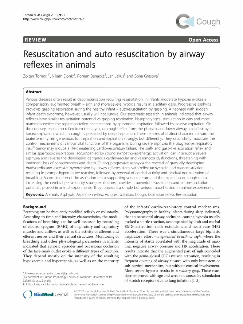
Figure 1 Autoresuscitation by gasping in healthy infant and its failure in neonate with SIDS. In a normal infant an inhalation of lower O2
and/or higher CO2 content in air evokes hypoxia with occurrence of solitary sighs or gasps. The sigh is accompanied by a startle reaction, whichmanifests with neck extension and upper airway dilation, resulting in normalization of breathing. Severe hypoxia provokes solitary gaspsaccompanied by cortical arousal and movements. Progressive asphyxia provokes a period of gasping respiration contributing to autoresuscitationby gasping. Similar asphyxia in a risky preterm baby (including SIDS during a critical period after birth), does not evoke solitary sighs with startleor gasps, but results in silent death. Reproduced with permission and modified from [31].
Tomori et al. Cough 2013, 9:21 Page 2 of 12http://www.coughjournal.com/content/9/1/21
many types of receptors for 5-HT, indicating independ-ence of gasping on levels of serotonin [11]. On the otherside, HR recovery failed at a critical age in 5 HT deficientmice exposed to episodic anoxia [12].The neuro-genesis of gasping is dependent on the dis-
charge of neurons in the rostro-ventral medulla, overlap-ping “the pre-Bötzinger complex” (preBötC). Neuronalactivities of this complex, characterized in an in vitrobrainstem-spinal cord preparation of the neonatal rat,have been hypothesized to underlie respiratory rhythmgeneration. The rhythmic activity of this in vitro prepar-ation is markedly different from eupnoea, but identical withgasping in vivo. Medullary neuronal activities generatingthe gasp and the identical rhythm of the in vitro prepar-ation are incorporated into the ponto-medullary circuitdefining ventilatory activity [13].EMG activity of upper airway (UA) genio-glossal
muscle -GG and the diaphragm (D) were studied inanaesthetized rabbits during progressive asphyxia in-duced by airway occlusion. Peak activity of GG increasedmore than that of the D during hyperpnoea andgasping. These data indicate differences in the controlmechanism of the GG and D during acute severe as-phyxia. Increased UA muscle activity seen during gaspingshould help preserve UA patency, and facilitateautoresuscitation by gasping. These observations supportthe concept that gasping is a highly organized function ofthe respiratory center [14,15]. During severe hypoxia thenetwork properties within the preBötC are reconfigured,whereby it no longer generates eupnoea, but instead gen-erates gasping. Such reconfiguration includes changes insynaptic and intrinsic properties triggered by hypoxiaitself, as well as the influence of differentneuromodulators released during hypoxia. Therefore,gasping respiration has been considered an importantmechanism, that triggers autoresuscitation. Deregulationof gasping has been proposed to result in failure toautoresuscitate and has been hypothesized to contributeto development of SIDS [7,8]. Precisely which synapticand/or neuronal intrinsic membrane properties are criticalto central respiratory rhythmogenesis, in either normoxiaor hypoxia, is still the subject of considerable discussion[13-16].
Protective and defensive airway reflexesFrom several reflexes evoked by stimulation of variousareas of the airways, the following three, illustrated inFigure 2, are especially important for the protection anddefence of the respiratory system. A gasp-like aspirationreflex (AspR) can be regularly evoked by various mech-anical, electrical, and other methods of stimulation ofthe nasopharynx (NPh) in both anaesthetized and non-anaesthetized cats and other mammals. It manifests as asolitary spasmodic inspiration (SI) that only lasts for
150–230 ms, and for its tendency to inhibit expiratoryefforts it is usually not followed by an immediate activeexpiration [17-21]. However, the irritant substances may betransported by the rapid and strong inspiratory airflow, anda muco-ciliary transport from NPh to the hypopharynx,from where they may provoke reflex swallowing or cough.Accidental input of irritants into the larynx usually pro-vokes laryngo-constriction and the expiration reflex (ExpR)or a prompt cough reflex (CR), providing expulsion ofirritants, and so preventing their aspiration into thelungs [22]. The tendency to provoke SIs is high at thebeginning of spontaneous inspiration for ventilatory drive,but it gradually decreases during the inspiratory phase[23]. The AspR can be induced also by negative or positivepressure air puff stimulation of the upper airways [24]. Inparalyzed cats, stimulation of the „irritant” rapidly adaptingreceptors (RARs) of the NPh by mechanical contactsand pressure pulses or air-puffs provokes a very strongactivity in the glossopharyngeal afferent fibers, resultingin AspR. The frequency of afferent impulses is very high(mean 197/s, range 67-330/s) [25], which is >10 timeshigher than during control inspiration. They strongly acti-vate many brainstem inspiratory neurons, including “theinspiratory pattern generator”, described in the brainstemof juvenile rats [26], and the respiratory central patterngenerator (CPG), was analyzed more in detail in severalpapers [12,27,28]. Strong activation of various inspiratoryneurons of adult cats by AspR was identified by c-Fos
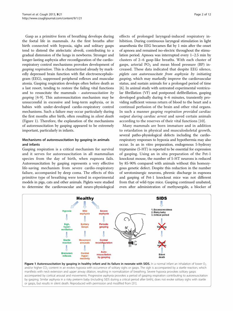
Figure 2 Schematic presentation of the main protective anddefensive reflexes in cats. Airflow record (V’) with inspirationdownward (↓). Stimulation of specific airway areas using mechanicalcontacts with a nylon fiber (———) and presentation of odoroussubstances (□□□) are indicated in anaesthetized cat. In addition togasp- and sniff-like aspiration reflex elicited from the nasopharynx,sniffing from the upper part of nasal cavity, expiration reflex from thelarynx, the cough reflex from the trachea-bronchial region and sneezingfrom the nasal mucosa. Modified from our earlier publications [22,31,40].
Tomori et al. Cough 2013, 9:21 Page 3 of 12http://www.coughjournal.com/content/9/1/21
immuno-reactive method in 14 of 35 tested brainstemnuclei [29].The AspR is characterized by a specific time-
frequency distribution of powerful energy in the phrenicnerve, manifesting with high frequency oscillations [30].Such powerful activation of the “presupposed inspira-tory generator” by the AspR [31] can very effectivelymodify (facilitate or inhibit through dense synaptic con-nections [32] and several mediators the “central mecha-nisms” of various vital functions [31]. Recording andpower spectral analysis of the phrenic and the hypoglossalnerve activities in paralyzed cats, indicated very similarcharacter and peaks during both the hypoxic medullarygasping and the AspR evoked by mechanical stimulationof NPh in normoxic conditions [33,34]. Such close similar-ity of AspR with gasping suggests their similar resuscita-tion potential, resembling autoresuscitation of humaninfants by gasping [4,5,7]. Great similarity and vitality ofAspR with gasping was supported by its persistence atthe absence of CR and ExpR in premortal gasping stageafter medullary transsection 5 mm above the obex in cats[35], fitting the localization of “inspiratory generator”. Theresuscitation potential of AspR was proved by terminationof progressive hypotension and atrio-ventricular (A-V)blockade in a cat, during gasping stage caused by severe
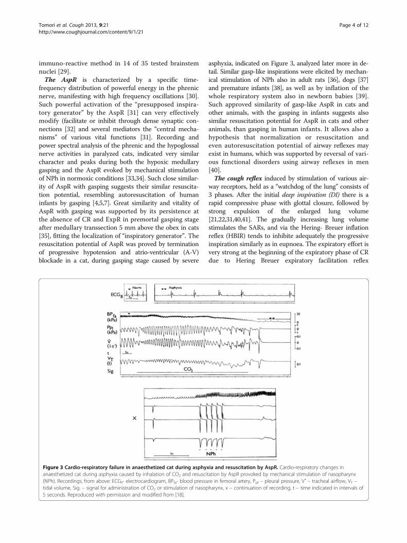
asphyxia, indicated on Figure 3, analyzed later more in de-tail. Similar gasp-like inspirations were elicited by mechan-ical stimulation of NPh also in adult rats [36], dogs [37]and premature infants [38], as well as by inflation of thewhole respiratory system also in newborn babies [39].Such approved similarity of gasp-like AspR in cats andother animals, with the gasping in infants suggests alsosimilar resuscitation potential for AspR in cats and otheranimals, than gasping in human infants. It allows also ahypothesis that normalization or resuscitation andeven autoresuscitation potential of airway reflexes mayexist in humans, which was supported by reversal of vari-ous functional disorders using airway reflexes in men[40].The cough reflex induced by stimulation of various air-
way receptors, held as a “watchdog of the lung” consists of3 phases. After the initial deep inspiration (DI) there is arapid compressive phase with glottal closure, followed bystrong expulsion of the enlarged lung volume[21,22,31,40,41]. The gradually increasing lung volumestimulates the SARs, and via the Hering- Breuer inflationreflex (HBIR) tends to inhibite adequately the progressiveinspiration similarly as in eupnoea. The expiratory effort isvery strong at the beginning of the expiratory phase of CRdue to Hering Breuer expiratory facilitation reflex
Figure 3 Cardio-respiratory failure in anaesthetized cat during asphyxia and resuscitation by AspR. Cardio-respiratory changes inanaesthetized cat during asphyxia caused by inhalation of CO2 and resuscitation by AspR provoked by mechanical stimulation of nasopharynx(NPh). Recordings, from above: ECGII- electrocardiogram, BPfa- blood pressure in femoral artery, Ppl – pleural pressure, V´ – tracheal airflow, VT –tidal volume, Sig. – signal for administration of CO2 or stimulation of nasopharynx, x – continuation of recording, t – time indicated in intervals of5 seconds. Reproduced with permission and modified from [18].
Tomori et al. Cough 2013, 9:21 Page 4 of 12http://www.coughjournal.com/content/9/1/21
(HBEFR), caused by enlarged lung volume [23]. Therefore,the deeper the initial DI, the more powerful or effective isusually the subsequent expiratory effort. During a series ofcough efforts persisting after the end of stimulation,their intensity gradually decreases with the lowering oflung volume, ventilatory drive, peak expiratory flow, andwith prolongation of the second non-active part of the ex-piratory phase of cough. In non-paralyzed subjects, themyotatic reflexes of the respiratory muscles and stimula-tion of various thoracic and abdominal proprioceptors, aswell as irritant RARs and C fibres in the airways and lungs,caused by flow and volume changes, can also contribute tothe modification of the successive cough efforts [31,41].Somato-sensory nerves innervating the chest wall, D andabdominal muscles, as well as nerves of visceral organsalso likely play important role in regulating cough [42].Cough is not a stereotyped output from the medullary“cough center”, but its pattern and strength depend onmany afferent inputs on the “cough center” [43].Experiments in paralyzed cats indicated close functional
connectivity of ventro-lateral medullary neurons withphrenic, lumbar and recurrent laryngeal nerves duringfictive coughing, induced by mechanical stimulation ofthe intrathoracic trachea. During the inspiratory phase,excitation of medullary inspiratory augmenting (IAug)neurons, are connected with activation of phrenic andrecurrent laryngeal nerves. In expiratory phase, the activityof expiratory augmenting (EAug) neurons, are accompaniedby activation of the lumbar and laryngeal nerves [27]. TheCPG undergoes reconfiguration to produce cough. About1/3 of medullary inspiratory decrementing (IDec) neuronsof the caudal ventral respiratory column (cVRC), duringthe first cycle (C1) of cough attack changes to IAug type.This is accompanied by increases of phrenic activity,esophageal pressure and the inspiratory phase duration.Coughing is a rhythmic process persisting also after theending of stimulation, with lower intensity and prolonga-tion. AspR and ExpR are solitary non-rhythmic respiratoryprocesses and for their short durations they often do notdisturbe the fictive cough attack [27,44,45]. Also preventionof proprioceptive reflexes in paralyzed animals eliminatessome modification effects of airway reflexes, describedin anaesthetized non-paralyzed animals [31,46] and theirmodel [47], or chronic cough in patients [48,49].The expiration reflex induced by stimulation of the
larynx is characterized by laryngo-constriction and promptpowerful expiratory effort without any inspiratory in-crease of actual lung volume [50,51]. The ExpR supportsthe CR to protect the airways and lungs from airborneand inhaled pathogens, allergens, aspirate and otherirritants. Acute and chronic cough is a frequent symptomof respiratory tract irritations and disease, such as gastro -oesophageal reflux disease, asthma and COPD in humans[52]. Aspiration of pathogens present in foreign materials
or secretions into the lungs, represents the most danger-ous complication in patients with acute stroke, Parkinson’sdisease, comatose states, long-term invasive artificialventilation, nasogastric feeding, recurrent respiratoryinfections and dysphagia, resulting in aspiration pneumo-nia, particularly in the elderly [48,52-54]. Foreign materialsand secretes containing viruses can be ejected by astrong expiratory effort up to 9 meters, and a patientcan infect many co-passengers during airplane traveland can disseminate influenza and other respiratoryinfections to various destinations. Feeding through anasogastric tube offers only limited protection againstaspiration pneumonia, developing already after few daysof illness in patients with dysphagia after acute stroke [55].This may result from unwanted reflex aspiration provokedby nasopharyngeal stimulation with the catheter. There-fore, the down- and up- regulation of CR and ExpR mayhave a very positive, both preventive and therapeuticeffects in patients. Testing the efficacy of CR in variouspathological conditions can be useful for the assessment ofthe risk, as well as prevention and treatment of aspirationpneumonia [53-55]. Particularly the ExpR induced fromthe larynx, accompanied by glottal closure, is the mainreflex that prevents aspiration [54,55]. Reliable ambulatorycounter for cough have been developed for evaluation ofcough count and intensity. Together with self-scoringevaluation of cough severity and impact on quality of life,it can be useful for diagnostic and antitussive therapy [56].Similar short reflex expirations (ExpR) can be induced alsoby mechanical stimulation of the trachea in anaesthetizedcats [57].
Function of respiratory central pattern generator inairway reflexesIn defensive airway behaviours the laryngeal motoneuronsare multifunctional in cats. During fictive CR the inspiratorylaryngeal motoneurons (ILM) and expiratory laryngealmotoneurons (ELM), responsible for successive glottaldilation, closure, opening, and narrowing are active, parallelwith the phrenic (PHR) or abdominal (ABD) motoneurons,contributing to the inspiratory or expiratory phase ofCR, respectively. During the fictive AspR there is a strongactivation not only of the PHR but also the ILM controllingthe glottal dilators, and also the styloglossus muscle,providing tongue-back elevation. These activities providean explosive inspiratory airflow during the AspR [58].According to a recent computational biomechanical modelmany components of the raphe’-ponto-medullary systemparticipate in the realization of the CR. This model basedon successive transsections in rats and complementarycalculations using 64 equations, resembles in vivo condi-tions. The DI caused by activation of PHR is accompaniedby glottal dilation, provided by ILM. The expiratory phaseof CR starts by activation of ELM and ABD motoneurons
Tomori et al. Cough 2013, 9:21 Page 5 of 12http://www.coughjournal.com/content/9/1/21
for glottal closure and production of strong expiratoryeffort, followed by activation of both ILM responsible forglottal opening, and ABD motoneurons providing rapidexpiratory airflow. The intensity of second and furthercough efforts gradually decreases, because of inactivationof high-pressure ILM, connected with successive loweringof peak lung volume, peak alveolar pressure, peak abdom-inal pressure and drive, resulting in lower peak expiratoryflow [59].Gradual transsection experiments in 4 weak old rats
indicated, that the respiratory CPG has 3 rhythmogeniccapabilities, realized by various neurons in 3 interactingstructures. The intact ponto-medullary complex producesa three-phase rhythm, consisting of inspiration, post-in-spiration and expiration. After elimination of the pons thePreBötC and the BötC of the medullary complex producea two-phase rhythm, consisting from inspiration and expir-ation. After elimination of BötC the remaining PreBötC(proposed to be a “kernel” of respiratory rhythmogenesis),together with the rostral Ventral Respiratory Column(rVRC) produces one-phase rhythm of rarer solitaryinspirations, influenced by hypercapnia [60], resemblinggasping respiration.A new computational model indicates that the regulation
of respiratory rate and breathing pattern, provided by
brainstem respiratory network, can be substantiallyinfluenced also by pulmonary and pontine feedback loops[61]. Similar model would be very useful for explanationof reconfiguration of breathing control by AspR duringthe stage of premortal gasping respiration, oftenresulting from asphyxia and providing autoresuscitationin both animals [6] and infants [7]. Mechanical stimulationof the NPh provokes AspR not only in severe hypoxia,manifesting by gasping, but also during eupnoea. Thevery similar character of AspR and gasping suggests thatAspR transiently suppresses the brainstem mechanism re-sponsible for neurogenesis of eupnoea, and activates thosefor gasping. The intensity of repeated AspR-es and accom-panying inspiratory processes decrease only for ~4 cycles,but rapidly normalizes [62]. This special ability mightallow use of AspR for experimental and model studies ofautoresuscitation in general. It would be useful to ex-plain the mechanism of reconfiguration of CPG pro-voking AspR by simple NPh stimulation, even in thestage of premortal gasping respiration in cats [35]. Prac-tical application of powerful resuscitation potential ofAspR in animals [19,31,35], and various revitalisation ef-fects of its voluntary surrogate, represented by sniff inhumans [40], seem to be very perspective.
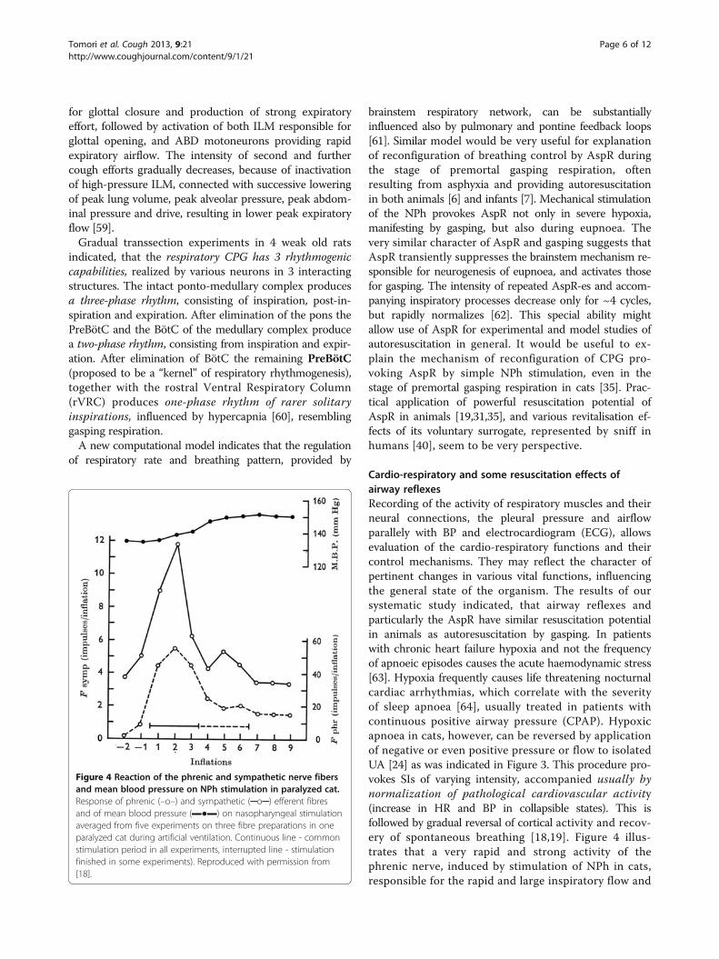
Cardio-respiratory and some resuscitation effects ofairway reflexesRecording of the activity of respiratory muscles and theirneural connections, the pleural pressure and airflowparallely with BP and electrocardiogram (ECG), allowsevaluation of the cardio-respiratory functions and theircontrol mechanisms. They may reflect the character ofpertinent changes in various vital functions, influencingthe general state of the organism. The results of oursystematic study indicated, that airway reflexes andparticularly the AspR have similar resuscitation potentialin animals as autoresuscitation by gasping. In patientswith chronic heart failure hypoxia and not the frequencyof apnoeic episodes causes the acute haemodynamic stress[63]. Hypoxia frequently causes life threatening nocturnalcardiac arrhythmias, which correlate with the severityof sleep apnoea [64], usually treated in patients withcontinuous positive airway pressure (CPAP). Hypoxicapnoea in cats, however, can be reversed by applicationof negative or even positive pressure or flow to isolatedUA [24] as was indicated in Figure 3. This procedure pro-vokes SIs of varying intensity, accompanied usually bynormalization of pathological cardiovascular activity(increase in HR and BP in collapsible states). This isfollowed by gradual reversal of cortical activity and recov-ery of spontaneous breathing [18,19]. Figure 4 illus-trates that a very rapid and strong activity of thephrenic nerve, induced by stimulation of NPh in cats,responsible for the rapid and large inspiratory flow and
Figure 4 Reaction of the phrenic and sympathetic nerve fibersand mean blood pressure on NPh stimulation in paralyzed cat.Response of phrenic (–o–) and sympathetic (─o─) efferent fibresand of mean blood pressure (▬●▬) on nasopharyngeal stimulationaveraged from five experiments on three fibre preparations in oneparalyzed cat during artificial ventilation. Continuous line - commonstimulation period in all experiments, interrupted line - stimulationfinished in some experiments). Reproduced with permission from[18].
Tomori et al. Cough 2013, 9:21 Page 6 of 12http://www.coughjournal.com/content/9/1/21
volume of AspR, is accompanied by a strong activationof the sympathetic efferent fibers. This results in reflextachycardia and severe vasoconstriction, causing amarked hypertensive reaction in paralyzed cats [18],demonstrating the revitalization effects of AspR.Similar vasomotor reflexes can be provoked easily also
by stimulation of cold receptors of the face and upperairways. Apnoea and bradycardia, followed usually by deepinspiration, peripheral vasoconstriction, and a systemichypertensive reaction with redistribution of the blood tovital organs, are the main effects of this diving reflex.Stimulation of the orbito-frontal region of the face withcold water of 5°C for 5 s, reversed a supra-ventriculartachycardia from 300/min to 120/min in a neonate [65].The diving reflex has a strong sympathetic componentaccompanying DI, in addition to other effects. Therefore, itproved to be very useful as a basic life support under
collapsible states, to postpone and/or prevent an imminentloss of consciousness and a subsequent ischemic-hypoxicbrain damage. From various methods of therapeutichypothermia, widely used in polytraumatic patients theNPh balloon technique proved to be very effective [66]. Inaddition to direct cooling of caudal brain structures,also the NPh cold and mechanoreceptor stimulationmay participate in the strong cerebral vasoconstriction,preventing ischemic and hypoxic brain damages.Figure 3 illustrates a powerful resuscitation effect of
AspR in a cat. Inhalation of CO2 after reactive transienthyperventilation evoked a gradual development of asphyxiaand cardio-respiratory failure. It manifested by SIs andasphyxia culminating in apnoea, followed by repeatedgasps. The asphyxia provoked severe bradycardia andextreme hypotension, accompanied by a progressive devel-opment of A-V blockade. In this agonal state each contact
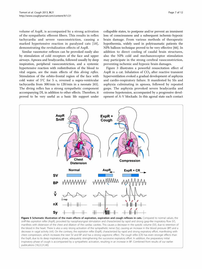
Figure 5 Schematic illustration of the main effects of aspiration, expiration and cough reflexes in cats. Compared to normal values, thesniff-like aspiration reflex (AspR), provoked by nasopharyngeal stimulation and characterized by rapid and strong gasp-like inspiratory flow (V’),manifests with distension of the chest and dilation of the cardiac cavities. This causes a decrease in the systolic volume (SV), due to retention ofthe blood in the heart. There is also a very strong activation of the sympathetic nerve (Sy), causing an increase in the blood pressure (BP) and adecrease in vagal activity (nX). On the contrary, the expiration reflex (ExpR), characterized by rapid and strong expiratory effort, manifesting withchest compression, which increases the next SV and BP and has a strong vagotonic effect. The cough reflex (CR) has even stronger effects thanthe ExpR, due to its deep inspiratory phase, adequately strengthening the successive expiratory effort. In addition, the preparatory initialinspiratory phase of cough is accompanied by a sympathetic activation, resulting in an increase in BP. Combined from results of our earlierpublications [18,22,31,40].
Tomori et al. Cough 2013, 9:21 Page 7 of 12http://www.coughjournal.com/content/9/1/21
stimulation of the NPh mucosa elicited AspR-es, whichwere even stronger than the preceding repeated gaspsand resulted in cardio-respiratory resuscitation [19].Similar interesting case report published in 1991 indicated,that a present paroxysmal supraventricular tachycardia wasterminated during introduction of a nasogastric catheterfor gastric juice collection in a patient [67]. Recently, NPhaspiration by a catheter proved to be a treatment optionfor supraventricular paroxysmal arrhythmia in infants [68].Clinical observations indicate, that also hiccough attackscan be easily terminated by introduction of a NPh catheterin patients [69]. Voluntary “coughing on demand”, pro-posed by Criley et al. [70] is frequently used for preventionand treatment of anaphylactic collapse, occurring duringfunctional magnetic resonance (FMR) examination and inother collapsible states.Mechanical stimulations of NPh by a nylon fibre through
a pharyngostomic opening, in anaesthetized adult ratsalso evoked SIs, very similar to AspR of cats. They werecharacterized by increase in tidal volume, airflow andminute volume, compared to control breaths. SuchAspR-es provoked by mechanical stimulations, repeated
in cycles of 16 s, significantly decreased the higher HRand the number of extrasystoles, caused by intraperitonealapplication of aconitin [36]. AspR has various powerfulnormalization and restorative effects, which can inhibitdifferent spastic events, such as bronchoconstriction andlaryngospasm, as well as interrupts hypoxic apnoea, andinhibits the number and intensity of the cough efforts, atleast in cats. Therefore, AspR as a model of SIs alone orcombined with forced expiration (ExpR) or prompt cougheffort (without preceding inspiration), can serve for rever-sal of many functional disorders, as indicated in our recentreview [40]. The main effects of AspR, ExpR and CR areschematically illustrated in Figure 5.
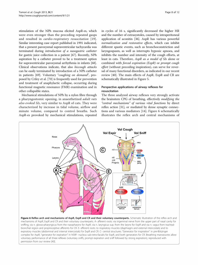
Perspective applications of airway reflexes forresuscitationThe three analyzed airway reflexes very strongly activatethe brainstem CPG of breathing, effectively modifying the“central mechanisms” of various vital functions by directreflex action [31], or mediated by dense synaptic connec-tions and various mediators [14]. Figure 6 schematicallyillustrates the reflex arch and central mechanisms of
Figure 6 Reflex arch and mechanisms of AspR, ExpR and CR and their voluntary counterparts. Schematic illustration of the reflex arch andmechanisms of AspR, ExpR and CR and their voluntary counterparts. A- afferent roots: via trigeminal nerve from the upper part of nasal cavity forsniffing, via n. glosso-pharyngicus from the nasopharynx for AspR, via n. laryngicus sup. from the larynx for ExpR and via n. vagus from tracheal-bronchial region and proprioceptive afferents for CR. E- efferent roots: to inspiratory muscles (diaphragm and external intercostals) and toexpiratory muscles (abdominal and internal intercostals) for ExpR and CR. C- central structures: “Generator for inspiration” in pre-Bötzingercomplex for AspR, “generator for expiration” in NSRF- nucleus sub-retro-facialis for ExpR, and both generators for CR. Breathing manoeuvres allowvoluntary performance of all three reflexes (voluntary sniffs, prompt expiration and sniff followed by strong expiration), reproduced withpermission from our review [40].
Tomori et al. Cough 2013, 9:21 Page 8 of 12http://www.coughjournal.com/content/9/1/21
AspR, ExpR and CR. The effects of airway reflexes maypromote normalization of hypo- and hyper-functionaldisorders, if not hindered by a presence of severe or fixedchanges (e.g., acute stroke or recent myocardial infarction),both in animal experiments and probably also in humanstudies. AspR and ExpR in cats reversed several life-threatening disorders of functional character, manifestingas hypoxic apnoea termination and cardio-pulmonary-cere-bral-resuscitation (CPCR) [19,31]. Therefore, the powerfulAspR might influence positively also the disbalance be-tween the excitatory and inhibitory cardio-respiratoryimpulses, deciding for survival or death in the patho-mechanism of SIDS, according to the hypothesis of Leiterand Böhm [71]. The powerful stimulatory effect of AspR,therefore, might support the survival of dying animals, aswell as SIDS infants and adult patients, particularly inemergency situations and when there is no immediatehealth-care service on site. This effect results in a decreaseof the number and intensity of disturbing cough efforts inanaesthetized cats [46]. In addition to strong gasp-likeinspirations provoked by NPh mechanical stimulation,mediated by brainstem central control mechanismsparticipating in gasping, AspR is characterized also byreciprocal inhibition of expiratory activity.Stimulation of the larynx both in cats and humans
strongly activates the higher located brainstem expira-tory mechanisms, causing laryngo-constriction andprompt expiratory effort without preceding inspiration.Similar short reflex expirations can be elicited also by
tracheal stimulation in anaesthetized cats [57]. These ef-forts prevent intrusion of irritant materials and secre-tions into the lungs and provide their expulsion.Stimulation of the trachea-bronchial mucous membraneactivates both the inspiratory and expiratory centralmechanisms, provoking DI followed by powerful expira-tory effort. The DI of CR provides venous return to theheart, and the successive strong expiratory effort sup-ports brain perfusion, preventing and/or terminatingvarious collapsible states [31]. Such “on demand” provo-cations of CR might be very useful for study of CPCR inmodel experiments with dysrhythmias, including VF,particularly in animals, but supposedly also in clinicalstudies. Recent results indicate that already spontaneousgasps restored the cerebral blood flow (CBF) to 59% ofthe control values in animals. There was a very signifi-cant correlation of the CBF with the decreases in intra-thoracic pressure during the inspiratory phase of gaspsand with the increases of aortic pressure during theirexpiratory phase. Spontaneous gasps producing signifi-cant increases in the CBF during untreated cardiac arrest,confirmed the beneficial resuscitative effect of gaspingduring the cardiac arrest [72]. Experiences obtained instudies with CR in animals have relevance for humancough research [52]. The gradual decrease in intensity ofsuccessive cough efforts can be explained by HBEFR,which is very strong at the beginning of the expiratoryperiod, reflecting the momentary relatively large lungvolume [23].
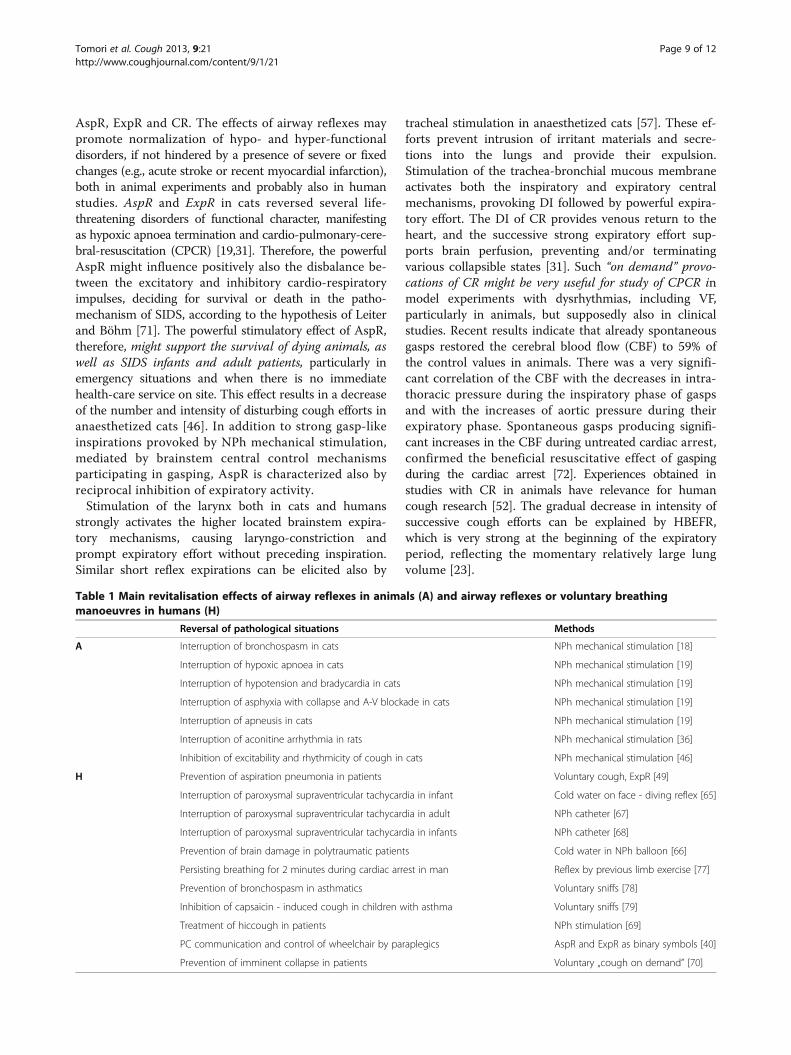
Table 1 Main revitalisation effects of airway reflexes in animals (A) and airway reflexes or voluntary breathingmanoeuvres in humans (H)
Reversal of pathological situations Methods
A Interruption of bronchospasm in cats NPh mechanical stimulation [18]
Interruption of hypoxic apnoea in cats NPh mechanical stimulation [19]
Interruption of hypotension and bradycardia in cats NPh mechanical stimulation [19]
Interruption of asphyxia with collapse and A-V blockade in cats NPh mechanical stimulation [19]
Interruption of apneusis in cats NPh mechanical stimulation [19]
Interruption of aconitine arrhythmia in rats NPh mechanical stimulation [36]
Inhibition of excitability and rhythmicity of cough in cats NPh mechanical stimulation [46]
H Prevention of aspiration pneumonia in patients Voluntary cough, ExpR [49]
Interruption of paroxysmal supraventricular tachycardia in infant Cold water on face - diving reflex [65]
Interruption of paroxysmal supraventricular tachycardia in adult NPh catheter [67]
Interruption of paroxysmal supraventricular tachycardia in infants NPh catheter [68]
Prevention of brain damage in polytraumatic patients Cold water in NPh balloon [66]
Persisting breathing for 2 minutes during cardiac arrest in man Reflex by previous limb exercise [77]
Prevention of bronchospasm in asthmatics Voluntary sniffs [78]
Inhibition of capsaicin - induced cough in children with asthma Voluntary sniffs [79]
Treatment of hiccough in patients NPh stimulation [69]
PC communication and control of wheelchair by paraplegics AspR and ExpR as binary symbols [40]
Prevention of imminent collapse in patients Voluntary „cough on demand” [70]
Tomori et al. Cough 2013, 9:21 Page 9 of 12http://www.coughjournal.com/content/9/1/21
Long-term resuscitation effects of airway reflexesLonger-lasting asphyxia may result in gradual decom-pensation of separate vital functions in general. Severeinspiratory resistive loading induces cardio-respiratoryfailure and not just an initial respiratory decompensationin anaesthetized rats [73]. A reduced respiratory activityresults in increased ventilatory drive and evokes aninactivity induced respiratory facilitation with gradualrestoration of breathing. Such breathing efforts wereconnected with an increase of mean BP, which proved tobe the principal compensating factor in response to thiscardio-respiratory failure, and supported generation ofpeak tracheal pressure [74,75]. Respiratory facilitationmanifests also after spinal cord injury as crossed phrenicphenomenon [76]. Similar complex cardiovascular effectsof resuscitation provided the required blood pressureincrease, which started the reversal of cardio-respiratoryfailure, induced by severe hypoxia also in anaesthetizedcats [31]. Similar facilitation was observed also during ex-perimental VF in anaesthetized sheeps. Previous reflectoricmovements of limbs caused that the thoraco-abdominalpump persisted to function for about 2 minutes in spite oftotal cardiac arrest, and caused synchronous changes inBP [77]. Coughing comprising initial DI, can allow boththe maintenance of the venous return to the heart aswell as brain perfusion, at least several minutes, whichis sufficient for persistence of wakefulness, if needed.Therefore, voluntary rapid SIs each followed by promptforced expiration (ExpR), might provide a powerful poten-tial for cardio-pulmonary- cerebral resuscitation particularlyin syncopal states, various collapses and even for preventionof sudden cardiac arrest [31,40]. Application of airwayreflexes proved to reverse pathological situations (inanimals 7 disorders) and (in humans 11 disorders), includ-ing asthma [78,79], indicated in a Table 1. However,there are still many open questions in this importanttopic.
Abbreviations5HT: 5 hydroxy tryptamine; AspR: Aspiration reflex; A-V: Atrio-ventricular;BötzC: Bötzinger complex; CBF: Cerebral blood flow; CD: Cardiac death;CPG: Central pattern generator; CPCR: Cardio pulmonary cerebralresuscitation; CR: Cough reflex; DI: Deep inspiration; ECG: Electrocardiogram;EEG: Electroencephalogram; EMG: Electromyogram; ExpR: Expiration reflex;FMRI: Functional magnetic resonance imaging; HBEFR: Hering-Breuerexpiration facilitating reflex; HBIR: Hering-Breuer inflation reflex; GG: Genioglossus muscle; NPh: Nasopharynx; NSRF: Nucleus sub-retro-facialis;Ppl: Pleural pressure; preBötzC: preBötzinger complex; RARs: Rapidly adaptingreceptors; SARs: Slowly adapting receptors; SI: Spasmodic inspiration;SIDS: Sudden infant death syndrome; UA: Upper airways; V’: Airflow;VF: Ventricular fibrillation; VT: Tidal volume.
Competing interestsZoltan Tomori and Viliam Donic were consultants of Nasophlex Slovakia, s.r.o.from January 2007 for testing of some effects of airway reflexes, which are inprogress with use of 3 patent applications of a resuscitation devicestimulating noninvasively the nasopharynx, sensitive points of the nasal filterand the external ear in sleep apnoea patients. All authors declare that theyhave no competing interests with the preparation of this paper. The patent
“Resuscitation device and method for resuscitation” was approved forAustralia no: 2006351860, for Canada no: 2,672,731, for Europe no:12167089.3-2305, in progress for USA no: 2010/0042179 A1, and Internationalno: WO 2008/072948-A1.
Authors’ contributionsZT developed the design of the experiments and the manuscript and VD, RB,SG and JJ equally helped him to study the mechanisms of the cough,aspiration and expiration reflexes and to complete the manuscript. Allauthors read and approved the final manuscript and informed thecorresponding author with their consent.
Authors’ informationViliam Donic, Roman Benacka, Jan Jakus and Sona Gresova are co-authors.
AcknowledgementsThe implementation of experiences obtained during the complex study ofairway reflexes summarized in a concentrated form in this review waspossible by close cooperation with prof. emeritus Imrich Ivanco MD, PhDand late prof. Juraj Korpas, MD, DrSc. at the Faculty of Medicine PJ SafarikUniversity, Kosice, Slovakia and later with Juraj Korpas, Prof. Jan Jakus, MD,DrSc, Prof. Kamil Javorka, MD, DrSc and others at the Jessenius Faculty ofMedicine in Martin, Comenius University, Bratislava, Slovakia, as well as withlate prof. John G. Widdicombe from Oxford University during one yearsabbatical study state, granted by Welcome Foundation, and later at LondonUniversity, UK. Many thanks for their valuable collegial cooperation. We thankIng. Martin Kundrík and Assist. prof. Maria Pallayova, MD, PhD for technicalhelp with preparation of the manuscript. The research was supported andapproved by Slovak Grant Agency for Research (APVV) No. 20.047705 andNo. 0189–11, as well as COST action B26. The Ethical committees of theFaculty of Medicine, PJ Safarik University and of the University Hospital,Kosice, and of the Jessenius Faculty of Medicine in Martin, Slovakia gave awritten consent to the analyzed research activities.
Author details1Department of Human Physiology Faculty of Medicine, University of PJSafarik, Kosice, Slovakia. 2Department of Pathophysiology, Faculty ofMedicine, University of PJ Safarik, Kosice, Slovakia. 3Jessenius Faculty ofMedicine in Martin, Comenius University, Bratislava, Slovakia.
Received: 6 September 2012 Accepted: 19 August 2013Published: 22 August 2013
References1. Wulbrand H, Von Zezschwitz G, Bentele KH: Submental and diaphragmatic
muscle activity during and at resolution of mixed and obstructiveapnoeas and cardiorespiratory arousal in preterm infants. Pediatr Res1995, 38:298–305.
2. Wulbrand H, Mc Namara F, Thach BT: The role of arousal relatedbrainstem reflexes in causing recovery from upper airway occlusion ininfants. Sleep 2008, 31:833–840.
3. Sanchez I, Vega-Briceño L, Muñoz C, Mobarec S, Brockman P, Mesa T, HarrisP: Polysomnographic findings in 320 infants evaluated for apneic events.Pediatr Pulmonol 2006, 41:215–221.
4. Jacobi MS, Thach BT: Effect of maturation on spontaneous recovery fromhypoxic apnea by gasping. J Appl Physiol 1989, 66:2384–2390.
5. Thach BT, Gershan WR, Jacobi MS: Control of breathing during asphyxiaand autoresuscitation. In Developmental neurobiology of breathing. Editedby Haddad GG, Farber JP. Basel: Marcel Dekker, New York; 1991:681–699.
6. Sanocka UM, Donnelly DF, Haddad GG: Autoresuscitation: a survivalmechanism in piglets. J Appl Physiol 1992, 73:749–753.
7. Sridhar R, Thach BT, Kelly DH, Henslee JA: Characterization of successfuland failed autoresuscitation in human infants, including those dying ofSIDS. Pediatr Pulmon 2003, 36:113–122.
8. Thach BT: The role of respiratory control disorders in SIDS. Respir PhysiolNeurobiol 2005, 149:343–353.
9. McNamara F, Lijovska AS, Thach BT: Spontaneous arousal activity ininfants during NREM and REM sleep. J Physiol 2002, 538:263–269.
10. Xie J, Weil MH, Sun S, Yu T, Tang W: Spontaneous gasping generatescardiac output during cardiac arrest. Crit Care Med 2004, 32:238–240.
Tomori et al. Cough 2013, 9:21 Page 10 of 12http://www.coughjournal.com/content/9/1/21
11. St-John WM, Li A, Leiter JC: Genesis of gasping is independent of levels ofserotonin in the Pet-1 knockout mouse. J Appl Physiol 2009, 107:679–685.
12. Onimaru H, Homma I: Two modes of respiratory rhythm generation inthe newborn rat brainstem-spinal cord preparation. Adv Exp Med Biol2008, 605:104–108.
13. St John WM: Medullary regions for neurogenesis of gasping: noeud vitalor noeuds vitals? J Appl Physiol 1996, 81:1865–1877.
14. Mathew OP, Thach BT, Abu-Osba YK, Brouillette RT, Roberts JL: Regulationof upper airway maintaining muscles during progressive asphyxia.Pediatr Res 1984, 18:819–822.
15. Peña F: Neuronal network properties underlying the generation ofgasping. Clin Exp Pharmacol Physiol 2009, 36:1218–1228.
16. Fong AY: Postnatal changes in the cardiorespiratory response and abilityto autoresuscitate from hypoxic and hypothermic exposure in mammals.Respir Physiol Neurobiol 2010, 174:146–155.
17. Tomori Z: Pleural, tracheal and abdominal pressure variations indefensive and pathologic reflexes of the respiratory tract. PhysiolBohemoslov 1965, 14:84–95.
18. Tomori Z, Widdicombe JG: Muscular, bronchomotor and cardiovascularreflexes elicited by mechanical stimulation of the respiratory tract.J Physiol 1969, 200:25–49.
19. Tomori Z: The sniff-like aspiration reflex. In Korpas J, Tomori Z: Cough andother respiratory reflexes, Progress in Respiratory Research Vol. 12. Edited byKarger company Basel. München, Paris, London, New York, Sydney: KargerBasel; 1979:234–250.
20. Benacka R, Tomori Z: The sniff-like aspiration reflex evoked by electricalstimulation of the nasopharynx. Respir Physiol 1995, 102:163–174.
21. Jakus J, Tomori Z, Stransky A: Neuronal determinants of breathing, coughingand other motor behaviours. Wist: Martin; 2004.
22. Korpas J, Tomori Z: Cough and other respiratory reflexes. Basel: Kargercompany; 1979.
23. Marek W, Muckenhoff K, Prabhakar NR: Significance of pulmonary vagalafferents for respiratory muscle activity in the cat. J Physiol Pharmacol2008, 59(Suppl. 6):407–420.
24. Tomori Z, Donic V, Kurpas J, Palenikova R: Sniff-like aspiration reflexevoked by pressure pulses from the upper airways in cats. Respir Physiol1994, 96:163–175.
25. Nail BS, Sterling GM, Widdicombe JG: Epipharyngeal receptors respondingto mechanical stimulation. J Physiol 1969, 204:91–98.
26. Janczewski WA, Feldman JL: Distinct rhythm generators forinspiration and expiration in the juvenile rat. J Physiol 2006,570:407–420.
27. Bolser DC, Poliacek I, Jakus J, Fuller DD, Davenport PW: Neurogenesis ofcough, other airway defensive behaviors and breathing: a holarchicalsystem? Respir Physiol Neurobiol 2006, 152:255–265.
28. Rubin JE, Shevtsova NA, Ermentrout GB, Smith JC, Rybak IA: Multiplerhythmic states in a model of the respiratory central pattern generator.J Neurophysiol 2009, 101:2146–2165.
29. Jakus J, Halasova E, Poliacek I, Tomori Z, Stransky A: Brainstem areasinvolved in the aspiration reflex: c-Fos study in anaesthetized cats.Physiol Res 2004, 33:703–717.
30. Knociková J: Time-frequency energy distribution of phrenic nervedischarges during aspiration reflex, cough and quiet inspiration.Comput Programs, methods Biomed 2010, 102:81–90.
31. Tomori Z, Poliacek I, Jakus J, Widdicombe JG, Donic V, Benacka R,Gresova S: Distinct generators for aspiration and expirationreflexes: localization, mechanisms and effects. J Physiol Pharmacol2010, 61:5–12.
32. Tang W, Pagliardiny S, Yang P, Janczewski WA, Feldman JL: Projections ofpreBötzinger complex neurons in adults rats. J Comp Neurol 2010,518:1862–1878.
33. Fung ML, StJohn WM, Tomori Z: Reflex recruitment of medullary gaspingmechanisms in eupnoea by pharyngeal stimulation in cats. J Physiol1994, 475:519–529.
34. Tomori Z, Fung ML, Donic V, Donicova V, St.John WM: Powerspectral analysis of respiratory reflexes to pharyngeal stimulationin cats: comparisons with eupnoea and gasping. J Physiol 1995,485:551–559.
35. Jakus J, Tomori Z, Bosel’ova L, Nagyova B, Kubinec V: Respiration andairway reflexes after transversal brainstem lesions in cats. PhysiolBohemoslov 1987, 36:329–340.
36. Gresova S, Vrbenska A, Tomori Z, Donic V, Kurpas M: Provocation ofaspiration reflex and confirmation of its antiarrhythmic effect in rats(in Slovak), In proceedings of Slovak and Czech conference on sleep. Martin;2007. Abstract p 13. ISBN 978-80-88866-46-6.
37. Tomori Z, Lemáková S, Holecyová A: Defensive reflexes of the respiratorytract in dogs. Physiol Bohemoslov 1977, 26:49–54.
38. Javorka K, Tomori Z, Zavarská L: Protective and defensive airway reflexesin premature infants. Physiol Bohemoslov 1980, 29:29–35.
39. Cross K, Klaus M, Tooley WH, Weisser K: The response of the new-bornbaby to inflation of the lungs. J Physiol (Lond). 1960, 151:551–565.
40. Tomori Z, Donic V, Benacka R, Gresova S, Peregrim I, Kundrik M, Pallayova M,Jakus J: Reversal of functional disorders by aspiration, expiration, andcough reflexes and their voluntary counterparts. Front. Respir. Physio.2012, article 00467:1–14. doi:10.3389/fphys. 2012.
41. Widdicombe J: Historical perspective: reflexes from the lungs andairways. J Appl Physiol 2006, 101:628–634.
42. Canning BJ: Anatomy and neurophysiology of the cough reflex: ACCPevidence-based clinical practice guidelines. Chest 2006, 129(1):33S–47S.
43. Widdicombe JG, Tatar M, Fontana G, Hanacek J, Davenport P, Lavorini F,Bolser D et al: Workshop: tuning the ‘cough center’. Pulm Pharmacol Ther2011, 24:344–352.
44. Shannon R, Baekey DM, Morris KF, Li Z, Lindsey BG: Functional connectivityamong ventrolateral medullary respiratory neurones and responsesduring fictive cough in the cat. J Physiol 2000, 525(Pt 1):207–224.
45. Segers LS, Nuding SC, Vovk A, Pitts T, Baekey DM, O’Connor R, Morris KF,Lindsey BG, Shannon R, Bolser DC: Discharge identity of medullaryinspiratory neurons is altered during repetitive fictive cough. FrontPhysiol 2012, 3:223.
46. Poliacek I, Jakus J, Simera M, Barani H, Visnovcova N, Halasova E,Tomori Z: Excitability and rhythmicity of tracheobronchial cough isaltered by aspiration reflex in cats. J Physiol Pharmacol 2009, 60(Suppl 5):105–110.
47. Poliacek I, Morris KF, Lindsey BG, Segers LS, Rose MJ, Corrie LW, Wang C,Pitts TE, Davenport PW, Bolser DC: Blood pressure changes altertracheobronchial cough: computational model of the respiratory-coughnetwork and in vivo experiments in anesthetized cats. J Appl Physiol2011, 111:861–873.
48. Teramoto S, Ishii T, Yamamoto Y, et al: Significance of chronic cough as adefense mechanism or a symptom of elderly patients with aspirationand aspiration pneumonia. Eur Respir J 2005, 24:10–12.
49. Widdicombe JG, Addington WR, Fontana RA, Stephens RE: Voluntary andreflex cough and the expiration reflex, implications for aspiration. PulmPharmacol Ther 2011, 3:312–317.
50. Korpas J: Expiration reflex from the vocal folds. Physiol bohemoslov 1972,21:671–675.
51. Korpas J: The expiration reflex. In Korpas J, Tomori Z: Cough and otherrespiratory reflexes, Progress in Respiratory Research Vol. 12. Edited by Kargercompany Basel. München, Paris, London, New York, Sydney: Karger Basel;1979:189–217.
52. Canning BJ: The cough reflex in animals: relevance to human coughresearch. Lung 2008, 186(Suppl 1):S23–S28.
53. Addington WR, Stephens RE, Widdicombe JG, Rekab K: Effect of strokelocation on the laryngeal cough reflex and pneumonia risk. Cough 2005,1:4. doi:10.1186/1745-9974-1-4.
54. Ebihara S, Ebihara T: Cough in the elderly: a novel strategy for preventingaspiration pneumonia. Pulm Pharmacol Ther 2011, 24:318–323.
55. Dziewas R, Ritter M, Schilling M, Konrad C, Oelenberg S, Nobavi DG,Stogbauer S, Ringestein EB, Ludemann P: Pneumonia in acute strokepatients fed by nasogastric tube. J Neurol Neurosurg Psychiatry 2004,75:852–856.
56. Chung KF: Assessment and measurement of cough: the value of newtools. Pulm Pharmacol Ther 2002, 15:267–272.
57. Poliacek I, Rose MJ, Corrie LW, Wang C, Jakus J, Barani H, Stransky A, PolacekH, Halasova E, Bolser DC: Short reflex expirations (expiration reflexes)induced by mechanical stimulation of the trachea in anesthetized cats.Cough 2008, 4:1. doi:10.1186/1745-9974-4-1.
58. Shiba K, Satoh I, Kobayashi N, Hayashi F: Multifunctional laryngealmotoneurons: an intracellular study in the cat. J Neurosci 1999,19:2717–2727.
59. O’Connor R, Segers LS, Morris KF, Nuding SC, Pitts T, Bolser DC, DavenportPW, Lindsey BG: A joint computational respiratory neural network -
Tomori et al. Cough 2013, 9:21 Page 11 of 12http://www.coughjournal.com/content/9/1/21
biomechanical model for breathing and airway defensive behaviors.Front Physiol 2012, 3:264. doi:10.3389/fphys.2012.00264.
60. Smith JC, Abdala AP, Koizumi H, Rybak IA, Paton JFJ: Spatial and functionalarchitecture of the mammalian brain stem respiratory network: ahierarchy of three oscillatory mechanisms. Neurophysiol 2007,98(6):3370–3387.
61. Molkov YI, Bacak BJ, Dick TE, Rybak IA: Control of breathing by interactingpontine and pulmonary feedback loops. Front Neural Circuits. 2013, 7:16.doi:10.3389/fncir.2013.00016.
62. Fung ML, St John WM: Expiratory neural activities in gasping induced bypharyngeal stimulation and hypoxia. Respir Physiol 1995, 100:119–127.
63. Gottlieb JD, Schwartz AR, Marshall J, Ouyang P, Kern L, Shetty V, Trois M,Punjabi NM, Brown C, Najjar SS, Gottlieb SS: Hypoxia, not the frequency ofapnea, induces acute hemodynamic stress in patients with chronic heartfailure. J Am Coll Cardiol 2009, 54:1706–1712.
64. Szaboova E, Holoubek D, Tomori Z, Szabo P, Donic V, Stancak B: Severity ofnocturnal cardiac arrhythmias correlates with intensity of sleep apnea inmen. Adv Exp Med Biol 2013, 755:155–168.
65. Hamilton J, Moody D, Levy J: The use of the diving reflex to terminatesupraventricular tachycardia in a 2-week-old infant. Am Heart J 1979,97:371–374.
66. Varon J, Acosta P: Therapeutic hypothermia: past, present, and future.Chest 2008, 133:1267–1274.
67. Gupta A, Lennmarken C, Lemming D, Lindqvist J: Termination ofparoxysmal supraventricular tachycardia with a nasogastric tube–a casereport. Acta Anaesthesiol Scand 1991, 35:786–787.
68. Bjelakovic B, Vukovic B, Vojinovic J, Saranac L, Savic D, Zivanovi S, Petrovic A,Velickovic A, Miljkovic M: Deep nasopharyngeal aspiration as a treatmentoption for conversion of supraventricular paroxysmal tachycardia ininfants. Pediatr Crit Care Med 2010, 12:1–2.
69. Salem MR, Baraka A, Rattenborg CC, Holaday DA: Treatment of hiccups bypharyngeal stimulation in anaesthetized and conscious subjects.J Amer Med Ass 1967, 202:32–36.
70. Criley JM, Blaufuss AH, Kissel GL: Cough-induced cardiac compression.J Amer Med Ass 1976, 236:1246–1250.
71. Leiter JC, Böhm I: Mechanisms of pathogenesis in the sudden infantdeath syndrome. Respir Physiol Neurobiol 2007, 159:127–138.
72. Ristagno G, Tang W, Sun S, Weil MH: Spontaneous gasping producescarotid blood flow during untreated cardiac arrest. Resuscitation 2007,75:366–371.
73. Simpson JA, Iscoe S: Cardiorespiratory failure in rat induced by severeinspiratory resistive loading. J Appl Physiol 2007, 102:1556–1564.
74. Ross EZ, Nowicky AV, McConnel AK: Influence of acute inspiratory loadingupon diaphragm motor-evoked potentials in healthy humans.J Appl Physiol 2007, 102:1883–1890.
75. Mahamed S, Strey KA, Mitchell GS, Baker-Herman TL: Reduced respiratoryneural activity elicits phrenic motor facilitation. Respir Physiol Neurobiol2011, 175:303–309.
76. Goshgarian HG: The crossed phrenic phenomenon: a model for plasticityin the respiratory pathways following spinal cord injury. J Appl Physiol2003, 94:795–810.
77. Haouzi P, Van De Louw A, Haouzi A: Breathing during cardiac arrestfollowing exercise: a new function of the respiratory system? RespirPhysiol Neurobiol 2012, 181:220–227.
78. Legath L, Perecinsky S, Varga M, Orolin M, Tomori Z, Legath J: Latent airwayhyperresponsiveness: a phenomenon bordering bronchial asthmadefinition. Adv Exp Med Biol 2013, 755:97–101.
79. Pecova R, Michnova T, Fabry J, Zatko T, Neuschlova M, Klco P, Hanacek J,Tatar M, Tomori Z: Deep nasal inspirations increase the cough thresholdin children with mild asthma. Adv Exp Med Biol 2013, 755:65–69.
doi:10.1186/1745-9974-9-21Cite this article as: Tomori et al.: Resuscitation and auto resuscitation byairway reflexes in animals. Cough 2013 9:21.
Submit your next manuscript to BioMed Centraland take full advantage of:
• Convenient online submission
• Thorough peer review
• No space constraints or color figure charges
• Immediate publication on acceptance
• Inclusion in PubMed, CAS, Scopus and Google Scholar
• Research which is freely available for redistribution
Submit your manuscript at www.biomedcentral.com/submit
Tomori et al. Cough 2013, 9:21 Page 12 of 12http://www.coughjournal.com/content/9/1/21