Embed Size (px)
Citation preview
Resume of 206 anatomy
Anatomy of cardiovascular system
CVS is formed of
• the heart is the pump • blood vessels which formed of arteries, veins, and capillaries.
Heart
Location • A person closed hand • Lies in the middle of the chest cavity • Psteriorly it lies against 5-8th thoracic vertebrae
The cardiac wall is formed of 3 layers • Epicardium : is the outer layer • Myocardium : is a thick, muscular, contract automatically and it doesnot fatigue. • Enndocardium: is lining of the myocardial wall.
Chambers of the heart • There are 4chambers • 2 above called atria • 2 below called ventricles.
Atria • Are left and right. • Separated by a septum • Receiving blood from the big veins • Atrial wall not thick
Ventricles • Pumping chambers right and left • Their wall is thicker than the atrial wall • left ventricale pumps blood to every part of the body, so its wall is the thicker one.
Cardiac valves • permit blood flow only in one direction.
A.atrioventricular (AV) : two valves • Tricusped valve:is right AV valve, has 3 flaps • Mitral valves: is left AV valve, has 2 flaps.

Semilunar valve (SL): Two valve • aortic valve: presents between left ventricale and aorta. • pulmonary valve:presents between right ventricle and pulmonary artery.
Blood flow through the heart: • Blood will be collected by superior and inferior vena cavae to right atrium • then its flows through tricuspid valve into right ventricle, • then through pulmonary valve to pulmonary artery to the lung , • here the blood will be oxygenated, • then it flow back through 4pulmonary veins to the left atrium, • and pass through mitral valve into left ventricale to pass through aortic valve to the aorta.
Blood supply of heart tissue • Right and left coronary artries supply myocardiac cells with blood.
Nerve supply of the heart: • Sympathetic fibers:3 branches, increase cardiac activity • Parasympathetic: part of vagus nerve, depress cardiac activity
Blood vessels Types of blood vessels
Artery: • carries blood from heart to periphery • conducts oxygenated blood, except the pulmonary artery.
Vein: • carries blood from periphery to the heart. • conduct nonoxygenated blood, except 4 pulmonary veines.
Capillary: • Carries blood from arteriole to venule
Function of blood vessels The main function is blood transport.
• Arteries o distribution o blood o keep blood pressure proper to his unction.
• Veins o collect the blood , their wall-------distend
- Capillary o it decrease the blood pressure. o it makes the blood flow is slow.

9. Right Atrium 10. Right Ventricle 11. Left Atrium 12. Left Ventricle 13. Papillary Muscles 14. Chordae Tendineae 15. Tricuspid Valve 16. Mitral Valve
Chapter(2)
Physiologyof the cardiovascular System(CVS)
Physiology of cardiovascular system The role of CVS depending on 2 factors • Pumping of heart • Peripheral resistance
Conducting system of the heart is formed of • SA node • AV bundle • Purkinjie fibres • AV node
It beginning in SA node (pacemaker) to make rhythmic activity without any stimulation from CNS, each impulse travels to both atria, reaching AV nod,this allows full contraction of both atria, the velocity of impulse will increase through AV bundle and purkinjie fibers to ventricular muscle to contract at the same time.
Ellectrocardiogram (ECG waves) • Record the electral activity • Its not record heart contraction.
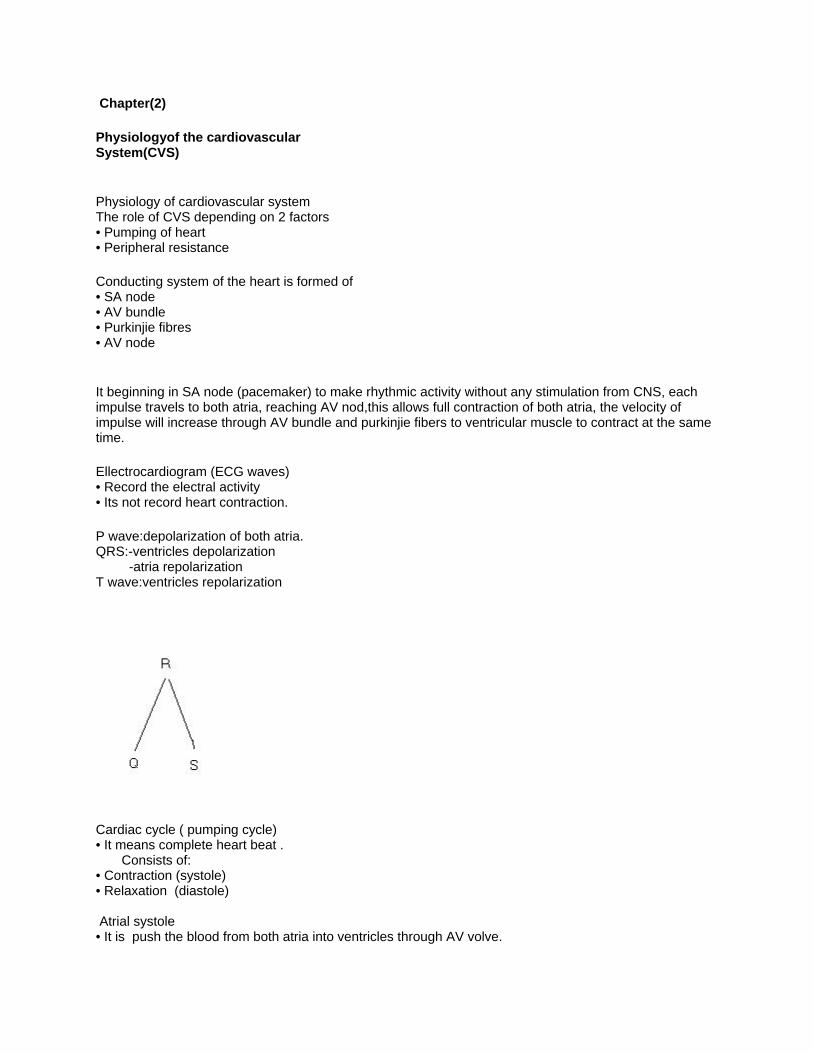
P wave:depolarization of both atria. QRS:-ventricles depolarization -atria repolarization T wave:ventricles repolarization
Cardiac cycle ( pumping cycle) • It means complete heart beat . Consists of: • Contraction (systole) • Relaxation (diastole) Atrial systole • It is push the blood from both atria into ventricles through AV volve.

Ventricular systole • The ventricles contract and push the blood through aorta and pulmonary artries (QRS wave)
Natural blood pressure
1.In systemic circulation (120/80) • Systole(120):ventricles contractions. • Diastol(80): ventricles relaxations.
2. In pulmonary circulation (25/8) • Systole(25):ventricles contractions. • Diastol(8): ventricles relaxations.
Heart Sounds
• The heart makes sounds during cardiac cycle(dob,lop) First sound: • Caused by contraction of ventricles and closure AV valve (systole) Second sound: • Cause by closure of semilunar valve (SL)
Factors affecting blood pressure • Cardiac output • Perphral resistance. Pulse • Expantion and recoil of artery Depends in 2 factors: 1. Cardiac pumping. 2. Elasticity of arteriolar wall.
Chapter(3) The blood
Blood: • One of the body fluid compartment • Forms 6-8% of total body weight
Function of blood • Transport medium (oxygen, hormones& enzymes) • Heat regulation system • Water balance
Blood volume • Young male about 5-6 liters • Young female about 4-5 liters

Two elements of blood
1. Plasma • flouid part of blood represents 55%. • 90% water& 10% solutes.
2. Formed elements • Red blood cells(RBC) One third of the cell formed hemoglobin(HB)♣ In man RBC number 5.5 million/mm3♣ In woman 4.8 million/mm3♣
The functhion of RBC transport of O2♣& CO2 Average life RBC 105-120 days.♣
Note:- each 100ml of blood in male contains 14-16 grams of HB♣ while in woman each 100ml contains 12-14 grams of HB♣
Anemia:- means each 100ml of the blood contains less than 10gr HB♣
• White blood cell(WBC) 5000-11000/mm3♣ 5 types of RBC according to the presence of the cytoplasmic granules♣
I- Granulocytes:- a.Neutrophil b. Basophil c. Essinophil.
II- Agranulocytes:- a.Lymphocyte b.Monocytes The main function of WBC is immunity.♣
Notes:- Leukopenia:- The total count of WBC is less than♣ 5000/mm3 Leukocytosis:-The total count of WBC is more than 11000/mm3♣
• Platelets Count 150.000- 350.000mm3♣ The main function of platelets is blood clotting.♣ Average life 7 days.♣

Blood group Blood group means the type of antigens present in RBC membrane, in other hand there are antibodys in plasma that react in specific group antigen.

ABO system Blood types are named according to the antigens present on RBC membranes. Group types⎬ Type A:-♣ - RBC contains antigen A - Plasma contains anti B antibodies Type B:-♣ - RBC contains antigen B - plasma contains anti A antibodies Type AB:-♣ - RBC contains antigen AB - Plasma contains no antibodies Type O:-♣ - RBC contains no antigen
RH system RH positive :- RBC contains antigen on its membranesϖ RH negative:- RBC has no antigen on its membranesϖ
Notes:- O –ve :- universal donarϖ AB +ve:-uneversal recepiantϖ
Chapter(4)
Lymphatic system
Lymphatic system

Lymphatic system is formed of 3 elements • Lymph vessels • Lymph • Lymph nodes
Functions of the lymphatic system • Immunity • Fluid balance
Lymph and interstitial fluid • Lymph is clear watery fluid found in the lymphatic vessels

Chapter(5) Anatomy of respiratory System(RS)
Anatomy of respiratory system (RS)
Structure:- two tracts Upper respiratory tract(present outside chest cavity)ϖ a. Nose b. Pharynx c. Larynx
Lower respiratory tract( present in chest cavity)ϖ a. Trachea b. Bronchial c. Lungs
Functions of the respiratory system a. Air dirbution. b. Gas exchanges c. Filtration, warming& humidification d. Sense of smell e. Influencing sound production
Upper respiratory tract:-ϖ A. Nose Two portions:- - external nose - internal nose(nasal cavity)
functions of the nose a. air passage b. filtration c. warming d. smelling e. chemical examination

Para nasal sinuses:- contains air spaces, that open or drain into nasal cavity.
Types of sinuses a. frontal sinus b. maxillary sinuses c. ethmoid sinuses d. sphenoid sinuses Sinusitis:- Infection of the Para nasal sinuses.

B. pharynx functions of pharynxϖ a. pathway for respiratory & digestive tracts b. change of the phonation
C. Larynx( voice box) Its triangular shape formed of cartilages. Cartilages of the larynx a. Thyroid cartilage (Adams apple), it’s the largest larynx cartilage b. Epiglottis, behind the tongue c. Cricoid, posterior wall of larynx
Functions of larynx a. Vital airway of the lungs b. Voice production c. Humidification & warming of the air
Lower respiratory tract:-ϖ A. Trachea:- - tube like 11cm long - diameter, 2.5cm - extends from larynx to the primary bronchi
Function of trachea is air passage
Bronchi:- The trachea divides into tow primary bronchus

RT bronchus:- larger -divides into three secondary bronchi
LT bronchus:- smaller - divides into tow secondary bronchi
Notes:- bronchi divides to many bronchioles to be lastly small tube ended as alveoli
Alveoli:- These are the primar gaz exchanges.
Functions of alveoliϖ Gas exchanges ♣ Distribution of the air♣ Cleaning of the ear ♣ Warming of the air♣

Lungs:- Each lung has three surfaces Costal surface:- direct contact with the chest wall♣ Medial surface:- the room of mediastinal structure♣ Inferior surface:- diaphragmatic♣
Rt lung:- is divided into three lobes, upper, middle & lower lobes. Lt lung:- is divided into upper& lower lobes.
Functions of lunges:- Gas exchange, getting O2 and giving CO2
Thorax cavity:- Chest cavity is lined by serious membrane called pleura.
The pleura:- Parietal pleura♣ Visceral pleura♣

Chapter(6)
Physiology of respiratory system
Physiology of the respiratory system Process of respiratory system
Breathing (pulmonary ventilation)♣ Gas exchanges♣ Gas transport by blood.♣ Breathing:-⎬ Two phase a. inspiration b. expiration
a. Inspiration:- During inspiration the air will rush from atmosphere to the lung according to the pressure gradient.
b. Expiration:- During the expiration the air will rush out the chest cavity according to passive process
Gas exchange⎬ a. exchange of gases in the lung:- occur between the alveolar air and the blood capillaries for oxygenation of the blood.
b. transportation of gases by blood O2 is carried in two forms:- • dissolved O2 in plasma, its only 0.3ml per 100ml of blood. • Oxyhemoglobin, each each gram of heamoglobin can unite with 1.34ml of O2
CO2 is carried by:- • Dissolved CO2 in the plasma = 10% • Carbainine compound 20-25 with NH3 (amine). • Bicarbonate, more than 66% of CO2 is carried by form HCO3 and some of CO2 by form H2CO3.
Regulation of brathing Regulation centers:- • Rhythmic center(in medulla oblongata) a. Inspiration :-by inspiratory centre. b. Expiration :- by expiratory centre. • Apneustic centre( in the pons) This centre stimulates the inspiratory centre in the medulla to increase the length and depth of inspiration.
• Pneumotaxic centre(in the pons) This centre inhibits both apenustic and inspiratory centres, to prevent over inflation of the lung.
common problems &diseases in RS Pneumonia Inflammation in the lung
Sleep Apnea Sleep Apnea is the intermittent absence of breathing during sleep
COPD Chronic (continual, permanent, incurable) Obstructive (blocks) Pulmonary (pertaining to the lungs)

Disease (condition with signs and symptoms) COPD or chronic obstructive pulmonary disease has no single definition.
Chronic bronchitis Inflammation in the bronchus occurs when the airways in your lungs have become narrow and partly clogged with mucus.
Emphysema Emphysema is an enlargement and destruction of the alveoli (air sacs) in the lungs. This causes the surrounding airways to collapse. Asthma Asthma is a chronic lung condition characterized by: one or a combination of the following breathing problems: cough, wheeze, shortness of breath, or chest tightness.
Chapter(7)
Anatomy of the digestive system
Anatomy of the digestive system
Structure:- • GIT, Gastrointestinal tract • Digestive glands
Digetive system is the system of:- • Alteration of the physical and chemical composition of the food. • Digestion • Elimination through defecation
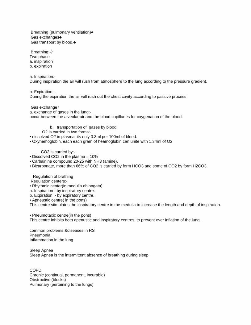
GIT structure:- • Mouth • Pharynx • Esophagus • Stomach • Small intestine • Large intestine
1) Mouth:- Structure of oral cavity • Lips • Cheeks • Tongue • Palate
Teeth Teeth are organ of chewing
Types of teeth • Decidous teeth:- -20 teeth -Appears at age 6 months & completed at age 2 years • Permanent teeth:- - 32 teeth - Appear at age 6 years
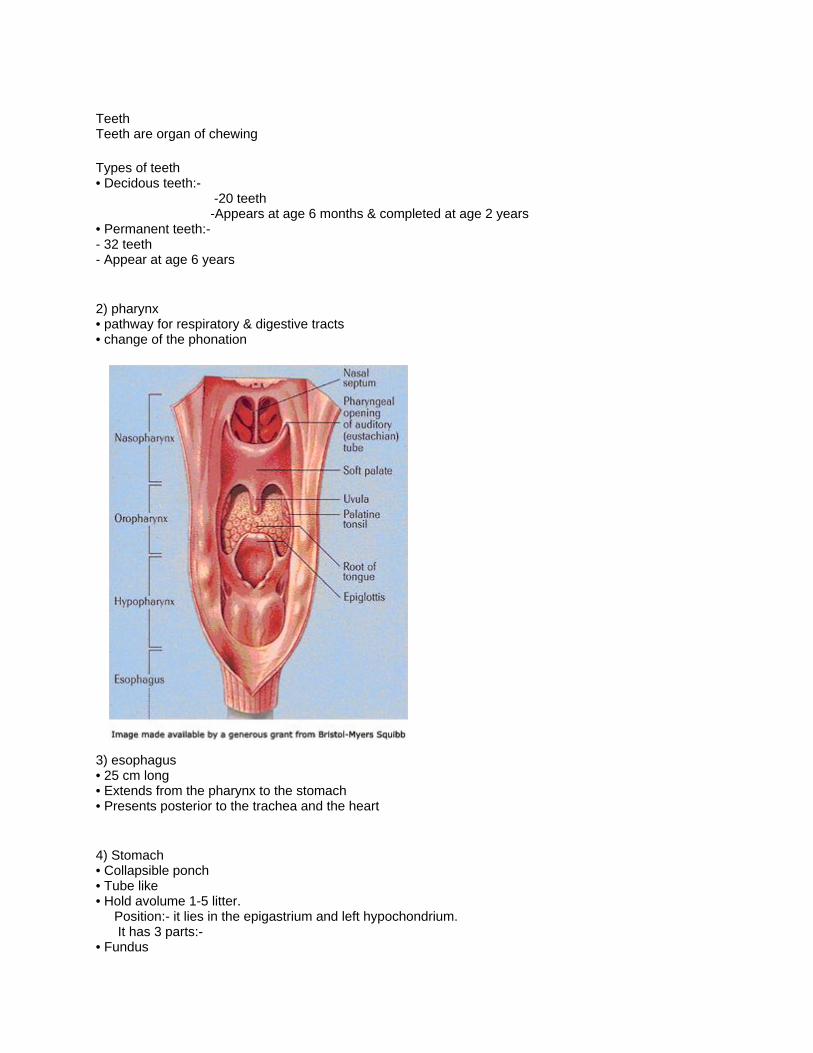
2) pharynx • pathway for respiratory & digestive tracts • change of the phonation
3) esophagus • 25 cm long • Extends from the pharynx to the stomach • Presents posterior to the trachea and the heart
4) Stomach • Collapsible ponch • Tube like • Hold avolume 1-5 litter. Position:- it lies in the epigastrium and left hypochondrium. It has 3 parts:- • Fundus

• Body • Pylorus
Sphincters • Lower esophageal • Pyloric
Functions of stomach:- • Reservoir for food till it can be partially digested • Secreation of gastric juice enzymes & acid • Secretes gastrin hormone, that regulating digestive function. • Anti microbial function
5) Small intestine Its a tube 2.5cm wide and 6 meter long Formed of 3 parts:- • Duodenum • Jejunum • Ileum
Small intestine wall • Mucosa • Sub mucosa • Muscular • Serosa
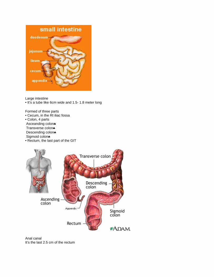
Large intestine • It’s a tube like 6cm wide and 1.5- 1.8 meter long
Formed of three parts • Cecum, in the Rt iliac fossa • Colon, 4 parts Asceanding colon♣ Transverse colon♣ Descending colon♣ Sigmoid colon♣ • Rectum, the last part of the GIT
Anal canal It’s the last 2.5 cm of the rectum

Anus The opening of the anal canal.
Hemorrhoid (pils) Are the varicose of anal veins( of the anal canal)
Apendix • Atube like 8-10 cm long • Presents at the Rt iliac fossa • Just behind the cecum Fuction • Defense mechanism • Medium for intestinal bacterial growth
Digestive glands 1) Salivary glands • Paroted:- between the skin and underlying masseter muscle below externalear. • Submandibular:- located below the mandibular angle • Sublingeal
2) Liver:- • Largest gland in the body • Lies under the diaphragum
Functions of liver • Detoxification of various substances e.g Alcohol
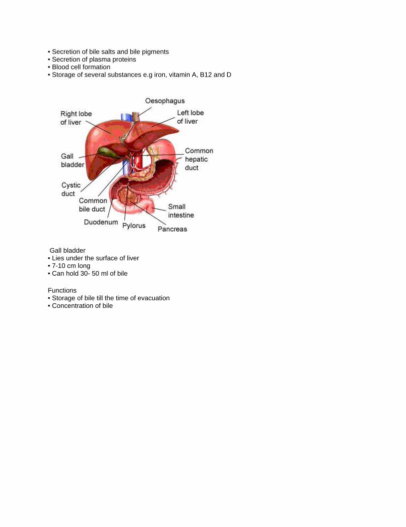
• Secretion of bile salts and bile pigments • Secretion of plasma proteins • Blood cell formation • Storage of several substances e.g iron, vitamin A, B12 and D
Gall bladder • Lies under the surface of liver • 7-10 cm long • Can hold 30- 50 ml of bile
Functions • Storage of bile till the time of evacuation • Concentration of bile

Pancreas Parts of pancreas • The head:- lies between the duodenum • The body:- lies behind stomach • The tail:-touch the spleen in the left side
Size:- 12-15cm long
Structures of pancreas Pancreas formed of two glandular tissue • Exocrine gland:- represents the majority of pancreas, - the tissue is acinar

• Endocrine gland:-between the exocrine gland. - pancreas islets ( alpha& beta cells)
functions of pancreas • the acinar units:- secrete the digestive enzymes in the pancreatic juice • beta cells:- secrete insulin (hypoglycemic hormone) • alpha cells:- secrete glucagons( hyperglycemic hormone)
Chapter(8) Physiology of digestive system
Physiology of digestive system The primary function of digestive system is to make the essential nutrients available for every cell in the body .
Digestion Includes tow process 1) Mechanical digestion 2) Chemical digestion
1) Mechanical digestion Includes the mastication, mixing with digestive juice, till the elimination
A- Mastication • Oral stage:- voluntary -tongue will press the bolus, pushing it backward to the oropharynx • pharyngeal & esoohageal stages:- involuntary - will push the bolus to the --- - - stomach During this stage three opening wll be closed: - • the mouth:- by the tongue • the oropharynx:- by the soft palate • larynx:- by the epiglottis
B- Motility:- • Peristalsis:- wave like - step by step - contraction & relaxation • Segmentation:-forward& backward - movements occur within a single region(segments)

Regulation of motility a. Gastric motility b. Hormonal c. Nervous d. Intestinal motility
2) Chemical digestion Means changes in food composition through the GIT
• Protein digestion:- by proteases (enzymes) that hydrolyze proteins - proteases are, pepsin(gastri)& typsin(pancreatic) • Fat digestion:- Emulisification, change fat to very small droplets - Pancreatic lipease--- fat digesting enzyme. Secretion:- The digestive secretion includes the release of saliva, gastric, pancreatic, And intestinal juices.
Absorption The passage of ingested food, water, salts and vitamins through the intestinal mucosa to the blood.
Vomiting Expulsion of food through mouth.
Defecation:- Is the process of expulsion the waste product of food
Constipation Occurs when the lower colon content move slower than normal so the water will be absorbed
Diarrhea Occurs when the movements through the small intestine too quickly
Epigastric pain Pain in the stomach
Peptic ulcer Ulceration in the stomach
Deodenum ulcer Ulceration in the duodenum
Hepatitis Inflammation in the liver
Cholicystitis Inflammation in Gall bladder
Appendicitis Inflammation of appendix

Chapter(9)
Anatomy of urinary system
The urinary system
Structure:- 1) Kidney:- the principle organ of the urinary system 2) Ureters 3) Urinary bladder 4) Urethra
Kidney Size:- 11 cm by 7 cm by 3 cm Site:- along the sides of the vertebral column, extend from the level of T12 to the level of L3.
The right kidney:- is slightly lower because of the liver.

Kidney tissue:- Each kidney is formed of:- a. Cortex:- the outer region b. Medulla:- the inner region
Renal blood vessels:- a. Renal artery b. Renal vain c. Interlobar arteries d. Interlobar vein

Ureter:- • It’s a tube about 25cm long • Its formed 3 layers: -Mucosa -smooth muscle layer -Fibrous layer
Urinary bladder:- • It’s a collapsible bage • Present just behined the symphysis pubis • Have 3 opening
Function • Reservior for urine, average volume is about 250cc • Expels urine from the body.
Urethra:- • Its ashort atube • Extends from trigone to the bladder exterior • Its length in female is 3cm & in male 20 cm
Micturation(voiding):- starts by • Voluntary relaxation of the external sphincter of the bladder • Bladder muscle contraction, internal sphincter relaxation.
Incontinence:- means involuntary urination
Nephron:- Is the structurel unit of the kidney, there are 1.25 million per kidney

Chapter(10)
Physiology of urinary system
Physiology of the urinary system
Functions of the kidney:- • plasma homeostasis • keeping fluid electrolyte and acid base • influence the rate of secretion of antidiretic hormone(ADH) and aldestrone. • The synthesis of active form of vit D
Kidneys perform their function through 3 processes • Filtration • Reabsorption • Tubular secretion
1) Filtration:- • Is a physical process occurs in renal corpuscles.
• Water and small solutes filter out of blood into bowmans capsules. • the only blood cells and most plasma proteins that don’t move out.
2) Reabsorption:- • Reabsorption of Na through active transport • Glucose and aminoacid passively absorped in accompanying with Na. • Water, passage of Na and Cl from tubules to the blood will create osmatic pressure, drives water out the tubules to the blood • Urea, will be reabsorped passively
3) Secretion:- is the movement of substances out of blood into tubular fluid
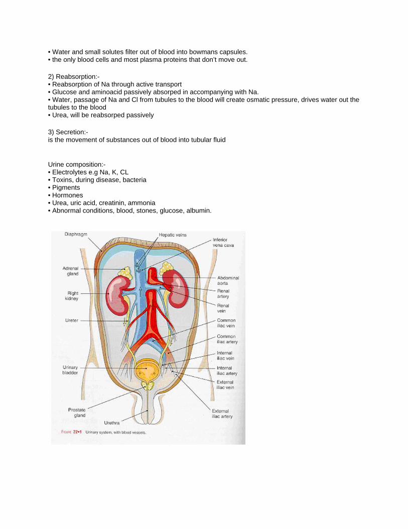
Urine composition:- • Electrolytes e.g Na, K, CL • Toxins, during disease, bacteria • Pigments • Hormones • Urea, uric acid, creatinin, ammonia • Abnormal conditions, blood, stones, glucose, albumin.


![Broadway - Capital Pacific · 206 Broadway E Seattle, WA 98102 206 Broadway [ ] 206 Broadway . 206 Broadway Broadway is a generational ... TRANSIT SCORE 91*](https://img.pdfslide.us/doc/110x75/5ac3885d7f8b9aae1b8c7cb8/broadway-capital-broadway-e-seattle-wa-98102-206-broadway-206-broadway-.jpg)