Embed Size (px)
Citation preview

Archives of Disease in Childhood, 1981, 56, 822-830
Results of selective treatment of spina bifida cysticaJOHN LORBER AND STEPHEN A W SALFIELD
Department ofPaediatrics, University of Sheffield
SUMMARY The results of selective treatment in 120 infants with open spina bifida, admitted betweenMay 1971 and December 1976, were prospectively studied. Seventy-one infants had adverse criteriaat birth and were not treated. They all died, more than 90% of them within 6 months of birth.Seven had meningocele. All were treated and survived without handicap. Forty-two infants withmyelomeningocele were actively treated. Thirty-six survive at follow-up after 3 to 9 years. Thequality of survival is much better than when selection was not used but 8 children have moderateor severe handicaps. The parents were fully informed and consulted at every decision-makingstep; they fully supported the principle of selection and the action taken on behalf of their ownchild.
The severity of handicap suffered by childrentreated unselectively for spina bifida cystica variesfrom no handicap in a fortunate few, to extensive,crippling, multisystem handicaps in many children.1 2One-third of the survivors are mentally retarded.There was a high mortality rate in the initiallyseverely affected infants, which continued wellbeyond infancy. Studies in the UK and elsewhereshow that most severely affected babies who arenot operated on in the newborn period die inthe first few months.39
Patients who survive with severe handicaps andwho require constant medical and surgical care foras long as they live have immense social, psycho-logical, and financial difficulties, and so too do theirfamilies.'>'2 The prospects for open employment,marriage, or anything approaching a normal life arepoor, and the cost to the community is immense.
In two large retrospective surveys1 2 of un-selectively treated children, a set of adverse criteriawas identified which, if present at birth, wasassociated with an unfavourable prognosis: (1) grossparalysis of the legs (paralysis below 3rd lumbarsegmental level, with at most hip flexors, adductors,and quadriceps being active); (2) a thoracolumbaror thoracolumbosacral lesion; (3) clinically evidentkyphosis or scoliosis; (4) grossly enlarged head withoccipitofrontal head circumference at least 2 cmabove the 90th centile; (5) birth injury; (6) othergross congenital abnormalities.
Since May 1971, a policy of selective treatment ofnewborn infants with spina bifida cystica has beenused in all infants referred to one of us (JL). Theearly results of this policy3 showed that 25 untreatedinfants had died before they reached age 9 months.
One of the 12 treated infants died and the otherswere at most moderately handicapped.
In recent years these adverse criteria have beenaccepted as a basis for selection in theUK and in manyother countries. The present study, which includesthe earlier series, was therefore undertaken toreview our results using these adverse criteria, 9years after starting a policy of selection.
Patients and methods
Between May 1971 and December 1976, 120 new-born infants with open spina bifida cystica werereferred to one of us (JL). Seven of these hadmeningocele and 113 had myelomeningocele. Mostpatients were referred from within the Trent region.
After full assessment of the infant the father, andif possible the mother too, was interviewed. If themother was not present the father was interviewedtogether with members of his wife's family ifpossible, and the father was asked to discuss thematter with his wife, and return with a final decision.Subsequently, all the mothers were personallyinterviewed.Almost all the assessments and interviews were
conducted by one of us (JL) at any time of the dayor night, or occasionally it was the duty of thesenior assistant. A member of the junior medicalstaff together with a member of the nursing staffwho would be looking after the baby were alwayspresent at the interview.The infant's condition was discussed and the
choice between active and non-active managementwas simply and repeatedly explained. A prognosiswas given of the minimum handicap the child would
822
copyright. on S
eptember 9, 2019 by guest. P
rotected byhttp://adc.bm
j.com/
Arch D
is Child: first published as 10.1136/adc.56.11.822 on 1 N
ovember 1981. D
ownloaded from

Results of selective treatment ofspina bifida cystica 823
have if total treatment were to be given, or the like-lihood or approximate length of survival if thechild were not treated. Frequently, in mildlyaffected babies, strong reassurance was givenbecause the parents tended to assume the worstwhen told their baby had spina bifida.At the end of the interview, which could last up
to 2 hours, a recommendation for or againsttreatment was made. If the advice was againstactive treatment the parents were offered a secondopinion. No parents wanted a second opinion, andnone wanted treatment against our advice.
In 2 cases near the 'borderline', the parents didnot agree to the recommended active treatment andtheir decision was accepted. Several parents of lessseverely affected patients were initially hesitant butagreed to treatment when the fairly good prognosisand the probability of survival, even in the absenceof treatment, were explained.
Infants who were not treated actively were eitherlooked after in the medical ward or were transferredback to the referring hospital. No further investiga-tions were performed. Normal nursing care withfeeds on demand were given, but these infants werenot tube fed or treated with antibiotics. Chloralhydrate was used to treat irritability or discomfort.It was the parents' universal wish that everythingshould be done to prevent suffering. A few parentsdecided to take their untreated infants home.
If treatment was decided on, the infant wasimmediately referred to our Paediatric SurgicalUnit for active treatment.
Hydrocephalus, if present in infants beingactively treated, was assessed by air ventriculographyand intraventricular pressure measurements soonafter the closure of the back. Computerised tomo-graphy (CT) was not available during this period.The initial treatment of hydrocephalus was generallywith isosorbidet314 given orally or by nasogastrictube. Only if there was evidence that the hydro-cephalus could not be controlled by isosorbide, orif the hydrocephalus was severe at birth, was shuntsurgery embarked upon.
All the treated patients were followed up in acombined clinic where they were seen by paedi-atrician, paediatric surgeon and orthopaedic surgeon,with full supporting staff. Initially the children wereseen frequently; after their second birthday theywere seen at least once a year. The children whoattend special schools were often accompanied bytheir nurses and physiotherapists as well as theirparents. Problem-orientated records have been keptsince birth on every patient for prospective study.Four patients have been discharged from the
clinic as each was normal and there were no fore-seeable problems. Their parents were contacted by
letter in 1979 and no new problems have arisen.No patient has been lost to follow-up.The degree of handicap was classified as slight,
moderate, or severe at the last assessment (near thechild's birthday in 1979/80, Table 1). These lists ofpossible handicaps are not complete, but are usedas a guide to the overall degree of handicap.
Results
Treated infants (n = 49). Seven infants each had asimple meningocele. They recovered without sequelaeand none had hydrocephalus. Their treatmentpresented neither clinical nor ethical problems.
All the subsequent analysis relates to the 42 withmyelomeningocele.Three had thoracolumbar lesions. Their treatment
was advised, against the protocol, for variousreasons (see Severely handicapped children). Allsurvive with severe sequelae and one is mentallyretarded.The remaining 39 infants had either small thoracic,
low lumbar, lumbosacral, or sacral lesions (Table 2).The lesions were closed on the first day in 38 and
were closed later in 3 patients. In one infant thelesion was too broad to close and it was allowed to
Table 1 Classification of degree ofhandicapSlight handicap (at least one of the following)Below knee weakness, slight enough to allow locomotion without aids.
Urinary incontinence with a normal upper urinary tract. Hydro-cephalus, successfully treated by a shunt*
Moderate handicap (at least one of the following)Moderate weakness of the legs, allowing walking with, at most, below
knee calipers, and sticks. Only occasional use of a wheelchair.Urinary diversion with normal upper urinary tract. Faecal in-continence with 'moderate' social control. 'Moderate' mentalretardation with IQ 60-75. Partial visual defects. Well-controlledepilepsy
Severe handicapt (at least one of the following)Gross paraplegia. Locomotion possible only with extensive bracing.A wheelchair frequently used. Urinary incontinence with per-manently damaged upper urinary tract. Frequent and embarrassingfaecal incontinence. Severe mental retardation with IQ less than60. Gross deformities such as gross kyphoscoliosis. Blindness.Severe, poorly controlled epilepsy
*Hydrocephalus arrested spontaneously or after treatment withisosorbide is not here considered to constitute a handicap.tIn practice all the severely handicapped children had more than oneof these factors.
Table 2 Site of lesion in 42 treated cases ofmyelomeningoceleThoracic 5Lumbar 3Lumbosacral 22Sacral 9Thoracolumbar* 3
*All three survive with severe sequelae and one is mentally retarded.
copyright. on S
eptember 9, 2019 by guest. P
rotected byhttp://adc.bm
j.com/
Arch D
is Child: first published as 10.1136/adc.56.11.822 on 1 N
ovember 1981. D
ownloaded from

824 Lorber and Salfield
epithelialise spontaneously. He was given all othernecessary surgical and medical treatment andsurvives.
Thirty-six survive and each was reviewed betweenages 3 and 9 years.
DeathsSix treated children died. Three died of ventriculitlsin the newborn period. Two of them had hydro-cephalus but this was not treated because they diedearly.An exceptionally extensive Arnold-Chiari mal-
formation was found at necropsy in an infant withhydrocephalus who had a fatal respiratory arrest on
the second day of life.The fifth, a baby with a small sacral lesion and
apparently good power in his legs, was remarkablyimmobile. The parents were fearful, but were
persuaded to agree to treatment. The parents'instinct was right however, as the baby never movedhis legs after surgery and became severely retardedwith a normal-sized head. He developed Escherichiacoli meningitis at age 4 months; the parents refusedfurther specific treatment and the infant died.The sixth child, who had hydrocephalus, was of
normal intelligence with normal bladder and renalfunction, and nearlynormal legs. He died ofa blockedshunt with cerebellar coning, resulting from alumbar puncture done at another hospital. Lumbarpuncture in a child with a shunt and suspectedraised intracranial pressure is dangerous and iscontraindicated.
Survivors (n = 36) (Table 3)
Without handicap (n = 10). Ten now have no
handicap although 6 of them have hydrocephaluswhich has been managed without a shunt.
Slightly handicapped children (n = 8). In 3 childrenthe main handicap is the presence of a shunt. One ofthem, who had prolonged ventriculitis after closureof her sacral lesion, is now a normal 6-year old.Despite intensive antibiotic treatment, she did notrecover from her ventriculitis until continuousventricular drainage was established. Another hadclosure of her spinal lesion delayed until age 3months. She had a shunt inserted before closure of
Table 3 Degree ofhandicap among the 36 survivors
Age (years) Handicapat 1979 or 1980review None Slight Moderate Severe
6-9 (n=15) 5 2 3 5
3-5 (n=21) 5 6 5 5
the back, not primarily for her hydrocephalus whichwas well controlled by isosorbide, but to facilitatethe closure of the spinal lesion and prevent break-down of the wound.One child has multiple thoracic hemivertebrae and
Sprengel's deformity, with a slight scoliosis, and aslight limp. His hydrocephalus was treated withisosorbide and he has no shunt. A recent CT scanshows gross hydrocephalus. He has normal intel-ligence and a head circumference 3 cm above the97th centile. This causes him no handicap or socialinconvenience.Three children have below knee weakness allowing
walking without aids. Two are incontinent with anormal upper urinary tract.
Moderately handicapped children (n = 8). All havemoderate weakness of the legs, allowing walkingwith, at most, below knee calipers and sticks. Threehave moderate talipes. Four are incontinent with anormal upper urinary tract.
Seven have hydrocephalus, but only 4 haveshunts. All are of normal intelligence, but 2 aresubject to fits.
Severely handicapped children (n = 10) (Table 4).Three had thoracolumbar lesions as their onlyadverse criteria, but were treated despite this. Inone the lesion was fairly small and his legs werenearly normal at birth. He was expected to surviveeven if he were not operated on. Unfortunately,this infant developed spastic quadriplegia despiteshunt treatment of his hydrocephalus. He also hasbilateral hip dislocation, a spastic bladder, andfaecal incontinence.The second had good muscle power at birth but
Table 4 Clinical details in 10 severely handicappedchildrenWith adverse criteria at birth (thoracolumbar lesions)(1) Good legs at birth. Spastic quadriplegia within months. Hydro-
cephalus with shunt. Incontinence of urine and faeces. Dislocatedhips
(2) Apparently good legs at birth. Paraplegic by 18 months. Grosshydronephrosis. Incontinence. Hemivertebrae. Gross kypho-scoliosis. Hydrocephalus treated with isosorbide
(3) Paraplegia. Absent right kidney. Gross left hydronephrosis. Grosshydrocephalus with shunt. Mental retardation. Incontinence
Without adverse criteria at birth(4) Paraplegia. Incontinence. Hydrocephalus with shunt(5) Paraplegia. Hydrocephalus with shunt. Incontinence. Upper
urinary tract damage(6) Severe mental retardation. Blindness after delayed recognition of
shunt blockage (elsewhere). Incontinence(1) Microcephaly. Mental retardation. Incontinence(8) Microcephaly. Mental retardation. Paraplegia. Incontinence(9) Lipoma of cauda equina. Severe scoliosis. Progressive neuro-
logical deterioration in legs(10) Paralysis. Incontinence
copyright. on S
eptember 9, 2019 by guest. P
rotected byhttp://adc.bm
j.com/
Arch D
is Child: first published as 10.1136/adc.56.11.822 on 1 N
ovember 1981. D
ownloaded from

Results of selective treatment of spina bifida cystica 825
gradually lessening spontaneous movement in herlegs until she became paraplegic by age 18 months.She has gross hydronephrosis and is incontinent ofurine. She has hemivertebrae and developed kypho-scoliosis by 2 years. Her hydrocephalus was success-fully treated with isosorbide and she is of normalintelligence.The third had extensively paralysed legs at birth
and was treated because of over-optimism on thepart of those assessing her. She had no right kidney.Later she developed gross left-sided hydronephrosiswith frequent urinary infections. She had grosshydrocephalus. Further treatment was refused bythe parents and she returned to the referring hospital.She survived, but by the time she returned to us atage 2 years she had a huge head with a circum-ference of 62 5 cm. A shunt was inserted but she isretarded with an IQ of 65, and is severelyhandicapped.
Seven severely handicapped children had noadverse criteria at birth. Of the 3 with shunts, 2 havegross paralysis, 1 with upper urinary tract damage,but both are of normal intelligence. The third isblind and retarded after shunt blockage which wasnot recognised in time. By this time he was no longerour patient.Four severely handicapped children who had no
adverse criteria at birth do not have shunts. Two aremicrocephalic and severely mentally retarded, butthe other handicaps in one ofthem are only moderate.The third, who had one paralysed and one normalleg at birth, had a lipoma of the cauda equina as
well as a lumbosacral myelomeningocele and a
thoracic meningocele. She later developed a severescoliosis and moderate spasticity in the previouslynormal leg. Lipoma of the cauda equina is wellknown to produce progressive neurological dam-age'5 16 but is not associated with mortality, even ifleft untreated. The fourth with a lumbosacralmyelomeningocele had nearly normal legs at birthbut his condition gradually deteriorated and hebecame extensively paralysed, as did 3 children whohad shunt treatment (see above).
Hydrocephalus and its treatment (Table 5). Hydro-cephalus was confirmed in 29 survivors and in a
shunt-treated child who died.
Table 5 Treatment of hydrocephalus in 30 infantsDegree of Not Isosorbide Isosorbide Shunthydrocephalus treated only then shunt only
Slight (n =7) 2 4 1 -
Moderate (n= 16) 3 7 6* -
Severe (n-7) - 1 3 3
*One died of shunt complications (see text).
The severity of the hydrocephalus was gradedmild (n=7), moderate (n=16), or severe (n=7),according to the amount of cerebral mantle found atpneumoventriculography.17No active treatment was given to those 5 whose
hydrocephalus was not severe on ventriculographyand whose head grew at a normal or slightlyincreased rate. One of these had mild hydrocephalusshown by ventriculography. However, her headcircumference increased gradually from below the3rd centile at birth to 1 cm above the 97th centileat 5 years. A CT scan shows moderately dilatedventricles. She has no neurological deficit attribut-able to her hydrocephalus, and her IQ is 112.The primary treatment was isosorbide in 22
infants. Twelve responded well, including one withsevere hydrocephalus, and no surgery was necessary.Ten initially treated with isosorbide needed a shuntlater, including 3 with severe hydrocephalus. The 3in whom ventricular dilatation was particularlypronounced each had a shunt as the primarytreatment.
Altogether 17 children with hydrocephalus had nosurgical treatment and all survived. In one, shunttreatment was necessary before closure of a largespina bifida lesion to aid healing of the wound andnot because of hydrocephalus which was slight.
Five of the 7 with severe hydrocephalus are ofnormal intelligence. Six have shunts. The seventhhad severe hydrocephalus first demonstrated at 6months. She responded to isosorbide alone and hasnormal intelligence (WISC full scale 92, verbal 92,performance 89), although a CT scan shows thather ventricles are just as dilated at age 7 years asthey were when isosorbide was started, and aregrossly asymmetrical. She has no neurologicaldeficit attributable to her hydrocephalus.
Thirteen children had shunts. Ten had between 1and 4 revisions. During a total of 65 'shunt-years' ofobservation there were 21 shunt revisions. One childdied and another became blind and severely retardedbecause of shunt blockage (see above).
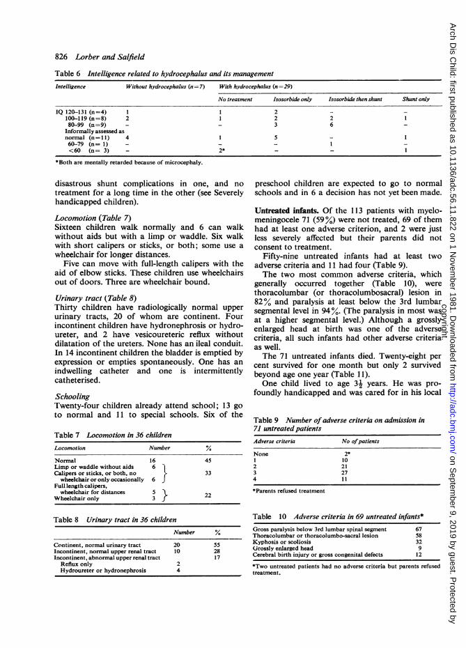
Intelligence (Table 6)Twenty-five children have had formal IQ assessment(Wechsler in 15 children, Stanford-Binet in 10), and11 have been assessed by school teachers, parents,and us. All the informally assessed are normal andsome probably are very intelligent. All 7 withouthydrocephalus are of normal intelligence.
Twenty-five of the 29 children with hydrocephalusare of normal or superior intelligence. The 3 with anIQ of 120 or above have no shunts.Two children without shunts are severely retarded
because of microcephaly. Two with severe shunt-treated hydrocephalus are retarded because of
copyright. on S
eptember 9, 2019 by guest. P
rotected byhttp://adc.bm
j.com/
Arch D
is Child: first published as 10.1136/adc.56.11.822 on 1 N
ovember 1981. D
ownloaded from
826 Lorber and Salfield
Table 6 Intelligence related to hydrocephalus and its management
Intelligence Without hydrocephalus (n =7) With hydrocephalus (n =29)
No treatment Isosorbide only Isosorbide then shunt Shunt only
IQ 120-131 (n=4) 1 1 2 - _100-119 (n=8) 2 1 2 2 180-99 (n=9) - 3 6
Informally assessed asnormal (n= 11) 4 1 5 -60-79 (n= 1) - - - 1< 60 (n= 3) - 2* - -
*Both are mentally retarded because of microcephaly.
disastrous shunt complications in one, and no
treatment for a long time in the other (see Severelyhandicapped children).
Locomotion (Table 7)Sixteen children walk normally and 6 can walkwithout aids but with a limp or waddle. Six walkwith short calipers or sticks, or both; some use awheelchair for longer distances.
Five can move with full-length calipers with theaid of elbow sticks. These children use wheelchairsout of doors. Three are wheelchair bound.
Urinary tract (Table 8)Thirty children have radiologically normal upperurinary tracts, 20 of whom are continent. Fourincontinent children have hydronephrosis or hydro-ureter, and 2 have vesicoureteric reflux withoutdilatation of the ureters. None has an ileal conduit.In 14 incontinent children the bladder is emptied byexpression or empties spontaneously. One has anindwelling catheter and one is intermittentlycatheterised.
SchoolingTwenty-four children already attend school; 13 goto normal and 11 to special schools. Six of the
Table 7 Locomotion in 36 childrenLocomotion Number %
Normal 16 45Limp or waddle without aids 6Calipers or sticks, or both, no 33
wheelchair or only occasionally 6 JFull length calipers,
wheelchair for distances S 22Wheelchair only 3
Table 8 Urinary tract in 36 childrenNumber %
Continent, normal urinary tract 20 55Incontinent, normal upper renal tract 10 28Incontinent, abnormal upper renal tract 17
Reflux only 2Hydroureter or hydronephrosis 4
preschool children are expected to go to normalschools and in 6 a decision has not yet been made.
Untreated indants. Of the 113 patients with myelo-meningocele 71 (59%) were not treated, 69 of themhad at least one adverse criterion, and 2 were justless severely affected but their parents did notconsent to treatment.
Fifty-nine untreated infants had at least twoadverse criteria and 11 had four (Table 9).The two most common adverse criteria, which
generally occurred together (Table 10), werethoracolumbar (or thoracolumbosacral) lesion in82% and paralysis at least below the 3rd lumbarsegmental level in 94%. (The paralysis in most wasat a higher segmental level.) Although a grosslyenlarged head at birth was one of the adversecriteria, all such infants had other adverse criteriaas well.The 71 untreated infants died. Twenty-eight per
cent survived for one month but only 2 survivedbeyond age one year (Table 11).One child lived to age 31 years. He was pro-
foundly handicapped and was cared for in his local
Table 9 Number ofadverse criteria on admission in71 untreated patients
Adverse criteria No ofpatients
None 2*1 102 213 274 11
*Parents refused treatment
Table 10 Adverse criteria in 69 untreated infants*Gross paralysis below 3rd lumbar spinal segment 67Thoracolumbar or thoracolumbo-sacral lesion 58Kyphosis or scoliosis 32Grossly enlarged head 9Cerebral birth injury or gross congenital defects 12
*Two untreated patients had no adverse criteria but parents refusedtreatment.
copyright. on S
eptember 9, 2019 by guest. P
rotected byhttp://adc.bm
j.com/
Arch D
is Child: first published as 10.1136/adc.56.11.822 on 1 N
ovember 1981. D
ownloaded from

Results of selective treatment of spina bifida cystica 827
Table 11 Life table of 71 untreated infants withmyelomeningoceleAlive at Number %
I day 711 week 52 731 month 223 months 12 176 months 69 months 31 year 2 32 years 23 years 14 years 0
hospital. He was not referred back when it becameapparent that he would live for some time.Another infant lived to 2j years. As she was in
good health at age 6 months she was treated with ashunt and back closure, despite her severe handicaps.Two years later she developed pneumococcalmeningitis and died in spite of full antibiotictreatment.The most common causes of death were ventricu-
litis, hydrocephalus, multiple congenital abnormali-ties, and respiratory infections. The parents of eachuntreated infant were seen some 3 months after thedeath of the infant. They were reassured that theyhad done nothing wrong either by omission or com-
mission which had resulted in their baby having spinabifida and that neither partner was more 'responsible'than the other, even if there were other cases on one
side of the family. They were also given appropriategenetic advice which during most of this periodincluded advice on antenatal diagnosis.
Discussion
It must be made clear that the results in this paper
are those obtained for all the patients referred toone of us (JL) between May 1971, when a policy ofselective treatment was started, and December 1976.During the same period paediatricians couldalternatively refer patients directly to the PaediatricSurgical Unit (PSU) which at that time did notpractise selection.During this 5-year period, the proportion of
infants referred to our medical unit progressivelyrose (Table 12), while the proportion sent directlyto the surgical unit fell, indicating the views of thereferring paediatricians on selection. Initially some
paediatricians were not aware of the difference inpolicy and continued the previous practice ofreferring to the PSU for that reason. Other paedi-atricians practised selection themselves, and in theearlier years referred the less handicapped infantsfor surgery direct to the PSU. A few infants withadverse criteria were referred directly to the PSUif the parents insisted on treatment.
Table 12 Surgical and medical admissions ofnewbornbabies with spina bifida cystica to SheffieldChildren's HospitalYear Surgical (n=89) Medical (n=120)
No %oftotal No Y. oftotal
1971 (May onwards) 31 72 12 281972 25 53 22 471973 18 50 18 501974 7 30 16 701975 5 15 28 851976 3 11 24 89
Thus the series described does not reflect the totalpopulation of spina bifida babies born in the areaor admitted to the Sheffield Children's Hospital,and the 59% of infants with adverse criteria in thisseries may be different from those found elsewhere.The survival of 86% of treated infants in this
series is similar to results obtained in other reportsof selective treatment (Table 13). The variationsare probably due to differences in the application ofselection procedures. Lorber2 retrospectively showedthat in an unselected treated series, 84% of thosewithout adverse criteria survived.Not surprisingly, when all infants were treated
unselectively in this hospital, the survival rate wasconsiderably lower than for treated infants inselected groups. Spain,18 in a community survey forthe whole of London, when almost all were un-selectively treated, found that only 47% survived toone year. Ames and Schut19 in Philadelphia, on theother hand, reported 78% survival to between ages3 and 8 years. They had however, a lower incidenceof thoracolumbar lesions in their series than inSheffield and their intake rmay well have differed inother ways (Table 14).-The great improvement in the quality of survivors
in the present series compared with previousunselected series in the same unit is shown in Table15. Twenty-four per cent have no handicap, com-pared with 1% in unselected groups. Unfortunately
Table 13 Survival of infants selectedfor treatment invarious series
Year Number Survival Age at reporting(%)
Present series 1971-76 42 86 3-9 yearsGuiney et al.,9Dublin 1973-75 77 84 3 months-3 years
Smith andSmith,7Melbourne 1961-69 216 78 3-11 years
Rickham andMawdsley,6Liverpool 1960-62 100 71 3 years
Stark andDrummond,5Edinburgh 1965-71 78 >70 6 years
copyright. on S
eptember 9, 2019 by guest. P
rotected byhttp://adc.bm
j.com/
Arch D
is Child: first published as 10.1136/adc.56.11.822 on 1 N
ovember 1981. D
ownloaded from

828 Lorber and Salfield
Table 14 Survival of infants treated unselectively invarious series
Year Number Survival Age at reporting(%) (years)
Lorber,I 2Sheffield a 1959-63 323 41 7-12
b 1962-64 200 51 7-9c 1967-68 201 63 2-4
Spain,18London 1967-69 377 47 1
Ames and Schut,19Philadelphia 1963-68 148 78 3-8
Table 15 Quality ofsurvivors in the selected series,compared with previous unselected series in thesame unit1Handicap Selected Unselected series
series1971-76 1967-68 1959-63
No %' No % No %
None 10 24 2 1 4 1Slightormoderate 16 38 11 5-5 20 6Severe with IQ 80+ 6 14 56 28 66 20-5Severe with IQ 60-79 1 2 25 12-5 28 9ExtremelQ<60 3 7 20 10 16 5
Total survivors 36 86 114 57 134 41-5
Dead 6 14 87 43 189 58*5
Total treated 42 100 201 100 323 100
23 % still have severe handicaps but this compares
with 34 to 50% in unselected series. Some of thisimprovement could be due to greater experience andnew technology, and especially to the reduced use
of shunts since isosorbide became available.13 14However, the main factor is that most of thechildren who would have been severely handicappedwere not treated, and died in infancy.
Earlier publicationsl'3 advocating selection weregreeted with enthusiasm by numerous people con-
cerned with the care of severely handicappedchildren with spina bifida in most parts of theworld. When a policy of selection was started byone of us (JL) in 1971, most paediatricians andparents of spina bifida children were in full agree-ment with the selection procedure. Many unitsaround the world now practise selection, althoughthe criteria vary.Some have found that when selection was used,
surprisingly large numbers of babies who were notoperated on in the newborn period survived.However, many infants were treated with shunts inone series,20 and in another21 there was difficulty insticking to protocols of selection and management,and active treatment-such as antibiotics-wassometimes used. Shurtleff et al.22 in Seattle, using
different selection criteria, found that of 36 infantstreated only symptomatically because of adversecriteria, 10% were surviving at 2 years. All theuntreated children in the present series died and only2 out of 71 lived up to one year. In our view if it isdecided to withhold closure of the back then it isessential also to withhold other treatment-such asantibiotics, tube feeds, incubator care, shunts, andresuscitation-in the newborn period. The infantshould receive normal nursing care, feeding ondemand, and analgesia and sedation to relievediscomfort. Many authors have now confirmed thatwhen this is done most severely affected infants diein the first few weeks or months (Table 16).We believe that parents of infants not selected for
treatment should, if possible, be dissuaded fromtaking their infant home because of the likelihoodthat they will then receive 'intensive' treatmentincluding antibiotics.
Occasionally, a severely affected infant notchosen for treatment survives, even when all formsof active treatment are withheld. Such a child canstill be treated later. This happened with one of ourpatients whose back was closed and shunt insertedat age 6 months (see Results). The delay in treatmentneed not worsen the long-term prognosis as suchchildren would have been paraplegic, incontinent,and probably would have needed shunt treatmenteven if treated in the newborn period. On thecontrary, hydrocephalus is often less rapidlyprogressive if the back is not closed.There will always be some who object to selection
on the grounds that life should be saved at all costs,even if the result is a life of suffering and severehandicaps for the patient and his family. We stronglydisagree with this view, as does the predominantopinion of our society and the medical profession.A Department of Health and Social Securitymemorandum23 states . . . 'As physical and in-tellectual outcome are related to the severity of theinitial malformation certain contraindications toactive treatment have been suggested' and adds. . . 'Adecision not to operate implies the existence of aco-ordinated medical and nursing policy whichrecognises the emotional and ethical problemsinvolved'. A working party under the auspices of
Table 16 Survival of untreated infants withmyelomeningocele in various series
Authors Noof Percentage who diedpatients by 6 months of age
Present study 71 92Rickham and Mawdsley6 57 96Hide, Williams, and Ellis4 99 79Stark and DrummondS 85 85Guiney et al.9 50 82
copyright. on S
eptember 9, 2019 by guest. P
rotected byhttp://adc.bm
j.com/
Arch D
is Child: first published as 10.1136/adc.56.11.822 on 1 N
ovember 1981. D
ownloaded from

Results of selective treatment ofspina bifida cystica 829
the Newcastle Regional Hospital Board24 states . . . 'inthe present state of medical knowledge the policy ofselection for treatment of spina bifida is in ouropinion justified'.
In our opinion a decision to withhold activetreatment should be taken with the parents, afterexpert advice on the expected outcome of treatingor not treating the infant.That the presence of adverse criteria at birth is
reliably associated with severe handicap later hasbeen repeatedly demonstrated.1 2 7 25 The argumentthat severe handicap does not necessarily ensue inthe presence of adverse criteria is not tenable, evengiven expert treatment.
However, the absence of adverse criteria does notnecessarily ensure a good quality of survival,although it makes it more likely. Seven of 39 infantswithout adverse criteria at birth became severelyhandicapped later. The criteria for selection fortreatment are sufficiently wide to ensure that anychild with a chance of survival with moderatehandicap or less will be treated. It is thereforeinevitable that some treated infants will becomeseverely handicapped as a result of postnatalevents-such as shunt complications, meningitis,ventriculitis, deteriorating neurological status aftersurgery, or renal problems-or due to problems-such as microcephaly-which are not predictable inthe newborn.The optimal answer to the problem of myelo-
meningocele is obviously not selection. The processof selection is extremely painful and distressing forparents, families, paediatricians, nurses and allconcerned, both at the decision-making stage andduring the life of the infant whether treated or not,and as already stated, appreciable numbers ofseverely handicapped children still result.No matter how well they are counselled, it is
inevitable that parents of some infants, both treatedand untreated, will have doubts about the rightnessof their decisions.Thus the true solution to the problem is pre-
vention. Whether vitamin supplements in earlypregnancy26 or other means will prove to be effective,remains to be seen. At present antenatal diagnosisand termination of affected pregnancies is the bestapproach. This has greatly reduced the number ofliveborn infants with severe spina bifida in areaslike Sheffield, where antenatal screening of allpregnant women has been implemented. In 1979 inSheffield only one baby was born with myelo-meningocele-that is one in 6000-compared withthe national figure of about one in 700. All pregnantwomen who have had previous babies with neuraltube defects should have estimation of amniotic fluidalpha-fetoprotein27 or acetyl-cholinesterase.28
It should be an urgent priority to make routineantenatal screening of serum alpha-fetoprotein,29followed when indicated by amniocentesis, nationallyavailable to all pregnant women.
We thank the ward sisters, nursing staff, and juniormedical staff for unstinting patient care andunderstanding of the difficult issues, our surgical col-leagues for surgical management, the many paedi-atricians who referred patients, Mr A Lonton whodid the IQ assessments, and the secretaries fortyping the manuscript.
References
Lorber J. Results of treatment of myelomeningocele. Ananalysis of 524 unselected cases, with special reference topossible selection for treatment. Dev Med Child Neurol1971; 13: 279-303.
2 Lorber J. Spina bifida cystica. Results of treatment of 270consecutive cases with criteria for selection for the future.Arch Dis Child 1972; 47: 854-73.
3 Lorber J. Early results of selective treatment of spinabifida cystica. Br MedJ 1973; iv: 201-4.
4 Hide D W, Williams H P, Ellis H L. The outlook for thechild with a myelomeningocele for whom early surgerywas considered inadvisable. Dev Med Child Neurol 1972;14: 304-7.
5 Stark G D, Drummond M. Results of selective earlyoperation in myelomeningocele. Arch Dis Child 1973;48: 676-83.
6 Rickham P P, Mawdsley T. The effect of early operationon the survival of spina bifida cystica. Dev Med ChildNeurol 1966; 8: Supplement 11, 20-6.
7 Smith G K, Smith E D. Selection for treatment in spinabifida cystica. Br Med J 1973; iv: 189-97.
8 Colliss V R. The effects of selective treatment of myelo-meningocele on a neonatal unit. Dev Med Child Neurol1972; 14: Supplement 27, 34-7.Guiney E J, Fitzgerald R J, Mehigan D, Purl P, Sundar B.Surgical closure of myelomeningocele: problems andconsequences of the introduction of a policy of selection.Ir J Med Sci 1977; 146: 260-2.
10 Freeston B M. An enquiry into the effect of a spinabifida child upon family life. Dev Med Child Neurol 1971;13:456-61.
11 Walker J H, Thomas M, Russell I T. Spina bifida-andthe parents. Dev Med Child Neurol 1971 ; 13: 462-76.
12 Lorber J, Schloss A L. The adolescent with myelomening-ocele. Dev Med Child Neurol 1973; 15: Supplement 29,113-4.
13 Lorber J. Isosorbide in treatment of infantile hydro-cephalus. Arch Dis Child 1975; 50: 431-6.
14 Hayden P W, Shurtleff D B. The medical management ofhydrocephalus. Dev Med Child Neurol 1972; 14: Supple-ment 27, 52-8.
15 Dubowitz V, Lorber J, Zachary R B. Lipoma of thecauda equina. Arch Dis Child 1965; 40: 207-13.
16 Rickwood A M K, Hemalatha V, Zachary R B. Lipomaof the cauda equina (lumbosacral lipoma): a study of 74cases operated in childhood. Z Kinderchir 1979; 27:159-69.
17 Lorber J. Systemic ventriculographic studies in infantsborn with meningomyelocele and encephalocele. ArchDis Child 1961; 36: 381-9.
18 Spain B. Spina bifida survey. GLC Intelligence UnitQuarterly Bulletin 1970; 12: 5-12.
copyright. on S
eptember 9, 2019 by guest. P
rotected byhttp://adc.bm
j.com/
Arch D
is Child: first published as 10.1136/adc.56.11.822 on 1 N
ovember 1981. D
ownloaded from

830 Lorber and Salfield
9 Ames M D, Schut L. Results of treatment of 171 con-secutive myelomeningoceles-1963 to 1968. Pediatrics1972; 50: 466-70.
20 Robards M F, Thomas G G, Rosenbloom L. Survival ofinfants with unoperated myeloceles. Br Med J 1975;iv: 12-3.
21 Feetham S L, Tweed H, Perrin J S. Practical problems inselection of spina bifida infants for treatment in theUSA. ZKinderchir 1979; 28: 301-6.
22 Shurtleff D B, Hayden P W, Loeser J D, Kronmal R A.Myelodysplasia: decision for death or disability. N EnglJMed 1974; 291: 1005-1 1.
23 The Standing Medical Advisory Committee for theCentral Health Services Council, the Secretary of Statefor Social Services, and the Secretary of State for Wales.Care of the child with spina bifida. London: HMSO, 1973.
24 Newcastle Regional Hospital Board. Ethics of selectivetreatment of spina bifida. Report by a working party.Lancet 1975; i: 85-8.
25 Stein S C, Schut L, Ames M D. Selection for earlytreatment in myelomeningocele: a retrospective analysisof various selection procedures. Pediatrics 1974; 54:553-7.
26 Smithells R W, Sheppard S, Schorah C J, et al. Possibleprevention of neural-tube defects by periconceptionalvitamin supplementation. Lancet 1980; i: 339-40.
27 Brock D J H, Sutcliffe R G. Alpha-fetoprotein in theantenatal diagnosis of anencephaly and spina bifida.Lancet 1972; ii: 197-9.
28 Smith A D, Wald N J, Cuckle H S, Stirrat G M,Bobrow M, Lagercrantz H. Amniotic-fluid acetylcho-linesterase as a possible diagnostic test for neural-tubedefects in early pregnancy. Lancet 1979; i: 685-8.
29 Wald N J, Brock D J H, Bonnar J. Prenatal diagnosis ofspina bifida and anencephaly by maternal serum-alpha-fetoprotein measurement. Lancet 1974; i: 765-7.
Correspondence to Professor John Lorber, Depart-ment of Paediatrics, Children's Hospital, SheffieldS10 2TH.
Received 17 October 1980
copyright. on S
eptember 9, 2019 by guest. P
rotected byhttp://adc.bm
j.com/
Arch D
is Child: first published as 10.1136/adc.56.11.822 on 1 N
ovember 1981. D
ownloaded from