Embed Size (px)
Citation preview

© 2006 Nature Publishing Group
Anyone who has ever taken a plant cutting and put it in some water to grow knows that plants have the ability to regenerate. But what are the molecular mechanisms behind this phenomenon? Ben Scheres and co-workers, reporting in Science, investigated regeneration in Arabidopsis thaliana roots and revealed the interaction network that re-establishes a new root tip.
The A. thaliana root has a simple structure that consists of concentric cylinders of cells. Within a growing root, this radial pattern emanates from a small group of stem cells that surround a quiescent centre (QC) — a region of mitotically inactive cells that functions to maintain the stem-cell status of adjacent cells — which itself is found at the heart
of the root apical meristem. Some of the new cells that originate at the meristem are root-cap cells, which cover the distal tips of the roots and protect the QC and stem-cell niche as the root grows.
Xu et al. used a laser to wound the QC to disrupt the flow of the plant hormone auxin, which regulates diverse aspects of plant growth and development. This resulted in the rapid upregulation of the auxin response, followed by cell-fate changes, which indicates that the disruption of auxin flow stimulates regeneration. Additionally, the authors investigated whether auxin disruption influences the localization of the transmembrane PIN proteins, which are putative auxin-efflux trans-porters. By monitoring functional PIN–green-fluorescent-protein fusions after QC ablation, they showed that new PIN-protein expres-sion and localization is dependent on renewed cell specification. It is only after renewed cell specification, there-fore, that PIN proteins can become correctly polarized in response to changes in auxin distribution.
So, what role might the key patterning genes have in the induced regeneration process? The GRAS-family transcription factors SHORTROOT (SHR) and SCARECROW (SCR) are required for QC-cell specification, and the genes that encode PLETHORA1 (PLT1) and PLT2 can be induced
by auxin and, when expressed ectopically, convey root identity. Examination of plt1–4 plt2–2 mutants showed defects in QC specification, as expected, as well as in nuclear SHR targeting — both events are associ-ated with root regeneration. The role of SCR in QC specification, which was investigated by examining the scr-4 null allele, was also shown to be crucial during regeneration.
Xu and colleagues have proposed a model for root regeneration in which the cell-fate changes that are induced by auxin are dependent on new expression patterns and the activity of PLT, SCR and SHR transcription factors. Whereas clas-sical canalization hypotheses, which were proposed to explain vascular pattern formation, are based on the assumption of positive-feedback regulation between auxin flow and transport, the authors have shown here that, surprisingly, in the root, cell-fate changes are first induced by auxin redistribution and, only con-sequently, changes in the polarity of auxin flow are induced. These excit-ing findings provide new insight into plant regeneration, but might mean a bit of re-wording for the nursery rhyme ‘Mary, Mary quite contrary, how does your garden grow?’
Sharon Ahmad
ORIGINAL RESEARCH PAPER Xu, J. et al. A molecular framework for plant regeneration. Science 311, 385–388 (2006)
R E G E N E R AT I O N
How does your garden grow?
These exciting findings provide new insight into plant regeneration
154 | MARCH 2006 | VOLUME 7 www.nature.com/reviews/molcellbio
GENEVIÈVE ALMOUZNIInstitut Curie, Paris, FranceJOAN S. BRUGGE Harvard Medical School, Boston, MA, USAIVAN DIKIC Goethe University Medical School, Frankfurt, GermanyTOREN FINKEL National Institutes of Health, Bethesda, MD, USA
PAMELA GANNON Cell and Molecular Biology Online YOSEF GRUENBAUM The Hebrew University of Jerusalem, Jerusalem, IsraelULRICH HARTL Max Planck Institute, Martinsried, GermanyELISA IZAURRALDE Max Planck Institute, Tübingen, Germany
STEPHEN P. JACKSON Wellcome Trust/Cancer Research UK Gurdon Institute, Cambridge, UKJENNIFER LIPPINCOTT-SCHWARTZ National Institutes of Health, Bethesda, MD, USAMATTHIAS MANN Max Planck Institute, Martinsried, GermanyNORBERT PERRIMON Harvard Medical School, Boston, MA, USA
NATASHA RAIKHELUniversity of California, CA, USAANNE RIDLEY Ludwig Institute for Cancer Research, London, UKKAREN VOUSDEN Beatson Institute for Cancer Research, Glasgow, UK
RESEARCH HIGHLIGHTS ADVISORS
HIGHLIGHTSRESEARCH
© 2006 Nature Publishing Group
AG E I N G
Genomic instability and aging-like phenotype in the absence of mammalian SIRT6.Mostoslavsky, R. et al. Cell 124, 315–329 (2006)
The yeast Sir2 protein regulates DNA recombination and, thereby, ageing in budding yeast. Knockout of the mammalian Sir2 homologue Sirt6 results in mice with ageing-related, degenerative pathologies, and genomic instability that is caused by defective base-excision repair. The mice also suffer from metabolic abnormalities. So, although there are functional analogies between SIRT6 and Sir2, the pathways that are affected by their deficiency seem to differ.
T E C H N O LO GY
In vivo identification of ribonucleoprotein–RNA interactions.Zielinski, J. et al. Proc. Natl Acad. Sci. USA 103, 1557–1562 (2006)
The authors developed a method for the in vivo identification of protein–RNA interactions by using a stable nucleic-acid homologue, peptide nucleic acid (PNA). PNA oligomers were linked to a peptide that can cross the extracellular-membrane lipid bilayer to ensure the efficient delivery of the compound into living cells. PNA hybridizes to its complementary mRNA target, and can subsequently be activated by ultraviolet irradiation, which causes it to crosslink to nearby RNA-binding proteins.
D E V E LO P M E N T
Developmental control of nuclear morphogenesis and anchoring by charleston, identified in a functional genomic screen of Drosophila cellularisation.Pilot, F. et al. Development 133, 711–723 (2006)
Developmental control of nuclear size and shape by kugelkern and kurzkern.Brandt, A. et al. Curr. Biol. 1 Feb 2006 (doi:10.1016/j.cub.200601.051)
Two groups have identified a novel nuclear envelope protein, Charleston/Kugelkern, that contains a farnesylation site, which is required for its anchorage to the inner nuclear membrane. Nuclear-envelope-associated Charleston/Kugelkern helps regulate the size, shape and position of nuclei in the Drosophila melanogaster embryo.
S M A L L R N A S
Functional proteomics reveals the biochemical niche of C. elegans DCR-1 in multiple small-RNA-mediated pathways.Duchaine, T. F. et al. Cell 124, 343–354 (2006)
The Dicer enzyme can process double-stranded RNA into small RNAs that initiate different gene-silencing mechanisms. Duchaine and colleagues used a proteomics approach to identify proteins that interacted with the Caenorhabditis elegans Dicer DCR-1. By characterizing deletion alleles of the 20 interacting proteins, 12 were linked to the function of DCR-1 or its small-RNA products. The RNA phosphatase homologue PIR-1 and the Dicer-related helicase DRH-2 are required for RNA interference, whereas four other proteins are needed for the accumulation of several endogenous small RNAs. These findings imply that Dicer requires pathway-specific factors.
IN BRIEF
The proteins of the Hedgehog (Hh) family function during development as morphogens, so their concentra-tion and distribution must be tightly regulated. Using Drosophila mela-nogaster, two groups now report in Development that lipid modifications have an important role in Hh signal-ling by affecting multimerization, Hh spreading and signalling activity.
Establishing the molecular mechanisms that generate the Hh gradient is essential for our under-standing of how the Hh signal elicits multiple responses in a temporally and spatially specific manner. The Hh spreading is particularly intrigu-ing, because Hh is a lipid-modified molecule, and lipid-modified proteins are usually membrane tethered. Gallet et al. and Callejo et al. sought to investigate the contradictory findings that have been previously reported about the role of lipid modifications in Hh signalling, in particular between vertebrates and D. melanogaster.
Gallet et al. investigated whether the cholesterol group of Hh is impor-tant for its long-range activity. They compared the behaviour of lipidated Hh with that of a non-cholesterol-modified Hh (Hh-N) form in three different epithelial tissues and found that the absence of cholesterol affects the secretion of Hh, its multimeriza-tion and its long-range signalling activity. For example, distant cell types
in the dorsal ectoderm, which require low Hh levels, are absent in Hh-N-expressing embryos, which indicates that the range of activity of Hh-N is limited. Based on these and other findings, the authors proposed that, as in vertebrates, cholesterol modifica-tion is required for the controlled planar movement of Hh, thereby preventing the unrestricted spreading of the protein in D. melanogaster.
In another study, Callejo and colleagues analysed the role of Hh lipid modifications (cholest-erol and palmitic acid) during Hh-gradient formation in the D. melanogaster imaginal discs. They found that dual lipid modifications are essential for the interaction between Hh with heparan sulphate proteoglycans (HSPGs), and that this interaction retains and stabilizes wild-type Hh within the epithelium to control spreading and proper signalling. Unlipidated Hh formed more extensive gradients, spreading for many more cell diameters than wild-type Hh. Cholesterol-unmodified Hh induced the same targets that respond to low levels of Hh as wild-type Hh independently of HSPGs. However, the activation of the targets that respond to high Hh levels required both HSPGs func-tion and lipid modifications. The Hh forms are also internalized differ-ently — wild-type Hh is taken up by the Hh receptor Patched through the basolateral membrane, whereas unlipidated Hh is taken up mainly by an unknown receptor through the apical membrane.
Further work is now required to resolve how lipid modification affects the precise range and activity of Hh in different D. melanogaster, as well as mammalian tissues.
Ekat Kritikou
ORIGINAL RESEARCH PAPERS Gallet, A. et al. Cholesterol modification is necessary for controlled planar long-range activity of Hedgehog in Drosophila epithelia. Development 133, 407–418 (2006) | Callejo, A. et al. Hedgehog lipid modifications are required for Hedgehog stabilization in the extracellular matrix. Development 133, 471–483 (2006)
D E V E LO P M E N T
Restricting the spreading
R E S E A R C H H I G H L I G H T S
NATURE REVIEWS | MOLECULAR CELL BIOLOGY VOLUME 7 | MARCH 2006 | 155
© 2006 Nature Publishing Group
P R OT E O M I C S
The yeast proteome: say cheese!Proteins rarely act alone in cells, but instead are components of larger ‘molecular machines’. These dynamic complexes of proteins are the key to biological function, and understand-ing the networks of reactions that are mediated in these complexes is crucial for deciphering how a biologi-cal system responds to change. Now, reporting in Nature, Gavin et al. describe the first complete genome-wide screen for protein complexes, and provide a de novo characteri-zation of the composition and organization of the cellular machinery of the yeast Saccharomyces cerevisiae.
Using tandem affinity purifica-tion coupled to mass spectrometry, the authors tagged all of the open reading frames of S. cerevisiae,
P O S T- T R A N S L AT I O N A L M O D I F I C AT I O N
A modern modificationBrownlee and colleagues now show that methylglyoxal — a highly reactive α-oxoaldehyde of previously unknown function that is mainly formed in cells from the triose phosphate intermediates of glycolysis — can post-translationally modify a coregulator protein to alter gene expression. Increased methylglyoxal levels have been implicated in various diseases, for example, diabetic vascular disease. Hyperglycaemia results in increased retinal levels of methylglyoxal-modified proteins, as well as in an increased retinal expression of angiopoietin-2, which causes vascular disease. So, in Cell, the authors proposed that methylglyoxal might directly regulate the angiopoietin-2 gene by covalently modifying proteins that bind to its promoter.
Working in retinal Müller cells, Brownlee and co-workers first showed that overexpressing glyoxalase I — which reduces methylglyoxal to d-lactate — prevented the hyperglycaemia-induced increased transcription of the angiopoietin-2
gene. By generating promoter-deletion constructs, they identified a GC-box sequence in the angiopoietin-2 promoter that is required for its glucose responsiveness. Searching for nuclear proteins that bind to this sequence, they showed that Sp1 binding increased under hyperglycaemic conditions, whereas Sp3 binding decreased. Both effects were prevented by overexpressing glyoxalase I, although neither Sp1 nor Sp3 were modified by methylglyoxal.
Instead, the authors found that mSin3A — a corepressor that interacts with Sp3 — is modified by methylglyoxal: hyperglycaemia increased the modification of mSin3A, and the overexpression of glyoxalase I blocked this increase. mSin3A has been reported to bind to O-linked N-acetylglucosamine transferase (OGT), and Brownlee and colleagues showed that hyperglycaemia resulted in an increased association between mSin3A and OGT — an effect that was blocked by overexpressing glyoxalase I.
The increased association of mSin3A and OGT led to an increased interaction between OGT and Sp3, and to Sp3 glycosylation. Overexpressing glyoxalase I abolished this effect. Hyperglycaemia led to a reduced binding of mSin3A–Sp3 to the angiopoietin-2 promoter, but to increased Sp1 binding — effects that were again blocked by the overexpression of glyoxalase I. The glycosylation of Sp3 was shown to be the cause of the reduced binding, because binding could be restored by inhibiting glycosylation.
isoforms, and ‘attachments’, which are found only in some isoforms. Additionally, the authors noted sev-eral instances in which two or more proteins in the attachments were always together and were present in multiple complexes — they called these complexes ‘modules’. Gavin et al. found that, overall, proteins that were within cores or modules showed the greatest degree of functional similarity and physical association, which strongly supports the view that core components represent functional units.
The architectural details of known complexes that were captured in the analysis provided new information — the data provided a dynamic view of cellular processes, hints of novel regulatory mechanisms, and allowed the specification of subtle differences in function among modules. In addition, by deriving a matrix that represented a global view of the con-nections between cores and modules,
Brownlee and colleagues now show that methylglyoxal ... can post-translationally modify a coregulator protein to alter gene expression.
thereby extracting complete protein complexes — constituting 73% of all known complexes — from the cells. The authors then derived a ‘socio-affinity’ index to quantify the likelihood of proteins to form partnerships — this represents the first attempt to recreate numbers that approximate physical measurements from proteomics data alone. A collec-tion of 491 complexes was generated, and a comparison with the complete collection of known complexes showed that 257 were novel; interest-ingly, all but 20 of the previously known complexes were shown to contain novel components.
This purification procedure divided proteins into core compo-nents, which are present in most
R E S E A R C H H I G H L I G H T S
156 | MARCH 2006 | VOLUME 7 www.nature.com/reviews/molcellbio
© 2006 Nature Publishing Group
the authors were able to suggest functions for modules, and highlight many known connections.
Gavin and colleagues extrapolated that there might be a total of 800 core machines in yeast and ~3,000 in humans. Yet, they noted that these numbers are small in comparison with the myriad cellular processes that these protein-complex cores mediate. The high modularity is therefore an efficient means to mul-tiply functionality while simplifying temporal and spatial regulation. The next step will be to integrate the data that were generated in this study into rational models of entire systems. But in the meantime, the authors have provided a fascinating snapshot of the yeast proteome from which to start.
Sharon Ahmad
ORIGINAL RESEARCH PAPER Gavin, A.-C. et al. Proteome survey reveals modularity of the yeast cell machinery. Nature 22 Jan 2006 (doi:10.1038/nature04532)
For accurate chromosome segregation to occur during mitosis, sister chromatids connect at their kinetochores to bundles of spindle microtubules, known as kinetochore (K) fibres, that originate from opposite spindle poles. Such bi-oriented chromosomes then ‘congress’ in the metaphase plate in preparation for segregation. Kapoor et al. now show that chromosomes in mammalian cells can congress before becoming bi-oriented — mono-oriented chromosomes can ‘hitch a ride’ along the K fibres of other, already bi-oriented chromosomes.
Using differential interference contrast (DIC) time-lapse microscopy, the authors frequently observed mono-oriented chromosomes that were moving towards the metaphase plate as if they were trying to congress. To determine whether these chromosomes were indeed mono-oriented, Kapoor and co-workers followed mitotic cells until one of the chromosomes had almost reached the metaphase plate, at which point the cells were fixed. Indeed, electron microscopy (EM) analysis of these fixed cells showed several instances in which the leading kinetochore of several chromosomes was not connected to microtubules from the opposite spindle pole. Instead, the kinetochore laterally interacted with a K fibre that was attached to a kinetochore of another, bi-oriented chromosome in the metaphase plate.
By simultaneously imaging microtubules and kinetochores using live-cell dual-channel fluorescence, Kapoor et al. were able to show that at least one chromosome per cell was mono-oriented (in 12 of 49 cells) and followed the same trajectory of K fibres of other, already bi-oriented cells. This pattern indicates that this mode of transportation of mono-oriented chromosomes towards the metaphase plate is functional and regulated.
To investigate this process further, the authors devised a clever assay for inducing the synchronized congression of mono-oriented chromosomes by treating the cells with several cell-permeable chemical inhibitors. Using this assay, they imaged individual cells by time-lapse DIC and spinning-disk confocal microscopy. Once several mono-oriented chromosomes initiated their movement towards the metaphase plate, the cell was fixed for EM analysis. For as many as six out of seven congressing mono-oriented chromosomes, the leading kinetochore was laterally associated with a K fibre from another, bi-oriented chromosome. As predicted, the lagging kinetochore was attached to a K fibre from the proximal spindle pole.
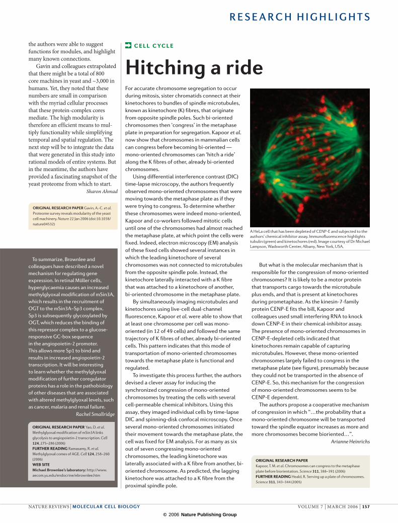
But what is the molecular mechanism that is responsible for the congression of mono-oriented chromosomes? It is likely to be a motor protein that transports cargo towards the microtubule plus ends, and that is present at kinetochores during prometaphase. As the kinesin-7-family protein CENP-E fits the bill, Kapoor and colleagues used small interfering RNA to knock down CENP-E in their chemical-inhibitor assay. The presence of mono-oriented chromosomes in CENP-E-depleted cells indicated that kinetochores remain capable of capturing microtubules. However, these mono-oriented chromosomes largely failed to congress in the metaphase plate (see figure), presumably because they could not be transported in the absence of CENP-E. So, this mechanism for the congression of mono-oriented chromosomes seems to be CENP-E dependent.
The authors propose a cooperative mechanism of congression in which “…the probability that a mono-oriented chromosome will be transported toward the spindle equator increases as more and more chromosomes become bioriented…”.
Arianne Heinrichs
ORIGINAL RESEARCH PAPER Kapoor, T. M. et al. Chromosomes can congress to the metaphase plate before biorientation. Science 311, 388–391 (2006)FURTHER READING Heald, R. Serving up a plate of chromosomes. Science 311, 343–344 (2005)
C E L L CYC L E
Hitching a ride
A HeLa cell that has been depleted of CENP-E and subjected to the authors’ chemical inhibitor assay. Immunofluorescence highlights tubulin (green) and kinetochores (red). Image courtesy of Dr Michael Lampson, Wadsworth Center, Albany, New York, USA.
To summarize, Brownlee and colleagues have described a novel mechanism for regulating gene expression. In retinal Müller cells, hyperglycaemia causes an increased methylglyoxal modification of mSin3A, which results in the recruitment of OGT to the mSin3A–Sp3 complex. Sp3 is subsequently glycosylated by OGT, which reduces the binding of this repressor complex to a glucose-responsive GC-box sequence in the angiopoietin-2 promoter. This allows more Sp1 to bind and results in increased angiopoietin-2 transcription. It will be interesting to learn whether the methylglyoxal modification of further coregulator proteins has a role in the pathobiology of other diseases that are associated with altered methylglyoxal levels, such as cancer, malaria and renal failure.
Rachel Smallridge
ORIGINAL RESEARCH PAPER Yao, D. et al. Methylglyoxal modification of mSin3A links glycolysis to angiopoietin-2 transcription. Cell 124, 275–286 (2006)FURTHER READING Ramasamy, R. et al. Methylglyoxal comes of AGE. Cell 124, 258–260 (2006)WEB SITEMichael Brownlee’s laboratory: http://www.aecom.yu.edu/endocrine/ebrownlee.htm
R E S E A R C H H I G H L I G H T S
NATURE REVIEWS | MOLECULAR CELL BIOLOGY VOLUME 7 | MARCH 2006 | 157
© 2006 Nature Publishing Group
In the news
MIRIAM, FOR FINE MODELLING
The computational systems-biology community has made enormous progress in improving access to models. Scientists now share programming languages for encoding models and have built public repositories to share them. However, the current challenge is quality control; systems biologists won’t use publicly available models if they can’t search them properly, or if the reuse of a model is hampered by mistakes.
To address these issues, an international group of scientists from 14 organizations have devised a new standard for defining biochemical models, known as MIRIAM (minimum information requested in the annotation of biochemical models) to help researchers to exchange and understand computer-generated biochemical models.
“MIRIAM is divided into two parts. The first part is a set of rules that describe how we should present a model … the second part explains how to annotate every model component so that we can identify them”, said Nicolas Le Novère, group leader at European Molecular Biology Laboratory–European Bioinformatics Institute (6 December 2005, EMBL press release).
Four biochemical model databases are in the process of updating their repositories to meet MIRIAM’s criteria.
MIRIAM provides users with a reasonable level of quality assurance. Not only academic researchers will benefit from this new system, but companies might also use biochemical models for the purpose of drug development.
“We also hope that journal editors will adopt MIRIAM as a quality control measure for papers that describe models. This approach has worked well for other fields, by enabling authors, publishers and data providers to work together to improve access to meaningful biological information”, Le Novère concluded (6 December 2005, EMBL press release).
Ekat Kritikou
Scaffold proteins are known to bind and organize many proteins in a signalling pathway. However, a report in Science now reveals that we have underestimated the function of the scaffold proteins. Bhattacharyya and colleagues now show that the Ste5 scaffold not only functions as an interaction assembly point for the mating-pathway components in yeast, but also serves as a regulatory node that actively participates in tun-ing pathway flux.
The scaffold protein Ste5 is involved in the mating (or pherom-one) response mitogen-activated protein kinase (MAPK) pathway in Saccharomyces cerevisiae. Ste5 inter-acts with the different components of the mating pathway — the MAPK Fus3, the MAPK kinase (MAPKK)
Ste7 and the MAPKK kinase Ste11 — and has separate binding sites for each of these proteins. But how does Ste5 interact with these kinases, especially with Fus3, and how does it promote proper signalling?
The authors first mapped the interaction of Fus3 with Ste5 and, through a series of deletion con-structs, refined this binding region to an ~30-residue polypeptide in Ste5. This Ste5 fragment not only binds to Fus3 in a non-canonical manner, but also allosterically stimulates the rate of Fus3 autophosphorylation by ~50-fold. However, Fus3 is phospho-rylated on only one of the two key residues (Tyr182) in the activation loop that are normally phosphor-ylated following full activation of the MAPK.
…other scaffolds might also have many roles in shaping signalling responses …controlling and coordinating their behaviour to precisely tune the amplitude and dynamics of the response.
Three reports in Nature address some pertinent questions regarding prokaryotic DNA replication — how the DNA-synthesis machinery replicates past DNA damage, how continuous DNA synthesis on the leading strand is coordinated with the discontinuous replication of the lagging strand, and how RNA polymerases can produce RNA primers for DNA replication.
How the cell ensures accurate DNA replication when one or both of the strands are damaged has puzzled scientists for decades. It has been shown that ultraviolet irradiation can lead to single-stranded gaps on both leading and lagging strands, which indicates that leading-strand synthesis is not continuous. Heller and Marians now provide mechanistic insights into how leading-strand synthesis can restart downstream of an unrepaired strand, leaving a gap that is presumably filled after the damage has been repaired.
The authors showed that in the presence of a blocking lesion on the
leading strand, replication-protein PriC-dependent loading of the replication fork helicase, DnaB, was sufficient to allow the reassembly of the replication machinery, to unwind the replication-fork duplex and to coordinate the repriming of both the leading and lagging strands. In fact, a single
D N A R E P L I C AT I O N
The double helix unzipped
S I G N A L T R A N S D U C T I O N
Fine tuning
R E S E A R C H H I G H L I G H T S
158 | MARCH 2006 | VOLUME 7 www.nature.com/reviews/molcellbio
© 2006 Nature Publishing Group
So, how does this interaction between Ste5 and Fus3 contribute to pathway output? The authors disrupted the Ste5–Fus3 inter-action and, to their surprise, they observed a two-fold increase in the transcriptional output of the pathway in vivo. Further analysis showed that autophosphorylated Fus3 promotes a decrease in the transcriptional output by phosphorylating Ste5 that contrib-utes to pathway downregulation.
The authors proposed that other scaffolds might also have many roles in shaping signalling responses,
DnaB hexamer on the lagging-strand template coordinated the priming of both strands, thereby coupling the ‘unzipping’ and priming activities.
In a second report, Lee, van Oijen and colleagues studied the kinetics of leading- and lagging-strand synthesis at the single-molecule level by monitor-ing the length of individual DNA molecules during DNA replication. They noticed short pauses in the synthesis of the leading strand, which were primase dependent and preceded the formation and release of a replication loop on the lagging strand. Primases synthesize RNA primers on the lagging strand at a much slower rate than the rate of DNA synthesis on the leading strand. The authors propose that, as a way of coordinating the two events, the primase activity on the lagging strand temporarily halts the progression of the replication fork, thereby preventing the synthesis of the leading strand from progressing too far ahead of that of the lagging strand.
Several viral and plasmid DNA-replication systems use bacterial RNA polymerase to synthesize a replication primer, but how the primer’s 3′ end anneals to the DNA template was unknown. Reporting in a third paper, Zenkin, Severinov and co-workers
not only wiring together specific sets of signalling components, but also controlling and coordinating their behaviour to precisely tune the amplitude and dynamics of the response.
Ekat Kritikou
ORIGINAL RESEARCH PAPER Bhattacharyya, R. P. et al. The Ste5 scaffold allosterically modulates signaling output of the yeast mating pathway. Science 19 Jan 2006 (doi:10.1126/science.1120941)FURTHER READING Kolch, W. Coordinating ERK/MAPK signalling through scaffolds and inhibitors. Nature Rev. Mol. Cell Biol. 6, 827–837 (2005)
carried out an in vitro primer RNA- synthesis reaction on DNA from bacteriophage M13. The replication primer co-eluted with Escherichia coli RNA polymerase as part of the so-called priming complex.
The authors noted that the properties of the replication-priming complex differed from that of known transcription-elongation complexes. The complex contained an overextended RNA–DNA hybrid, which caused the termination of transcription, and the topology of the complex left the 3′ end of the RNA available for interaction with the DNA polymerase to initiate DNA synthesis. Transcription complexes with similar topologies in other bacterial systems might use the same replication-priming mechanism.
Arianne Heinrichs
ORIGINAL RESEARCH PAPERS Heller, R. C. & Marians, K. J. Replication fork reactivation downstream of a blocked nascent leading strand. Nature 439, 557–562 (2006) | Zenkin, N. et al. The mechanism of DNA replication primer synthesis by RNA polymerase. Nature 439, 617–620 (2006) | Lee, J.-B. et al. DNA primase acts as a molecular brake in DNA replication. Nature 439, 621–624 (2006)FURTHER READING Bell, S. D. Molecular cell biology: prime-time progress. Nature 439, 542–543 (2006)
Structure watch A MOLECULAR RULER
In the initiation step of RNA interference, the multidomain ribonuclease Dicer processes double-stranded RNA (dsRNA) into small fragments that are ~21–25 nucleotides long. Several models have been put forward to explain how Dicer produces RNA fragments of such an exact size, but structural insights have been lacking. However, in Science, Doudna and colleagues now describe the crystal structure of full-length Giardia intestinalis Dicer at 3.3-Å resolution.
The G. intestinalis Dicer contains the PAZ domain, which binds to the end of dsRNA, and the tandem RNase III catalytic domains that are characteristic of Dicer, but it lacks other domains that are found in Dicer in higher eukaryotes. However, G. intestinalis Dicer substituted for Schizosaccharomyces pombe Dicer in vivo, which indicates a conserved mechanism of Dicer-catalysed dsRNA processing. Using Er3+, which has an inhibitory effect on G. intestinalis Dicer, the authors showed that its RNase III domains use a two-metal-ion catalytic mechanism. Furthermore, the structure of G. intestinalis Dicer, which seems to be conserved, indicates how Dicer generates specifically sized RNA fragments. A flat, positively charged platform domain separates the PAZ domain from the RNase III domains by a distance of 65 Å. This distance matches the length of 25 base pairs of RNA. So, Dicer works as “…a molecular ruler that measures and cleaves ~25 nucleotides from the end of a dsRNA.”
REFERENCE MacRae, I. J. et al. Structural basis for double-stranded RNA processing by Dicer. Science 311, 195–198 (2006)
A RIVETING TOXIN
The bacterial toxin aerolysin kills cells by forming heptameric channels in plasma membranes, but the channel structure and the mechanisms of membrane insertion and channel formation are poorly understood. van der Goot and colleagues focused on a loop in domain 3 (DIII loop) of aerolysin, because it contains alternating charged and neutral residues that could form a transmembrane β-hairpin (the channel might therefore be a β-barrel). In The EMBO Journal, they describe their study of the topology of the DIII loop and its role in channel formation.
The authors restrained the DIII loop using disulphide bonds, and showed that this did not affect aerolysin heptamerization. However, it affected membrane insertion and channel formation. By carrying out cysteine-scanning mutagenesis coupled with channel analysis using planar lipid bilayers and cysteine-specific labelling, they identified residues in the DIII loop that line the channel lumen and the sequence that forms the turn of the β-hairpin. By mutating this hydrophobic WPLVG sequence, they showed that this turn drives membrane insertion, and they propose that once it has crossed a bilayer, it folds back perpendicular to the barrel axis. They modelled this rivet-like conformation for the membrane anchoring of aerolysin, and sequence alignments indicate that many other pore-forming toxins might also use this type of channel riveting.
REFERENCE Iacovache, I. et al. A rivet model for channel formation by aerolysin-like pore-forming toxins. EMBO J. 25, 457–466 (2006)
R E S E A R C H H I G H L I G H T S
NATURE REVIEWS | MOLECULAR CELL BIOLOGY VOLUME 7 | MARCH 2006 | 159