Embed Size (px)
Citation preview
Restricted 12p Amplification and RAS Mutation inHuman Germ Cell Tumors of the Adult Testis
Helene Roelofs,* Marijke C. Mostert,*Kirsten Pompe,*† Gaetano Zafarana,*Monique van Oorschot,*Ruud J. H. L. M. van Gurp,* Ad J. M. Gillis,*Hans Stoop,* Berna Beverloo,‡
J. Wolter Oosterhuis,* Carsten Bokemeyer,† andLeendert H. J. Looijenga*From the Pathology/Laboratory for Experimental Patho-
Oncology,* University Hospital Rotterdam/Daniel, Josephine
Nefkens Institute, Rotterdam, The Netherlands; the Department of
Hematology and Oncology,† University of Tubingen, Tubingen,
Germany; and the Department of Cell Biology and Genetics,‡
Erasmus University Rotterdam, Rotterdam, The Netherlands
Human testicular germ-cell tumors of young adults(TGCTs), both seminomas and nonseminomas, arecharacterized by 12p overrepresentation, mostly asisochromosomes, of which the biological and clinicalsignificance is still unclear. A limited number ofTGCTs has been identified with an additional high-level amplification of a restricted region of 12p in-cluding the K-RAS proto-oncogene. Here we showthat the incidence of these restricted 12p amplifica-tions is ;8% in primary TGCTs. Within a single cellformation of i(12p) and restricted 12p amplificationis mutually exclusive. The borders of the ampliconscluster in short regions, and the amplicon was neverfound in the adjacent carcinoma in situ cells. Semi-nomas with the restricted 12p amplification virtuallylacked apoptosis and the tumor cells showed pro-longed in vitro survival like seminoma cells with amutated RAS gene. However, no differences in prolif-eration index between these different groups of sem-inomas were found. Although patients with a semi-noma containing a homogeneous restricted 12pamplification presented at a significantly younger agethan those lacking it, the presence of a restricted 12pamplification/RAS mutation did not predict the stageof the disease at clinical presentation and the treat-ment response of primary seminomas. In 55 primaryand metastatic tumors from 44 different patients whofailed cisplatinum-based chemotherapy, the re-stricted 12p amplification and RAS mutations had thesame incidence as in the consecutive series of re-sponding patients. These data support the model thatgain of 12p in TGCTs is related to invasive growth. Itallows tumor cells, in particular those showing char-acteristics of early germ cells (ie, the seminoma
cells), to survive outside their specific microenviron-ment. Overexpression of certain genes on 12p prob-ably inhibits apoptosis in these tumor cells. However,the copy numbers of the restricted amplification of12p and K-RAS mutations do not predict response totherapy and survival of the patients. (Am J Pathol2000, 157:1155–1166)
Malignant transformation is a complex, multistep pro-cess.1 Although involvement of several genes has beensuggested in the development of testicular germ-cell tu-mors of young adults (TGCTs), histologically seminomaand nonseminomas,2 evidence is lacking.
All TGCTs originate from carcinoma in situ (CIS).3,4 CISis frequently found in the parenchyma adjacent to aninvasive TGCT, being located on the inner side of thebasal membrane of the seminiferous tubules, in closeassociation with Sertoli cells.5 The mechanisms involvedin the development from CIS, via the microinvasive stage,to overt invasive tumors are still unclear. Obviously tumorcells are selected that are capable of surviving and grow-ing outside of the specific microenvironment of the sem-iniferous tubule.
The only consistent chromosomal anomaly in TGCTs isthe gain of the short arm of chromosome 12, mostlybecause of isochromosomes of 12p.6,7 The copy numberof 12p reportedly predicts prognosis.8–10 K-RAS hasbeen proposed as the relevant gene on 12p,11–13 ofwhich the encoding protein (p21) is involved in signaltransduction. Mutated RAS has been found to be corre-lated with poor prognosis in childhood acute lymphocyticleukemia,14 and non-small-cell lung cancer.15,16 Re-cently, activation of RAS has been shown to be involvedin tumor maintenance17,18 and in inducing anchorage-independent growth because of inhibition of apopto-sis.19,20 Indeed, a correlation between activated RAS andmetastatic capacity has been reported.21,22 Besides ac-tivation by means of mutations, RAS can also be involvedin malignant transformation because of increased copynumbers of the wild-type gene, resulting in overexpres-
Supported by Dutch Cancer Society grant KWF, DDHK 94 836 and afortune grant of Tubingen University.
Accepted for publication June 14, 2000.
Address reprint requests to L. H. J. Looijenga, Ph.D., Pathology/Labo-ratory for Experimental Patho-Oncology, University Hospital Rotterdam/Daniel, Josephine Nefkens Institute, FGG/EUR Building Be, Room 430b,P. O. Box 1738, 3000 DR Rotterdam, The Netherlands. E-mail:[email protected].
American Journal of Pathology, Vol. 157, No. 4, October 2000
Copyright © American Society for Investigative Pathology
1155
sion of wild-type mRNA and accumulation of the wild-type protein.23–28
Seminomas are highly sensitive to irradiation and cis-platinum-based chemotherapy. The vast majority of pa-tients with metastatic nonseminomas is cured by cisplati-num-based combination chemotherapy.29 Because theclinical course of these tumors can still not be predictedfor individual patients, additional prognostic markers areneeded. Interestingly, activated RAS genes increase theintrinsic resistance to radiation and cisplatinum thera-py.30–34 It is unclear, however, whether amplification ofwild-type RAS has the same effect.
A small percentage of TGCTs has RAS mutations,35–37
of which the clinical relevance was not studied. Weshowed previously that seminomas with a mutated RASgene have survival advantage in vitro and have reducedapoptosis in the primary tumor.38,39 Noteworthy, in vitrosurvival of tumor cells correlates with poor prognosis inpatients with metastatic TGCTs40 and adult acute my-eloid leukemia.41 Recently, a number of invasive TGCTswith amplification of a restricted region of 12p have beenidentified.42–45 We showed that the shortest region ofoverlap of amplification (SROA) is ;1.7 Mbases, contain-ing three known genes, ie, SOX5, JAW1, and K-RAS.45 Itis unknown so far whether amplification of wild-type K-RAS in these tumors has the same effects as RAS muta-tions. Finally, the clinical relevance of a restricted 12pamplification has not been conclusively investigated inTGCTs.
The goal of this study is to further investigate the bio-logical and clinical significance of gain of 12p sequencesin TGCTs. The incidence of restricted 12p amplificationwas studied in a consecutive series of 76 untreated pri-mary TGCTs. The newly found six tumors, as well as thepreviously identified nine cases, were studied for the distri-bution of the restricted 12p amplification within the tumor(homogeneous or heterogeneous pattern), the borders ofthe SROA, the presence of i(12p), the proliferative activ-ity, the presence of apoptosis, and capacity of the tumorcells to survive in vitro. In addition, corresponding CISand microinvasive seminoma were tested for the pres-ence of the restricted 12p amplification. The clinical im-portance of the restricted 12p amplification and RASmutation was further studied in 44 patients who failedcisplatinum-based chemotherapy.
Materials and Methods
Samples
The freshly obtained tumor samples included in this studywere collected in close collaboration with urologists andpathologists in the southwestern part of the Netherlands.All tumors were obtained before chemotherapy and/orirradiation. Directly after surgical removal, representativeparts of the tumor and adjacent normal tissue (whenavailable) were snap-frozen and other pieces were fixedovernight in 10% buffered formalin and embedded inparaffin. The sizes of the testis and the tumor were mea-sured in three dimensions. The tumors were diagnosed
according to the World Health Organization classificationfor testicular tumors.46 Nonseminomas containing both aseminoma and a nonseminoma component were classi-fied as combined tumors according to the British classi-fication,47 instead of as nonseminomas according to theWorld Health Organization classification system. Identifi-cation of CIS, seminoma, and embryonal carcinoma wasaided by direct enzyme histochemical detection of alka-line phosphatase activity on representative frozen tissuesections, as reported before.48 The consecutive seriestested for the presence of a restricted 12p amplification(see below) consisted of 46 seminomas, 23 nonsemino-mas (14 embryonal carcinomas/yolk-sac tumors/terato-mas; three teratomas; two embryonal carcinomas/yolk-sac tumors; two yolk-sac tumors, one teratoma/yolk-sactumor; one embryonal carcinoma) and seven combinedtumors (three embryonal carcinomas/seminomas; threeembryonal carcinomas/yolk-sac/seminomas; one teratoma/yolk-sac tumor/seminoma). The newly identified cases witha restricted 12p amplification, and the previously foundcases45 and unpublished observations, were studied forthe borders of the amplicon (see below), the distributionwithin the tumor (see below), presence of i(12p) by karyo-typing (when available), proliferation index (see below), ap-optosis, and in vitro survival, as described before (see be-low).38,39 In addition, the formerly identified seminomas witha mutated RAS gene37 were included in this analysis.
The analysis of the possible clinical impact of the re-stricted 12p amplification/K-RAS mutation was studied ona series of patients who failed cisplatinum-based chemo-therapy. These cases were collected in collaboration withthe Departments of Hematology and Oncology, Univer-sity of Tubingen, Germany, and Internal Medicine, Neth-erlands Cancer Institute, Amsterdam, The Netherlands.In total, samples of 44 different patients were included, ofwhom 22 primary TGCTs and 33 metastasis were studied(of 11 patients both the primary and metastasis wasavailable).
Immunohistochemical Detection of Ki-67
Sections were cut from one representative paraffin blockper tumor, which was mounted on 3-aminopropyl-tri-ethoxysilane-coated slides and dried at 50°C overnight.Subsequently, the sections were heated to 120°C in so-dium citrate solution (0.01 mol/L, pH 6.0) using an auto-clave.49 Endogenous peroxidase reactivity was blockedwith H2O2/methanol. The Ki-67 antigen was demon-strated using the polyclonal antibody A0047 (DAKO,Glostrup, Denmark), diluted 1:100. Incubation was donefor 1 hour at room temperature after blocking of nonspe-cific binding sites with 5% bovine serum albumin. Afterextensive washing, biotinylated swine anti-rabbit (1:200)(DAKO) was used as second step, which was detectedusing the horseradish-labeled streptavidin-biotin com-plex (DAKO) diluted 1:100. Peroxidase was visualizedwith diaminobenzidine, after which the sections werecounterstained lightly with Mayer’s hemalum. Of eachtumor, 3 3 50 tumor cells were independently counted bytwo observers in one tissue section. The results were
1156 Roelofs et alAJP October 2000, Vol. 157, No. 4
statistically analyzed using the Student’s t-test. Positiveand negative (excluding the first antibody) controls wereincluded in each experiment.
Metaphase Preparations
After surgical removal representative parts of nonsemi-nomas were enzymatically digested (collagenase; SigmaChemical Co., St. Louis, MO), and cultured in T75 flasks(Corning Costar, Europe, Schiphol-Rijk, The Netherlands)for several days under standard conditions, ie, 37°C in ahumidified atmosphere with 5% CO2 in air in Dulbecco’smodified Eagle’s medium/HF12 culture medium with 10%heat-inactivated fetal calf serum (Gibco-BRL, Life Tech-nologies BV, Brueda, The Netherlands) as describedbefore.50 Mitotic cells were harvested after 2 to 4 hours ofcolcemid treatment, swollen in hypotonic solution, andfixed in methanol/acetic acid (3:1). Representative partsof seminoma were directly processed to isolate meta-phase spreads as described before.51 Briefly, the mitoticcells were, after mechanical dissociation of the tumor,directly harvested in the presence of colcemid. The cellswere subsequently swollen in hypotonic KCl/EGTA/Hepes solution and fixed with methanol:acetic acid (3:1).For conventional G-band karyotyping the air-dried prep-arations were digested with pancreatin. The chromosomeconstitution was described according to the InternationalSystem for Human Cytogenetic Nomenclature 1995.52
Restricted 12p Amplification
For the random screen the in situ hybridization experi-ments on the methanol/acetic acid-fixed nuclei suspen-sions on the consecutive series were performed as de-scribed earlier45 using YAC#5 (mapped to 12p11.2–12.1;kindly provided by Dr. B. Gemmill, Denver, CO). Thisprobe is known to map to the SROA as determined in ourearlier study.45 To make this probe suitable for the in situhybridization approach, it was purified by pulsed-field gelelectrophoresis, amplified, and labeled with digoxigenin-11-dUTP (Roche Diagnostics Nedevland BV, Almeve,The Netherlands) using a nick-translation kit (Gibco-BRL). It was visualized with fluorescein isothiocyanate-conjugated sheep-a-digoxigenin (Roche). The presenceof a 12p amplification was defined as reported before45
as being ;15 to 30 signals per interphase nucleus.TGCTs identified with a restricted 12p amplification
were studied in more detail regarding the breakpointsand distribution. Of these cases, frozen tissue sections of16-mm thickness, containing tumor, microinvasive semi-noma, and CIS (when available), were cut and air-driedovernight at 37°C on microscope slides treated with3-aminopropyl-triethoxysilane. In addition, one parallelsection (4-mm thickness) was stained with hematoxylinand eosin (H&E) and the other for alkaline phosphatasereactivity for histological examination. The slides for thein situ hybridization were submerged in 70% ethanol(220°C) for 1 hour and dehydrated in an increasingethanol series (80%, 90%, 100%, 2 minutes each) andair-dried. Subsequently, the tissue sections underwent
protein digestion with 0.0005% pepsin (Sigma) in 0.01mol/L HCl in water, 1 minute at 37°C, followed by a washstep (phosphate-buffered saline, 5 minutes) and dehy-dration. Hybridization was performed as described forthe methanol/acetic acid fixed nuclei.45 YAC#5 was usedas control probe in combination with another probe (testprobe) (Figure 1). In particular, probes positive for se-quence-taq-sites D12S1945, D12S1688, and AFM267yc9(on the distal side) and D12S1350E, KRAS2, D12S1313,and D12S1411 (on the proximal border) were used as testprobes. YAC#5 was labeled as described above withdigoxigenin-11-dUTP, and visualized using fluorescein iso-thiocyanate-conjugated sheep anti-digoxigenin (Roche).The test probe was also labeled using nick-translation withbiotin-11-dUTP and detected using avidine-CY3 (JacksonImmunoResearch, West Grove, PA). A restricted 12p am-plification was defined as the presence of nuclei with atleast 15 hybridization signals. The pattern was identifiedas heterogeneous when the positive nuclei are scatteredthroughout the tissue section: both regions with and with-out restricted 12p amplification are present. In contrast,the pattern is defined as homogeneous when all regionsshowed tumor nuclei with restricted 12p amplification.Because of tissue cutting artifacts, this does not excludethe presence of single nuclei without such an amplifica-tion. The criteria used to determine the borders of thebreakpoints was similar to that used in our former study.45
Briefly, the test probe was scored as part of the ampliconwhen paired hybridization signals with the control probe(YAC#5) was observed, and scored as outside the ampli-con when less hybridization signals were found comparedto the control probe. Higher copy numbers of the testprobes (see above) compared to the control probe have notbeen found so far.
The paraffin-embedded tissue sections of the treat-ment-resistant TGCTs (4-mm thickness) were preincu-bated overnight at 50°C, and subsequently baked for 10minutes at 80°C. The sections were deparaffinized usingxylene, washed in 100% methanol four times at roomtemperature, and air-dried. Sodium thiocyanate (1 mol/L)pretreatment was done for 10 minutes at 80°C, afterwhich the slides were thoroughly rinsed in water. Diges-tion was done using 8000 U pepsin (Sigma) in 0.2 mol/LHCl in phosphate-buffered saline at 37°C for 30 to 40minutes, depending on the tissue under investigation.After rinsing in water at 4°C, dehydration was done usinga series of increasing ethanol concentrations (70%, 80%,90%, and 100%). The hybridization with a K-RAS-specificdigoxigenin-labeled probe, washing, and detection pro-cedures were performed as described for the methanol/acetic acid-fixed nuclei (see above). A restricted 12pamplification was defined as the presence of at least 10hybridization signals of the K-RAS probe. Because of thethickness of the tissue sections used, the tumors were notscored for the presence of a homogeneous or heteroge-neous pattern.
Visualization was done with a Zeiss Axioskop epifluo-rescence microscope (Carl Zeiss, BV, Weesp, The Neth-erlands) with a Pinkel filter in combination with a tripleband-pass filter, which enabled the observation of fluo-rescein isothiocyanate, CY3, and DAPI in one view.
12p and Testicular Germ Cell Tumors 1157AJP October 2000, Vol. 157, No. 4
Comparative Genomic Hybridization (CGH)
For CGH, metaphase spreads were prepared using stan-dard procedures from in vitro phytohemagglutinin-stimu-lated peripheral blood lymphocyte cultures of a healthymale as reported previously.53 High molecular weightDNA was isolated from the snap-frozen tissue samples(test DNA) and from peripheral blood of a normal male(reference DNA) using standard procedures.54 The CGHprocedure and analysis were performed as describedbefore.55 Both the control male DNA and the tumor DNAwere directly labeled by nick-translation with lissamineand fluorescein isothiocyanate, respectively. The datawere analyzed using Quips XL software from Vysis(Downers Grove, IL). Normalization was performed usingthe average of the green-to-red fluorescent intensities forthe entire metaphase. At least 10 metaphases were stud-ied for each case. Losses of DNA sequences were de-fined as chromosomal regions where the average green-to-red ratio and its 95% confidence intervals are below0.9 whereas gains are .1.1.56 The heterochromaticblocks of chromosomes 1, 9, 16, and Y, and the imme-diate telomeric regions were excluded from the analysis
because these regions present variable results in exper-iments with normal control DNAs.
Spectral Karyotyping
Spectral karyotyping was performed on a single semi-noma with a restricted 12p amplification, demonstratedboth by CGH and in situ hybridization, using a spectralkaryotyping kit and analysis system (Vysis) with a slightlymodified procedure as described by the supplier. Inparticular, proteinase K digestion was performed in 1mol/L Tris-HCl, pH 7.5, and after denaturation of theprobe, the mix was kept on ice. Washing during thedetection procedure was performed in 55% formamide at39°C instead of 45°C. In addition, 0.05% instead of 0.1%Tween was used in 43 standard saline citrate, and thelast washing step was performed at room temperature.
RAS Gene Mutation Detection
Mutations in codon 12 or 13 of the N- and K-RAS gene wereanalyzed by direct sequencing (Amplicycle; Amersham,
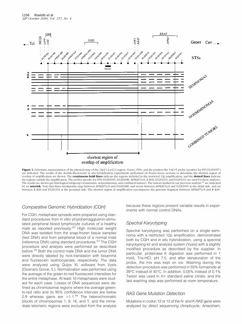
Figure 1. Schematic representation of the physical map of the 12p11.2-p12.1 region. Genes, STSs, and the position the YAC#5 probe (positive for STS D12S1057)are indicated. The results of the double-fluorescent in situ hybridization experiments performed on frozen tissue sections to determine the shortest region ofoverlap of amplification are shown. The continuous bold lines indicate the regions included in the restricted 12p amplification, and the dotted lines indicatethe regions outside the amplification. The probes specific for STSs D12S1945, D12S1688, AFM267yc9, K-RAS, D12S1313, and D12S1411 are used for these analyses.The results are shown per histological subgroup (seminomas, nonseminomas, and combined tumors). The tumors studied in our previous analysis,45 are indicatedby an asterisk. Note that three breakpoints map between AFM267yc9 and D12S1688, and seven between AFM267yc9 and D12S1945 at the distal side, and sixbetween K-RAS and D12S1313 at the proximal side. The shortest region of amplification encompasses the genomic fragment between AFM267yc9 and K-RAS.
1158 Roelofs et alAJP October 2000, Vol. 157, No. 4
Arlington Heights, IL), using primer NA (59-GACTGAGTA-CAAACTGGTGG-39)/NB (59-CTCTATGGTGGGATCATATT-39) and KA (59-GACTGAATATAAACTTGTGG-39)/KB (59-CTATTGTTGGATCATATTCC-39), respectively, on DNAisolated from snap-frozen seminomas tested for in vitrosurvival and presence of apoptosis. DNA was isolated asdescribed above. Only histological areas containing.70% tumor cells were used. In addition, the presence ofK-RAS codon 12 mutations was investigated in the seriesof paraffin-embedded tumors of the nonresponding pa-tients as follows: two 15-mm thickness sections from eachsample were first deparaffinized with xylene and thendehydrated with absolute ethanol. A parallel section(4-mm thickness) was stained with H&E to confirm thepresence of tumor and to check for histology. Only sec-tions with a major tumor component were used for DNAisolation. DNA was eluted in 30 ml of water by heating theair-dried tissues at 95°C for 5 minutes. Typically 50 to 100ng of DNA was used for each polymerase chain reaction(PCR). Each sample was first tested with HLA-dQ prim-ers57 to assess the quality of the eluted DNA. Only sam-ples showing proper amplification were subsequentlyused. The PCR reactions to detect mutations in codon 12of K-RAS were performed essentially as described be-fore58 with the following modifications. The K-RAS 59primer used in the two rounds of amplifications carried abiotin label at the 59 end. After the first MvaI digestion, thePCR products were affinity purified on streptavidin-coated paramagnetic beads (Dynal AS, Oslo, Norway)and the bound PCR products were redigested on thebeads in 50 ml of MvaI buffer containing 10 U of enzymefor 3 hours at 37°C. Subsequently the beads were affinitypurified and washed on the magnet to remove the bufferand the unbound fraction. Finally, single-stranded DNAwas eluted from the bound fraction by boiling the beadswith 10 ml of water. One half of each fraction was used inthe second amplification step. Samples positive for mu-tations in codon 12 were further characterized by cyclesequencing the single-stranded DNA obtained by affinitypurification of the PCR products retained on the beadsafter the final MvaI digestion. The procedure was con-trolled at every stage with DNA samples obtained fromarchival paraffin-embedded primary TGCTs with andwithout codon 12 mutations.37
Detection of DNA Laddering
High molecular weight DNA was isolated from snap-frozen histologically checked samples using standardprocedures (see above). DNA laddering was visualizedusing ethidium bromide staining after electrophoresis of 1mg as described previously.39
Results
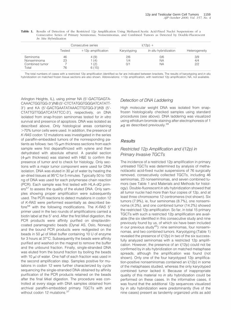
Restricted 12p Amplification and i(12p) inPrimary Invasive TGCTs
The incidence of a restricted 12p amplification in primaryuntreated TGCTs was determined by analysis of metha-nol/acetic acid-fixed nuclei suspensions of 76 surgicallyremoved, consecutively collected TGCTs, including 46seminomas, 23 nonseminomas, and seven combined tu-mors (see Table 1 and Materials and Methods for histol-ogy). Double-fluorescent in situ hybridization showed thatall tumor nuclei had more than four copies of 12p, and atleast three chromosome 12 centromeres (not shown). Sixtumors (7.9%), ie, four seminomas (8.7%), one nonsemi-noma (4.3%), and one combined tumor (14.2%) showedthe restricted 12p amplification. So far, in total 15 primaryTGCTs with such a restricted 12p amplification are avail-able (the six identified in this consecutive study and ninepreviously found by us, of which six have been includedin our previous study45): nine seminomas, four nonsemi-nomas, and two combined tumors. Karyotyping (Table 1)revealed the presence of i(12p) in two of the six success-fully analyzed seminomas with a restricted 12p amplifi-cation. However, the presence of an i(12p) could not beconfirmed by in situ hybridization on matched metaphasespreads, although the amplification was found (notshown). Only one of the four karyotyped 12p amplifica-tion-positive nonseminomas contained an i(12p) in someof the metaphases studied, whereas the only karyotypedcombined tumor lacked it. Because of inappropriatequality of this material no in situ hybridization could beperformed on these cases. In the informative cases, itwas found that the additional 12p sequences visualizedby in situ hybridization were predominantly (five of thenine cases) present as tandemly organized units as add
Table 1. Results of Detection of the Restricted 12p Amplification Using Methanol/Acetic Acid-Fixed Nuclei Suspensions of aConsecutive Series of Primary Seminomas, Nonseminomas, and Combined Tumors as Detected by Double-FluorescentIn Situ Hybridization
Consecutive series i(12p) 1
HeterogeneityTested 112p amplification Karyotyping In situ hybridization
Seminoma 46 4 (9) 2/6 0/6 3/9Nonseminoma 23 1 (4) 1/4 NA 4/4Combined tumor 7 1 (2) 0/1 NA 2/2Total 76 6 (15)
The total numbers of cases with a restricted 12p amplification identified so far are indicated between brackets. The results of karyotyping and in situhybridization on matched frozen tissue sections are also shown. Abbreviations: 112p amplification, with restricted 12p amplification; NA, not available.
12p and Testicular Germ Cell Tumors 1159AJP October 2000, Vol. 157, No. 4
(12)(p11), add(p12), or add(p13), although other siteswere also found to contain 12p-specific sequences, in-cluding parts of chromosomes 6, 8, and 11 (not shown).
Breakpoints Involved in the Restricted 12pAmplification
In addition to our earlier report consisting of six cases,45
three seminomas, two nonseminomas, and one com-bined tumor (indicated by an asterisk in Figure 1), in situhybridization was applied on the newly identified TGCTs(n 5 9) with a restricted 12p amplification. Because thisanalysis was done on frozen tissue sections instead ofmethanol/acetic acid-fixed nuclei suspension, as in ourearlier study, all cases could be analyzed, including thetwo previously identified noninformative cases.45 AgainYAC#5 was used in combination with probes specific forthe more proximal and distal regions of the contig (seeFigure 1 and Materials and Methods section). In accor-dance with our previous findings, the amplified regionalways includes the genomic fragment between STSAFM267yc9 at the distal end and K-RAS at the proximalend. Therefore, K-RAS, JAW1, and SOX5 are consistentlyamplified in all TGCTs with a restricted 12p amplification,irrespective of histology. In addition to this finding, theborders of the amplicon appeared to cluster in narrow re-gions: 40% between K-RAS and STS D12S1313 at theproximal side (44.4% for seminoma and 25% for nonsemi-noma), and 20% between AFM267yc9 and D12S1688 atthe distal side (11% for seminoma and 50% for nonsemi-noma). In addition, 46.7% of the breakpoints map betweenAFM267yc9 and D12S1945 (44.4% for seminoma and 75%for nonseminoma) (Figure 1).
Intratumor 12p Amplification and TumorHeterogeneity
In situ hybridization results on the suspensions of nucleialready indicated that the restricted 12p amplificationcan be heterogeneously distributed in one tumor (notshown). This was verified by in situ hybridization on frozentissue sections. Six out of the nine seminomas showedthe amplification homogeneously throughout the tumor(Table 1). The other three cases showed regions with andwithout amplification, of which two contained i(12p) bykaryotyping. All nonseminomas and combined tumorsshowed a heterogeneous pattern. In two cases the am-plification was present in a subpopulation of cells of theyolk-sac tumor component, and in one in a subpopulationof both the embryonal carcinoma and yolk-sac tumorcomponent. One of the combined tumors with the re-stricted 12p amplification was a mixture of seminoma andembryonal carcinoma. The amplification was found in asubpopulation of both components. No i(12p) was iden-tified in this case. The other combined tumor showedamplification in a minority of the seminoma cells only andkaryotyping failed.
One of the nonseminomas, initially diagnosed asmixed nonseminoma showed no i(12p) by karyotyping.
This tumor was xenografted (orthotopically into a nuderat), and after ;10 months, a tumor completely com-posed of yolk sac tumor was formed. Karyotyping afterdirect harvesting revealed i(12p) in seven out of 14 meta-phase spreads, confirmed by in situ hybridization (notshown). These metaphase spreads did not contain the12p amplification. However, a limited number of inter-phase nuclei isolated from the original tumor and thexenograft showed the presence of 12p amplification byin situ hybridization (not shown). In situ hybridization onfrozen tissue sections of these tumors demonstrated that,5% of the tumor cells from the primary tumor showedthe 12p amplification, preferentially in the embryonal car-cinoma and yolk-sac tumor component. This percentagewas constant during subsequent xenograft passages.
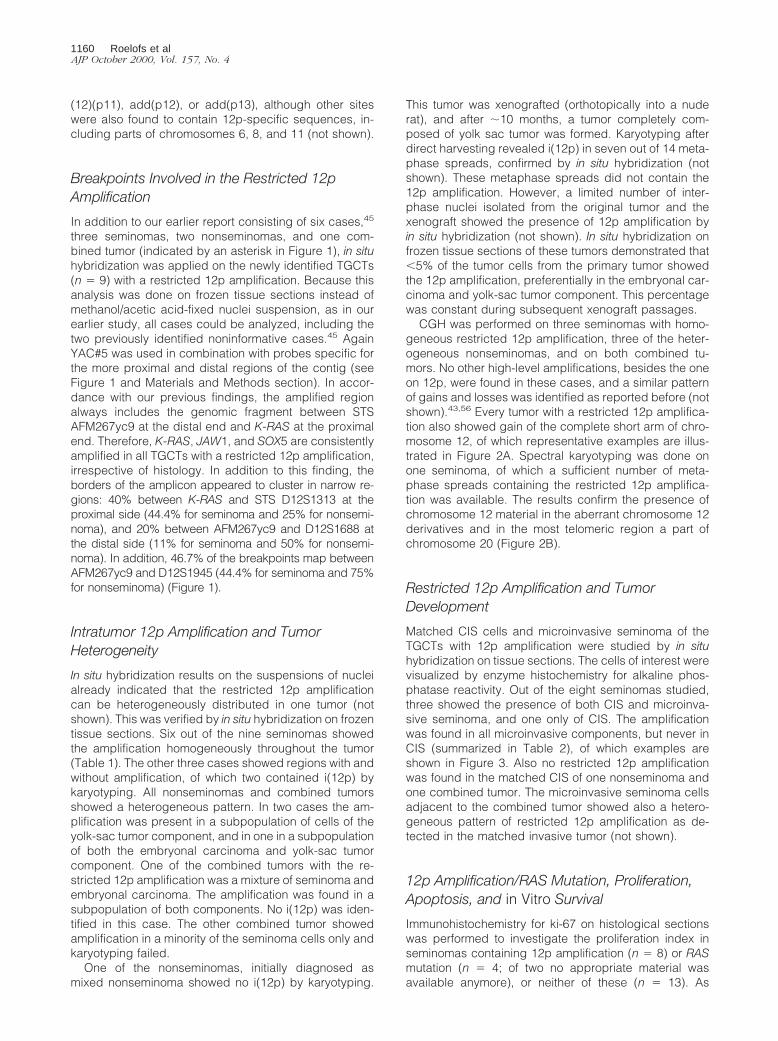
CGH was performed on three seminomas with homo-geneous restricted 12p amplification, three of the heter-ogeneous nonseminomas, and on both combined tu-mors. No other high-level amplifications, besides the oneon 12p, were found in these cases, and a similar patternof gains and losses was identified as reported before (notshown).43,56 Every tumor with a restricted 12p amplifica-tion also showed gain of the complete short arm of chro-mosome 12, of which representative examples are illus-trated in Figure 2A. Spectral karyotyping was done onone seminoma, of which a sufficient number of meta-phase spreads containing the restricted 12p amplifica-tion was available. The results confirm the presence ofchromosome 12 material in the aberrant chromosome 12derivatives and in the most telomeric region a part ofchromosome 20 (Figure 2B).
Restricted 12p Amplification and TumorDevelopment
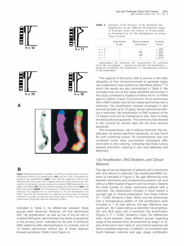
Matched CIS cells and microinvasive seminoma of theTGCTs with 12p amplification were studied by in situhybridization on tissue sections. The cells of interest werevisualized by enzyme histochemistry for alkaline phos-phatase reactivity. Out of the eight seminomas studied,three showed the presence of both CIS and microinva-sive seminoma, and one only of CIS. The amplificationwas found in all microinvasive components, but never inCIS (summarized in Table 2), of which examples areshown in Figure 3. Also no restricted 12p amplificationwas found in the matched CIS of one nonseminoma andone combined tumor. The microinvasive seminoma cellsadjacent to the combined tumor showed also a hetero-geneous pattern of restricted 12p amplification as de-tected in the matched invasive tumor (not shown).
12p Amplification/RAS Mutation, Proliferation,Apoptosis, and in Vitro Survival
Immunohistochemistry for ki-67 on histological sectionswas performed to investigate the proliferation index inseminomas containing 12p amplification (n 5 8) or RASmutation (n 5 4; of two no appropriate material wasavailable anymore), or neither of these (n 5 13). As
1160 Roelofs et alAJP October 2000, Vol. 157, No. 4
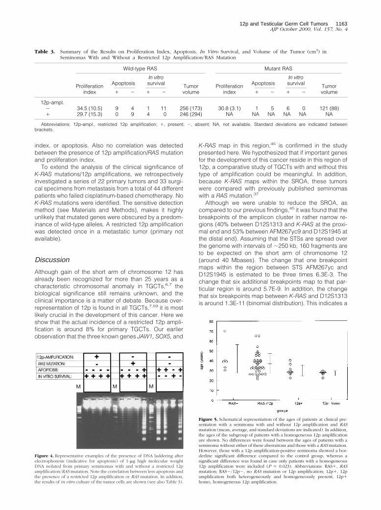
indicated in Table 3, no differences between thesegroups were observed. However, all nine seminomaswith 12p amplification, as well as five of the six with amutated RAS gene, demonstrated low levels of apoptosisin the primary tumor, indicated by the lack of profoundDNA laddering after electrophoresis. In contrast, nine of13 tested seminomas without any of these featuresshowed apoptosis (Table 3 and Figure 4).
The capacity of the tumor cells to survive in vitro afterdisruption of their microenvironment to generate singlecell suspensions was studied as described before,38 ofwhich the results are also summarized in Table 3. Weexcluded that one of the newly identified seminomas inthis study contained a mutation of either the N- or K-RASgene in codons 12 and 13 (not shown). All six seminomaswith a RAS mutation and all four tested seminomas with arestricted 12p amplification showed prolonged in vitrosurvival (at least up to 10 days). Eleven seminomas with-out a restricted 12p amplification or RAS mutation of the12 tested could not be maintained in vitro. Nine of theseshowed profound apoptosis. The seminoma that showedin vitro survival for several days did not show obviousapoptosis.
The nonseminomas, with or without restricted 12p am-plification all lacked significant apoptosis, as was foundfor both combined tumors. All nonseminomas and onecombined tumor were successfully karyotyped aftershort-term in vitro culturing, indicating that these tumorsallowed short-term culturing in vitro (see Materials andMethods).
12p Amplification, RAS Mutation, and ClinicalBehavior
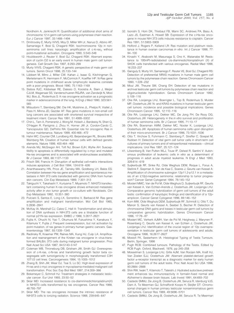
The age at clinical diagnosis of patients with a seminomawith and without a restricted 12p amplification/RAS mu-tation is indicated in Figure 5. No age differences existbetween seminoma and nonseminoma patients with andwithout a RAS mutation (Figure 5 and not shown). Despitethe small number of cases, seminoma patients with arestricted 12p amplification showed a trend toward ayounger age at clinical presentation than those without(P 5 0.055, Student’s t-test). When only the seminomaswith a homogeneous pattern of 12p amplification wereincluded (n 5 6) (see above), the age difference wassignificant: 36.7 years without a restricted 12p amplifica-tion and 26.8 years with a restricted 12p amplification(Figure 5; P 5 0.023, Student’s t-test). No differenceswere found between these different groups regardingstage of the disease (they all presented as stage I or II),size of the tumor (see Table 3), and clinical response (allhad a complete response). In addition, no correlation wasfound between outcome and age, stage, proliferation
Figure 2. A: Representative examples of CGH for chromosome 12 on twoseminomas without 12p amplification (left) and two with a homogeneousrestricted 12p amplification (right). Note that the seminomas with an am-plification also show gain of the complete short arm of this chromosome. Thenumber of metaphase spreads analyzed is indicated. B: Representative ex-ample of banding (left) and spectral karyotyping (real color at the right, andfalse color in the middle) of a chromosome 12 derivative known to containrestricted 12p amplification (as determined by in situ hybridization andCGH). Note that the chromosome is composed of chromosome 12 material(indicated in purple), except the most distal region of the p arm, showingchromosome 20-specific material (indicated in blue).
Table 2. Summary of the Presence of the Restricted 12pAmplification in the Different Development Stagesof Testicular Germ Cell Tumors of Young Adults,as Determined by In Situ Hybridization on FrozenTissue Sections
Carcinomain situ
Micro-invasiveseminoma
Invasivetumor
n 5 3 2 1 1 (SE)n 5 1 2 NA 1 (SE)n 5 1 2 NA 6 (NS)n 5 1 2 6 6 (CT)
Abbreviations: SE, seminoma; NS, nonseminoma; CT, combinedtumor; NA, not available; 2, absence of restricted 12p amplification; 1,presence of restricted 12p- amplification; 6, heterogeneous presenceof 12p amplification.
12p and Testicular Germ Cell Tumors 1161AJP October 2000, Vol. 157, No. 4
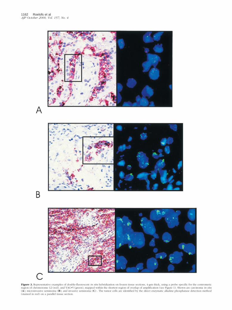
Figure 3. Representative examples of double-fluorescent in situ hybridization on frozen tissue sections, 4-mm thick, using a probe specific for the centromericregion of chromosome 12 (red), and YAC#5 (green), mapped within the shortest region of overlap of amplification (see Figure 1). Shown are carcinoma in situ(A); microinvasive seminoma (B); and invasive seminoma (C) . The tumor cells are identified by the direct enzymatic alkaline phosphatase detection method(stained in red) on a parallel tissue section.
1162 Roelofs et alAJP October 2000, Vol. 157, No. 4
index, or apoptosis. Also no correlation was detectedbetween the presence of 12p amplification/RAS mutationand proliferation index.
To extend the analysis of the clinical significance ofK-RAS mutations/12p amplifications, we retrospectivelyinvestigated a series of 22 primary tumors and 33 surgi-cal specimens from metastasis from a total of 44 differentpatients who failed cisplatinum-based chemotherapy. NoK-RAS mutations were identified. The sensitive detectionmethod (see Materials and Methods), makes it highlyunlikely that mutated genes were obscured by a predom-inance of wild-type alleles. A restricted 12p amplificationwas detected once in a metastatic tumor (primary notavailable).
Discussion
Although gain of the short arm of chromosome 12 hasalready been recognized for more than 25 years as acharacteristic chromosomal anomaly in TGCTs,6,7 thebiological significance still remains unknown, and theclinical importance is a matter of debate. Because over-representation of 12p is found in all TGCTs,7,59 it is mostlikely crucial in the development of this cancer. Here weshow that the actual incidence of a restricted 12p ampli-fication is around 8% for primary TGCTs. Our earlierobservation that the three known genes JAW1, SOX5, and
K-RAS map in this region,45 is confirmed in the studypresented here. We hypothesized that if important genesfor the development of this cancer reside in this region of12p, a comparative study of TGCTs with and without thistype of amplification could be meaningful. In addition,because K-RAS maps within the SROA, these tumorswere compared with previously published seminomaswith a RAS mutation.37
Although we were unable to reduce the SROA, ascompared to our previous findings,45 it was found that thebreakpoints of the amplicon cluster in rather narrow re-gions (40% between D12S1313 and K-RAS at the proxi-mal end and 53% between AFM267yc9 and D12S1945 atthe distal end). Assuming that the STSs are spread overthe genome with intervals of ;250 kb, 160 fragments areto be expected on the short arm of chromosome 12(around 40 Mbases). The change that one breakpointmaps within the region between STS AFM267yc andD12S1945 is estimated to be three times 6.3E-3. Thechange that six additional breakpoints map to that par-ticular region is around 5.7E-9. In addition, the changethat six breakpoints map between K-RAS and D12S1313is around 1.3E-11 (binomial distribution). This indicates a
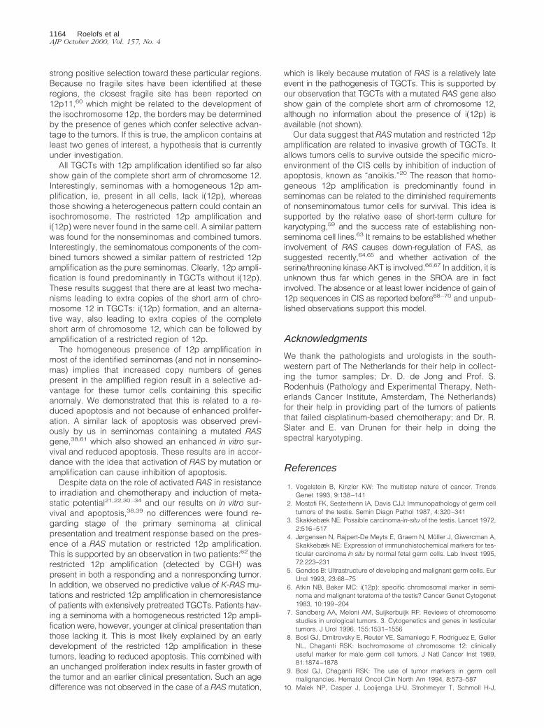
Figure 4. Representative examples of the presence of DNA laddering afterelectrophoresis (indicative for apoptosis) of 1-mg high molecular weightDNA isolated from primary seminomas with and without a restricted 12pamplification/RAS mutation. Note the correlation between less apoptosis andthe presence of a restricted 12p amplification or RAS mutation. In addition,the results of in vitro culture of the tumor cells are shown (see also Table 3).
Figure 5. Schematical representation of the ages of patients at clinical pre-sentation with a seminoma with and without 12p amplification and RASmutation (mean, average, and standard deviations are indicated). In addition,the ages of the subgroup of patients with a homogeneous 12p amplificationare shown. No differences were found between the ages of patients with aseminoma without either of these aberrations and those with a RAS mutation.However, those with a 12p amplification-positive seminoma showed a bor-derline significant difference compared to the control group, whereas asignificant difference was found in case only patients with a homogeneous12p amplification were included (P 5 0.023). Abbreviations: RAS1, RASmutation; RAS2/12p2, no RAS mutation or 12p amplification; 12p1, 12pamplification both heterogeneously and homogeneously present; 12p1homo, homogeneous 12p amplification.
Table 3. Summary of the Results on Proliferation Index, Apoptosis, In Vitro Survival, and Volume of the Tumor (cm3) inSeminomas With and Without a Restricted 12p Amplification/RAS Mutation
Wild-type RAS Mutant RAS
Proliferationindex
ApoptosisIn vitrosurvival Tumor
volumeProliferation
index
ApoptosisIn vitrosurvival Tumor
volume1 2 1 2 1 2 1 2
12p-ampl.2 34.5 (10.5) 9 4 1 11 256 (173) 30.8 (3.1) 1 5 6 0 121 (88)1 29.7 (15.3) 0 9 4 0 246 (294) NA NA NA NA NA NA
Abbreviations: 12p-ampl., restricted 12p amplification; 1, present; 2, absent; NA, not available. Standard deviations are indicated betweenbrackets.
12p and Testicular Germ Cell Tumors 1163AJP October 2000, Vol. 157, No. 4
strong positive selection toward these particular regions.Because no fragile sites have been identified at theseregions, the closest fragile site has been reported on12p11,60 which might be related to the development ofthe isochromosome 12p, the borders may be determinedby the presence of genes which confer selective advan-tage to the tumors. If this is true, the amplicon contains atleast two genes of interest, a hypothesis that is currentlyunder investigation.
All TGCTs with 12p amplification identified so far alsoshow gain of the complete short arm of chromosome 12.Interestingly, seminomas with a homogeneous 12p am-plification, ie, present in all cells, lack i(12p), whereasthose showing a heterogeneous pattern could contain anisochromosome. The restricted 12p amplification andi(12p) were never found in the same cell. A similar patternwas found for the nonseminomas and combined tumors.Interestingly, the seminomatous components of the com-bined tumors showed a similar pattern of restricted 12pamplification as the pure seminomas. Clearly, 12p ampli-fication is found predominantly in TGCTs without i(12p).These results suggest that there are at least two mecha-nisms leading to extra copies of the short arm of chro-mosome 12 in TGCTs: i(12p) formation, and an alterna-tive way, also leading to extra copies of the completeshort arm of chromosome 12, which can be followed byamplification of a restricted region of 12p.
The homogeneous presence of 12p amplification inmost of the identified seminomas (and not in nonsemino-mas) implies that increased copy numbers of genespresent in the amplified region result in a selective ad-vantage for these tumor cells containing this specificanomaly. We demonstrated that this is related to a re-duced apoptosis and not because of enhanced prolifer-ation. A similar lack of apoptosis was observed previ-ously by us in seminomas containing a mutated RASgene,38,61 which also showed an enhanced in vitro sur-vival and reduced apoptosis. These results are in accor-dance with the idea that activation of RAS by mutation oramplification can cause inhibition of apoptosis.
Despite data on the role of activated RAS in resistanceto irradiation and chemotherapy and induction of meta-static potential21,22,30–34 and our results on in vitro sur-vival and apoptosis,38,39 no differences were found re-garding stage of the primary seminoma at clinicalpresentation and treatment response based on the pres-ence of a RAS mutation or restricted 12p amplification.This is supported by an observation in two patients:62 therestricted 12p amplification (detected by CGH) waspresent in both a responding and a nonresponding tumor.In addition, we observed no predictive value of K-RAS mu-tations and restricted 12p amplification in chemoresistanceof patients with extensively pretreated TGCTs. Patients hav-ing a seminoma with a homogeneous restricted 12p ampli-fication were, however, younger at clinical presentation thanthose lacking it. This is most likely explained by an earlydevelopment of the restricted 12p amplification in thesetumors, leading to reduced apoptosis. This combined withan unchanged proliferation index results in faster growth ofthe tumor and an earlier clinical presentation. Such an agedifference was not observed in the case of a RAS mutation,
which is likely because mutation of RAS is a relatively lateevent in the pathogenesis of TGCTs. This is supported byour observation that TGCTs with a mutated RAS gene alsoshow gain of the complete short arm of chromosome 12,although no information about the presence of i(12p) isavailable (not shown).
Our data suggest that RAS mutation and restricted 12pamplification are related to invasive growth of TGCTs. Itallows tumors cells to survive outside the specific micro-environment of the CIS cells by inhibition of induction ofapoptosis, known as “anoikis.”20 The reason that homo-geneous 12p amplification is predominantly found inseminomas can be related to the diminished requirementsof nonseminomatous tumor cells for survival. This idea issupported by the relative ease of short-term culture forkaryotyping,59 and the success rate of establishing non-seminoma cell lines.63 It remains to be established whetherinvolvement of RAS causes down-regulation of FAS, assuggested recently,64,65 and whether activation of theserine/threonine kinase AKT is involved.66,67 In addition, it isunknown thus far which genes in the SROA are in factinvolved. The absence or at least lower incidence of gain of12p sequences in CIS as reported before68–70 and unpub-lished observations support this model.
Acknowledgments
We thank the pathologists and urologists in the south-western part of The Netherlands for their help in collect-ing the tumor samples; Dr. D. de Jong and Prof. S.Rodenhuis (Pathology and Experimental Therapy, Neth-erlands Cancer Institute, Amsterdam, The Netherlands)for their help in providing part of the tumors of patientsthat failed cisplatinum-based chemotherapy; and Dr. R.Slater and E. van Drunen for their help in doing thespectral karyotyping.
References
1. Vogelstein B, Kinzler KW: The multistep nature of cancer. TrendsGenet 1993, 9:138–141
2. Mostofi FK, Sesterhenn IA, Davis CJJ: Immunopathology of germ celltumors of the testis. Semin Diagn Pathol 1987, 4:320–341
3. Skakkebæk NE: Possible carcinoma-in-situ of the testis. Lancet 1972,2:516–517
4. Jørgensen N, Rajpert-De Meyts E, Graem N, Muller J, Giwercman A,Skakkebæk NE: Expression of immunohistochemical markers for tes-ticular carcinoma in situ by normal fetal germ cells. Lab Invest 1995,72:223–231
5. Gondos B: Ultrastructure of developing and malignant germ cells. EurUrol 1993, 23:68–75
6. Atkin NB, Baker MC: i(12p): specific chromosomal marker in semi-noma and malignant teratoma of the testis? Cancer Genet Cytogenet1983, 10:199–204
7. Sandberg AA, Meloni AM, Suijkerbuijk RF: Reviews of chromosomestudies in urological tumors. 3. Cytogenetics and genes in testiculartumors. J Urol 1996, 155:1531–1556
8. Bosl GJ, Dmitrovsky E, Reuter VE, Samaniego F, Rodriguez E, GellerNL, Chaganti RSK: Isochromosome of chromosome 12: clinicallyuseful marker for male germ cell tumors. J Natl Cancer Inst 1989,81:1874–1878
9. Bosl GJ, Chaganti RSK: The use of tumor markers in germ cellmalignancies. Hematol Oncol Clin North Am 1994, 8:573–587
10. Malek NP, Casper J, Looijenga LHJ, Strohmeyer T, Schmoll H-J,
1164 Roelofs et alAJP October 2000, Vol. 157, No. 4
Nordheim A, Janknecht R: Quantification of additional short arms ofchromosome 12 in germ cell tumors using polymerase chain reaction.Eur J Cancer 1997, 33:1488–1494
11. Dmitrovsky E, Murty VVVS, Moy D, Miller WH Jr, Nanus D, Albino AP,Samaniego F, Bosl G, Chaganti RSK: Isochromosome 12p in non-seminoma cell lines: karyologic amplification of c-ki-ras2 withoutpoint-mutational activation. Oncogene 1990, 5:543–548
12. Houldsworth J, Reuter V, Bosl GJ, Chaganti RSK: Aberrant expres-sion of cyclin D2 is an early event in human male germ cell tumori-genesis. Cell Growth Dev 1997, 8:293–299
13. Murty VVVS, Chaganti RSK: A genetic perspective of male germ celltumors. Semin Oncol 1998, 25:133–144
14. Lubbert M, Mirro J, Miller CW, Kahan J, Isaac G, Kitchingman G,Mertelsmann R, Herrmann F, McCormick F, Koeffler HP: N-Ras genepoint mutations in childhood acute lymphocytic leukemia correlatewith a poor prognosis. Blood 1990, 75:1163–1169
15. Slebos RJC, Kibbelaar RE, Dalesio O, Kooistra A, Stam J, MeijerCJLM, Wagenaar SS, Vanderschueren RGJRA, van Zandwijk N, MooiWJ, Bos JL, Rodenhuis S: K-ras oncogene activation as a prognosticmarker in adenocarcinoma of the lung. N Engl J Med 1990, 323:561–565
16. Mitsudomi T, Steinberg SM, Oie HK, Mulshine JL, Phelps R, Viallet J,Pass H, Minna JD, Gazdar AF: Ras gene mutations in non-small celllung cancers are associated with shortened survival irrespective oftreatment intent. Cancer Res 1991, 51:4999–5002
17. Chin L, Tam A, Pomerantz J, Wong M, Holash J, Bardeesy N, Shen Q,O’Hagan R, Pantginis J, Zhou H, Horner JW Jr, Cordon-Cardo C,Yancopoulos GD, DePinho RA: Essential role for oncogenic Ras intumour maintenance. Nature 1999, 400:468–472
18. Hahn WC, Counter CM, Lundberg AS, Beijersbergen RL, Brooks MW,Weinberg RA: Creation of human tumour cells with defined geneticelements. Nature 1999, 400:464–468
19. Arends MJ, McGregor AH, Toft NJ, Brown EJH, Wyllie AH: Suscep-tibility to apoptosis is differentially regulated by c-myc and mutatedHa-ras oncogenes and is associated with endonuclease availability.Br J Cancer 1993, 68:1127–1133
20. Frisch SM, Francis H: Disruption of epithelial cell-matrix interactionsinduces apoptosis. J Cell Biol 1994, 124:619–626
21. Ananthaswamy HN, Price JE, Tainsky MA, Goldberg LH, Bales ES:Correlation between Ha-ras gene amplification and spontaneous me-tastasis in NIH 3T3 cells transfected with genomic DNA from humanskin cancers. Clin Exp Metastasis 1989, 7:301–313
22. Takiguchi Y, Takahashi Y, Kuriyama T, Miyamoto T: NIH3T3 transfec-tant containing human K-ras oncogene shows enhanced metastaticactivity after in vivo tumor growth or co-culture with fibroblasts. ClinExp Metastasis 1992, 10:351–360
23. Pulciani S, Santos E, Long LK, Sorrentino V, Barbacid M: Ras geneamplification and malignant transformation. Mol Cell Biol 1985,5:2836–2841
24. McKay IA, Marshall CJ, Cales C, Hall A: Transformation and stimula-tion of DNA synthesis in NIH-3T3 cells are a titratable function ofnormal p21N-ras expression. EMBO J 1986, 5:2617–2621
25. Fujita K, Ohuchi N, Yao T, Okumura M, Fukushima Y, Kanakura Y,Kitamura Y, Fujita J: Frequent overexpression, but not activation bypoint mutation, of ras genes in primary human gastric cancers. Gas-troenterology 1987, 93:1339–1345
26. Radinsky R, Kraemer PM, Raines MA, Kung HJ, Culp LA: Amplifica-tion and rearrangement of the Kirsten ras oncogene in virus-trans-formed BALB/c 3T3 cells during malignant tumor progression. ProcNatl Acad Sci USA 1987, 84:5143–5147
27. Coleman WB, Throneburg DB, Grisham JW, Smith GJ: Overexpres-sion of c-K-ras, c-N-ras and transforming growth factor beta co-segregate with tumorigenicity in morphologically transformed C3H10T1/2 cell lines. Carcinogenesis 1994, 15:1005–1012
28. Zhang B, Shih JW, Wear DJ, Tsai S, Lo SC: High-level expression ofH-ras and c-myc oncogenes in mycoplasma-mediated malignant celltransformation. Proc Soc Exp Biol Med 1997, 214:359–366
29. Bokemeyer C, Schmoll HJ: Treatment strategies in metastatic testic-ular cancer. Eur Urol 1993, 23:50–52
30. Sklar MD: Increased resistance to cis-diamminedichloroplatinum(II)in NIH3T3 cells transformed by ras oncogenes. Cancer Res 1988,48:793–797
31. Sklar MD: The ras oncogenes increase the intrinsic resistance ofNIH3T3 cells to ionizing radiation. Science 1988, 239:645–647
32. Isonishi S, Hom DK, Thiebaut FB, Mann SC, Andrews PA, Basu A,Lazo JS, Eastman A, Howell SB: Expression of the c-Ha-ras onco-gene in mouse NIH 3T3 cells induces resistance to cisplatin. CancerRes 1991, 51:5903–5909
33. Holford J, Rogers P, Kelland LR: Ras mutation and platinum resis-tance in human ovarian carcinomas in vitro. Int J Cancer 1998, 77:94–100
34. Kinashi Y, Akaboshi M, Masunaga S, Ono K, Watanabe M: Resis-tance to 195mPt-radiolabeled cis-diaminedichloroplatinum (II) ofSHOK cells transfected with various oncogenes. Radiat Med 1998,16:233–237
35. Ganguly S, Murty VV, Samaniego F, Reuter VE, Bosl GJ, Chaganti RS:Detection of preferential NRAS mutations in human male germ celltumors by the polymerase chain reaction. Genes Chromosom Cancer1990, 1:228–232
36. Moul JW, Theune SM, Chang EH: Detection of ras mutations inarchival testicular germ cell tumors by polymerase chain reaction andoligonucleotide hybridization. Genes Chromosom Cancer 1992,5:109–118
37. Olie RA, Looijenga LHJ, Boerrigter L, Top B, Rodenhuis S, MulderMP, Oosterhuis JW: N- and KRAS mutations in human testicular germcell tumors: incidence and possible biological implications. GenesChromosom Cancer 1995, 12:110–116
38. Olie RA, Looijenga LHJ, Dekker MC, De Jong FH, De Rooy DG,Oosterhuis JW: Heterogeneity in the in vitro survival and proliferationof human seminoma cells. Br J Cancer 1995, 71:13–17
39. Olie RA, Boersman AWM, Dekker MC, Nooter K, Looijenga LHJ,Oosterhuis JW: Apoptosis of human seminoma cells upon disruptionof their micro-environment. Br J Cancer 1996, 73:1031–1036
40. Otto T, Virchow S, Fuhrmann C, Steinberg F, Streffer C, Goepel M,Rubben H: Detection of vital germ cell tumor cells in short-term cellcultures of primary tumors and of retroperitoneal metastasis—clinicalimplications. Urol Res 1997, 25:121–124
41. Lowenberg B, Van Putten WLJ, Touw IP, Delwel R, Santini V: Auton-omous proliferation of leukemic cells in vitro as a determinant ofprognosis in adult acute myeloid leukemia. N Engl J Med 1993,328:614–619
42. Suijkerbuijk RF, Sinke RJ, Olde Weghuis DEM, Roque L, Forus A,Stellink F, Siepman A, Van de Kaa C, Soares J, Geurts van Kessel A:Amplification of chromosome subregion 12p11.2-p12.1 in a metasta-sis of an i(12p)-negative seminoma: relationship to tumor progres-sion? Cancer Genet Cytogenet 1994, 78:145–152
43. Mostert MMC, Van de Pol M, Olde Weghuis D, Suijkerbuijk RF, Geurtsvan Kessel A, Van Echten-Arends J, Oosterhuis JW, Looijenga LHJ:Comparative genomic hybridization of germ cell tumors of the adulttestis: confirmation of karyotypic findings and identification of a 12p-amplicon. Cancer Genet Cytogenet 1996, 89:146–152
44. Korn MW, Olde Weghuis DEM, Suijkerbuijk RF, Schmidt U, Otto T, DuManoir S, Geurts van Kessel A, Seeber S, Becher R: Detection ofchromosomal DNA gains and losses in testicular germ cell tumors bycomparative genomic hybridization. Genes Chromosom Cancer1996, 17:78–87
45. Mostert MC, Verkerk AJMH, Van de Pol M, Heighway J, Marynen P,Rosenberg C, Geurts van Kessel A, van Echten J, Oosterhuis JW,Looijenga LHJ: Identification of the crucial region of 12p overrepre-sentation in testicular germ cell tumors of adolescents and adults.Oncogene 1998, 16:2617–2627
46. Mostofi FK, Sesterhenn IA: Histological Typing of Testis Tumours.Berlin, Springer, 1998
47. Pugh RCB: Combined tumours. Pathology of the Testis. Edited byRCB Pugh. Oxford, Blackwell, 1976, pp 245–258
48. Mosselman S, Looijenga LHJ, Gillis AJM, Van Rooijen MA, Kraft HJ,Van Zoelen EJJ, Oosterhuis JW: Aberrant platelet-derived growthfactor a-receptor transcript as a diagnostic marker for early humangerm cell tumors of the adult testis. Proc Natl Acad Sci USA 1996,93:2884–2888
49. Shin RW, Iwaki T, Kitamoto T, Tateishi J: Hydrated autoclave pretreat-ment enhances tau immunoreactivity in formalin-fixed normal andAlzheimer’s disease brain tissues. Lab Invest 1991, 64:693–702
50. Castedo SMMJ, De Jong B, Oosterhuis JW, Seruca R, Idenburg VJS,Dam A, Te Meerman GJ, Schraffordt Koops H, Sleijfer DT: Chromo-somal changes in human primary testicular nonseminomatous germcell tumors. Cancer Res 1989, 49:5696–5701
51. Castedo SMMJ, De Jong B, Oosterhuis JW, Seruca R, Te Meerman
12p and Testicular Germ Cell Tumors 1165AJP October 2000, Vol. 157, No. 4
GJ, Dam A, Schraffordt Koops H: Cytogenetic analysis of ten humanseminomas (two of them lacking the i(12p)). Cancer Res 1989, 49:439–443
52. Mitelman F: ISCN An International System for Human CytogeneticNomenclature. Basel, S. Karger, 1995
53. Rosenberg C, Mostert MC, Bakker Schut T, Van de Pol M, VanEchten-Arends J, De Jong B, Raap T, Tanke H, Oosterhuis JW,Looijenga LHJ: Chromosomal constitution of human spermatocyticseminomas: comparative genomic hybridization supported by con-ventional and interphase cytogenetics. Genes Chromosom Cancer1998, 23:286–291
54. Maniatis T, Fritsch EF, Sambrook J: Isolation of high molecular-weight, eukaryotic DNA from cells grown in tissue culture. MolecularCloning. New York, Cold Spring Harbor Laboratory, 1982, pp 280–281
55. Rosenberg C, Van Gijlswijk RP, Vos CBJ, Wiegant J, Cornelisse CJ,Tanke HJ, Raap AK: Comparative genomic hybridization with lissa-mine- and fluorescein-labelled nucleotides. Cytometry 1998, 32:337–341
56. Rosenberg C, Bakker Schut T, Mostert MC, Tanke HJ, Raap AK,Oosterhuis JW, Looijenga LHJ: Chromosomal gains and losses intesticular germ cell tumors of adolescents and adults investigated bya modified CGH approach. Lab Invest 1999, 79:1447–1451
57. Benhattar J, Losi L, Chaubert P, Givel JC, Costa J: Prognostic signif-icance of K-ras mutations in colorectal carcinoma. Gastroenterology1993, 104:1044–1048
58. Kahn SM, Jiang W, Culbertson TA, Weinstein IB, Williams GM, TomitaN, Ronai Z: Rapid and sensitive nonradioactive detection of mutantK-ras genes via “enriched” PCR amplification. Oncogene 1991,6:1079–1083
59. Van Echten-Arends J, Oosterhuis JW, Looijenga LHJ, Wiersma J, TeMeerman G, Schraffordt Koops H, Sleijfer DT, De Jong B: No recur-rent structural abnormalities in germ cell tumors of the adult testisapart from i(12p). Genes Chromosom Cancer 1995, 14:133–144
60. Ford JH: Translocations of chromosome 12. II. A comparison of thedistribution of sites of spontaneous and induced breakages. HumGenet 1981, 58:279–281
61. Oosterhuis JW, Gillis AJM, Looijenga LHJ: In Vitro Survival, RASMutations, Apoptosis and Activation of the SAPK-Pathway in HumanSeminoma Cells. Edited by I Appleyard. London, John Libbey &Company Ltd., 1997, pp 51–57
62. Rao PH, Houldsworth J, Palanisamy N, Murty VV, Reuter VE, MotzerRJ, Bosl GJ, Chaganti RS: Chromosomal amplification is associatedwith cisplatin resistance of human male germ cell tumors. Cancer Res1998, 58:4260–4263
63. Andrews PW, Casper J, Damjanov I, Duggan-Keen M, Giwercman A,Hata J-i, Von Keitz A, Looijenga LHJ, Oosterhuis JW, Pera M, SawadaM, Schmoll H-J, Skakkebæk NE, Van Putten W, Stern P: A compara-tive analysis of cell surface antigens expressed by cell lines derivedfrom human germ cell tumors. Int J Cancer 1996, 66:806–816
64. Fenton RG, Hixon JA, Wright PW, Brooks AD, Sayers TJ: Inhibition offas (CD95) expression and fas-mediated apoptosis by oncogenicras. Cancer Res 1998, 58:3391–3400
65. Peli J, Schroter M, Rudaz C, Hahne M, Meyer C, Reichmann E,Tschopp J: Oncogenic Ras inhibits Fas ligand-mediated apoptosisby downregulating the expression of Fas. EMBO J 1999, 18:1824–1831
66. Aoki M, Batista O, Bellacosa A, Tsichlis P, Vogt PK: The akt kinase:molecular determinants of oncogenicity. Proc Natl Acad Sci USA1998, 95:14950–14955
67. Brunet A, Bonni A, Zigmond MJ, Lin MZ, Juo P, Hu LS, Anderson MJ,Arden KC, Blenis J, Greenberg ME: Akt promotes cell survival byphosphorylating and inhibiting a Forkhead transcription factor. Cell1999, 96:857–868
68. Vorechovsky I, Mazanec K: Is isochromosome i(12p) present in go-nadal precancerous tissue? Neoplasma 1989, 36:697–700
69. Vos A, Oosterhuis JW, De Jong B, Buist J, Schraffordt Koops H:Cytogenetics of carcinoma in situ of the testis. Cancer Genet Cyto-genet 1990, 46:75–81
70. Looijenga LHJ, Rosenberg C, Van Gurp RJHLM, Geelen E, VanEchten-Arends J, De Jong B, Mostert MC, Oosterhuis JW: Compar-ative genomic hybridization of microdissected samples from differentstages in the development of a seminoma and nonseminoma.J Pathol 2000, 191:187–192
1166 Roelofs et alAJP October 2000, Vol. 157, No. 4