Embed Size (px)
Citation preview
University of New EnglandDUNE: DigitalUNE
Case Report Papers Physical Therapy Student Papers
12-4-2015
Restoring Functional Mobility In A Patient WithDelayed Onset Of Physical RehabilitationFollowing A Hemorrhagic Stroke: A Case ReportBettie KrugerUniversity of New England
Follow this and additional works at: http://dune.une.edu/pt_studcrpaper
Part of the Physical Therapy Commons
© 2015 Bettie Kruger
This Course Paper is brought to you for free and open access by the Physical Therapy Student Papers at DUNE: DigitalUNE. It has been accepted forinclusion in Case Report Papers by an authorized administrator of DUNE: DigitalUNE. For more information, please contact [email protected].
Recommended CitationKruger, Bettie, "Restoring Functional Mobility In A Patient With Delayed Onset Of Physical Rehabilitation Following A HemorrhagicStroke: A Case Report" (2015). Case Report Papers. 36.http://dune.une.edu/pt_studcrpaper/36
brought to you by COREView metadata, citation and similar papers at core.ac.uk
provided by University of New England
1
Restoring Functional Mobility in a Patient with Delayed 1
Onset of Physical Rehabilitation Following a 2
Hemorrhagic Stroke: A Case Report 3
4
Bettie Kruger 5
6
7
B Kruger, BS, is a DPT student at the University of New England, 716 Stevens Ave., 8
Portland, ME 04103 9
Address all correspondence to Bettie Kruger at: [email protected] 10
11
12
The patient signed an informed consent allowing the use of medical information and 13
video footage for this report and received information on the institution’s policies 14
regarding the Health Insurance Portability and Accountability Act. 15
16
17
The author acknowledges Jill Walsh, DPT, for supervision and assistance with 18
collecting data and treatment, and Noel Squires, PT, DPT, for assistance with this 19
case report conceptualization 20
21
2
Abstract 22
Background and Purpose: Hemorrhagic stroke accounts for 10% to 15% of all 23
strokes and is associated with a high mortality rate1. A decompressive craniectomy 24
is a neurosurgical procedure to reduce intracranial pressure in order to minimize 25
risk of mortality and disability. It has been suggested that early and frequent 26
mobilization following stroke onset is optimal for motor recovery.2 The purpose of 27
this case report is to provide a framework of sub-‐acute rehabilitation that facilitated 28
motor recovery and functional mobility in a patient with delayed participation in 29
physical therapy (PT) following a stroke. 30
Case Description: A 54 year-‐old female underwent an intracranial hemorrhage (ICH), 31
subsequent decompressive craniectomy, and a cranioplasty. Sub acute PT occurred 32
6 days a week for 45-‐60 minute sessions over the course of 11 weeks. Initial 33
findings included impaired spatial orientation, cognition, and impaired mobility. 34
Treatment included therapeutic exercise, therapeutic activities, neuromuscular 35
reeducation, and gait training to address all functional impairments. 36
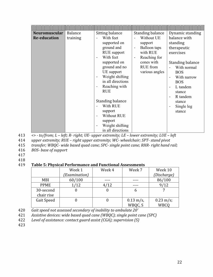
Outcome Measures: A Physical Performance and Mobility Examination (PPME) score 37
at baseline was 1/12; Modified Barthel Index (MBI) was 60/100, however, 38
ambulation was scored a 12/15 (minimum assistance) in the parallel bars for 7’; 30-‐39
second chair rise and gait speed were unable to be performed. At discharge: PPME 40
score improved to 9/12; MBI was 86/100; 30-‐second chair rise was 7; and gait 41
speed was 0.23m/s using a wide based quad cane (WBQC). 42
Discussion: Functional gains were noted over the course of care. The patient’s 43
improved outcomes, and increasing level of independence could be a result of 44
3
consistency and gradually progressing interventions. Further research should 45
investigate optimal time of rest and return to activity following ICH in order to 46
regain optimal functional mobility. 47
48 Manuscript word count: 3,124 49 50
Background 51
Stroke, also known as cerebrovascular accident (CVA), is a sudden loss of 52
neurological function caused by an interruption of blood flow to the brain. It is the 53
fourth leading cause of death and the leading cause of long-‐term disability in the 54
United States.1 An ischemic stroke is caused by a blocked artery interrupting blood 55
flow to the brain, while a hemorrhagic stroke is defined by a ruptured blood vessel 56
causing a leakage of blood in or around the brain.1 57
Hemorrhagic strokes account for only 10% to15% of first ever strokes and 58
approximately 35% to 50% are fatal strokes within 30-‐days.3 Signs, symptoms and 59
resulting impairments of an ICH depend on the location, size, and extent of bleeding. 60
Signs and symptoms include headache, vomiting, increased blood pressure, and 61
impaired level of consciousness.3 ICH is considered a medical emergency and is 62
associated with early risk of neurological deterioration and cardiopulmonary 63
instability.3 It has also been identified to result in greater disability and a higher risk 64
of mortality, compared to ischemic stroke. Depending on the severity of the 65
hemorrhage, surgical treatment may be necessary. A decompressive craniectomy is 66
a neurosurgical procedure which consists of removing a portion of the skull to 67
reduce intracranial pressure (ICP). This procedure is performed to reduce the risk 68
4
of mortality and minimize disability until the ICP has returned to normal and the 69
surgeon deems it appropriate to perform a cranioplasty, which replaces the skull 70
flap usually one to three months later.5 71
It has been suggested that early and frequent out of bed activity within the 72
first 24 hours following stroke onset will enhance motor recovery.2 Limited data is 73
available describing the outcomes of individuals who do not receive care within that 74
timeframe, but instead receive delayed physical rehabilitation. Therefore, the 75
purpose of this case report is to describe the motor recovery and functional gains 76
for a patient who received delayed rehabilitation following a hemorrhagic stroke. 77
78
Case Description: patient history and system review 79
The case patient, DH, was a 54 year-‐old female taken to the emergency 80
department with signs and symptoms of spontaneous stroke. There she was 81
diagnosed with a right-‐sided ICH, secondary to supratherapeutic INR with 82
subsequent right decompressive hemicraniectomy, tracheostomy and jejunostomy 83
tube procedures. Following these operations, she had recurrent jejunal tube 84
dislodgement and a partial small bowel obstruction, secondary to jejunal tube 85
balloon. Right-‐sided brain lesions are often associated with left-‐sided hemiplegia; 86
hemi-‐sensory loss; impaired visual-‐perception, organization and problem solving; 87
and poor judgment, impulsivity and difficulty sustaining movement1; all of which 88
DH presented. DH presented with a medical history of hypertension; atrial 89
fibrillation, Coumadin use; open gastric bypass (her pre-‐operation weight was 90
5
279lbs, and her resulting weight was 149lbs); aspiration pneumonitis; history of 91
delirium, tobacco use for more than 40 pack year; and depression. 92
Prior to her stroke, DH worked full time for the city for more than 20 years to 93
keep drinking water and waterways clean and safe. She was mostly sedentary, cared 94
for her three year-‐old grandson a few days a week and enjoyed traveling and 95
attending concerts. DH lives in a private residence with 10 stairs to enter and 18-‐20 96
stairs within the home. Each set of stairs contains two handrails. She has two sons, 97
one of whom lives with her and is able to assist DH upon discharge. 98
DH was referred to sub-‐acute physical therapy, three months after her 99
hemorrhagic stroke and subsequent right decompressive hemicraniectomy. Per 100
patient report, DH had been in and out of short-‐term rehabilitation facilities and the 101
hospital since the onset of her stroke and she participated in minimal activity out of 102
bed. At the time of admission, DH was required to wear a head orthotic when out of 103
bed to protect her open craniectomy, per surgeon orders. She reported mild 104
episodes of discomfort in her abdomen where the skull flap was stored to remain 105
viable and sterile until the cranioplasty. Medications taken at this time included: 106
Aspirin, Atorvastatin, Cyanocobalamin (vitamin V-‐12), Ergocalciferol, Lidocaine, 107
Metroprolol, Multivitamin, QUEtiapine (Seroquel), Senna, and Sertraline (Zoloft). 108
Her current level of activity was very low due to minimal activity since onset of her 109
stroke. Per chart review, DH presented with significantly impaired left-‐sided 110
strength, motor control and coordination. She required moderate to maximum 111
assistance for bed mobility, static stance, and transfers, and was unable to ambulate 112
or negotiate stairs at this time. Her goals were to get out of bed as much as possible, 113
6
regain the ability to ambulate and be independent with activities of daily living 114
(ADLs), specifically using the restroom without assistance. A systems review can be 115
found in Table 1. 116
The patient signed an informed consent allowing the use of medical information, in 117
adherence to the HIPPA policy. 118
119
Clinical Impression #1 120
DH was admitted to sub-‐acute rehabilitation three months following the 121
onset of her stroke. Prior to PT examination, a medical pre-‐admission review 122
revealed the patient demonstrated left knee buckling, and required moderate 123
assistance to stand and maintain static standing. Transfer from bed to wheelchair to 124
the right required moderate assistance of two people. The patient was able to take 125
one large step with the right lower extremity (LE), completing 50% of the transfer, 126
but required maximum assistance to pivot the rest of the way. Based on this report, 127
it was hypothesized these findings would be demonstrated during the initial PT 128
examination and functional assessment. Given the patient’s medical status and post-‐129
surgical condition, there was no need for differential diagnoses. DH was a good 130
candidate for a case report because of her unusually delayed return to physical 131
activity and her potential for functional improvement. Further examination to 132
establish functional limitations and provide a baseline of impairments included: 133
assessments of strength, motor control, balance and posture, as well as, her ability 134
transfer and ambulate. It is important to have case reports providing the framework 135
of PT management for patients who have had a stroke because each occurrence 136
7
presents with varying impairments, disability and rate of recovery. The most 137
dramatic neurological recovery occurs within the first three to six months following 138
medical stabilization.1 139
140
Examination 141
Primary examination focused on functional mobility including bed mobility, 142
transfers, and ambulation. Findings were based upon visual observation and patient 143
demonstration. DH required moderate assistance of one for all bed mobility, 144
moderate assistance of one to two people for transfers, including sit to and from 145
stand and stand pivot transfers between surfaces. She was non-‐ambulatory, and 146
subsequently unable to ascend or descend stairs at this time. Objective measures 147
such as, strength, posture, and balance can be found in Table 2. 148
Four functional outcome assessments were used throughout the episode of 149
care (EOC) to track the patient’s progress. This data was collected at baseline, every 150
10th visit for recertification when necessary, and again at discharge. The MBI is an 151
assessment of functional activity consisting of 10 ADLs to determine a patient’s level 152
of physical dependency. It is reported to be highly responsive to change, have 153
excellent reliability, and has a minimal detectable change (MCD) of 4.02.6,7,8 The 30-‐154
second chair rise is an assessment of functional LE strength. It requires a patient to 155
perform as many sit-‐to-‐stands possible from a standard chair and no upper 156
extremity (UE) support in 30-‐seconds. Although this test is not specific to stroke, it 157
has been suggested to have excellent validity and responsiveness, by Jones et al. in 158
community dwelling older adults.9 The gait speed assessment, also known as the 10-159
8
Meter Walk Test, is a measure of functional mobility and ambulation. It has a minimal 160
clinically important different (MCID) of a small meaningful change of 0.06m/s and a 161
substantial change of 0.14m/s, as well as, an excellent correlation with the Barthel 162
Index.10, 11 The PPME is a functional and mobility measurement, consisting of six 163
domains, initially developed for hospitalized elderly. 164
165
Clinical Impression #2 166
Upon evaluation, the initial impression was confirmed and the patient was 167
determined an appropriate candidate for PT. The patient’s primary problems 168
included left-‐sided hemiparesis, and impaired cognition, perception, posture, 169
balance and all forms of mobility, which were consistent with signs and symptoms 170
of a stroke. Potential barriers for patient progress include comorbidities such as a 171
history of obesity, hypertension, atrial fibrillation, depression and tobacco use1; all 172
of which DH presented. Additionally, the patient’s cognitive impairment and 173
negative expectations, influenced by her previous rehabilitation experiences since 174
onset of injury, may affect her recovery. Aside from these barriers, the patient had 175
potential for functional improvements. Although DH has also had a prolonged 176
sedentary state following onset of stroke, she is good candidate for a case report 177
because she is within three to six months post-‐stroke onset. There is limited 178
literature pertaining to the functional recovery following delayed initiation of 179
physical rehabilitation after stroke. Furthermore, DH is motivated to regain her 180
independence with ADLs and ambulation. She is hopeful to return to work and her 181
prior level functioning. 182
9
The patient also participated in occupational therapy (OT) to address ADLs 183
and UE impairments, and speech language therapy (SLP) to focus on cognitive 184
impairments. The plan of care included coordination, communication and 185
documentation with OT, SLP, and the attending nurses and medical physicians. 186
Patient education was provided to the patient and family regarding the medical 187
condition, impairments, activity limitations, interventions, management of risk 188
factors and safety concerns. Procedural interventions addressed physical 189
impairments. PT short-‐term and long-‐goals that were discussed and agreed upon 190
with the patient can be found in Table 3. 191
192
Interventions 193
Coordination, communication, and documentation 194
Communication was maintained among all treating therapists throughout 195
the patient’s EOC. PT, OT and SLP coordinated times of treatment that would be 196
optimal for the patient in order to reduce fatigue and enhance recovery. Weekly 197
meetings were held with all therapists and the director of rehabilitation to discuss 198
patient progress and discharge plans. Any areas of concern outside our scope of 199
practice were communicated to nursing, attending physicians and social workers, as 200
appropriate. Family meetings were available upon request, which included all 201
treating healthcare professionals, the patient and their family. Documentation was 202
maintained on a daily basis, which included interventions performed, patient 203
response, and any pertinent communication among other health professionals, in 204
addition to, patient evaluation, recertification notes and discharge summary. 205
10
206
Patient/client related instructions 207
Patient education focused on proper instruction to self-‐maintain WBQC with 208
ambulation and stair negotiation, as well as, techniques to enhance performance to 209
decrease her impairments. Spatial orientation, self-‐awareness to her left hemiplegic 210
UE and safety with all forms of mobility were stressed daily to prevent further 211
injury. Interactive instruction and education were provided to the patient’s family to 212
ensure safety and proper technique when assisting the patient with ADLs and 213
functional mobility in order to prepare an optimal living environment upon 214
discharge. 215
216
Procedural interventions 217
Interventions were determined based on the patient’s physical, cognitive and 218
perceptual impairments and a thorough activity-‐based task analysis of the patient’s 219
functional ability. Activity based task analysis is the process of breaking down an 220
activity into its components to understand and evaluate the demands required to 221
complete a task.12 Procedural interventions included therapeutic exercise, 222
neuromuscular reeducation, therapeutic activity, and gait training in order to 223
complete activities of functional mobility. Therapeutic exercises were implemented 224
to increase muscle strength, motor control and coordination. Neuromuscular 225
reeducation was used to improve proprioceptive awareness, spatial orientation, 226
facilitate optimal posture, and decrease fall risk. Therapeutic activities focused on 227
the ability to safely complete activities of bed mobility and transfers; gait training 228
11
facilitated a normalized gait pattern and negotiation of stairs. Refer to Table 4 for a 229
detailed framework of interventions implemented and amount of assistance 230
required during the EOC. 231
Interventions during the first few weeks focused on foundational aspects of 232
functional mobility, including static stability, postural control and controlled 233
movements to regain skills needed to perform ADLs. This is important because it 234
facilitates a neutral posture with the center of gravity (COG) over the base of 235
support (BOS) in order to improve motor control and reduce fall risk with static and 236
dynamic activities.13 Each activity incorporated proper technique and continuous 237
attention to her left UE. Static stance in the parallel bars, incorporating a mirror for 238
visual feedback, was used daily to maintain a neutral static stance, re-‐establish the 239
COG, and improve spatial orientation. As the patient began to demonstrate 240
improvements with transfers and balance, therapeutic exercises and gait training 241
were integrated more frequently during therapy. Therapeutic exercises initially 242
consisted of two sets of ten repetitions in order to accommodate DH’s limited 243
activity tolerance and avoid effects of fatigue. Exercises gradually progressed to 244
three sets of 15 repetitions, as the patient demonstrated increased strength and 245
motor control. Three sets of 15 repetitions is a conventional parameter of exercise 246
because it helps with muscular endurance, strengthening properties, and the 247
repetition contributes to muscle memory.14 248
Gait training was first initiated in the parallel bars and then progressed to 249
using a WBQC, as improvements were demonstrated with limb advancement and 250
sequencing. Within the first three weeks, DH was able to ambulate up to 30’ in the 251
12
parallel bars with minimal assistance. She required moderate verbal cues and rest 252
breaks every 10’. DH progressed to ambulating with a WBQC on even surfaces with 253
tactile and verbal cues to facilitate a proper heel strike at the initiation of stance 254
phase and toe clearance during the swing phase. Areas requiring continuous 255
attention included proper sequencing and placement of the cane; proper weight 256
shift; appropriate step length and width in order to prevent a scissoring gait 257
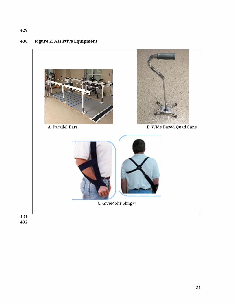
pattern; and the ability to maintain a forward gaze. Figure 2 represents a visual 258
presentation of assistive equipment DH used throughout her EOC. 259
Around week six,of treatment, therapy was put on hold for three days, as DH was 260
discharged to the hospital for a cranioplasty, which replaced her skull flap. Upon 261
return, DH demonstrated minimal functional regression. Therefore, the last six 262
weeks of therapy consisted of standing therapeutic exercises, advanced standing 263
balance exercises with various BOS, ambulation for further distances with WBCQ, 264
ambulation without an assistive device to reintegrate a reciprocal gait pattern, and 265
negotiation of stairs. Stair negotiation was initiated with 4-‐inch steps and 266
progressed to a 6-‐inch standard step. At week eight, the patient required a 267
supervision level of assistance with bed mobility, functional transfers (including sit 268
to and from stand from various surfaces), and toileting. She currently required 269
minimal assistance with standing therapeutic exercises that involve single leg stance 270
and minimal assistance with stair negotiation and ambulation without an assistive 271
device. 272
273
274
13
Outcomes 275
DH was able to progress from a physically dependent functional state to an 276
independent or modified independent level of mobility. She gradually achieved all 277
her goals over the course of treatment. DH demonstrated significant improvement 278
on the gait speed test, as she increased her walking speed from 0.0m/s to 0.23m/s, 279
surpassing the MCID of 0.14m/s. Table 5 further represents the patient’s functional 280
outcome measures throughout her EOC. At discharge, DH was able to perform bed 281
mobility and transfers independently; ambulate 300’ with a WBQC, on even 282
surfaces, modified independent; ambulate 250’ with WBQC on uneven ground, 283
supervision; ascend and descend up to 22 steps with the right handrail and self-‐284
management of WBQC with modified independence; negotiate curbs and ramps 285
using a WBQC with contact guard assistance; and was able to don and doff her 286
GiveMohr* sling with modified independence. The GiveMohr sling was specifically 287
ordered for DH because of its functional positioning of the UE with ambulation, in 288
addition to its ability to support the glenohumeral head and educe subluxation.14 289
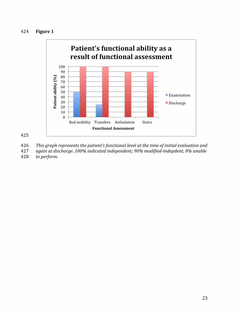
Figure 1 represents a visual presentation of DH’s functional level at the time of her 290
initial examination and again at discharge. 291
The patient was able to maintain independent sitting and standing balance 292
and demonstrated improved strength, coordination, activity tolerance and 293
endurance. Although the left LE continues to demonstrate impaired motor control, 294
improvements with ambulation were seen through her ability to clear the left LE 295
during swing phase, heel strike at the initial of stance phase, and an increased gait 296
* GiveMohr Corporation: 3600 Osuna Road NE, Suite 316, Albuquerque, N.M. 87109
14
velocity. Although DH was able to ambulate with a single point cane, it was agreed 297
upon between the patient and PT that the WBQC was more appropriate, as her 298
ambulation not only appeared more steady and safe, but she also stated, “feeling 299
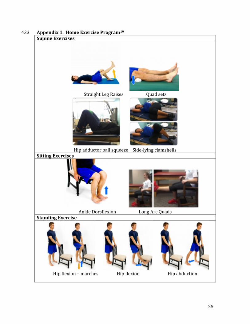
more stable and secure”. Appendix 1 represents the home exercise the patient was 300
given at discharge to maintain her functional improvements. 301
302
Discussion 303
Functional gains were noted over the course of care. The patient progressed 304
from a dependent functional state to a supervised to independent level of function. 305
She was also able to improve her strength, coordination and activity tolerance. It is 306
possible the patient’s improved outcomes and increased level of independence were 307
a result of the consistency, gradual progression of interventions and her willingness 308
to participate in daily PT. After 11 weeks of sub-‐acute therapy, the patient was 309
deemed appropriate for discharge, as she achieved an 86/100 on the MBI and 310
plateaued with functional mobility progression. Contributing factors to her overall 311
relatively low level of function could be related to her present comorbidities, prior 312
level of activity, severity of stoke, and delayed onset of rehabilitation. Her present 313
comorbidities and past medical history, such as obesity, hypertension, atrial 314
fibrillation, as well as, her relatively sedentary lifestyle, put her at a higher risk for 315
complications. Furthermore, the severity of her stroke and delay of admission to 316
rehabilitation dampened her potential for functional improvement. 317
Salter et al., compared the effects of early vs. delayed admission in a study 318
which revealed a significant and inverse relationship between the time passed from 319
15
the onset of the stoke until admission to a rehabilitation program with initial 320
severity of function and stroke related deficits.16 In other words, the more time 321
passed between the onset of stroke and admission to a rehabilitation program, the 322
greater an individuals deficits may present. The study also noted that delayed 323
admission to rehabilitation could further progress functional disability. Another 324
study reported by Biernaskie et al., suggested that earlier admission to 325
rehabilitation was associated with greater functional gains, regardless of functional 326
deficits.17 Congruently, a third study by Paolucci et al., revealed individuals involved 327
in stroke-‐specific rehabilitation experienced greater functional improvement, as 328
measured by the Barthel Index, compared to those who began rehabilitation more 329
than 20 days following their stroke.18 Based on the literature, it is suggested the 330
sooner an individual who has had a stroke participates in a rehabilitation program, 331
the greater functional improvements one can experience. 332
In conclusion, individuals who experience a hemorrhagic stroke present with 333
greater functional disability and require a longer recovery period before engaging in 334
a rehabilitation program. Although it may require more time to accomplish 335
functional goals, independence may be attained even with delayed initiation of 336
rehabilitation. Further research should investigate the optimal timing of rest, return 337
to activity and the initiation of physical rehabilitation following ICH in order to 338
regain optimal functional mobility. 339
340 341 342
16
References 343 344
1. O’Sullivan SB. Chapter 15: Stroke. In: O’Sullivan SB, Schmitz TJ, Fulk GD. 345
Physical Rehabilitation. 6th ed. Philadelphia, PA: F.A. Davis Company; 2014: 346
645-666. 347
2. Chippala P., Sharma R. Effect of very early mobilization on functional status in 348
patients wit acute stroke: a single-blind, randomized controlled trial. Clin Rehabil. 349
2015. doi: 10.1177/0269215515596054 350
3. Broderick J. et al. A guideline from the American heart association/American 351
stroke association stroke council, high blood pressure research council, and the 352
quality of care and outcomes in research interdisciplinary working group. Stroke. 353
2007;38:2001-2023.doi:10.1161/STROKEAHA.107.183689 354
4. Anderson K.K., Olsen T.S., Dehlendorff C., Kammersgaard L.R. Hemorrhagic 355
and ischemic strokes compared stroke severity, mortality, and risk factors. Stroke. 356
2009; 40:2068-2070. Doi:10.1161/STROKEAHA.108.540112 357
5. Kim K, Park J, Kang S, et al. Comparison of the effect of decompressive 358
craniectomy on different neurosurgical diseases. Acta Neurochir. 2009;151(1):21-359
30. doi:10.1007/s00701-008-0164-6 360
6. Houlden H, Edwards M, McNeil J, Greenwood R. Use of the Barthel Index and 361
the Functional Independence Measure during early inpatient rehabilitation after 362
single incident brain injury. Clin Rehabil. 2006;20(2):153-159. 363
doi:10.1191/0269215506cr917oa. 364
7. Rollnik J. The Early Rehabilitation Barthel Index (ERBI). Rehabilitation (Stuttg). 365
2011;50(06):408-411. doi:10.1055/s-0031-1273728. 366
17
8. Hsieh Y, Wang C, Wu S, Chen P, Sheu C, Hsieh C. Establishing the Minimal 367
Clinically Important Difference of the Barthel Index in Stroke 368
Patients. Neurorehabil Neural Repair. 2007;21(3):233-238. 369
doi:10.1177/1545968306294729. 370
9. Jones C, Rikli R, Beam W. A 30-s Chair-Stand Test as a Measure of Lower Body 371
Strength in Community-Residing Older Adults. Res Q Exerc Sport. 372
1999;70(2):113-119. doi:10.1080/02701367.1999.10608028. 373
10. Perera S, Mody S, Woodman R, Studenski S. Meaningful Change and 374
Responsiveness in Common Physical Performance Measures in Older Adults. J 375
Am Geriatr Soc. 2006;54(5):743-749. doi:10.1111/j.1532-5415.2006.00701.x. 376
11. Tyson S, Connell L. The psychometric properties and clinical utility of measures 377
of walking and mobility in neurological conditions: a systematic review. Clin 378
Rehabil 2009;23(11):1018-1033. doi:10.1177/0269215509339004. 379
12. O’Sullivan SB. Chapter 2: Framework for Clinical Decision-Making. In: 380
O’Sullivan SB, Schmitz TJ. Improving Functional Outcomes in Physical 381
Rehabilitation. Philadelphia, PA: F.A. Davis Company; 2010: 6-7. 382
13. O’Sullivan SB. Chapter 1: Interventions to Improve Motor Control and Motor 383
Learning. In: O’Sullivan SB, Schmitz TJ. Improving Functional Outcomes in 384
Physical Rehabilitation. Philadelphia, PA: F.A. Davis Company; 2010: 12-14. 385
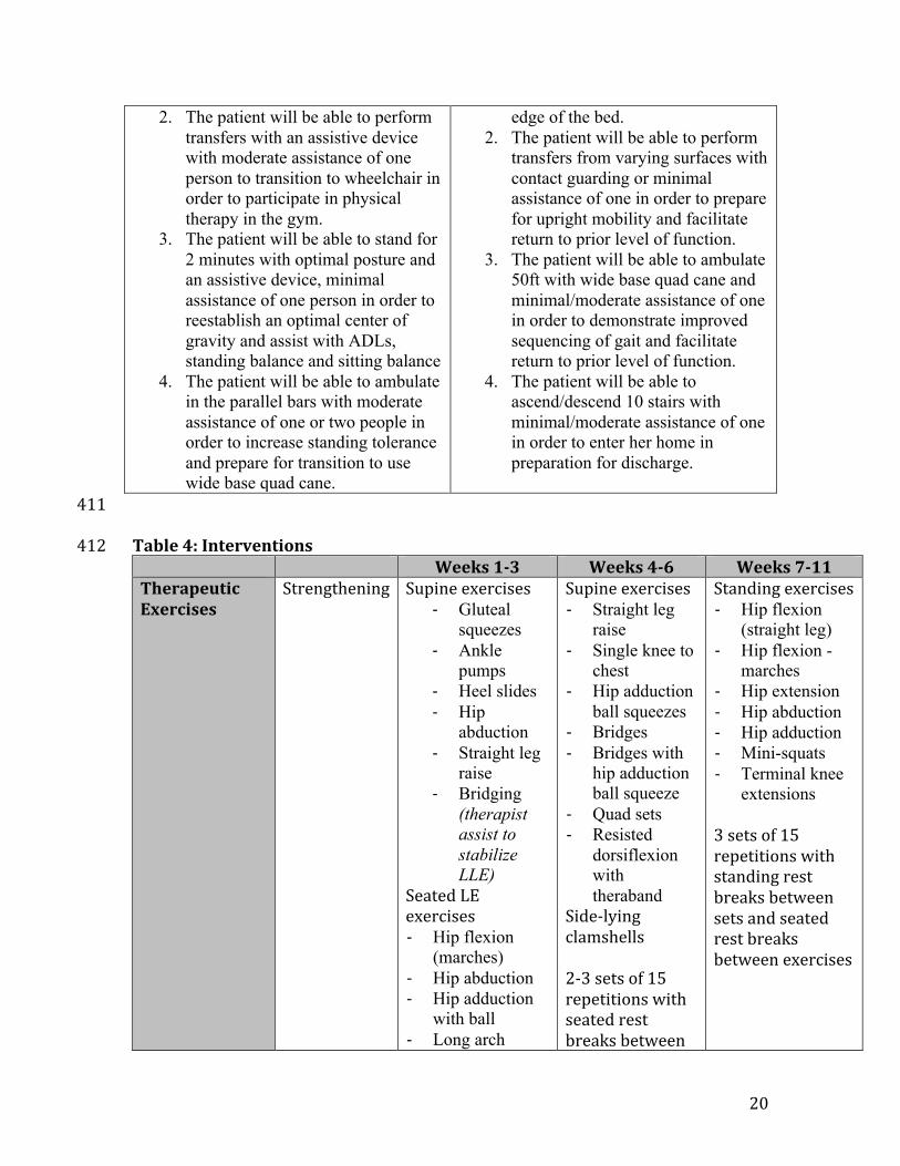
14. O’Sullivan SB. Chapter 10: Strategies to Improve Motor Function. In: O’Sullivan 386
SB, Schmitz TJ, Fulk GD. Physical Rehabilitation. 6th ed. Philadelphia, PA: F.A. 387
Davis Company; 2014: 418-419 388
18
15. GivMohr sling flaccid upper extremity positioning device. 389
http://www.givmohrsling.com/ Published 2005. Updated 2015. Accessed on 390
October 18, 2015. 391
16. Salter K, Jutai J, Hartley M et al. Impact of early vs delayed admission to 392
rehabilitation on functional outcomes in persons with stroke. J Rehabil Med. 393
2006; 38: 113-117. doi:10.1080/16501970500314350. 394
17. Biernaskie J, Chernenko G, Corbett D. Efficacy of rehabilitative experience 395
declines with time after focal ischemic brain injury. J Neurosci 2004; 24: 396
1245 397
18. Paolucci S, Antonucci G, Grasso MG, Morelli D, Troisi E, Coiro P, et al. Early 398
versus delayed inpatient stroke rehabilitation: a matched comparison conducted in 399
Italy. Arch Phys Med Rehabil 2000; 61: 695-700. 400
19. HEP2GO. https://www.hep2go.com/. Published 2010. Updated 2015. Accessed 401
on October18, 2015. 402
403
19
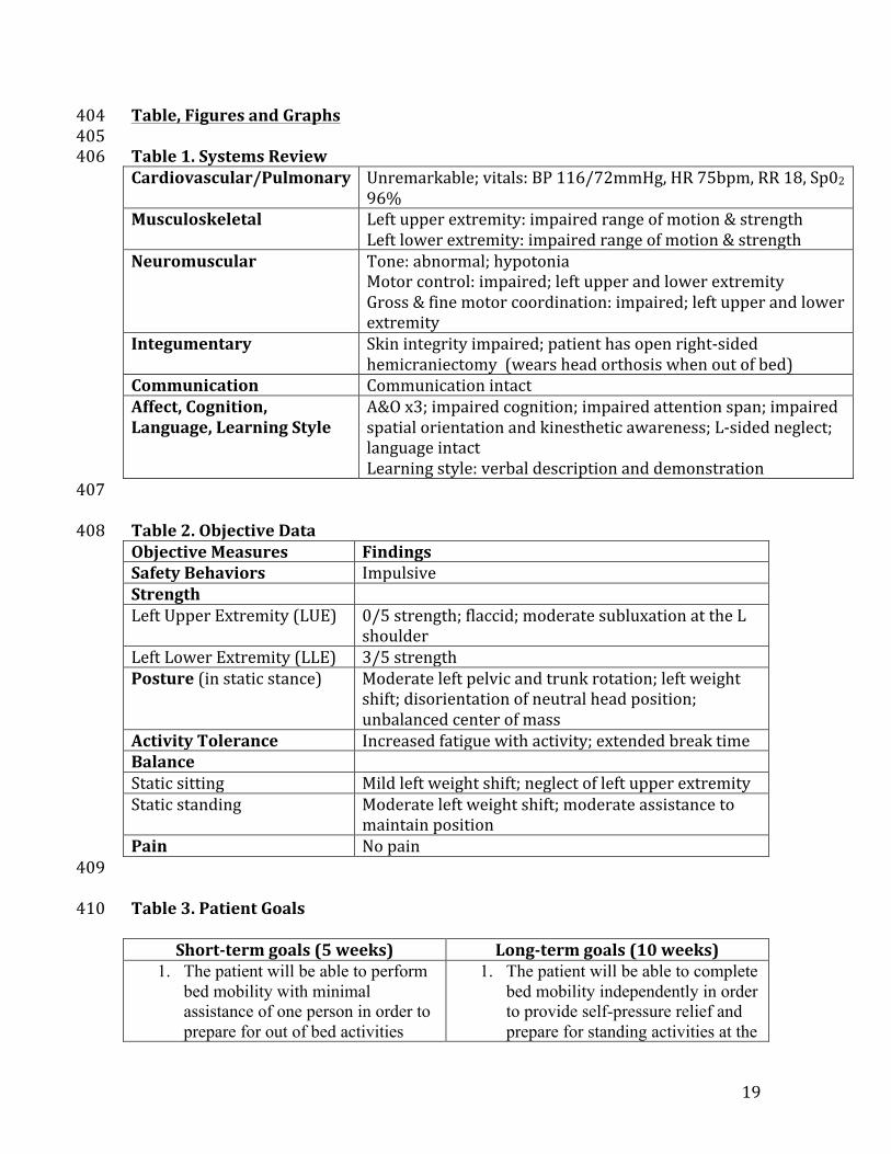
Table, Figures and Graphs 404 405 Table 1. Systems Review 406 Cardiovascular/Pulmonary Unremarkable; vitals: BP 116/72mmHg, HR 75bpm, RR 18, Sp02
96% Musculoskeletal Left upper extremity: impaired range of motion & strength
Left lower extremity: impaired range of motion & strength Neuromuscular Tone: abnormal; hypotonia
Motor control: impaired; left upper and lower extremity Gross & fine motor coordination: impaired; left upper and lower extremity
Integumentary Skin integrity impaired; patient has open right-‐sided hemicraniectomy (wears head orthosis when out of bed)
Communication Communication intact Affect, Cognition, Language, Learning Style
A&O x3; impaired cognition; impaired attention span; impaired spatial orientation and kinesthetic awareness; L-‐sided neglect; language intact Learning style: verbal description and demonstration
407
Table 2. Objective Data 408 Objective Measures Findings Safety Behaviors Impulsive Strength Left Upper Extremity (LUE) 0/5 strength; flaccid; moderate subluxation at the L
shoulder Left Lower Extremity (LLE) 3/5 strength Posture (in static stance) Moderate left pelvic and trunk rotation; left weight
shift; disorientation of neutral head position; unbalanced center of mass
Activity Tolerance Increased fatigue with activity; extended break time Balance Static sitting Mild left weight shift; neglect of left upper extremity Static standing Moderate left weight shift; moderate assistance to
maintain position Pain No pain 409
Table 3. Patient Goals 410
Short-‐term goals (5 weeks) Long-‐term goals (10 weeks) 1. The patient will be able to perform
bed mobility with minimal assistance of one person in order to prepare for out of bed activities
1. The patient will be able to complete bed mobility independently in order to provide self-pressure relief and prepare for standing activities at the
20
2. The patient will be able to perform transfers with an assistive device with moderate assistance of one person to transition to wheelchair in order to participate in physical therapy in the gym.
3. The patient will be able to stand for 2 minutes with optimal posture and an assistive device, minimal assistance of one person in order to reestablish an optimal center of gravity and assist with ADLs, standing balance and sitting balance
4. The patient will be able to ambulate in the parallel bars with moderate assistance of one or two people in order to increase standing tolerance and prepare for transition to use wide base quad cane.
edge of the bed. 2. The patient will be able to perform
transfers from varying surfaces with contact guarding or minimal assistance of one in order to prepare for upright mobility and facilitate return to prior level of function.
3. The patient will be able to ambulate 50ft with wide base quad cane and minimal/moderate assistance of one in order to demonstrate improved sequencing of gait and facilitate return to prior level of function.
4. The patient will be able to ascend/descend 10 stairs with minimal/moderate assistance of one in order to enter her home in preparation for discharge.
411
Table 4: Interventions 412 Weeks 1-‐3 Weeks 4-‐6 Weeks 7-‐11 Therapeutic Exercises
Strengthening Supine exercises -‐ Gluteal
squeezes -‐ Ankle
pumps -‐ Heel slides -‐ Hip
abduction -‐ Straight leg
raise -‐ Bridging
(therapist assist to stabilize LLE)
Seated LE exercises -‐ Hip flexion
(marches) -‐ Hip abduction -‐ Hip adduction
with ball -‐ Long arch
Supine exercises -‐ Straight leg
raise -‐ Single knee to
chest -‐ Hip adduction
ball squeezes -‐ Bridges -‐ Bridges with
hip adduction ball squeeze
-‐ Quad sets -‐ Resisted
dorsiflexion with theraband
Side-‐lying clamshells 2-‐3 sets of 15 repetitions with seated rest breaks between
Standing exercises -‐ Hip flexion
(straight leg) -‐ Hip flexion -
marches -‐ Hip extension -‐ Hip abduction -‐ Hip adduction -‐ Mini-squats -‐ Terminal knee
extensions 3 sets of 15 repetitions with standing rest breaks between sets and seated rest breaks between exercises
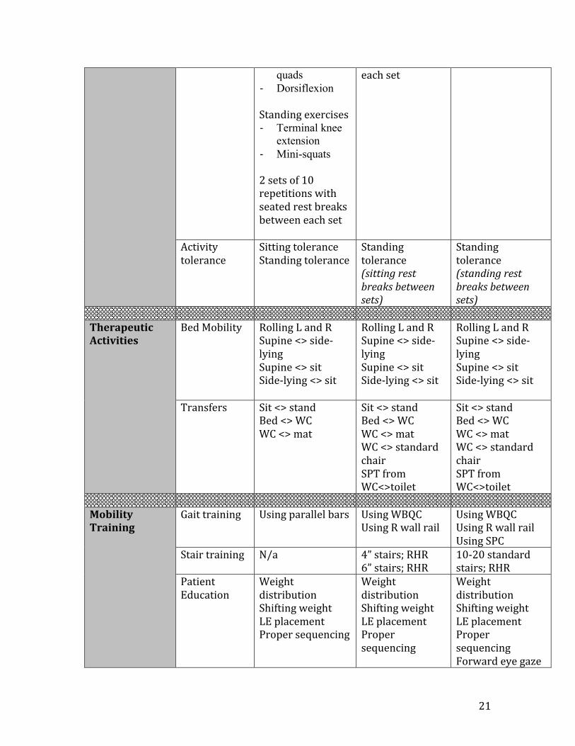
21
quads -‐ Dorsiflexion Standing exercises -‐ Terminal knee
extension -‐ Mini-squats
2 sets of 10 repetitions with seated rest breaks between each set
each set
Activity tolerance
Sitting tolerance Standing tolerance
Standing tolerance (sitting rest breaks between sets)
Standing tolerance (standing rest breaks between sets)
Therapeutic Activities
Bed Mobility Rolling L and R Supine <> side-‐lying Supine <> sit Side-‐lying <> sit
Rolling L and R Supine <> side-‐lying Supine <> sit Side-‐lying <> sit
Rolling L and R Supine <> side-‐lying Supine <> sit Side-‐lying <> sit
Transfers
Sit <> stand Bed <> WC WC <> mat
Sit <> stand Bed <> WC WC <> mat WC <> standard chair SPT from WC<>toilet
Sit <> stand Bed <> WC WC <> mat WC <> standard chair SPT from WC<>toilet
Mobility Training
Gait training Using parallel bars Using WBQC Using R wall rail
Using WBQC Using R wall rail Using SPC
Stair training N/a 4” stairs; RHR 6” stairs; RHR
10-‐20 standard stairs; RHR
Patient Education
Weight distribution Shifting weight LE placement Proper sequencing
Weight distribution Shifting weight LE placement Proper sequencing
Weight distribution Shifting weight LE placement Proper sequencing Forward eye gaze
22
Neuromuscular Re-‐education
Balance training
Sitting balance -‐ With feet
supported on ground and RUE support
-‐ With feet supported on ground and no UE support
-‐ Weight shifting in all directions
-‐ Reaching with RUE
Standing balance -‐ With RUE
support -‐ Without RUE
support -‐ Weight shifting
in all directions
Standing balance -‐ Without UE
support -‐ Balloon taps
with RUE -‐ Reaching for
cones with RUE from various angles
Dynamic standing balance with standing therapeutic exercises Standing balance -‐ With normal
BOS -‐ With narrow
BOS -‐ L tandem
stance -‐ R tandem
stance -‐ Single leg
stance
<> -‐ to/from; L – left; R-‐ right; UE-‐ upper extremity; LE – lower extremity; LUE – left 413 upper extremity; RUE – right upper extremity; WC-‐ wheelchair; SPT-‐ stand pivot 414 transfer; WBQC-‐ wide based quad cane; SPC-‐ single point cane; RHR-‐ right hand rail; 415 BOS-‐ base of support 416 417 418 Table 5: Physical Performance and Functional Assessments 419
Week 1 (Examination)
Week 4 Week 7 Week 10 (Discharge)
MBI 60/100 -‐-‐-‐-‐ -‐-‐-‐-‐ 86/100 PPME 1/12 4/12 -‐-‐-‐-‐ 9/12
30-‐second chair rise
0 0 6 7
Gait Speed 0 0 0.13 m/s, WBQC, S
0.23 m/s; WBCQ
Gait speed not assessed secondary of inability to ambulate 20’ 420 Assistive devices: wide based quad cane (WBQC); single point cane (SPC) 421 Level of assistance: contact guard assist (CGA); supervision (S) 422 423
23
Figure 1 424
425
This graph represents the patient’s functional level at the time of initial evaluation and 426 again at discharge. 100% indicated independent; 90% modified-‐indepdent; 0% unable 427 to perform. 428
0 10 20 30 40 50 60 70 80 90 100
Bed mobility Transfers Ambulation Stairs
Patient ability (%
)
Functional Assessment
Patient's functional ability as a result of functional assessment
Examination
Discharge
24
429
Figure 2. Assistive Equipment 430
A. Parallel Bars B. Wide Based Quad Cane
C. GiveMohr Sling14
431 432
25
Appendix 1. Home Exercise Program19 433 Supine Exercises
Straight Leg Raises Quad sets
Hip adductor ball squeeze Side-‐lying clamshells
Sitting Exercises
Ankle Dorsflexion Long Arc Quads
Standing Exercise
Hip flexion – marches Hip flexion Hip abduction
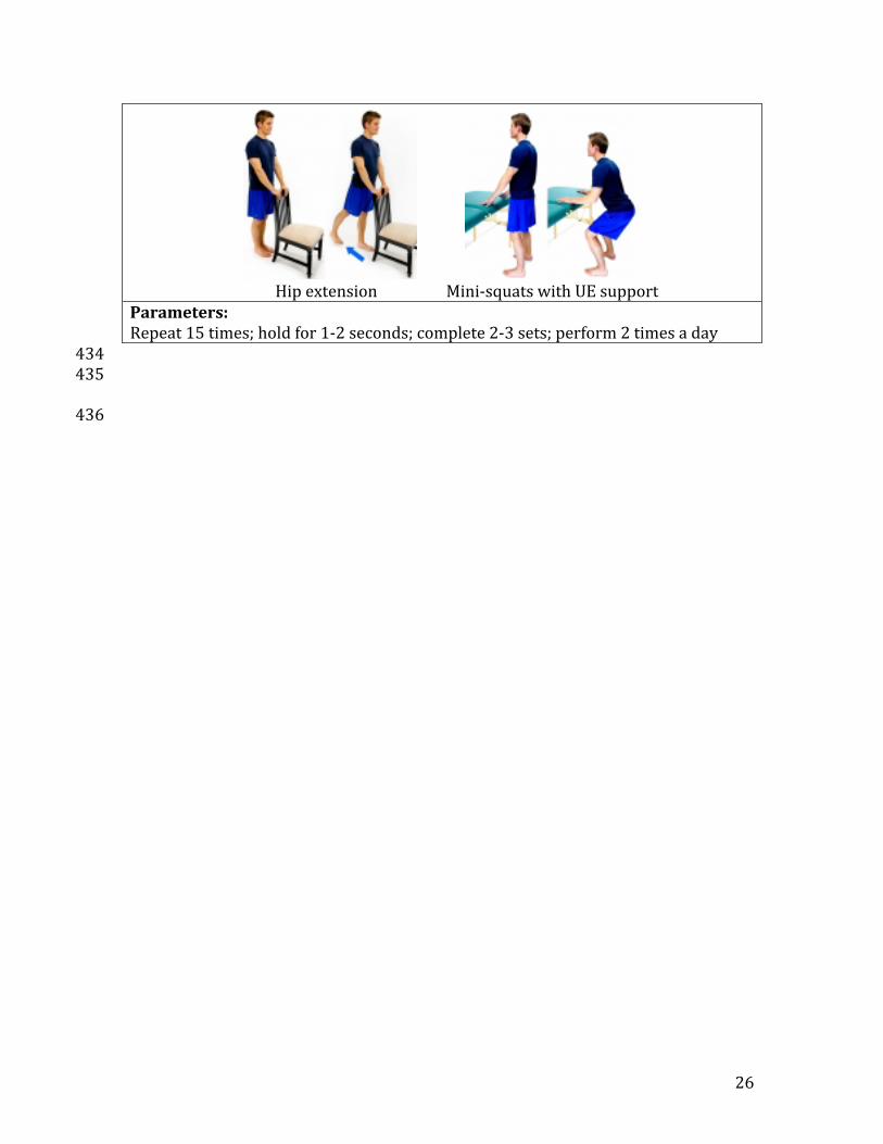
26
Hip extension Mini-‐squats with UE support
Parameters: Repeat 15 times; hold for 1-‐2 seconds; complete 2-‐3 sets; perform 2 times a day 434 435
436