Embed Size (px)
Citation preview

RESTORATIVE AND PULP
TREATMENT OF PRIMARY AND
YOUNG PERMANENT TEETHCharles Lekic DDM, MSc, PhD FRCD(C)
May 2015

Restorative and pulp treatment of
primary and young permanent teeth
Materials and techniques for restorative and pulp
treatment of primary and young permanent teeth
have improved ever since. Due to time constraints
this presentation will focus on:
a) Local anesthesia
b) Rubber dam
c) Restorative procedures
d) Pulp treatment options

Local anesthesia
Always inject slowly,
aspirating frequently
Use smaller needles (30-
gauge), thus reducing
discomfort
In patients with unerupted first
permanent molars and due to
greater deflection of smaller
needles, perform mandibular
block with a larger needle
(27-gauge)

Local anesthesia (cont’d)
Calculating maximum pediatric dose for local
anesthetic:
Maximum recommended dose (mg/Kg) x Child’s weight (Kg)
Anesthetic Concentration x Volume of cartridges
The maximum amount of Xylocaine (2%) for a child weighing
17 Kg would be:
4.4 mg/Kg x 17 Kg = 74.8 = 2.08 cartridges
2% x 1.8 cc 36

Rubber dam
Briefly, rubber dam is to be used at all times in restorative pediatric dentistry with the exception of:
a) Incompletely erupted tooth (inability to place the clamp)
b) When splinting the displaced tooth).
Restorative procedures for primary teeth
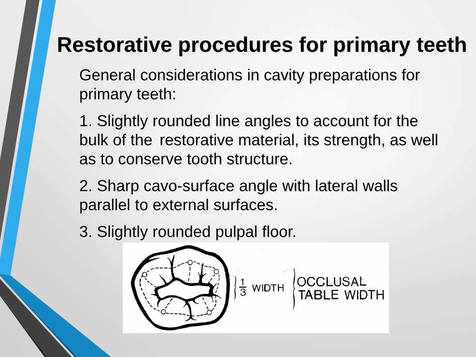
General considerations in cavity preparations for
primary teeth:
1. Slightly rounded line angles to account for the
bulk of the restorative material, its strength, as well
as to conserve tooth structure.
2. Sharp cavo-surface angle with lateral walls
parallel to external surfaces.
3. Slightly rounded pulpal floor.

Restorative procedures for primary teeth
General considerations in cavity preparations for primary teeth (cont’d):
1) Depth and width of the cavity preparation is usually 1 mm, unless dictated by the extent of the caries lesion.
2) Gingival preparation is slightly below gingival papilla.
3) Wedge (arrow) to reduce hemorrhage and prevent damaging of the adjacent surface

Restorative procedures for primary teeth
(cont’d)
Matrix application:
Highly recommended is the T-band as it allows
placement of multiple matrices, no specific
equipment is needed, easy to use, inexpensive

Composite vs. amalgam restorations in primary teeth
• In 1996 Health Canada has advised dentists
to consider the use of non-mercury filling
materials for restoring the primary teeth in
instances when high tensile/compressive
stresses are not encountered.
• Amalgams should not be placed in children
with impaired kidney function on
consultation with patient's physician or
hypersensitivity to amalgam.

Composite vs. amalgam restorations
in primary teeth (Cont’d) (Cont’d)
• Controversy between the use of amalgam
and/or composite resin has subsided.
• No solid scientific evidence has been
established regarding the overall amalgam
toxicity as a filling material
• Composite resins have similar success rate
over 5-10 years follow-up period
• Composite resins require minimum needed
removal of tooth structure

Condensation of Amalgam
• Rinse & Dry Prep
• Place pulp protection as needed
• Place small amount amalgam into proximal (class II)
• Use small condenser, condense thrusting against gingival, buccal and lingual walls
• Class II- after proximal box condensation, proceed with condensation of occlusal surface
• Slightly over pack
• Trim off excess with large round ball burnisher

Carving1. Use explorer at gingival embrasure, and the marginal ridge
2. Carve excess amalgam from cavo-surface using discoid-cleoid or T-3
3. Use #21 carving instrument & follow inclined planes into all grooves,
4. Restore accessory grooves and define occlusal anatomy using discoid-cleiod or #21
5. Carve excess amalgam
6. Remove matrix
7. Floss interproximal
8. Wipe amalgam with moist cotton pellet
9. Check occlusion

Composite restorations in primary dentition
• Etch, rinse and dry, leaving cavity slightly moistened
• Apply primer and adhesive material. With an air syringe gently spread the material over the cavity preparation, light cure and reapply.
• After condensing and light curing the resin material partially remove the matrix band and light cure the resin 10 sec on the buccal and then 10 sec on the lingual surface.

Composite restorations in primary dentition (cont’d)
• Remove the matrix band and with the flame-shaped finishing bur, contour the interproximal areas. Remove any excess, maintaining normal tooth morphology.
• Apply etchant to the surrounding enamel, bond and seal to close enamel cracks which may occur with polymerization shrinkage.
• Floss and remove the rubber dam and with the articulating paper, check for premature contacts. Contour and polish the occlusal surfaces.
Pulp therapy in primary and young
permanent teeth
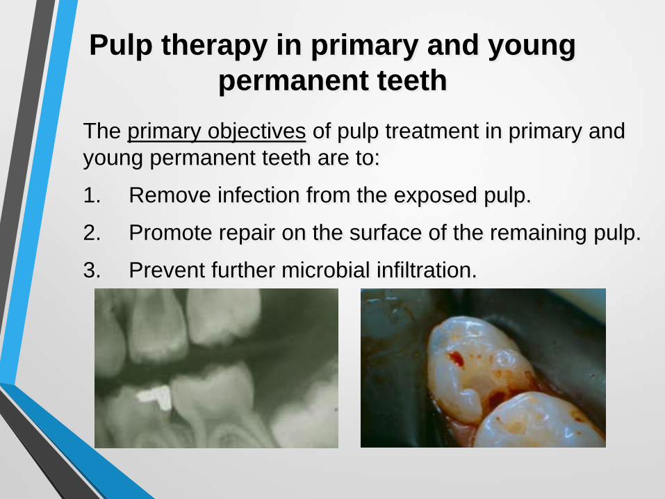
The primary objectives of pulp treatment in primary and
young permanent teeth are to:
1. Remove infection from the exposed pulp.
2. Promote repair on the surface of the remaining pulp.
3. Prevent further microbial infiltration.

Clinical Examination
• History of discomfort
• Inspection of tooth color and the condition of the surrounding soft tissues
• Assessment of tooth mobility and sensitivity to percussion

Pulp Treatment in Primary teeth
The following procedures could be implemented in
treating a pulpally involved tooth:
• Indirect and direct pulp
capping
• Pulpotomy
• Pulpectomy

Indirect and Direct Pulp Capping
Objectives:
• Preserve the vitality and
normal functions of the
pulp
• Act as a protective barrier
between the restorative
material and the tooth
• Promote tertiary dentin
formation and caries
dentin remineralization

Indirect Pulp Capping
Recommended due to high success rate
(~90%) in primary teeth
Indications:• Asymptomatic tooth
• Deep caries lesion
• Vital pulp
Google images, indirect pulp capping

Indirect Pulp Capping (Cont’d)
Indirect Pulp Capping:
• Remove soft, mushy dentin
with a #8 round bur
• Cap hard and discolored
dentin with Ca(OH)2 and/or
glass ionomer base
• Restore the cavity with a
conventional filling material
Google images, indirect pulp capping

Direct Pulp Capping
Direct Pulp Capping:
• Remove carious dentin with a #8 round bur
• Over a mechanical pinpoint pulp exposure place a Ca(OH)2 or MTA to stimulate dentin formation
• Place permanent restoration
Recommended only in the case of mechanical
pulp exposure of primary teeth

Direct Pulp Capping (Cont’d)
• MTA promotes cytokine
release leading to
osteoblast differentiation
• Found to maintain pulp
integrity
• Has dentinogenic effect
(dentin bridge formation) Dr. De Silva Modena, J Appl Oral
Sci, 2009

Pulpotomy for Primary Teeth
Indications:
• Exposure of the pulp
• Absence of unprovoked pain
• No sensitivity to percussion or palpation
• No apical, periapical and furcation radiolucencies

Pulpotomy for Primary Teeth (Cont’d)
• Absence of pulp suppuration
• No abnormal mobility of tooth
• Controllable hemorrhage after removal of coronal pulp
• Not more than 1/3 of resorbed root

Pulp therapy in primary and young
permanent teeth (cont’d)
Mineral trioxide aggregate or formocresol or ferric sulfate pulpotomy are the most common forms of pulp treatment for primary teeth.
It is recommended to use sterile round burr and the spoon excavator, when removing pulp tissue. This reduces hemorrhage and promotes healing.

Pulpotomy for Primary Teeth (Cont’d)
• Pulp obliteration (lower risk
than for MTA)
• Succedaneous tooth damage
a small risk
• Exfoliation accelerated
• Cellular toxicity (???)
• Immune sensitization risk
(type I allergic reaction, ???)
• Mutagenic and carcinogenic
potential (???)
Negative effects of formocresol pulpotomy:

Pulpotomy for Primary Teeth (Cont’d)
• Dental cement with discrete crystals and amorphous structure
• Method of action is mineralization
• Pulp canal obliteration common
• Multiple investigations to support efficacy
Procedure (Cont’d – mineral trioxide aggregate):

Pulpotomy for Primary Teeth (Cont’d)
• Mineral trioxide aggregate
(MTA) has 3 material’s
composition – calcium,
aluminum and selenium
• MTA contact with humid
environment produces high
pH – 12.5
• Antibacterial effect and good
marginal seal

Pulpotomy for Primary Teeth (Cont’d)
Advantage for the use of MTA:
• High success rate
• Less time needed for the procedure
• Disadvantage for the use of MTA:
• Very expensive (used for a single patient)
Google images, expensive
Pulp therapy in primary and young
permanent teeth (cont’d)
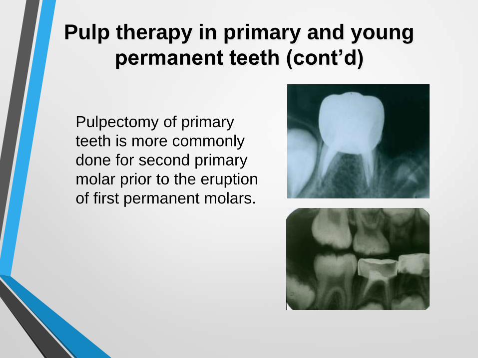
Pulpectomy of primary
teeth is more commonly
done for second primary
molar prior to the eruption
of first permanent molars.

Pulp therapy in primary and young
permanent teeth (cont’d)
Apexification of young
permanent teeth is used
to facilitate closure of the
apex of young permanent
teeth, exhibiting extensive
degeneration or necrosis
Pulp therapy in primary and young permanent teeth (cont’d)
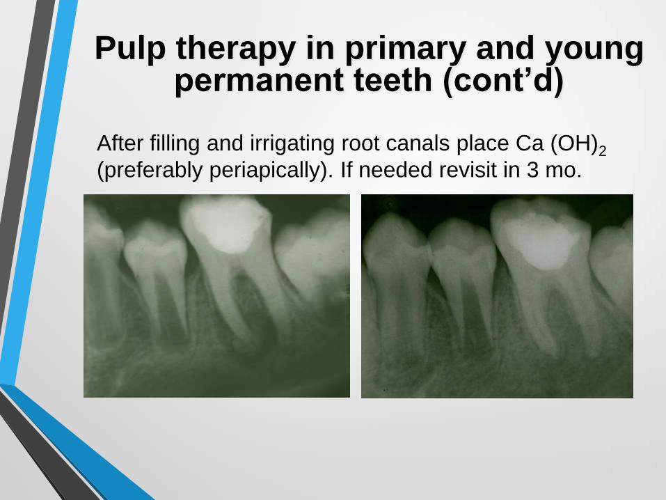
After filling and irrigating root canals place Ca (OH)2
(preferably periapically). If needed revisit in 3 mo.

Stainless steel crown procedures
The stainless steel crown
(SSC) is used to restore
form and function to teeth
which have lost structure
due to caries, pulp therapy,
or trauma.
Advantages:
• Simple preparation of tooth
• Easy to manipulate
• Inexpensive
• High success rate

Stainless steel crown procedures (cont’d)
Indications for primary teeth:
1. Severely broken down molars (involvement of 3
surfaces) where an amalgam or composite is
impractical or impossible.
2. For full coverage of a tooth where conditions are
such that decay would certainly re-occur.
3. Where a pulpotomy or pulpectomy has been done.

Stainless steel crown procedures
(cont’d)
Start preparing the
occlusal surface and
thus gain access to
caries lesion.
Tooth prep, fit and the
seat of the crown
should be completed
in 15 min.

Special Considerations for Stainless Steel Crown
Placing of Adjacent Crowns
1. Complete occlusal preparation of 1 tooth before beginning the other
2. Proximal reduction should produce a 1.5 mm of space at the gingival level
3. Cement both crowns at the same time

Special Considerations for Stainless Steel Crown (cont’d)
Preparing Crowns in Areas of Space Loss:
1. Contour pliers can be used (mesially and distally) to reduce mesiodistal dimension
2. Recontour proximal buccal and lingual walls

Special Considerations for Stainless Steel Crown (cont’d)
• Sometimes lower D tooth could be better restored with an upper, contra-lateral D crown.
• The scoop end of a T-3 is used to remove crowns when fitting them. At the same when removing the crown hold with other hand the occlusal surface to avoid pushing of the crown further back.
• The beaks of the crimping pliers can be used to force a crown on.

Complex restorative treatment in
pediatric dentistry
Patient K.M. age 12
has an advanced form
of amelogenesis
imperfecta.
Chief complaints:
extreme sensitivity to
cold as well as
esthetics concerns

Complex restorative treatment in
pediatric dentistry (cont’d)
Restorative treatment of severe form of enamel
defects may require resin build-up and placement
of stainless steel crowns.

What would you, what would I do?
P.C. age 7 presented with buccal swelling lower left.
What would you, what would I do?
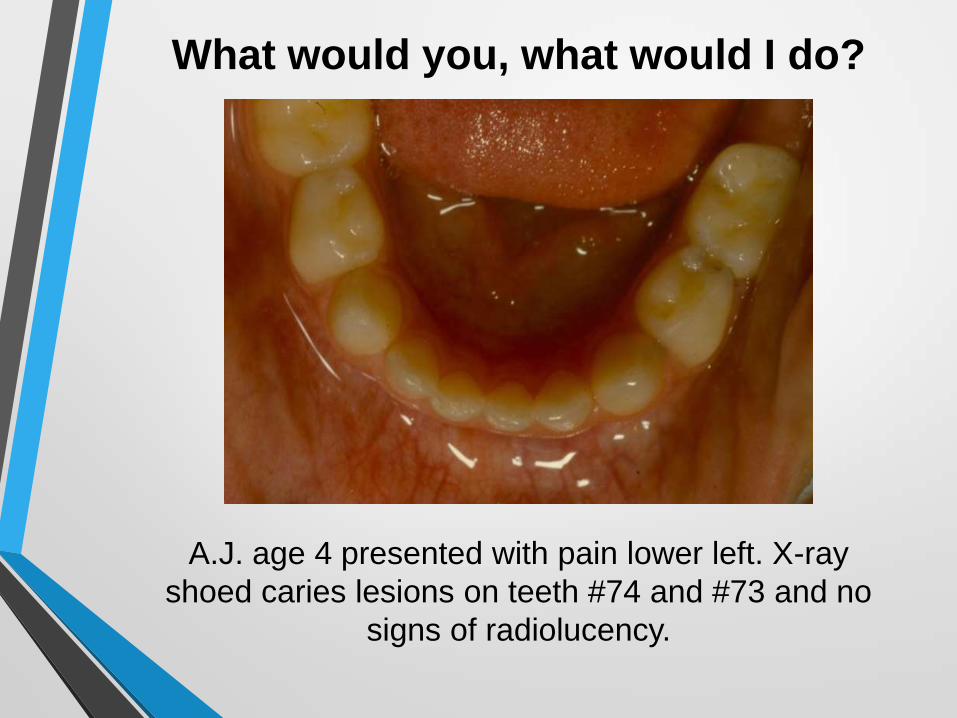
A.J. age 4 presented with pain lower left. X-ray
shoed caries lesions on teeth #74 and #73 and no
signs of radiolucency.

What would you, what would I do?
M.S. age 8 presented with caries lesions on teeth #55 and #54. X-rays showed deep caries, but no
signs of radiolucency.

What would you, what would I do?
A.E. age 11 presented with disto-occlusal caries on teeth #55 and #65 as well as deeply stained grooves
on tooth #16. X-rays showed no signs of radiolucency.

What would you, what would I do?
M.K. age 7 presented with celulitis upper left. Child was febrile (99.5º) and X-ray showed extensive caries and
radiolucency in the furcation area of tooth #64.

What would you, what would I do?
J.J. age 5 presented with pain from tooth #75. There was no swelling and percussion and
palpation was negative.
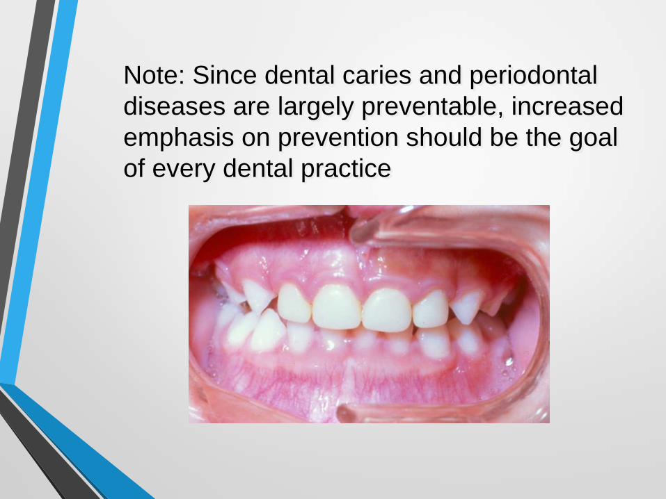
Note: Since dental caries and periodontal
diseases are largely preventable, increased
emphasis on prevention should be the goal
of every dental practice
Thank you for your attention