Embed Size (px)
Citation preview

Submitted 6 May 2014Accepted 31 July 2014Published 26 August 2014
Corresponding authorDanilo Garcia,[email protected],[email protected]
Academic editorJafri Abdullah
Additional Information andDeclarations can be found onpage 11
DOI 10.7717/peerj.531
Copyright2014 Archer et al.
Distributed underCreative Commons CC-BY 4.0
OPEN ACCESS
Restoration of MPTP-induced deficits byexercise and Milmed® co-treatmentTrevor Archer1,2, Danilo Garcia2,3 and Anders Fredriksson4
1 Department of Psychology, University of Gothenburg, Gothenburg, Sweden2 Network for Empowerment and Well-Being, Sweden3 Institute of Neuroscience and Physiology, Centre for Ethics, Law and Mental Health (CELAM),
University of Gothenburg, Gothenburg, Sweden4 Department of Neuroscience Psychiatry, Uppsala University, Uppsala, Sweden
ABSTRACT1-methyl-4-phenyl-1,2,3,6-tetrahydropyridine (MPTP) induces permanentneurochemical and functional deficits. Following the administration of either two orfour injections of the dopamine neurotoxin, MPTP, at a dose of 40 mg/kg, C57/BL6mice were given access to running-wheels (30-min sessions, four times/week,Monday–Thursday) and treatment with the treated yeast, Milmed® (fourtimes/week, Monday–Thursday), or simply running-wheel exercise by itself,over ten weeks. It was observed that the combination of physical exercise andMilmed® treatment, the MPTP + Exercise + Yeast (MC) group [MPTP + Ex-ercise + Milmed® (MC)], restored spontaneous motor activity markedly by testday 10, restored completely subthreshold L-Dopa-induced activity, and dopamineconcentration to 76% of control values, in the condition wherein two adminis-trations of MPTP (2 × 40 mg/kg) were given prior to initiation of exercise and/orMilmed® treatment. Physical exercise by itself, MPTP + Exercise (MC) group,attenuated these deficits only partially. Administration of MPTP four times (i.e.,40 mg/kg, s.c., once weekly over four weeks for a total of 160 mg/kg, MPTP + Exer-cise + Yeast (MC) group [MPTP + Exercise + Milmed® (SC)] and MPTP + Exercise(SC), induced a lesioning effect that was far too severe for either exercise alone or theexercise + Milmed® combination to ameliorate. Nevertheless, these findings indicatea powerful effect of physical exercise reinforced by Milmed® treatment in restoringMPTP-induced deficits of motor function and dopamine neurochemistry in mice.
Subjects Global Health, Neurology, Public HealthKeywords MPTP, Exercise, Milmed®, Parkinsonism, Running-wheels, Locomotion,Total activity, Dopamine, Attenuation, Restoration
Although physical exercise by itself ameliorates functional, motor activity, and
neurochemical, dopamine, deficits induced by the selective dopamine neurotoxin,
1-methyl-4-phenyl-1,2,3,6-tetrahydropyridine (MPTP, Archer & Fredriksson, 2010; Archer
& Fredriksson, 2012; Archer & Fredriksson, 2013a; Archer & Fredriksson, 2013b; Archer
& Fredriksson, 2013c; Fredriksson et al., 2011), the combination of physical exercise
with Milmed® provided evidence of complete restoration of these deficits (Archer &
Fredriksson, 2013a). The efficacy of physical exercise (Archer, 2011; Archer, Johansson &
Fredriksson, 2011; Earhart & Falvo, 2013) and physiotherapy (Cholewa, Boczarska-Jedynak
How to cite this article Archer et al. (2014), Restoration of MPTP-induced deficits by exercise and Milmed® co-treatment. PeerJ 2:e531;DOI 10.7717/peerj.531

& Opala, 2013) for Parkinsonism and other neurodegenerative conditions has been
well-documented with ever-increasing evidence from both clinical and laboratory studies
(Park et al., 2014; Wang et al., 2013; Zigmond & Smeyne, 2014). Three main conclusions
were obtained from a recent Milmed® study (Archer & Fredriksson, 2013a): (i) dopamine
integrity was observed to be a direct function of ability to express running exercise in
a treadmill running arrangement, (ii) dopamine integrity was observed to be a direct
function of the capacity for motor performance as measured by spontaneous motor
activity and subthreshold L-Dopa (5 ml/kg) induced activity in the motor activity test
chambers, and (iii) running exercise in the treadmill running wheel predicted later motor
performance in the motor activity test chambers to an extremely high degree. In this
respect, it has been demonstrated in rats that caffeine, both hyperthermic and ergogenic,
in combination with physical exercise increased extracellular dopamine and noradrenaline
in the preoptic area and anterior hypothalamus (Zheng et al., 2014). Treadmill exercise
ameliorated also the nigrostriatal dopaminergic neuronal loss in adolescent rats following
neonatal hypoxic brain ischemia which improved spatial learning ability (Park et al., 2013).
Finally, using the 10 × 25 mg/kg MPTP model of Parkinson’s disease it was indicated that
four weeks of treadmill running decreased the levels of the inducible form of nitric oxide
and neuronal nitric oxide in the brains of MPTP-treated mice (Al-Jarrah, 2013).
The preparation and application of treated yeast culture, Saccharoyces cerevisiae,
to provide the antiparkinson agent, Milmed®, in suspension form has been outlined
previously (Gedymin et al., 1999; Kolosova et al., 1998). The derivation of Milmed® for
cellular genesis and reparation has been reported elsewhere (Golant, 1994; Golant et
al., 1994; Ragimov et al., 1991). The description, preparation, and application of the
Milmed® suspension (oral administration, four times a week) in an animal MPTP
model of Parkinsonism has also been described comprehensible in earlier studies
(Archer & Fredriksson, 2013a; Archer & Fredriksson, 2013b). Generally, studies designed
to apply exercise intervention using MPTP lesioning to induce Parkinson symptoms,
e.g., hypokinesia, in the laboratory have introduced exercise, with or without (Archer &
Fredriksson, 2010) Milmed® co-treatment, prior to administration of the neurotoxin,
was shown to prevent any MPTP-induced deficits in several experiments. In the present
study, C57/BL6 mice were administered MPTP (40 mg/kg) either twice or four times
before access to the exercise (running-wheel, four 30 min/sessions/week, Mon.–Thurs.)
and Milmed® co-treatment intervention (once/day, four times/week). The purpose of the
present study was to ascertain the extent to which the exercise + Milmed® treatment
combination would restore MPTP-induced functional and neurochemical deficits
following either two (moderate condition; [MPTP + Exercise + Milmed® (MC)]) or
four (severe condition; [MPTP + Exercise + Milmed® (SC)]) injections of the neurotoxin.
MATERIALS AND METHODSEthics statementThe study was carried out in accordance with the European Communities Council Direc-
tive of 24th November 1986 (86/609/EEC) after approval from the local ethical Committee
Archer et al. (2014), PeerJ, DOI 10.7717/peerj.531 2/13

(Uppsala University and Agricultural Research Council), and by the Swedish Committee
for Ethical Experiments on Laboratory animals (License S93/92 and S77/94, Stockholm,
Sweden). Throughout all the series of experiments performed with Milmed® there is no
evidence of any intestinal problem or any other side effects. Additionally, Milmed® has
been administered, orally, to more than 1,000 human subjects for the alleviation of several
ailments, including Parkinson’s disease, borellia, cancer, and attention-deficit hyperactive
disorder, as well as a nutritional adjunct for elite-level athletes, over periods extending from
eight-to-sixteen weeks; no side effects have ever been reported.
AnimalsMale C57/BL6 mice, purchased from B&K, Sollentuna, Sweden, maintained five-to-ten in
a cage in plastic cages in an isolated room at 22 ± 1 ◦C and 12 h/12 h constant light/dark
cycle (lights on between 06.00 and 18.00 h), were acclimatized, housed and given access to
the running-wheels or holding-cages (30-min sessions) in an identical to that described
previously (Archer & Fredriksson, 2013a). Each group consisted of 10 mice.
DrugsMPTP (Research Biochemical Inc., MA, USA), injected 4 × 40 mg/kg, s.c. (1-week inter-
vals between injections, was dissolved in saline and administered in a volume of 2 ml/kg
body weight. Milmed® was prepared for administration according to a procedure identical
to that described previously (Archer & Fredriksson, 2013a). Mice were administered
oral injections of 0.5 ml/kg Milmed® containing a cell concentration of approximately
2 × 10⁶ yeast cells daily, according to the preparation protocol and design developed
from previous observations regarding stability and viability of the compound (each dose
contained 1 × 10⁶ yeast cells). Each mouse was administered Milmed® once each day
four times/week during the 10 weeks of exercise + Milmed® treatment with Design and
treatment maintained as described previously (cf. Archer & Fredriksson, 2013a).
Behavioural measurements and apparatusTesting of motor activity in the ADEA test chambers where Locomotion, Rearing and Total
activity were measured was performed in an identical manner to that described previously
(Archer et al., 1986). Activity test chambers: An automated device, consisting of macrolon
rodent test cages (40×25×15 cm) each placed within two series of infra-red beams (at two
different heights, one low and one high, 2 and 8 cm, respectively, above the surface of the
sawdust, 1 cm deep), was used to measure spontaneous motor activity (RAT-O-MATIC;
ADEA Elektronic AB, Uppsala, Sweden). The distance between the infra-red beams was as
follows: the low levels beams were 73 mm apart lengthwise and 58 mm apart breadthwise
in relation to the test chamber; the high level beams, placed only along each long side of the
test chamber were 28 mm apart. According to the procedures described previously (Archer
et al., 1986), the following parameters were measured: LOCOMOTION was measured
by the low grid of infra-red beams. Counts were registered only when the mouse in the
horizontal plane, ambulated around the test-cage. REARING was registered throughout
the time when at least one high level beam was interrupted, i.e., the number of counts
Archer et al. (2014), PeerJ, DOI 10.7717/peerj.531 3/13
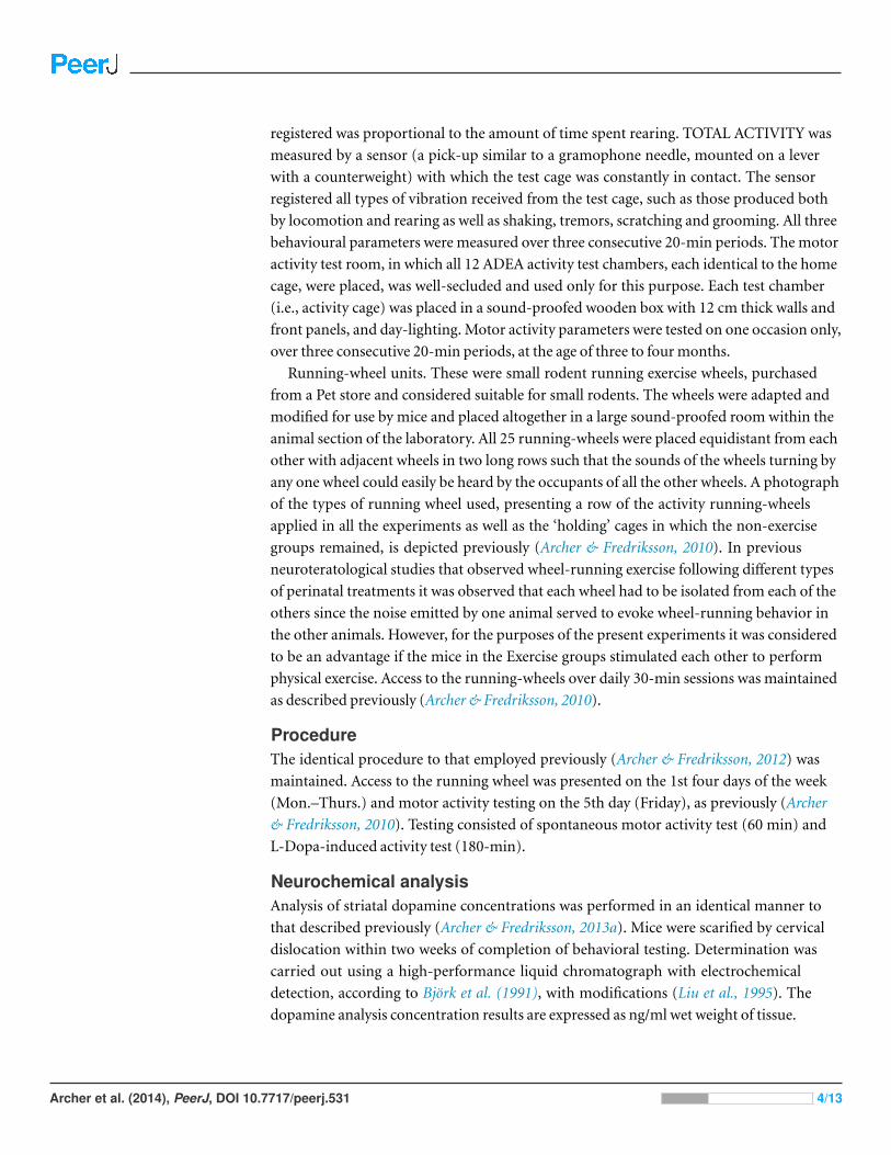
registered was proportional to the amount of time spent rearing. TOTAL ACTIVITY was
measured by a sensor (a pick-up similar to a gramophone needle, mounted on a lever
with a counterweight) with which the test cage was constantly in contact. The sensor
registered all types of vibration received from the test cage, such as those produced both
by locomotion and rearing as well as shaking, tremors, scratching and grooming. All three
behavioural parameters were measured over three consecutive 20-min periods. The motor
activity test room, in which all 12 ADEA activity test chambers, each identical to the home
cage, were placed, was well-secluded and used only for this purpose. Each test chamber
(i.e., activity cage) was placed in a sound-proofed wooden box with 12 cm thick walls and
front panels, and day-lighting. Motor activity parameters were tested on one occasion only,
over three consecutive 20-min periods, at the age of three to four months.
Running-wheel units. These were small rodent running exercise wheels, purchased
from a Pet store and considered suitable for small rodents. The wheels were adapted and
modified for use by mice and placed altogether in a large sound-proofed room within the
animal section of the laboratory. All 25 running-wheels were placed equidistant from each
other with adjacent wheels in two long rows such that the sounds of the wheels turning by
any one wheel could easily be heard by the occupants of all the other wheels. A photograph
of the types of running wheel used, presenting a row of the activity running-wheels
applied in all the experiments as well as the ‘holding’ cages in which the non-exercise
groups remained, is depicted previously (Archer & Fredriksson, 2010). In previous
neuroteratological studies that observed wheel-running exercise following different types
of perinatal treatments it was observed that each wheel had to be isolated from each of the
others since the noise emitted by one animal served to evoke wheel-running behavior in
the other animals. However, for the purposes of the present experiments it was considered
to be an advantage if the mice in the Exercise groups stimulated each other to perform
physical exercise. Access to the running-wheels over daily 30-min sessions was maintained
as described previously (Archer & Fredriksson, 2010).
ProcedureThe identical procedure to that employed previously (Archer & Fredriksson, 2012) was
maintained. Access to the running wheel was presented on the 1st four days of the week
(Mon.–Thurs.) and motor activity testing on the 5th day (Friday), as previously (Archer
& Fredriksson, 2010). Testing consisted of spontaneous motor activity test (60 min) and
L-Dopa-induced activity test (180-min).
Neurochemical analysisAnalysis of striatal dopamine concentrations was performed in an identical manner to
that described previously (Archer & Fredriksson, 2013a). Mice were scarified by cervical
dislocation within two weeks of completion of behavioral testing. Determination was
carried out using a high-performance liquid chromatograph with electrochemical
detection, according to Bjork et al. (1991), with modifications (Liu et al., 1995). The
dopamine analysis concentration results are expressed as ng/ml wet weight of tissue.
Archer et al. (2014), PeerJ, DOI 10.7717/peerj.531 4/13

Statistical analysisSpontaneous motor activity counts (60-min test sessions) and L-Dopa-induced motor
activity counts (180-min test sessions) were subjected Split-plot ANOVA whereas striatal
dopamine concentrations were subjected to one-way ANOVA (Kirk, 1995). Pairwise testing
between groups was performed using Tukey’s HSD tests.
RESULTSThe combination of physical exercise with Milmed® restored both function, spontaneous
motor activity and L-Dopa-induced activity, and neurochemical, dopamine, deficits
whereas exercise, by itself, attenuated the deficits.
Spontaneous motor activitySplit-plot ANOVA indicated a significant Groups x Test days interaction for locomotion,
rearing and total activity counts: F(41,419) = 97.61, p < 0.0001; F(41,419) = 69.23,
p < 0.0001; and, F(41,419) = 50.13, p < 0.0001, respectively. Figure 1 presents the mean
and standard deviation (SD) for locomotion, rearing and total activity counts for each
of the six groups: Vehicle, MPTP, MPTP + Exercise (MC), MPTP + Exercise + Yeast
(MC) [MPTP + Exercise + Milmed® (MC)], MPTP + Exercise (SC), and MPTP + Exer-
cise + Yeast (SC) [MPTP + Exercise + Milmed® (SC)], over 60-min test sessions for the
spontaneous activity tests.
Pairwise testing using Tukey’s HSD test indicated that over all three motor activity pa-
rameters that: (i) the MPTP + Exercise (MC) and MPTP + Exercise + Yeast (MC) groups
made more counts than the MPTP, MPTP + Exercise (SC) and MPTP + Exercise + Yeast
(SC) [MPTP + Exercise + Milmed® (SC)] groups but fewer counts than the Vehicle group,
(ii) the MPTP + Exercise + Yeast (MC) [MPTP + Exercise + Milmed® (MC)] group
made more counts than the MPTP + Exercise (MC) group over test days 6, 8, 10, and (iii)
the MPTP + Exercise (MC) and MPTP + Exercise + Yeast (MC) groups increased the
numbers of counts made from test days 3–10 days, an indication of gradual recovery.
L-Dopa-induced activitySplit-plot ANOVA indicated a significant Groups x Test days interaction for locomotion,
rearing and total activity counts: F(17,143) = 72.81, p < 0.0001; F(17,143) = 15.81,
p < 0.0001; and F(17,143) = 15.97, respectively. Figure 2 presents the mean and SD
for locomotion, rearing and total activity counts for each of the six groups: Vehicle,
MPTP, MPTP + Exercise (MC), MPTP + Exercise + Yeast (MC) [MPTP + Exer-
cise + Milmed® (MC)], MPTP + Exercise (SC), and MPTP + Exercise + Yeast (SC)
[MPTP + Exercise + Milmed® (SC)], over 180-min test sessions for the L-Dopa-induced
activity test.
Pairwise testing using Tukey’s HSD test indicated that over all three motor activity
parameters that: (i) the MPTP + Exercise + Yeast (MC) group made more counts
than all the other MPTP-injected groups and as many counts as the vehicle group,
(ii) the MPTP + Exercise (MC) [MPTP + Exercise + Milmed® (MC)] group made
more counts than the the MPTP, MPTP + Exercise (SC) and MPTP + Exercise + Yeast
Archer et al. (2014), PeerJ, DOI 10.7717/peerj.531 5/13

Figure 1 Spontaneous motor activity. Mean (SD) locomotion, rearing and total activity counts foreach of the six groups: Vehicle, MPTP, MPTP + Exercise (MC), MPTP + Exercise + Yeast (MC)[MPTP + Exercise + Milmed® (MC)], MPTP + Exercise (SC), and MPTP + Exercise + Yeast (SC)[MPTP + Exercise + Milmed® (SC)], over 60-min test sessions for the spontaneous activity tests. MPTPwas injected (40 mg/kg, s.c., single weekly injections) either twice or four times prior to initiation ofwheel-running exercise (30-min sessions/week, Mon.–Thurs.) + Milmed treatment (four injections, p.o.,each week). Pairwise comparisons: A versus MPTP group, p < 0.01, a versus MPTP group, p < 0.05; Bversus MPTP + Exercise (MC) group, p < 0.01 b versus MPTP + Exercise (MC) group, p < 0.05.
(SC) [MPTP + Exercise + Milmed® (SC)] groups, and (iii) the motor activity of the
MPTP + Exercise + Yeast (MC) group was restored completely.
Neurochemical analysisOne-way ANOVA indicated a significant between-groups effect for striatal dopamine
concentrations: F(5,30) = 55.53, p < 0.0001. Figure 3 presents the mean and SD in
Archer et al. (2014), PeerJ, DOI 10.7717/peerj.531 6/13

Figure 2 L-Dopa-induced activity. Mean (SD) locomotion, rearing and total activity counts for each ofthe six groups: Vehicle, MPTP, MPTP + Exercise (MC), MPTP + Exercise + Yeast (MC) [MPTP + Exer-cise + Milmed® (MC)], MPTP + Exercise (SC), and MPTP + Exercise + Yeast (SC) [MPTP + Ex-ercise + Milmed® (SC)], over 180-min test sessions for the L-Dopa-induced activity tests. MPTPwas injected (40 mg/kg, s.c., single weekly injections) either twice or four times prior to initiation ofwheel-running exercise (30-min sessions/week, Mon.–Thurs.) + Milmed treatment (four injections, p.o.,each week). Pairwise comparisons: A versus MPTP group, p < 0.01, a versus MPTP group, p < 0.05; Bversus MPTP + Exercise (MC) group, p < 0.01.
dopamine concentrations for each of the six groups: Vehicle, MPTP, MPTP + Exercise
(MC), MPTP + Exercise + Yeast (MC) [MPTP + Exercise + Milmed® (MC)],
MPTP + Exercise (SC), and MPTP + Exercise + Yeast (SC).
Pairwise testing using Tukey’s HSD test indicated the following differences: The MPTP
group that received the exercise—Milmed® combination, i.e., MPTP + Exercise + Yeast
Archer et al. (2014), PeerJ, DOI 10.7717/peerj.531 7/13

Figure 3 Neurochemical analysis. Mean (SD) dopamine concentrations for each of the six groups:Vehicle, MPTP, MPTP + Exercise (MC), MPTP + Exercise + Yeast (MC) [MPTP + Exer-cise + Milmed® (MC)], MPTP + Exercise (SC), and MPTP + Exercise + Yeast (SC) [MPTP + Ex-ercise + Milmed® (SC)]. MPTP was injected (40 mg/kg, s.c., single weekly injections) either twice orfour times prior to initiation of wheel-running exercise (30-min sessions/week, Mon.–Thurs.) + Milmedtreatment (four injections, p.o., each week). Pairwise comparisons: A versus MPTP group, p < 0.01, aversus MPTP group, p < 0.05.
(MC), showed higher dopamine concentrations than the MPTP + Exercise (MC)
[MPTP + Exercise + Milmed® (MC)] group which in turn showed higher dopamine
concentrations than the MPTP, MPTP + Exercise (SC), and MPTP + Exercise + Yeast
(SC) [MPTP + Exercise + Milmed® (SC)] groups. Expressed as percent of control
(Vehicle) values, the following were obtained: MPTP = 20%; MPTP + Exercise (MC)
= 40%; MPTP + Exercise + Yeast (MC) [MPTP + Exercise + Milmed® (MC)] = 76%;
MPTP + Exercise (SC) = 19%; and MPTP + Exercise + Yeast (SC) [MPTP + Exer-
cise + Milmed® (SC)] = 23%.
DISCUSSIONThe purpose of this study was to ascertain whether or not the combination of exercise
with Milmed® treatment would restore MPTP-induced functional and neurochemical
deficits. The results showed that wheel-running exercise (30-min sessions, 4 days/week)
combined with the treated yeast Milmed® suspension (administered 4 times/week),
the MPTP + Exercise + Yeast (MC) [MPTP + Exercise + Milmed® (MC)] group,
restored spontaneous motor activity markedly by test day 10, restored completely
subthreshold L-Dopa-induced activity, and dopamine concentration to 76% of control
values, in the condition wherein two administrations of MPTP (2 × 40 mg/kg) were
given prior to initiation of exercise and/or Milmed® treatment. Physical exercise by
Archer et al. (2014), PeerJ, DOI 10.7717/peerj.531 8/13

itself, MPTP + Exercise (MC) group, attenuated these deficits only partially, as has been
observed several times previously (Archer & Fredriksson, 2010; Archer & Fredriksson,
2012; Archer & Fredriksson, 2013a; Archer & Fredriksson, 2013b; Archer & Fredriksson,
2013c; Fredriksson et al., 2011). Administration of 4 injections of MPTP each week (4 × 40
mg/kg) induced deficits that were far too severe for amelioration by exercise and Milmed®:
i.e., groups MPTP + Exercise (SC) and MPTP + Exercise + Yeast (SC) whereas the MPTP
group received no exercise access.
Throughout the published series of experiments (Archer & Fredriksson, 2010; Archer
& Fredriksson, 2012; Archer & Fredriksson, 2013a; Archer & Fredriksson, 2013b; Archer &
Fredriksson, 2013c; Fredriksson et al., 2011) and (T Archer, 2014, unpublished data), apply-
ing different MPTP dose regimes and number of administrations, the percentage increase
in striatal dopamine levels, following the exercise invention, has varied as follows: 15% (5
weeks of exercise), 47% (14 weeks of exercise), 44% (7 weeks of exercise), 21% (14 weeks
of exercise), 20% (10 weeks of exercise), 42% (14 weeks of exercise), 27% (10 weeks of
exercise) and in the present experiment 20% (10 weeks of exercise). Despite this consistent
evidence that running-wheel exercise induced reliable elevations in striatal dopamine con-
centration, it is obvious that exercise by itself was not sufficient to ensure complete restora-
tion. Nevertheless, for the integrity of dopamine neurons, physical exercise throughout
exerted an essential and central role: “use it or lose it”. Combining running-wheel
exercise with Milmed® administration induced complete restoration of striatal dopamine
concentrations (Archer & Fredriksson, 2013a; Archer & Fredriksson, 2013b; Archer &
Fredriksson, 2013c). In the present study, the treatment with exercise + Milmed® induced
a striatal dopamine level that was 76% of the control value, or a percentage increase of
56% over the 10 weeks of the treatment combination. However, it must be considered that
prior to the treatment intervention a total of 80 (40 + 40) mg/kg of MPTP neurotoxin
had been administered, after introduction of the treatment intervention, a further 80
(40 + 40) mg/kg MPTP was administered. In the Archer & Fredriksson (2013b) and Archer
& Fredriksson (2013c) studies, the 1st two weeks of exercise + Milmed® treatment combi-
nation were incorporated prior to the 1st administration of MPTP whereas in the Archer
& Fredriksson (2013a) study, a 3 × 30 mg/kg dose regime of MPTP was applied and the
exercise + Milmed® treatment combination was introduced after the 1st administration
of MPTP. Thus, the MPTP dose regimes administered in all those studies was substantially
milder that employed in the present experiment; indeed, 2 × 40 mg/kg of MPTP induces
a substantial lesion (Archer & Fredriksson, 2003; Archer & Fredriksson, 2006), whether
followed by a further 2 × 40 mg/kg of the neurotoxin or not. Throughout the series of ex-
periments applying the physical exercise + Milmed® interventions to ameliorate or restore
the loss of dopamine, it has been indicated that both this intervention, and that of physical
exercise by itself, has generated neuroreparative and neurogenesis processes, likely medi-
ated through brain-derived neurotrophic factor (BDNF) (Archer & Fredriksson, 2013b).
The clinical implications of physical exercise for improving the patients’ condition in
Parkinsonism are multiple: e.g., progressive high-intensity locomotor training with body
weight support improved their clinical status, quality-of-life and gait capacity as well as
Archer et al. (2014), PeerJ, DOI 10.7717/peerj.531 9/13

being practicable and well-tolerated (Rose et al., 2013). A program of 12-week walking both
for Parkinson’s disease patients and community-dwelling older adults was shown to be
effective: it was found that there were velocity and step-length in the Parkinson’s disease
group (Cheng et al., 2013). In a review of implications for rehabilitation, Eriksson, Arne &
Ahlgren (2013) have forwarded the notion that physical exercise constitutes an essential
ingredient in the process of retaining the healthy self in older individuals with Parkinson’s
disease. Since L-Dopa remains the drug-of-choice in treatment of Parkinson’s disease, it
is important to observe that the exercise + Milmed® combination restored completely
motor activity after administration of the subthreshold dose (5 mg/kg) of L-Dopa (see
Fig. 2). Nevertheless, the emergence of side effects with L-Dopa remains a continual hazard
(Cerasa et al., 2014; Pietracupa et al., 2014; Shin et al., 2014). However, it has been shown
also in 6-hydroxydopamine-injected rats, an animal model of Parkinson’s disease, that
L-DOPA-induced dyskinesias were attenuated through the intervention with an exercise
schedule (Aguiar et al., 2013). Grazina & Massano (2013) have presented three conclusions
in conjunction with the putative influences of physical exercise upon the symptoms
expressions and prognosis in Parkinson’s disease: (i) exercise is associated with a lower
propensity for developing Parkinson’s disease symptoms, (ii) it has been demonstrated
that exercise ameliorates, but does not eliminate, disease symptoms, mobility loss, balance
problems, gait instability and lesser quality of life (it appears that walking training, tai-chi
and tango dancing have demonstrated the highest level of evidence of efficacy); and (iii)
that neuroprotective effects accumulating from elevated neuroplasticity may be expected
in Parkinsonism conditions, despite the occurrence of these observation from animal
studies exclusively. The present findings, taken together with previous observations (Archer
& Fredriksson, 2013a; Archer & Fredriksson, 2013b; Archer & Fredriksson, 2013c), both
underline these benefits and implicate the role of Milmed® combined with physical
exercise to produce more dramatic manifestations of reclaimed dopamine-integrity
following disorder onset.
In summary, the lesioning effects of MPTP upon dopamine neurons were introduced
either twice or four times before access to running-wheel exercise and/or administration
of the treated yeast, Milmed®. In the former condition, the co-treatment of exer-
cise + Milmed® restored both functional, motor activity, and neurochemical, dopamine
levels, integrity to a notable extent. Exercise, by itself, attenuated the motor activity deficit
and loss of dopamine. In the latter condition, the administration of 4 doses of MPTP
(40 mg/kg), a total of 160 mg/kg induced an extent of tissue destruction that proved to
be far too severe for later exercise + Milmed® intervention to affect. As we, and others,
have described previously (Archer & Fredriksson, 2013a; Archer & Fredriksson, 2013b),
exercise by itself mobilizes neurogenesis and neuroreparative processes in brain regions
that have suffered insult; in the present study, the restorative effects upon motor function
and dopamine integrity, with particular efficacy in combination with Milmed®, have
expressed this propensity of this intervention.
Archer et al. (2014), PeerJ, DOI 10.7717/peerj.531 10/13

ADDITIONAL INFORMATION AND DECLARATIONS
FundingThis research was supported by a grant from Milmed® AB to Uppsala University. The
funders had no role in study design, data collection and analysis, decision to publish, or
preparation of the manuscript.
Grant DisclosuresThe following grant information was disclosed by the authors:
Milmed® AB.
Competing InterestsThe authors declare that there are no competing interests.
Author Contributions• Trevor Archer conceived and designed the experiments, analyzed the data, contributed
reagents/materials/analysis tools, wrote the paper, reviewed drafts of the paper.
• Danilo Garcia wrote the paper, reviewed drafts of the paper.
• Anders Fredriksson conceived and designed the experiments, performed the experi-
ments, analyzed the data, contributed reagents/materials/analysis tools, wrote the paper,
prepared figures and/or tables, reviewed drafts of the paper.
Patent DisclosuresThe following patent dependencies were disclosed by the authors:
Milmed European patent number (Trevor Archer, Anders Fredricksson), EP 2470213
Animal EthicsThe following information was supplied relating to ethical approvals (i.e., approving body
and any reference numbers):
The study was carried out in accordance with the European Communities Council
Directive of 24th November 1986 (86/609/EEC) after approval from the local ethical
Committee (Uppsala University and Agricultural Research Council), and by the Swedish
Committee for Ethical Experiments on Laboratory animals (License S93/92 and S77/94,
Stockholm, Sweden).
REFERENCESAguiar Jr AS, Moreira EL, Hoeller AA, Oliveira PA, Cordova FM, Glaser V, Walz R,
Cunha RA, Leal RB, Latini A, Prediger RD. 2013. Exercise attenuates levodopa-induced dyskinesia in 6-hydroxydopamine-lesioned mice. Neuroscience 243:46–53DOI 10.1016/j.neuroscience.2013.03.039.
Al-Jarrah M. 2013. Exercise training and rehabilitation of the brain in Parkinson’s disease. ClinicalMedicine Research 2(2):11–17 DOI 10.11648/j.cmr.20130202.12.
Archer et al. (2014), PeerJ, DOI 10.7717/peerj.531 11/13

Archer T. 2011. Physical exercise alleviates debilities of normal aging and Alzheimer’s disease. ActaNeurologica Scandinavica 123(4):221–238 DOI 10.1111/j.1600-0404.2010.01412.x.
Archer T, Fredriksson A. 2003. An antihypokinesic action of α2-adrenoceptors uponMPTP-induced behavior deficits in mice. Journal of Neural Transmission 110:183–200DOI 10.1007/s00702-002-0777-5.
Archer T, Fredriksson A. 2006. Influence of noradrenaline denervation on MPTP-induced deficitsin mice. Journal of Neural Transmission 113(9):1119–1129 DOI 10.1007/s00702-005-0402-5.
Archer T, Fredriksson A. 2010. Physical exercise attenuates MPTP-induced deficits in mice.Neurotoxicity Research 18:313–327 DOI 10.1007/s12640-010-9168-0.
Archer T, Fredriksson A. 2012. Delayed exercise-induced functional and neurochemical partialrestoration following MPTP. Neurotoxicity Research 21(2):210–221DOI 10.1007/s12640-011-9261-z.
Archer T, Fredriksson A. 2013a. The yeast product Milmed enhances the effect of physical exerciseon motor performance and dopamine neurochemistry recovery in MPTP-lesioned mice.Neurotoxicity Research 24(3):393–406 DOI 10.1007/s12640-013-9405-4.
Archer T, Fredriksson A. 2013b. Pharmacogenomics and personalized medicine in Parkinsonism.In: Barh D, ed. Pharmacogenomics. Berlin: Springer.
Archer T, Fredriksson A. 2013c. Physical exercise as intervention in Parkinsonism. In: KostrzewaRM, ed. Handbook of neurotoxicity. New York: Springer.
Archer T, Fredriksson A, Jonsson G, Lewander T, Mohammed AK, Soderberg U. 1986. Centralnoradrenaline depletion antagonizes aspects of d-amphetamine-induced hyperactivity in therat. Psychopharmacology 88:141–146 DOI 10.1007/BF00652230.
Archer T, Johansson B, Fredriksson A. 2011. Exercise alleviates Parkinsonism: clinical andlaboratory evidence. ACTA Neurologica Scandinavica 123(2):73–84DOI 10.1111/j.1600-0404.2010.01360.x.
Bjork L, Lindgren S, Hacksell U, Lewander T. 1991. (S)-UH-301 antagonises R-8OH-DPAT-induced cardiovascular effects in the rat. European Journal of Pharmacology 199:367–370DOI 10.1016/0014-2999(91)90502-H.
Cerasa A, Fasano A, Morgante F, Koch G, Quattrone A. 2014. Maladaptive plasticity inlevodopa-induced dyskinesias and tardive dyskinesias: old and new insights on the effectsof dopamine receptor pharmacology. Frontiers in Neurology 9(5):49 eCollection 2014DOI 10.3389/fneur.2014.00049.
Cheng SP, Yang CY, Tang FI, Chen IJ. 2013. Training effects of a 12-week walking programon Parkinson disease patients and community-dwelling older adults. NeuroRehabilitation32(4):967–976 DOI 10.3233/NRE-130920.
Cholewa J, Boczarska-Jedynak M, Opala G. 2013. Influence of physiotherapy on severity of motorsymptoms and quality of life in patients with Parkinson disease. Neurologia I NeurochirurgiaPolska 47(3):256–262 DOI 10.5114/ninp.2013.35774.
Earhart GM, Falvo MJ. 2013. Parkinson disease and exercise. Comprehensive Physiology3(2):833–848 DOI 10.1002/cphy.c100047.
Eriksson BM, Arne M, Ahlgren C. 2013. Keep moving to retain the healthy self: the meaningof physical exercise in individuals with Parkinson’s disease. Disability and Rehabilitation35(26):2237–2244 DOI 10.3109/09638288.2013.775357.
Fredriksson A, Stigsdotter IM, Hurtig A, Ewalds-Kvist B, Archer T. 2011. Running wheel activityrestores MPTP-induced functional deficits. Journal of Neural Transmission 118(3):407–420DOI 10.1007/s00702-010-0474-8.
Archer et al. (2014), PeerJ, DOI 10.7717/peerj.531 12/13

Gedymin LE, Golant MB, Kuznetsov AP, Mudrik DG, Kolpikova TV, Balakireva LZ. 1999. Theuse of yeast living cells as a biological retranslation media for the therapeutic EHF effects.Millimeter Waves Biology and Medicine 16:10–15 (in Russian).
Golant MB. 1994. Physical laws of medicine and their use in the realization of living organismswith EHF radiation. Radiophysics and Quantum Electronics 37:45–47 DOI 10.1007/BF01039300.
Golant MB, Mudrik DG, Kruglyakova OP, Izvol’skaya VE. 1994. Effect of EHF-radiationpolarization on yeast cells. Radiophysics and Quantum Electronics 37:82–84DOI 10.1007/BF01039307.
Grazina R, Massano J. 2013. Physical exercise and Parkinson’s disease: influence onsymptoms, disease course and prevention. Reviews in the Neurosciences 24(2):139–152DOI 10.1515/revneuro-2012-0087.
Kirk R. 1995. Experimental design: procedures for the behavioral sciences. Belmont: Brooks/Cole.
Kolosova LI, Akoev GN, Ryabchikova OV, Avelev VD. 1998. Effect of low-intensitymillimeter-range electromagnetic irradiation on the recovery of function in lesioned sciaticnerves in rats. Neuroscience and Behavioral Physiology 28:26–30 DOI 10.1007/BF02461908.
Liu Y, Yu H, Mohell N, Nordvall G, Lewander T, Hachsell U. 1995. Derivatives ofcis-2-amino-8-hydroxy-1-methyltetralin: mixed 5-HT1A-receptor agonists and dopamineD2-antagonists. Journal of Medicinal Chemistry 38:150–160 DOI 10.1021/jm00001a020.
Park CY, Lee SH, Kim BK, Shin MS, Kim CJ, Kim H. 2013. Treadmill exercise amelioratesimpairment of spatial learning ability through enhancing dopamine expression in hypoxicischemia brain injury in neonatal rats. Journal of Exercise Rehabilitation 9(4):406–412DOI 10.12965/jer.130053.
Park A, Zid D, Russell J, Malone A, Rendon A, Wehr A, Li X. 2014. Effects of a formal exerciseprogram on Parkinson’s disease: a pilot study using a delayed start design. Parkinsonism &Related Disorders 20(1):106–111 DOI 10.1016/j.parkreldis.2013.10.003.
Pietracupa S, Latorre A, Berardelli A, Fabbrini G. 2014. Parkinsonian patients and poorawareness of dyskinesias. Frontiers in Neurology 5:32 eCollection 2014.
Ragimov CR, Ter-Asturov GP, Golant MB, Rogov KA, Balakireva LZ. 1991. Stimulation ofreparative osteogenesis by millimeter band electromagnetic radiation in experimentaltraumatic defects of the mandible. Bulletin of Experimental Biology and Medicine 111:562–565DOI 10.1007/BF00841505.
Rose MH, Løkkegaard A, Sonne-Holm S, Jensen BR. 2013. Improved clinical status, qualityof life, and walking capacity in Parkinson’s disease after body weight-supportedhigh-intensity locomotor training. Archives of Physical Medicine and Rehabilitation94(10):2033 DOI 10.1016/j.apmr.2013.03.031.
Shin E, Rogers JR, Devoto P, Bjorklund A, Carta M. 2014. Noradrenaline neurondegeneration contributes to motor impairments and development of L-DOPA-induceddyskinesia in a rat model of Parkinson’s disease. Experimental Neurology 257:25–38DOI 10.1016/j.expneurol.2014.04.011.
Wang Z, Myers KG, Guo Y, Ocampo MA, Pang RD, Jakowec MW, Holschneider DP. 2013.Functional reorganization of motor and limbic circuits after exercise training in a rat modelof bilatera parkinsonism. PLoS ONE 8(11):e80058 DOI 10.1371/journal.pone.0080058.
Zheng X, Takatsu S, Wang H, Hasegawa H. 2014. Acute intraperitoneal injection ofcaffeine improves endurance exercise performance in association with increasing braindopamine release during exercise. Pharmacology, Biochemistry and Behavior 122C:136–143DOI 10.1016/j.pbb.2014.03.027.
Zigmond MJ, Smeyne RJ. 2014. Exercise: is it a neuroprotective and if so, how does itwork? Parkinsonism & Related Disorders 20(Suppl 1):S123–S127DOI 10.1016/S1353-8020(13)70030-0.
Archer et al. (2014), PeerJ, DOI 10.7717/peerj.531 13/13

![Vascular parkinsonism · Vascular parkinsonism – REVIEW future science groupfuture science group 239 20%) suffered from parkinsonism with strong evidence of CVD [23]](https://img.pdfslide.us/doc/110x75/5c12e69c09d3f208438bb500/vascular-parkinsonism-vascular-parkinsonism-review-future-science-groupfuture.jpg)