Embed Size (px)
Citation preview
13
Respiratory Muscle Aids in the Management of Neuromuscular Respiratory Impairment
to Prevent Respiratory Failure and Need for Tracheostomy
A. J. Hon and J. R. Bach Department of Physical Medicine and Rehabilitation,
UMDNJ-New Jersey Medical School USA
1. Introduction
Respiratory impairment results from primary disease of the lungs / airways or from impairment of the respiratory muscles. The proper identification of the respiratory impairment allows for proper management to decrease morbidity and mortality. Patients with neuromuscular impairment typically have hypoventilation or ventilator insufficiency/failure resulting in hypercapnia, hypoxia and an ineffective cough. In contrast, lung and airway diseases are characterized by hypoxia with eucapnia or hypocapnia, which often occur during an exacerbation resulting in acute respiratory failure (ARF). Often physicians evaluate and treat both as respiratory insufficiency/failure.
Historically and even currently, when neuromuscular patients develop respiratory failure the traditional paradigm has been resorting to tracheostomy, resulting in increasing weakness of inspiratory muscles and loss of ventilator free breathing ability. In contrast, we and others with neuromuscular patient populations, successfully use a new management paradigm including noninvasive interfaces for intermittent positive pressure ventilation (NIV) in place of tracheostomy (TIV) to maintain not only life but also quality of life (Bach, 2010). It has been noted previously that patient populations prefer NIV over TIV both overall and specifically with regards to comfort, convenience, speech, swallowing, cosmesis, and safety (Bach, 1993).
NIV can provide from intermittent up to continuous ventilatory support for patients with advanced neuromuscular disease who have normal lung tissue but respiratory muscle weakness. These concepts and techniques can be used for any patient with respiratory muscle weakness, for example high level traumatic spinal cord injury and polio patients (Bach, 1991; Bach & Alba, 1990; Bach et al., 1989). Even in patients with severely dysfunctional expiratory muscles with little to no vital capacity or maximum expiratory pressures, noninvasive pressure aids can provide effective cough flows. The inability to cough up secretions, due to an ineffective cough, often results in mucous plugging of the airways, but when using noninvasive ventilatory muscle aids, a cough can be augmented for effective and sufficient secretion removal.
Neuromuscular Disorders 236
Inspiratory and expiratory muscle aids, including devices and manual assisted techniques, result in intermittent pressure changes to assist inspiratory and expiratory muscles in their natural function. Noninvasive inspiratory and expiratory muscle aids are used to maintain lung and chest wall compliance, maintain normal alveolar ventilation, and to maximize cough peak flows (CPF) thus preventing episodes of acute respiratory failure, especially during intercurrent chest infections. This allows for decreased hospitalizations and prolongs survival without tracheostomy for patients with Duchenne muscular dystrophy (DMD), Spinal Muscular Atrophy including type 1 (SMA), amyotrophic lateral sclerosis (ALS), and others.
2. Pathophysiology
Respiratory muscle groups include inspiratory muscles (predominately the diaphragm), expiratory muscles (predominately abdominal and chest wall muscles used for coughing) and bulbar-innervated muscles (used to protect the airway). Many patients with ventilatory insufficiency manage for years without ventilator use but at the cost of orthopnea and hypercapnia which can result in compensatory metabolic alkalosis which depresses central ventilatory drive. As a result the brain becomes accustomed to the hypercapnia without obvious symptoms of ventilatory failure. Patients not introduced to NIV are oftentimes prescribed supplemental oxygen which exacerbates hypercapnia and eventually results in the coma of carbon dioxide narcosis and ventilatory arrest.
Patients with inspiratory and expiratory muscle weakness can be sustained using NIV. Ventilatory insufficiency/failure spans the spectrum from those with only diaphragm dysfunction (resulting in nocturnal ventilatory insufficiency/failure when in bed) to complete inspiratory muscle failure. Patients with complete inspiratory and expiratory muscle failure (with as little as 0 mL of vital capacity) can be completely supported using NIV for over 50 years without tracheostomy (Bach, 2004). Some of them use only nocturnal ventilatory aids and use glossopharyngeal breathing (GPB) to maintain ventilation during the day (Bach, 2004).
3. Evaluating a patient
On initial presentation, ambulatory patients with hypercapnic ventilatory insufficiency often initially report exertional dyspnea and later morning headaches, fatigue, sleep disturbances and hypersomnolence (Bach & Alba, 1990). On the other hand wheelchair users may report wheelchair users may report very few symptoms, e.g., anxiety, dyspnea, and difficulty with sleep, except during a respiratory infection. Physical signs of tachypnea, paradoxical breathing, hypophonia, nasal flaring, accessory respiratory muscle use, cyanosis, flushing, pallor and airway secretion and congestion are signs of increasing carbon dioxide levels. Lethargy and confusion are indicative of carbon dioxide narcosis, which can be reversed with proper management through NIV.
Evaluation can include obtaining a vital capacity (VC), cough peak flows (CPF), capnography/oximetry and a polysomnogram. VC is measured in the supine and sitting positions. The VC supine is a superior indicator of ventilatory dysfunction as hypoventilation begins and is worse during sleep. The difference between the two should be less than 7 percent, while a value greater than 20 percent indicates need for nocturnal NIV. If the patient wears a thoracolumbar brace, a VC should be recorded with and without the brace as the fit can improve or worsen the VC.
Respiratory Muscle Aids in the Management of Neuromuscular Respiratory Impairment to Prevent Respiratory Failure and Need for Tracheostomy 237
Spirometry is also used for monitoring the maximum insufflation capacity (MIC), that is, the ability to “air stack”. The ability to air stack is holding with the glottis a maximal volume by holding consecutively delivered volumes of air delivered from a manual resuscitator or volume cycling ventilator. Interfaces that can be used for air stacking include a simple mouthpiece or when the lips are too weak for this, a nasal interface or lipseal. In patients who have learned glossopharyngeal breathing (GPB) techniques, this enables them to approach or attain the MIC independently.
Cough Peak Flow (CPF), measured with a peak flow meter, is an indicator of the patient’s cough effectiveness. A CPF of 160 L/m is the minimum needed for sufficiently effective coughing and airway clearance to reliably permit safe extubation (Bach & Saporito, 1996). This is also the best indicator for tracheostomy removal, regardless of pulmonary function, that is, the status of the inspiratory and expiratory muscles. Patients with a VC of less than 1,500 mL should have an assisted CPF measured using a maximal lung volume by air stacking and an abdominal thrust for a manually assisted cough. The abdominal thrust should be delivered simultaneously to the opening of the glottis.
In patients without significant intrinsic lung disease, arterial blood gas sampling is unnecessary. Many patients (25%) tend to hyperventilate due to discomfort or anxiety from the procedure (Currie et al., 1986), so accurate results can be difficult to obtain. Oximetry monitoring and capnography, which is the measurement of end tidal pCO2, provide more useful information. Most conveniently they can be performed in the home.
Patients with questionable symptoms, multiple hourly nocturnal oxyhemoglobin desaturations to below 95%, and elevated nocturnal PaCO2 should undergo a trial of nocturnal NIV. If the questionably symptomatic patient finds nocturnal NIV to be more burdensome than the symptoms of ventilatory insufficiency, the patient can discontinue NIV and should be reevaluated in 3 to 6 months.
In symptomatic patients with a normal VC, no carbon dioxide retention and no clear pattern of oxyhemoglobin desaturation, a polysomnogram is warranted to evaluate for sleep disordered breathing (Williams et al., 1991). Patients with obesity-hypoventilation also should be treated with NIV or pressure or volume control ventilation and not CPAP. Neuromuscular disease NMD patients with decreased VC have no indication for undergoing polysomnography as the device is programmed to interpret each apnea and hypopnea as having a central nervous system or obstructive etiology rather than being due to inspiratory muscle weakness. Treatment of asymptomatic NMD patients based solely on polysomnographic abnormalities with continuous positive airway pressure (CPAP) or low spans of bi-level PAP is ineffective or at the least, suboptimal.
4. Lung expansion therapy
Lung expansion therapy often results in increasing VC, increasing CPF, maintaining or increasing pulmonary compliance, decreasing atelectasis and facilitating introduction of NIV. Various devices and techniques have been developed to prolong survival without tracheotomy through maintaining pulmonary compliance, and augmenting inspiratory and expiratory muscle function. For example, researchers have found that Duchenne muscular dystrophy patients requiring up to continuous ventilator dependence as NIV, can avoid
Neuromuscular Disorders 238
hospitalizations, pulmonary morbidity and mortality for decades and tracheotomy indefinitely when properly managed with respiratory muscle aids (Gomez-Merino & Bach, 2002). Forty percent of ALS patients can survive for almost a year on average and up to 8 years without a tracheostomy tube despite continuous ventilator dependence supplied via NIV (Bach et al., 2004).
4.1 Maintaining pulmonary compliance
Maintaining pulmonary (chest wall and lung) compliance, constantly and consistently maintaining normal alveolar ventilation, and maximizing cough peak flows are essential to preventing episodes of ARF, avoiding hospitalizations and prolonging survival without tracheotomy. Particularly for children, maintaining chest wall/lung compliance promotes more normal lung and chest wall growth. Regular lung and chest wall mobilization is done by air stacking, receiving deep insufflations, or using nocturnal NIV for small children who cannot cooperate with air stacking (Bach et al., 2000). With increasing respiratory muscle weakness, the patient cannot expand the lungs to the predicted inspiratory capacity. This results in chest wall contractures and lung restriction (decreased pulmonary compliance).
Regular mobilization through deep insufflations, air stacking or NIV can result in an increase in VC and CPF. The degree to which the MIC exceeds the VC (MIC-VC) indicates glottis/bulbar innervated muscle integrity and correlates with the ability to use NIV rather than require tracheotomy. The Lung Insufflation Capacity (LIC) is the maximum passive insufflation volume using the assistance of a device (Bach & Kang, 2000).
In patients with inability to close the glottis, insufflation can be achieved passively through the use of a Cough AssistTM, pressure cycling ventilator with pressures 40 to 70 cm H2O, or using a manual resuscitator with the exhalation valve blocked to air stack (Figure 1). Patients are instructed to air stack at least 10 to 15 times, 2 to 3 times a day once the VC is found to have decreased below 80% of predicted normal. For NIV users, volume cycling is preferable to pressure cycling as it enables air stacking and this allows for increased volume of voice (Bach et al., 2008). In patients with primarily ventilatory impairment, CPAP is not useful since it does not aid inspiratory or expiratory muscles.
In addition, patients who are able to air stack are also able to use NIV. If at any point the patient is intubated, he or she can more easily be extubated directly to NIV regardless of ventilator free breathing ability (VFBA). Proper instruction is essential as the extubation of inexperienced patients without VFBA to NIV can at times result in panic, ventilator dyssynchrony, asphyxia or even reintubation.
In patients, such as infants, who can not cooperate with active insufflation therapy, that is, who are unable to air stack, oral-nasal interfaces are used for deep passive insufflations. Infants with spinal muscular atrophy (SMA) of any type or any neuromuscular disease with paradoxical chest wall movement require nocturnal NIV to promote normal chest wall/lung growth, prevent pectus excavatum, as well as possibly for ventilatory assistance (Bach et al., 2002). Often by 14 to 30 months children can become cooperative with deep insufflation therapy.

Respiratory Muscle Aids in the Management of Neuromuscular Respiratory Impairment to Prevent Respiratory Failure and Need for Tracheostomy 239
Fig. 1. Patient demonstrating air stacking using a manual resuscitator.
4.2 Inspiratory muscle assistance
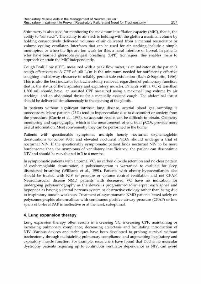
Historically, negative pressure body ventilators have been used to apply pressure to the body for ventilatory support, but this is less effective than NIV as they can cause obstructive apneas and are decreasingly efficacious with increasing age and decreasing pulmonary compliance (Bach et al, 1989). In figure 2, the intermittent abdominal pressure ventilator (IAPV) or “Exsufflation Belt” is a body ventilator that can augment tidal volumes from 300 to 1,200 mL. The intermittent inflation of the elastic air sac worn in a corset or belt under the patient’s clothing moves the diaphragm upward to assist in expiration. With bladder deflation, passive inspiration occurs with the diaphragm and abdominal contents returning to resting position with gravity. For effective use the trunk must be 30 degrees or more from the horizontal. Patients with inspiratory capacity or ability to glossopharyngeal breathe can add volumes of air to those mechanically taken in. Patients with less than one hour of breathing tolerance usually prefer it to NIV during daytime hours (Bach & Alba, 1991).
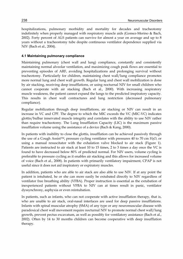
Inspiratory muscles can be assisted using NIV. There are no contraindications to long term use of NIV other than uncontrollable seizures. Multiple interfaces can be used and they can be introduced to the patients in a clinic or home setting. They include 15 mm angled mouth pieces, lipseals, and nasal and oral-nasal interfaces. Open NIV systems include mouthpiece or nasal NIV which rely on central nervous system reflexes to prevent insufflation air leakage during sleeping (Bach &Alba, 1990; Bach et al., 1955). A closed system, such as by using a lipseal-nasal prong system, can avoid excessive leakage by delivering air via mouth and nose during sleep (Figure 3). In addition to minimizing insufflation leakage, they maximize skin comfort. Using nocturnal NIV improves daytime O2 and CO2. Supplemental O2 and sedatives should be avoided to maintain maximal nocturnal NIV effectiveness.
Neuromuscular Disorders 240
Fig. 2. High level spinal cord injured patient with no measurable vital capacity or ventilator-free breathing ability using an intermittent abdominal pressure ventilator for daytime ventilatory assistance (Exsufflation BeltTM, Philips-Respironics International Inc., Murrysville, PA) and using a lipseal for nocturnal support for 15 years.
Fig. 3. Duchenne muscular dystrophy patient using an interface that includes nasal prongs with lip covering to provide a closed system of ventilatory support during sleep, here seen using it during surgery with general anesthesia.
Respiratory Muscle Aids in the Management of Neuromuscular Respiratory Impairment to Prevent Respiratory Failure and Need for Tracheostomy 241
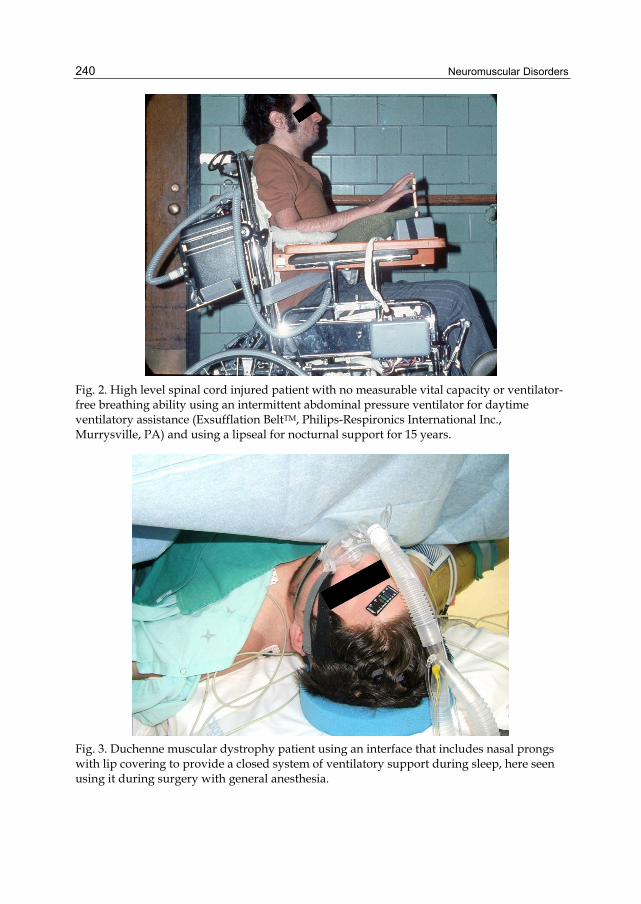
In patients with some neck movement and lip function, the 15 mm angled mouthpiece interface is the most useful, and is able to be used by some patients all day (Ishikawa, 2005). Often times patients can keep the mouthpiece near the mouth with a metal clamp attached to the wheelchair or affixed onto the motorized wheelchair controls, most commonly the sip and puff, chin or tongue controls. The patient can grab the mouthpiece for supplemental air or full breath support, as demonstrated in Figure 4.
The volume ventilator is set for large tidal volumes, commonly 800 to 1500 mL. Therefore, the patient can control the volume of air obtained and can use air stacking to cough, increase speech volumes, and maintain pulmonary compliance. To use mouthpiece NIV a patient must be able to move the soft palate posteriocranially to seal off the nasopharynx, open the glottis and vocal cords, and maintain hypopharynx and airway patency. These movements quickly become reflexive. They must and usually can be quickly relearned by patients who have been ventilated via tracheostomy and have lost them (Bach et al., 1993).
Fig. 4. A 66 year old woman with multiple sclerosis and continuous ventilator dependence using a 15 mm angled mouth piece for daytime support for 27 years despite having only 30 mL of vital capacity.
Neuromuscular Disorders 242
For those who are unable to grab or maintain a tight seal on a mouthpiece for daytime NIV, such as infants, nasal NIV using small nasal prong systems can be ideal and can be used continuously as a viable alternative to tracheostomy (Bach & Alba, 1990). To prevent oral insufflation leakage during nasal NIV, patients learn to close the mouth or seal the oropharynx with the soft palate and tongue. Humidifying the air is essential for nocturnal mouthpiece/lipseal ventilation but infrequently for nocturnal nasal NIV. Suboptimal humidification results in dry, irritated mucous membranes and increased airflow resistance to up to 8 cm H2O (Richards et al., 1996). The air can be warmed to body temperature and humidified using a hot water bath humidifier to decrease irritation of nasal membranes (Richards et al., 1996).
Complications from NIV are few. At times abdominal distension can occur sporadically. The air usually passes as flatus when the patient is mobilized in the morning. If severe, it can increase ventilator dependence and result in necessary placement of a gastrostomy or nasogastric tube to burp out the air or a rectal tube to decompress the colon. In 1000 NIV users there was noted to be one case of pneumothorax despite aggressive lung mobilization and expansion three times daily with NIV support for most and, indeed for over 50 years in many cases (Suri et al., 2008). Often mistakenly noted to be a complication or limiting aspect of NIV, secretion management remains the most important aspect of noninvasive management.
4.3 Cough Augmentation: Expiratory muscle assistance
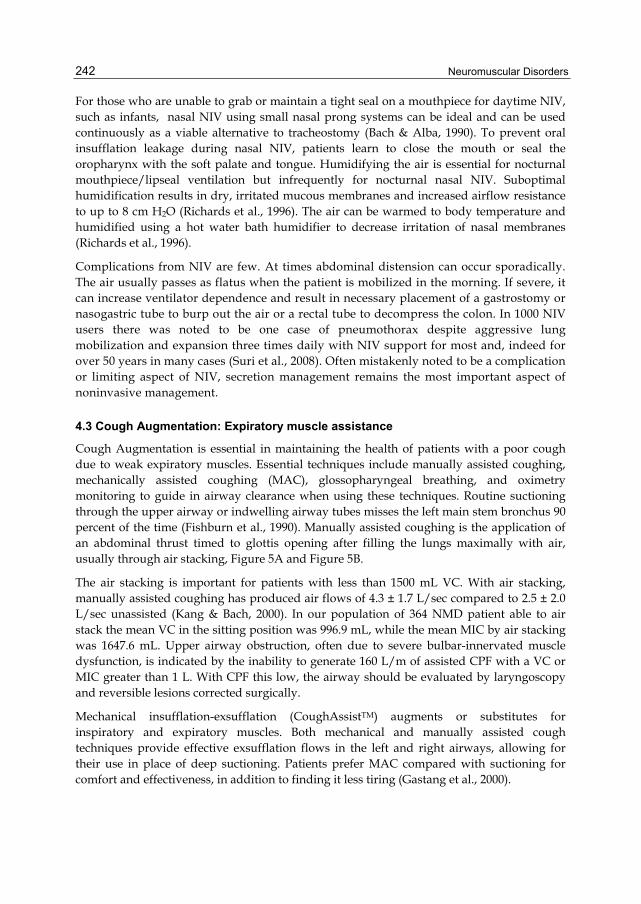
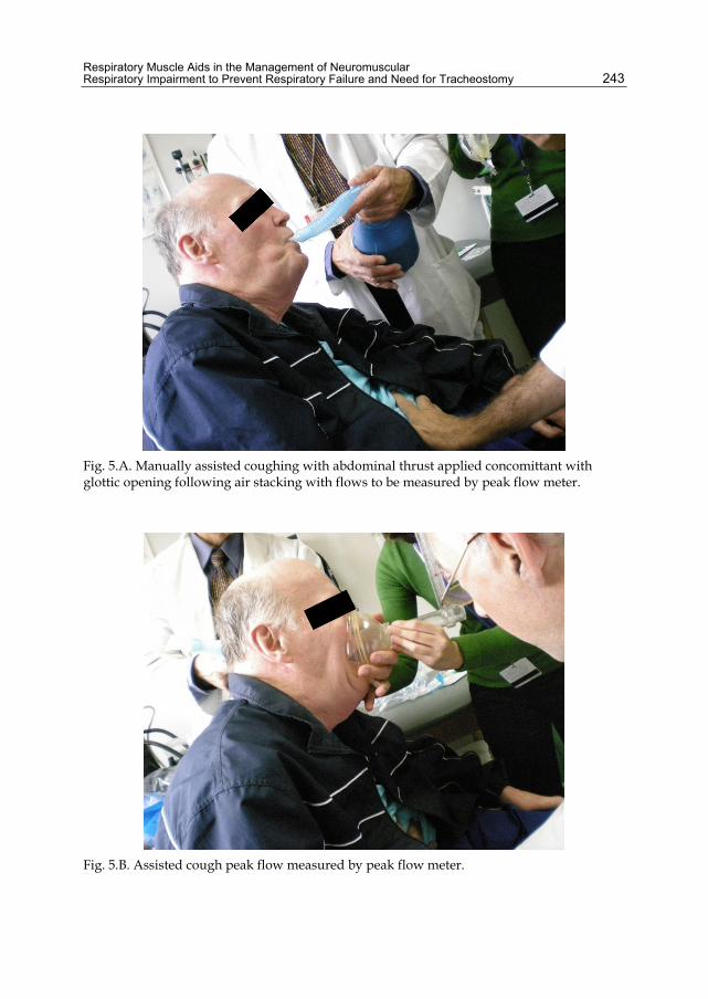
Cough Augmentation is essential in maintaining the health of patients with a poor cough due to weak expiratory muscles. Essential techniques include manually assisted coughing, mechanically assisted coughing (MAC), glossopharyngeal breathing, and oximetry monitoring to guide in airway clearance when using these techniques. Routine suctioning through the upper airway or indwelling airway tubes misses the left main stem bronchus 90 percent of the time (Fishburn et al., 1990). Manually assisted coughing is the application of an abdominal thrust timed to glottis opening after filling the lungs maximally with air, usually through air stacking, Figure 5A and Figure 5B.
The air stacking is important for patients with less than 1500 mL VC. With air stacking, manually assisted coughing has produced air flows of 4.3 ± 1.7 L/sec compared to 2.5 ± 2.0 L/sec unassisted (Kang & Bach, 2000). In our population of 364 NMD patient able to air stack the mean VC in the sitting position was 996.9 mL, while the mean MIC by air stacking was 1647.6 mL. Upper airway obstruction, often due to severe bulbar-innervated muscle dysfunction, is indicated by the inability to generate 160 L/m of assisted CPF with a VC or MIC greater than 1 L. With CPF this low, the airway should be evaluated by laryngoscopy and reversible lesions corrected surgically.
Mechanical insufflation-exsufflation (CoughAssistTM) augments or substitutes for inspiratory and expiratory muscles. Both mechanical and manually assisted cough techniques provide effective exsufflation flows in the left and right airways, allowing for their use in place of deep suctioning. Patients prefer MAC compared with suctioning for comfort and effectiveness, in addition to finding it less tiring (Gastang et al., 2000).
Respiratory Muscle Aids in the Management of Neuromuscular Respiratory Impairment to Prevent Respiratory Failure and Need for Tracheostomy 243
Fig. 5.A. Manually assisted coughing with abdominal thrust applied concomittant with glottic opening following air stacking with flows to be measured by peak flow meter.
Fig. 5.B. Assisted cough peak flow measured by peak flow meter.
Neuromuscular Disorders 244
4.3.1 Mechanically assisted coughing
Mechanically assisted coughing (MAC) combines mechanical insufflation-exsufflation with an exsufflation-timed abdominal thrust to increase CPF. The CoughAssistTM can be manually or automatically cycled with the deep insufflations followed immediately by deep exsufflations, ideally with pressures of 40 to 60 alternating with -40 to -60 cm H2O. Interfaces for MAC include oral-nasal masks, a simple mouthpiece, or translaryngeal or tracheostomy tubes. With use via a tracheostomy tube, the cuff, if present, should be inflated. Manual cycling allows caregiver-patient coordination during inspiration and expiration with the insufflation and exsufflation.
Treatments can be as frequent as up to every 30 minutes during respiratory infections and post-extubation. One treatment set includes about five cycles of MAC followed by a short period of normal breathing or ventilator use to avoid hyperventilation. When patients cannot cooperate with MAC, the timing of the insufflation and exsufflation is adjusted to the patient’s breathing to provide maximum chest expansion and rapid lung emptying, in general 2 to 4 seconds for adults but much more quickly for small children. Treatments continue until no further secretions are able to be removed and oxyhemoglobin desaturation due to mucus plugging is reversed. The abdominal thrust is also important for infants. By 2.5 to 5 years of age, children become accustomed to coughing simultaneously with the insufflation-exsufflation.
With the elimination of airway secretions and mucus using MAC, vital capacity, abnormal pulmonary flow rates and oxyhemoglobin saturation can improve immediately (Bach et al., 1993). Fifteen percent to 42 percent increase in VC was noted immediately following MAC for 67 patients with “obstructive dyspnea” and a 55 percent increase in VC was noted in patients with neuromuscular conditions (Barach, 1954). In ventilator assisted neuromuscular disease patients with chest infections, a 15 to 400 percent (200 to 800 mL) increase was noted following secretion removal with MAC (Bach, 1993).
Although MAC can augment and even substitute for the inspiratory and expiratory muscles, if bulbar innervated muscles, which prevent airway collapse and protect against food and saliva aspiration, are inadequately functioning, tracheotomy becomes indicated. However, this is generally only observed in advanced bulbar ALS patients. Patients with intact bulbar muscle function can usually air stack to volumes of 3 L or more, and, unless very scoliotic or obese, a properly delivered abdominal thrust can result in assisted CPF of 6 to 9 L/s, which is sufficient to clear secretions and prevent pneumonia and ARF without requiring MAC. Those with moderately impaired bulbar muscle function, e.g., non-ALS neuromuscular disease patients like those with Duchenne muscular dystrophy, that limits assisted CPF to less than 300 L/m benefit the most from MAC (Gomez-Merino, 2002).
4.3.2 Glossopharyngeal Breathing
Glossopharyngeal Breathing (GPB) is a technique that involves “gulping” boluses of air into the lungs using the glottis to add to the inspiratory effort. Six to 9 gulps (40 mL to 200 mL) total a full breath. The technique can be taught, to assist inspiratory and indirectly expiratory muscle function in patients with good bulbar-innervated musculature (Bach et al., 1987), allowing for discontinuation of ventilator use from minutes up to a whole day. Initial training can be supplemented with a training manual and videos (Dail et al., 1979;
Respiratory Muscle Aids in the Management of Neuromuscular Respiratory Impairment to Prevent Respiratory Failure and Need for Tracheostomy 245
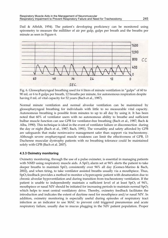
Dail & Affeldt, 1954). The patient’s developing proficiency can be monitored using spirometry to measure the milliliter of air per gulp, gulps per breath and the breaths per minute as seen in Figure 6.
Fig. 6. Glossopharyngeal breathing used for 6 liters of minute ventilation in “gulps” of 60 to 90 mL or 6 to 8 gulps per breath, 12 breaths per minute, for autonomous respiration despite having 0 mL of vital capacity for 52 years (Bach et al., 1987).
Normal minute ventilation and normal alveolar ventilation can be maintained by glossopharyngeal breathing for individuals with little to no measurable vital capacity. Autonomous breathing is possible from minutes to up to all day by using it. It has been noted that 60% of ventilator users with no autonomous ability to breathe and sufficient bulbar muscle function can use GPB for ventilator-free breathing (Bach et al., 1987; Bach & Alba, 1990). This technique is ideal in the event of ventilator failure or disconnection during the day or night (Bach et al., 1987; Bach, 1991). The versatility and safety afforded by GPB are safeguards that make noninvasive management safer than support via tracheostomy. Although severe oropharyngeal muscle weakness can limit the effectiveness of GPB, 13 Duchenne muscular dystrophy patients with no breathing tolerance could be maintained solely with GPB (Bach et al. 2007).
4.3.3 Oximetry monitoring
Oximetry monitoring, through the use of a pulse oximeter, is essential in managing patients with NMD using respiratory muscle aids. A SpO2 alarm set at 94% alerts the patient to take deeper breaths to maintain SpO2 consistently over 94% all day (Gomez-Marino & Bach, 2002), and when tiring, to take ventilator assisted breaths usually via a mouthpiece. Thus, SpO2 feedback provides a method to monitor a hypercapnic patient with desaturation due to chronic alveolar hypoventilation and during transition from tracheostomy ventilation. If the patient is unable to independently maintain a sufficient level of at least SpO2 of 94%, mouthpiece or nasal NIV should be initiated for increasing periods to maintain normal SpO2
which helps to reset central ventilatory drive. Thereby, oximetry feedback facilitates the introduction and indicates the extent of daytime need for mouthpiece and/or nasal NIV. In addition, oximetry monitoring is especially useful during episodes of respiratory tract infection as an indicator to use MAC to prevent cold triggered pneumonias and acute respiratory failure, usually due to mucus plugging in the NIV patient population. Proper
Neuromuscular Disorders 246
instruction of NIV and MAC, and rapid access to MAC during the onset of a chest cold may be all that is necessary to avert pneumonia, ARF and subsequent hospitalizations.
Especially in infants and small children, with often inadequate cough to prevent chest colds from triggering pneumonia and ARF, MAC should be used for any desaturation below 95 percent. In continuous NIV users, desaturations are usually due to bronchial mucus plugging, which can develop into atelectasis and pneumonia if the secretions are not quickly cleared.
5. Contraindications to noninvasive aids: Invasive ventilatory support
Certain patient populations are better managed with tracheostomy. Contraindications to noninvasive aid include ventilator dependence with depressed cognitive function, orthopedic conditions interfering with noninvasive interface use, restrictive pulmonary syndrome along with severe pulmonary disease necessitating high FiO2, or uncontrolled seizures or substance abuse (Waldhornet al., 1990). In addition, the presence of a nasogastric tube can hamper the fitting of a nasal interface and the use of mouthpiece or nasal NIV by limiting soft palate closure of the pharynx and the necessary seal at the nose. For neuromuscular disease patients, only those with severe bulbar dysfunction, as observed with severe bulbar ALS resulting in the inability for protect the airway, require tracheotomy (Bach et al., 2004). Other than for the occasional spinal muscular atrophy type 1 patient, tracheotomy is rarely if ever indicated for Duchenne muscular dystrophy or any other neuromuscular disease (Bach et al., 2009). Although tracheostomy ventilation can support alveolar ventilation and extend survival for many NMD patients (Bach, 1996), morbidity and mortality outcomes are not as favorable as by noninvasive approaches (Bach et al., 1998; Toussaint et al., 2006).
6. Long term outcomes
A number of centers have reported up to continuous NIV dependence to maintain patients with neuromuscular disease. One hundred and one of our nocturnal only NIV users became continuously NIV dependent for 7.6 ± 6.1 years to 30.1 ± 6.1 years of age with 56 patients still living. Twenty six became continuously dependent on NIV without requiring hospitalization, while eight continuous tracheostomy ventilation users were decanulated to noninvasive NIV. Using the above described techniques for the DMD patient population, we have extubated 31 “unweanable” intubated patients consecutively to NIV/MAC without resort to tracheotomy (Bach & Martinez, 2001). Also reported by Kohler et al., continuous NIV can prolong life in DMD. Seven of our DMD patients have lived to over 40 years of age requiring continuous NIV for 20 years or more.
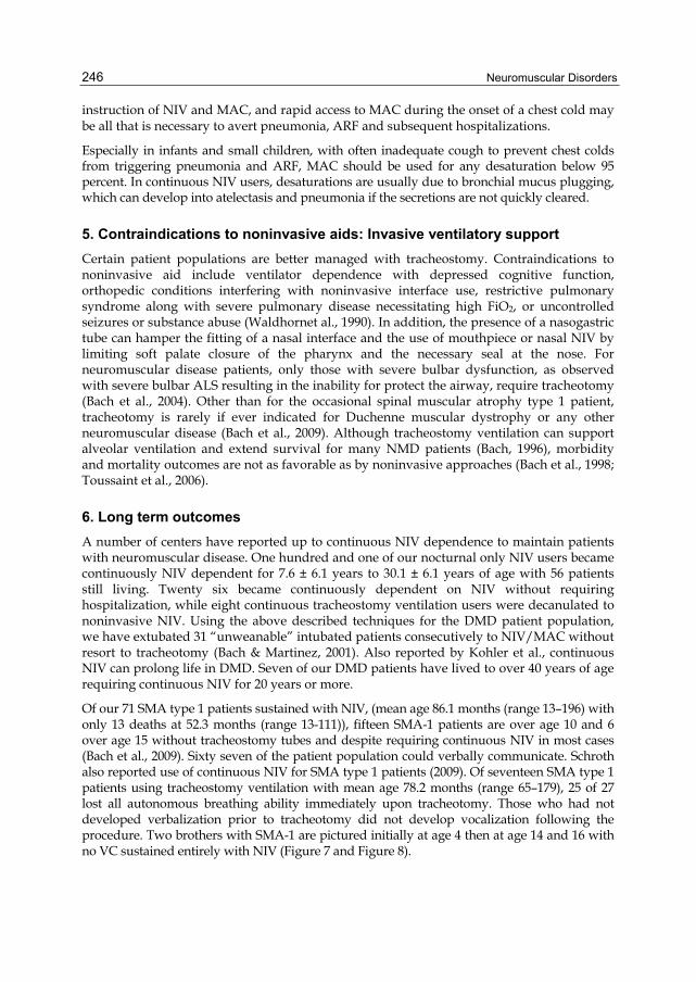
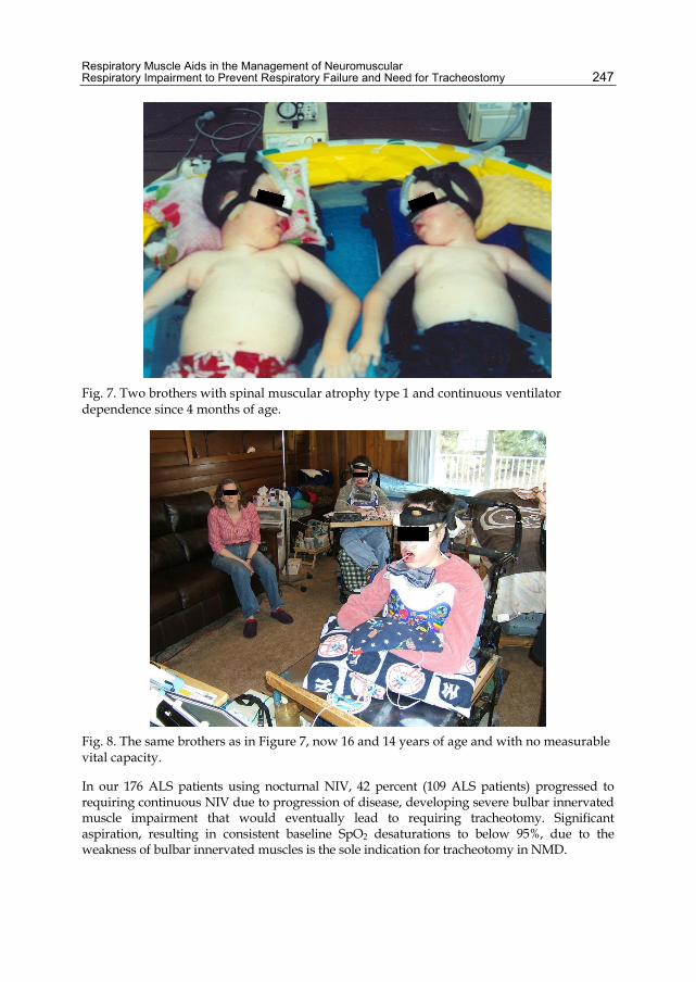
Of our 71 SMA type 1 patients sustained with NIV, (mean age 86.1 months (range 13–196) with only 13 deaths at 52.3 months (range 13-111)), fifteen SMA-1 patients are over age 10 and 6 over age 15 without tracheostomy tubes and despite requiring continuous NIV in most cases (Bach et al., 2009). Sixty seven of the patient population could verbally communicate. Schroth also reported use of continuous NIV for SMA type 1 patients (2009). Of seventeen SMA type 1 patients using tracheostomy ventilation with mean age 78.2 months (range 65–179), 25 of 27 lost all autonomous breathing ability immediately upon tracheotomy. Those who had not developed verbalization prior to tracheotomy did not develop vocalization following the procedure. Two brothers with SMA-1 are pictured initially at age 4 then at age 14 and 16 with no VC sustained entirely with NIV (Figure 7 and Figure 8).
Respiratory Muscle Aids in the Management of Neuromuscular Respiratory Impairment to Prevent Respiratory Failure and Need for Tracheostomy 247
Fig. 7. Two brothers with spinal muscular atrophy type 1 and continuous ventilator dependence since 4 months of age.
Fig. 8. The same brothers as in Figure 7, now 16 and 14 years of age and with no measurable vital capacity.
In our 176 ALS patients using nocturnal NIV, 42 percent (109 ALS patients) progressed to requiring continuous NIV due to progression of disease, developing severe bulbar innervated muscle impairment that would eventually lead to requiring tracheotomy. Significant aspiration, resulting in consistent baseline SpO2 desaturations to below 95%, due to the weakness of bulbar innervated muscles is the sole indication for tracheotomy in NMD.
Neuromuscular Disorders 248
7. Extubation of “unweanable” patient
Using new NMD specific extubation criteria and protocol, including MAC and pulse oximetry monitoring, “unweanable” patients with DMD, SMA, ALS, and other neuromuscular diseases, e.g., SCI and polio, were successfully extubated to NIV (Bach, 2010).
Extubation Criteria for Unweanable Ventilator Dependent Patients
1. Afebrile and normal white blood cell count
2. PaCO2 40 mm Hg or less at peak inspiratory pressures less than 30 cm H2O on full ventilatory support and normal breathing rate, as needed
3. Oxyhemoglobin saturation (SpO2) ≥ 95% for 12 hours or more in ambient air 4. All oxyhemoglobin desaturations below 95% reversed by mechanically assisted
coughing and suctioning via translaryngeal tube
5. Fully alert and cooperative, receiving no sedative medications
6. Chest radiograph abnormalities cleared or clearing 7. Air leakage via upper airway sufficient for vocalization upon cuff deflation
Table 1. Adapted from Bach, J.R. (2010). Extubation of Patients With Neuromuscular Weakness. Chest, 137( 5), 1033-1039
The extubation criteria and protocol have been developed for the neuromuscular disease specific patient population. Instead of spontaneous breathing trials which patients typically undergo prior to extubation attempts, once a NMD patient meets the criteria cited in Table 1, he or she can be directly extubated to nasal NIV, assist control 800 to 1500 mL and rate 10-14 breaths/minute in ambient air, with aggressive MAC. Ideally the orogastric or nasogastic tube should be removed to facilitate proper fitting of the NIV interface which can be nasal, oro-nasal and/or mouthpiece interfaces.
As the patient receives full volume support via NIV, the assisted CPF, or CPF obtained by abdominal thrust following air stacking, is measured within 3 hours of extubation. Patients with sufficient neck movement and lip function used the 15 mm angled mouthpiece and weaned themselves as tolerated by taking fewer and fewer positive pressure ventilations. For those unable to effectively use the 15 mm mouth piece, diurnal nasal NIV was used via nasal prongs, with a nasal or oronasal interface used for nocturnal ventilation. Patients were educated and trained in air stacking and manually assisted coughing and assisted and unassisted CPF were measured.
For SpO2<95%, ventilator positive inspiratory pressure (PIP), interface or tubing air leakage, CO2 retention, ventilator settings, and MAC were considered. Therapists, nurses, and especially family and personal care attendants were trained and provided with a CoughAssistTM to use MAC via oro-nasal interfaces up to every 30 min until airway secretions cleared and SpO2 could be maintained consistently above 94 percent. Open gastrostomies were performed under local anesthesia using NIV without complication in 7 patients with unsafe post-extubation oral intake.
Respiratory Muscle Aids in the Management of Neuromuscular Respiratory Impairment to Prevent Respiratory Failure and Need for Tracheostomy 249
One hundred and fifty seven consecutive “unweanable” patients were treated including 25 with SMA, 20 with DMD, 16 with ALS, 17 with spinal cord injury, 11 with postpolio syndrome, and 68 with other NMD. Eighty three of these were transferred from other hospitals after refusing tracheostomy after inability to pass spontaneous breathing trials. They were successfully extubated to NIV and MAC despite being unable to pass spontaneous breathing trials before or after extubation. Not requiring re-intubation during the hospitalization defined extubation success. Prior to hospitalization 96 (61%) patients had no experience with NIV, 41 (26%) used it part-time, and 20 (13%) were continuously NIV dependent.
There was an extubation success rate of 95% (149 patients) on first attempt. On patients with assisted CPF ≥ 160 L/m, all 98 extubations were successful. Six of 8 patients who had assisted CPF less than 160 L/m initially failed extubation but succeeded on subsequent attempts (Bach et al, 2010). Only two bulbar ALS patients with no measurable assisted CPF underwent tracheotomy (Bach et al., 2010). Multiple centers now routinely extubate DMD patients to NIV directly to avoid tracheotomy.
8. “Unweanable” patient decannulation
In 1996 we initially reported the decannulation of 50 unweanable patients with neuromuscular weakness (Bach & Saporito, 1996). Any ventilator dependent patient with sufficient bulbar-innervated musculature to prevent significant secretion aspiration is a candidate for decannulation to NIV. This is ideal as decanulation facilitates speech and swallowing. The basic principles for decannulation are essentially identical to those for extubation. Patients with tracheostomy tubes and with no VFBA, possessing VC of 250 mL or greater developed VFBA subsequent to decannulation. Within 3 weeks of decannulation, most weaned to only nocturnal NIV.
9. Conclusion
The ability to decannulate and maintain survival of neuromuscular disease patients using NIV in place of tracheostomies is indeed possible, and in fact preferable regarding aspects of convenience, speech, swallowing, cosmesis, comfort, safety, and is preferred overall (Bach, 1993). The extubation criteria set forth is a simple evaluation of patients with NMD (ventilatory muscle weakness) compared with the extensive “ventilator weaning parameters” used as criteria along with spontaneous breathing trials considered for the extubation of patients with intrinsic/obstructive lung diseases before extubating them.
To maintain a sufficient SpO2 of greater than 94 percent, inspiratory and expiratory muscle aid is used to prevent and manage desaturations and hypercapnia instead of supplemental oxygen. “Unweanable” patients, with sufficient glottic function to prevent significant aspiration of secretions, can be extubated to NIV and maintained on NIV for many years, averting tracheostomy. In the NMD patient population, with primarily ventilatory muscle weakness rather than intrinsic lung disease, this alternative paradigm provides optimal management and quality of life.
Neuromuscular Disorders 250
10. References
Bach, J.R. (1991). New approaches in the rehabilitation of the traumatic high level quadriplegic. Am J Phys Med Rehabil, 70:13-20
Bach, J.R. (1993). A comparison of long-term ventilatory support alternatives from the perspective of the patient and care giver. Chest, 104:1702-1706
Bach, J.R. (1993). Mechanical insufflation-exsufflation: comparison of peak expiratory flows with manually assisted and unassisted coughing techniques. Chest, 104:1553-1562
Bach, J.R. (1996). Conventional approaches to managing neuromuscular ventilatory failure. In: Pulmonary rehabilitation: the obstructive and paralytic conditions. Bach JR, ed. Philadelphia: Hanley & Belfus, 285-301
Bach, J.R. (2004). Management of patients with neuromuscular disease. Philadelphia: Hanley & Belfus
Bach, J.R. & Alba, A.S. (1990). Management of chronic alveolar hypoventilation by nasal ventilation. Chest, 97:52-57
Bach, J.R. & Alba, A.S. (1991). Intermittent abdominal pressure ventilator in a regimen of noninvasive ventilatory support. Chest, 99:630-636
Bach, J.R. & Alba, A.S. (1990). Noninvasive options for ventilatory support of the traumatic high level quadriplegic. Chest, 98:613-619
Bach, J.R., Alba, A.S., Bodofsky, E., Curran, FJ & Schultheiss, M. (1987). Glossopharyngeal breathing and noninvasive aids in the management of post-polio respiratory insufficiency. Birth Defects, 23:99-113
Bach, J.R., Alba, A.S. & Saporito, L.R. (1993). Intermittent positive pressure ventilation via the mouth as an alternative to tracheostomy for 257 ventilator users. Chest, 103:174-182
Bach, J.R., Alba, A.S. & Shin, D. (1989). Management alternatives for post-polio respiratory insufficiency: assisted ventilation by nasal or oral-nasal interface. Am J Phys Med Rehabil, 68:264-271
Bach, J.R., Baird, J.S., Plosky, D., Navado, J & Weaver, B. (2002). Spinal muscular atrophy type 1: management and outcomes. Pediatr Pulmonol, 34:16-22
Bach, J.R., Bianchi, C. & Aufiero, E. (2004). Oximetry and indications for tracheotomy in amyotrophic lateral sclerosis. Chest, 126:1502-07
Bach, J.R., Bianchi, C., Finder. J., Fragasso, T., Goncalves, M.R., Ishikawa, Y., Ramlall, A.K., McKim, D., Servera, E., Vianello, A., Villanova, M. & Winck, J.C. (2007). Tracheostomy tubes are not needed for Duchenne muscular dystrophy. Eur Respir J., 30:179-180
Bach, J.R., Bianchi, C., Vidigal-Lopes, M., Turi, S. & Felisari, G. (2007). Lung inflation by glossopharyngeal breathing and “air stacking” in Duchenne muscular dystrophy. Am J Phys Med Rehabil, 86:295-300
Bach, J.R., Gonçalves, M.R., Hamdani, I. & Winck, J.C. (2010). Extubation of unweanable patients with neuromuscular weakness: a new management paradigm. Chest, 137:1033-1039
Bach, J.R., Gupta, K., Reyna, M. & Hon, A. (2009). Spinal muscular atrophy type 1: prolongation of survival by noninvasive respiratory aids. Pediatric Asthma, Allergy & Immunology, 22:151-162
Respiratory Muscle Aids in the Management of Neuromuscular Respiratory Impairment to Prevent Respiratory Failure and Need for Tracheostomy 251
Bach, J.R. & Kang, S.W. (2000). Disorders of ventilation: weakness, stiffness, and mobilization. Chest, 117:301-303
Bach, J.R., Mahajan, K., Lipa, B., Saporito, L. & Komaroff, E. (2008). Lung insufflation capacity in neuromuscular disease. Am J Phys Med Rehabil, 87:720-725
Bach, J.R. & Martinez, D. (2011). Duchenne muscular dystrophy: continuous noninvasive ventilatory support prolongs survival. Respir Care, 56:744-750
Bach, J.R., Rajaraman, R., Ballanger, F., Tzeng, A.C., Ishikawa, Y., Kulessa, R. & Bansal, T. (1998). Neuromuscular ventilatory insufficiency: the effect of home mechanical ventilator use vs. oxygen therapy on pneumonia and hospitalization rates. Am J Phys Med Rehabil, 77:8-19
Bach, J.R., Robert, D., Leger, P. & Langevin, B. (1995). Sleep fragmentation in kyphoscoliotic individuals with alveolar hypoventilation treated by nasal IPPV. Chest, 107:1552-1558
Bach, J.R. & Saporito, L.R. (1996). Criteria for extubation and tracheostomy tube removal for patients with ventilatory failure. A different approach to weaning. Chest, 110:1566-1571
Bach, J.R., Smith, W.H., Michaels, J., Saporito, L., Alba, A.S., Dayal, R. & Pan, J. (1993). Airway secretion clearance by mechanical exsufflation for post-poliomyelitis ventilator assisted individuals. Arch Phys Med Rehabil, 74:170-177
Barach, A.L. & Beck, G.J. (1954). Exsufflation with negative pressure: physiologic and clinical studies in poliomyelitis, bronchial asthma, pulmonary emphysema and bronchiectasis. Arch Intern Med, 93:825-841
Currie, D.C., Munro, C., Gaskell, D. & Cole PJ. (1986). Practice, problems and compliance with postural drainage: a survey of chronic sputum producers. Br J Dis Chest, 80:249-253
Dail, C.W. & Affeldt, J.E. Glossopharyngeal breathing [video]. Los Angeles: Department of Visual Education, College of Medical Evangelists, 1954
Dail, C., Rodgers, M., Guess, V., et al. Glossopharyngeal breathing. Downey, CA: Rancho Los Amigos Department of Physical Therapy, 1979
Fishburn, M.J., Marino, R.J., Ditunno, J.F. Jr. (1990). Atelectasis and pneumonia in acute spinal cord injury. Arch Phys Med Rehabil, 71:197-200
Garstang, S.V., Kirshblum, S.C. & Wood, K.E. (2000). Patient preference for in-exsufflation for secretion management with spinal cord injury. J Spinal Cord Med, 23:80-85
Gomez-Merino, E. & Bach, J.R. (2002). Duchenne muscular dystrophy: prolongation of life by noninvasive respiratory muscle aids. Am J Phys Med Rehabil, 81:411-415
Ishikawa, Y. (2005). Manual for the care of patients using noninvasive ventilation. Japan Planning Center, Matsudo, Japan
Kang, S.W. & Bach, J.R. (2000). Maximum insufflation capacity. Chest, 118:61-65 Kohler, M., Clarenbach, C.F., Böni, L., Brack, T., Russi, E.W., Bloch, K.E. (2005). Quality of
life, physical disability, and respiratory impairment in Duchenne muscular dystrophy. Am J Respir Crit Care Med, 172:1032-1036
McKim, D.A. & LeBlanc, C. (2006). Maintaining an "oral tradition": specific equipment requirements for mouthpiece ventilation instead of tracheostomy for neuromuscular disease. Respiratory Care, 51:297-298
Neuromuscular Disorders 252
Richards, G.N., Cistulli, P.A., Gunnar Ungar, R., Berthon-Jones, M., Sullivan, C.E. (1996). Mouth leak with nasal continuous positive airway pressure increases nasal airway resistance. Am Respir Crit Care Med, 154:182-186
Schroth, M.K. (2009). Special considerations in the respiratory management of spinal muscular atrophy. Pediatrics, 123:S245-S249
Suri, P., Burns, S.P., Bach, J.R. (2008). Pneumothorax associated with mechanical insufflation-exsufflation and related factors. Am J Phys Med Rehabil, 87:(11)951-955
Toussaint, M., Steens, M., Wasteels, G. & Soudon, P. (2006). Diurnal ventilation via mouthpiece: survival in end-stage Duchenne patients. Eur Respir J, 28:549-555
Waldhorn, R.E., Herrick, T.W., Nguyen, M.C., O'Donnell, A.E., Sodero, J. & Potolicchio, S.J. (1990). Long-term compliance with nasal continuous positive airway pressure therapy of obstructive sleep apnea. Chest, 97:33-38
Webber, B. & Higgens, J. (1999). Glossopharyngeal breathing: what, when and how? [video] Aslan Studios Ltd., Holbrook, Horsham, West Sussex, England
Williams, A.J., Yu, G., Santiago, S., Stein, M. (1991). Screening for sleep apnea using pulse oximetry and a clinical score. Chest, 100:631-635