Embed Size (px)
Citation preview

Resolving Hot Spots in the C-Terminal DimerizationDomain that Determine the Stability of the MolecularChaperone Hsp90Emanuele Ciglia1., Janina Vergin2., Sven Reimann3, Sander H. J. Smits3, Lutz Schmitt3, Georg Groth2,
Holger Gohlke1*
1 Institute for Pharmaceutical and Medicinal Chemistry, Heinrich-Heine-University, Dusseldorf, Germany, 2 Institute for Biochemical Plant Physiology, Heinrich-Heine-
University, Dusseldorf, Germany, 3 Institute of Biochemistry, Heinrich-Heine-University, Dusseldorf, Germany
Abstract
Human heat shock protein of 90 kDa (hHsp90) is a homodimer that has an essential role in facilitating malignanttransformation at the molecular level. Inhibiting hHsp90 function is a validated approach for treating different types oftumors. Inhibiting the dimerization of hHsp90 via its C-terminal domain (CTD) should provide a novel way to therapeuticallyinterfere with hHsp90 function. Here, we predicted hot spot residues that cluster in the CTD dimerization interface by astructural decomposition of the effective energy of binding computed by the MM-GBSA approach and confirmed thesepredictions using in silico alanine scanning with DrugScorePPI. Mutation of these residues to alanine caused a significantdecrease in the melting temperature according to differential scanning fluorimetry experiments, indicating a reducedstability of the mutant hHsp90 complexes. Size exclusion chromatography and multi-angle light scattering studiesdemonstrate that the reduced stability of the mutant hHsp90 correlates with a lower complex stoichiometry due to thedisruption of the dimerization interface. These results suggest that the identified hot spot residues can be used as apharmacophoric template for identifying and designing small-molecule inhibitors of hHsp90 dimerization.
Citation: Ciglia E, Vergin J, Reimann S, Smits SHJ, Schmitt L, et al. (2014) Resolving Hot Spots in the C-Terminal Dimerization Domain that Determine the Stabilityof the Molecular Chaperone Hsp90. PLoS ONE 9(4): e96031. doi:10.1371/journal.pone.0096031
Editor: Suzannah Rutherford, Fred Hutchinson Cancer Research Center, United States of America
Received August 13, 2013; Accepted April 2, 2014; Published April 23, 2014
Copyright: � 2014 Ciglia et al. This is an open-access article distributed under the terms of the Creative Commons Attribution License, which permitsunrestricted use, distribution, and reproduction in any medium, provided the original author and source are credited.
Funding: This work was supported by funds from the Strategischer Forschungsfonds of the Heinrich-Heine-University, Dusseldorf. The funders had no role instudy design, data collection and analysis, decision to publish, or preparation of the manuscript.
Competing Interests: The authors have declared that no competing interests exist.
* E-mail: [email protected]
. These authors contributed equally to this work.
Introduction
Protein-protein complexes have gained increasing attention in
structural biology and drug discovery due to their ubiquitous
participation in fundamental cellular processes. As such, protein-
protein interactions (PPIs) are involved in a variety of physiological
regulatory mechanisms, e.g., signaling, cellular growth, and
apoptosis [1,2]. PPIs also play an important role in pathophysi-
ology [3,4] such that modulating PPIs is considered a valuable
approach for treating diseases [2,3,5–7]. Targeting PPIs is
considered difficult, however, because of the size, lack of deep
binding pockets, and stability of PPIs. Yet, protein-protein
interfaces have been shown to be energetically non-homogeneous
in that only a few ‘‘hot spot’’ residues account for most of the
binding affinity [8–10]. Accordingly, PPI modulators often target
only the functional epitope that contains these hot spots [11–13].
Thus, identifying such hot spots provides important insights into
the energetics of PPIs, which can be exploited for the identification
of PPI modulators [12].
Here, we aim at resolving hot spots in the C-terminal
dimerization domain of the human heat shock protein of
90 kDa (hHsp90). Hsp90 is a molecular chaperone that belongs
to a highly conserved family of proteins that are central to a
number of cellular functions, including protein (re)folding,
stabilization, and quality control [14–16]. Despite its high basal
expression in eukaryotes and prokaryotes [17,18], Hsp90 remains
mostly in a latent state under physiological conditions. In response
to environmental stress, the cellular activity of Hsp90 (along with
other heat shock proteins) is increased in order to protect the
exposed cell [16,19]. Recent data has also demonstrated essential
roles for chaperones in facilitating malignant transformation at the
molecular level: the chaperone allows tumor cells to tolerate
mutations in multiple critical signaling molecules that would
otherwise be lethal [20,21]. Accordingly, many studies have
validated Hsp90 inhibition as an approach for treating different
types of tumors [14,22–26].
Regarding its structure, Hsp90 is a flexible homodimeric
protein; each monomer consists of three major domains: an
amino terminal domain (NTD), a middle domain (M), and a
carboxy terminal domain (CTD) [17,27] (Figure 1, A). The NTD
contains a nucleotide binding pocket, responsible for Hsp90’s
ATPase activity, which is coupled to the chaperone activity
[28,29]. This pocket is the binding site of most of the known
Hsp90 inhibitors [30,31]. The M domain is the major interaction
site for Hsp90 clients, and bridges NTD and CTD [28]. In
addition to being involved in regulating ATPase activity and co-
chaperone recruitment, the CTD is responsible for Hsp90
dimerization [18,32]. The dimerization interface is formed by
PLOS ONE | www.plosone.org 1 April 2014 | Volume 9 | Issue 4 | e96031

two pairs of helices creating a characteristic four helix bundle
[17,33]. Recent results showed that the C-terminal dimer opens
and closes with fast kinetics [34] in contrast to previous
assumptions that the C-terminal interface is permanently dimer-
ized [17]. These findings led us to hypothesize that inhibiting the
C-terminal dimerization will be a viable way to interfere with
Hsp90 activity. Although some Hsp90 inhibitors have been
described that act on the CTD [35,36] to the best of our
knowledge none of these targets the dimerization interface.
In order to identify hot spots as a first step to define the
functional epitope in the dimerization interface, we conducted a
combined computational and experimental study. First, we
predicted potential hot spot candidates by two independent
computational approaches, MM-GB/SA [37] and DrugScorePPI
[38,39], using a homology model of the human C-terminal Hsp90
domain. A subset of these was mutated to alanine, and the stability
of wild type and mutant proteins was evaluated by a Thermofluor
assay [40], size exclusion chromatography (SEC), and multi-angle
light scattering (MALS). Our findings provide insights into the
energetics of CTD dimerization in Hsp90, which are valuable for
pursuing a novel approach that aims at therapeutically interfering
with Hsp90 activity.
Results
Homology modeling and molecular dynamicssimulations
When starting this study, neither a crystal structure of the
human full length Hsp90 (hHsp90) nor of its CTD was available,
which would be required for any later structure-based endeavor to
identify PPI modulators. Thus, we set out to generate a model of
the hHsp90 CTD by comparative modeling with MODELLER
[41] using crystal structures from S. cerevisiae and E. coli as
templates. The sequence identity (similarity) between the target
sequence and the template sequences is sufficiently high (S.
cerevisiae: 54% (74%); E. coli: 25% (43%); (Figure S1 in File S1).
The obtained model is of good structural quality as assessed with
the PROCHECK software [42] (Figure S2 in File S1). Recently, a
crystal structure of an M-CTD construct of hHsp90 has been
reported (PDB code: 3Q6M) [43]. Our model and the crystal
structure show very good structural agreement as demonstrated by
a root mean square deviation (RMSD) of all Ca atoms of ,0.8 A
(Figure 1, B). This value decreases to ,0.7 A when the Ca atoms
of only the amino acids located in the four helix bundle (helices
H4, H49 and H5, H59) are taken into account (Figure 1, C). The
orientation of the side chains in the dimerization interface agrees
almost perfectly between the model and the crystal structure
(Figure 1, C) such that the results of the hot spot prediction (see
below) should not depend on whether the prediction is based on
one or the other structure.
The homology model was subjected to molecular dynamics
simulations (MD) of 100 ns length in explicit water to generate a
conformational ensemble for the subsequent hot spots detection.
The CTD dimer remains stable during the simulation time: the
RMSD of the single domains is ,6.5 A, and the dimer shows
structural deviations of ,8 A (Figure S3, A in File S1). Relevant
conformational changes are only observed in the region of helices
H2 and H29 (Figure 1, B) and account for most of the structural
deviations observed. As such, not taking into account H2 and H29,
the RMSD of the single domains and the dimer drops to ,3 A
(Figure S3, B in File S1). This is in agreement with previous
experimental and computational findings according to which the
mobility of H2 and H29 is required for forming interactions
between the CTD and substrates and, hence, Hsp90 function
[33,44].
Hot spot predictionIn order to identify amino acids at the CTD interface that are
crucial for dimer stability, we performed MM-GB/SA calculations
combined with a decomposition of the effective energy (i.e., the
sum of gas phase and solvation free energy) of dimerization on a
per residue level. The approach mimics computational alanine
scanning and has been applied successfully by us in retro- and
prospective studies on the determinants of protein-protein
interactions [10–12,45]. The results reveal a distinctive interaction
profile, which is almost identical for the two monomers (Figure 2,
A). Residues are identified as hot spots if their contribution to the
effective energy of dimerization DG,22 kcal mol21 [46].
The hot spots are spatially clustered and are located on H4 and
H5, except for a single hot spot on H3. The main cluster is formed
by residues I688, Y689, I692, and L696 located on H5 at the inner
side of the four helix bundle (Figure 2, B). I692 and L696 form
Figure 1. Homology model. (A) Surface representation of the full length S. cerevisiae Hsp90 (PDB code 2CG9), showing the three different proteindomains (N-terminal domain: orange, middle domain: green, C-terminal domain: blue). (B) Homology model of hHsp90 C-terminal domain (blue)overlaid with a crystal structure (PDB code 3Q6M) of the same domain (red) (C) Blow-up of the overlay highlighting the side chain orientation ofresidues located at the interface of helices H5 and H49.doi:10.1371/journal.pone.0096031.g001
Hot Spots in the C-Terminal Domain of Human Hsp90
PLOS ONE | www.plosone.org 2 April 2014 | Volume 9 | Issue 4 | e96031

hydrophobic contacts with L6629, L6659, and L6669 located on
H49; Y689 establishes hydrophobic interactions with L6659 and
L6669 (Figure 2, B) but also forms hydrogen bonds with S6739 and
T6699 on H49 and H6409 on the loop located above the N-
terminal end of H3. The multiple hydrogen bond formation is
possible because the side chain of Y689 adopts two conformations
during the MD simulations, one where the aromatic ring points in
the direction of H39 and one where it points to H49, i.e., the
interior of the interface. In the latter case, an indentation in the
binding epitope of H49 accommodates the side chain. L672 and
T669 form a second, smaller cluster on H4. The latter residue is
involved in interactions with Y6899 (see above); L672 interacts
with P681 on H59. Finally, I642 on H3 is located in a peripheral
position with respect to the interface but forms hydrophobic
contacts with the C-terminal end of H5, that way apparently
contributing to the stabilization of this secondary structure
element.
As the crystal structure of an M-CTD construct of hHsp90
became available only recently (PDB code: 3Q6M) [43], we
repeated the hot spot prediction for a CTD dimer of that
structure, using the same settings for these computations and the
prior MD simulations as in the case of the homology model. The
resulting interaction profile (Figure S4 in File S1) is in very good
agreement with the one of the homology model (Figure 2, A) such
that all of the above mentioned hot spots are identified again.
While this may have been expected from the high structural
similarity between our model and the crystal structure (see above),
these findings validate, in an indirect manner, the quality of our
homology model and demonstrate the robustness of our MM-GB/
SA-based hot spot predictions. In order to independently confirm
the MM-GB/SA calculations, we performed in silico alanine
scanning on the homology model with the DrugScorePPI web
server developed by us [38,39]. The interaction profile obtained is
in good agreement with the above findings, pointing to essentially
the same hot spots that are crucial for hHsp90 CTD dimerization
(Table S1 in File S1). We also tested if the above hot spots could
have been identified by a simpler computational approach given
that these hot spots are largely buried upon complex formation.
For this, we computed the residue-wise relative change in the
solvent-accessible surface area upon complex formation (using the
SA values of the MM-GB/SA calculations starting from the CTD
dimer of the crystal structure; Figure S5 in File S1). This suggests
Figure 2. Hot spot and cold spot prediction. (A) Contribution to the dimer stabilization of each amino acid within the hHsp90 CTD. DG valuesare calculated by the MM-GB/SA approach [37,83] starting from the homology model, employing a structural decomposition of the effective energy[10]. The standard error in the mean is ,0.1 kcal mol21 in all cases. Amino acids contributing to the dimerization with DG,22 kcal mol21 areconsidered hot spots and are indicated in the graphic by red dots. In addition two ‘‘cold spots’’ mentioned in the text are marked with blue dots. Inthe upper part of the panel, the secondary structure of the CTD is shown. The amino acids are numbered according to the full length hHsp90 aisoform (UniProt code: P07900). (B) Hot spot residues localized on H5 (red) and interacting residues on H49 (green). (C) Helical wheel representationshowing the position of hot spots (red) and cold spots (blue) on helices H5 and H49.doi:10.1371/journal.pone.0096031.g002
Hot Spots in the C-Terminal Domain of Human Hsp90
PLOS ONE | www.plosone.org 3 April 2014 | Volume 9 | Issue 4 | e96031

essentially all residues in the dimer interface as hot spots,
indicating a pronounced loss of specificity in the predictions
compared to when additional energy contributions are considered.
In turn, L696, which is more peripheral to the dimer interface, is
not found among the top candidates anymore when using the
surface area-based approach. In our view, these findings
demonstrate the predictive value of the energy-based methods.
Finally, we selected residues S658 and Q682 as ‘‘cold spots’’
(DG = 0.06 and 20.67 kcal mol21, respectively, as calculated with
the MM-GB/SA approach starting from the homology model;
Figure 2, A and C), which will serve as negative controls in the
subsequent experiments. These amino acids are located at the
CTD interface between H5 and H49, but they are predicted to be
only marginally important for the dimerization (Figure 2, A and
C). Consequently, mutating these ‘‘cold spots’’ to alanine should
not impact the CTD stability.
Analysis of hHsp90 stability by Thermofluor assayThe stability of the CTD of hHsp90 wild type, cold spot and hot
spot alanine mutants was analyzed by differential scanning
fluorimetry (Table 1, Table 2) [40]. In this assay thermally-
induced protein unfolding is monitored by the binding of the
fluorescent dye SYPRO orange [47] to the hydrophobic core of
the protein that becomes exposed upon unfolding, and the related
increase in fluorescence emission. The temperature at the
midpoint of the unfolding transition is defined as melting
temperature (Tm) of the protein [48]. A shift in Tm of a protein
in its native state, or in site-specific or chemically modified forms,
indicates a change in the stability of the protein [49]. In order to
identify conditions under which the native state is most stable we
analyzed the thermal unfolding of the purified wild type hHsp90
CTD in the pH range from 3–10. These studies (Figure 3, A)
showed that the CTD of hHsp90 is most stable at mild neutral
conditions (pH 7.5). Thus, screening of all hot spot and cold spot
variants of hHsp90 was done at this pH condition.
The wild type form of hHsp90 is characterized by a melting
temperature of 73uC (Figure 3, B). In contrast, hot spot
substitution mutants carrying alanines at positions 689, 692 and
696 (CTDY689A/I692A/L696A, Figure 3, D) or at positions 688, 689
and 692 (CTDI688A/Y689A/I692A, Figure 3, E) show a significant
decrease (DTm$13uC) in their melting temperatures indicating a
substantial loss in the stability due to the substitution of the native
hHsp90 residues in these positions by the small, non-polar alanine.
These triple mutations were chosen, respectively, out of the four
hot spots of the main cluster because they result in patterns of
sequence localization that could be mimicked by non-peptidic a-
helix mimetics (see Discussion for further details).
Additionally, alanine single mutants were analyzed at the same
conditions to reveal the potential contribution of individual
positions at the dimerization interface on the stability of the
CTD dimer. To this end we substituted the predicted hot spots at
positions I688, Y689, I692 and L696 individually to alanines
(Table S2, Table S3 in File S1) and determined the melting
temperature and protein stability of the single mutants. Compared
to the alanine triple mutants, all of the single mutants showed a
much lower reduction (DTm,8uC) in their melting temperature
with respect to the wild type of hHsp90 (Figure S6, Table S4 in
File S1). Alanine substitutions at positions S658 and Q682
identified as cold spots (Figure 2, A–C) had no effect on the
stability of the protein. The related mutant (Figure 3, C) showed a
melting temperature corresponding to the hHsp90 wild type.
Analysis of hHsp90 multimer stability by size exclusionchromatography
The effect of alanine substitutions in predicted hot and cold
spots positions of hHsp90 with respect to disrupting the
oligomerization state of the protein was analyzed by SEC. In
order to relate the elution volume on the gel filtration column to
the molecular mass of the purified wild type and alanine mutants
of hHsp90 (Table 1), a number of protein standards (Lysozyme
14.000 Da, Carbonic Anhydrase 29.000 Da, BSA 66.000 Da,
Alcohol Dehydrogenase 150.000 Da, b-Amylase 200.000 Da,
Apoferritin 443.000 Da) were applied to the gel filtration column.
Based on the calibration proteins the main peak of the elution
profile (at a buffer volume of ,14 mL) of the wild type CTD
corresponds to a molecular mass (Mw) of 80–90 kDa. Assuming
that the overall fold of the recombinant CTD resembles the
globular form of the protein standards, the Mw calculated from
the SEC experiments implies that the wild type CTD exists as
tetramer in solution (Figure 4, A). However, if the protein analyzed
by SEC is asymmetrical or elongated - a condition met with the
CTD of hHsp90 - the protein can easily elute at a position twice
the Mw of a globular protein [50]. Thus, the apparent Mw
calculated from the SEC experiments and the complex stoichi-
ometry deduced from this should not be taken as absolute numbers
in the case of the hHSp90 CTD but rather as relative measures to
indicate whether mutations at the dimerization interface affect the
stability of the complex. At a lower elution volume of ,13 mL, a
small shoulder is visible in the elution profile that, based on the
calibration, corresponds to Mw = 160–190 kDa, i.e., an apparent
octamer. Figure 4, C–D shows the elution profile for the hot spot
mutants CTDY689A/I692A/L696A and CTDI688A/Y689A/I692A. Com-
pared to the wild type the main fractions of both hHsp90 mutants
elute at higher buffer volumes (,15.5 mL) from the column. This
clearly shows that these variants predominantly possess a lower
molecular mass than the wild type and form complexes of lower
subunit stoichiometry. Based on the calibration with the globular
protein standards each of the alanine substitution mutants has a
molecular mass of 58 kDa, which would indicate an apparent
trimeric or dimeric state. In addition, a smaller peak is visible in
the elution profile at a buffer volume similar to that found for the
wild type, indicating a low population of apparent tetramers. In
contrast, the elution profile of the cold spot mutant was virtually
identical to that of the CTD of wild type hHsp90 (Figure 4, B):
The main fraction of the mutant eluted at a buffer volume
comparable to that of the main peak of hHsp90 wild type, with a
small shoulder visible again at a lower buffer volume. This
indicates that the molecular masses of both variants are
comparable. Taking together the information of the different gel
filtrations, we conclude that the organizational state of the CTD of
hHsp90 is larger in the wild type and the cold spot variants,
whereas the hot spot variants predominantly form complexes of
lower stoichiometry. The data clearly indicate that the hot spot
mutations substantially affect the apparent Mw and the complex
stoichiometry.
Multi-angle light scatteringIn order to resolve whether the effects of the hot spot mutations
on the apparent Mw correspond to a shift from a tetramer to a
loosely associated dimer or from an elongated dimer to a
monomer, we performed MALS experiments on wild type CTD
and the two hot spot mutants. MALS allows determining the
absolute molar mass of particles in solution. For the wild type, the
experiments revealed a predominant species of 45.460.1 kDa
(Table 3; Figure 5, A), in almost perfect agreement with the
expected Mw of 43 kDa of the dimer CTD (Table 1). Additionally,
Hot Spots in the C-Terminal Domain of Human Hsp90
PLOS ONE | www.plosone.org 4 April 2014 | Volume 9 | Issue 4 | e96031

higher oligomeric species are present eluting earlier from the
column (Figure 5, A). Although we could not clearly assign a
specific mass for this peak and thereby the exact oligomeric state
due to a low population and an insufficient separation from the
dimer signal, this species might represent a tetramer. For both hot
spot mutants CTDY689A/I692A/L696A and CTDI688A/Y689A/I692A,
the predominant species detected had Mw = 23.560.2 and
23.260.2 kDa, respectively (Table 3; Figure 5, B and C),
corresponding to monomeric CTDs (Table 1). In addition, with
a population of ,23% and ,31%, respectively, species with
Mw = 48.760.5 kDa and Mw = 50.160.5 kDa were detected,
corresponding to residual dimeric CTDs. With a much lower
population, higher oligomeric species were detected again
(Figure 5, B and C). Taken together, the MALS experiments
reveal that hot spot mutations substantially influence the stability
of the CTD complex in that the predominant form of the wild type
CTD is a dimer whereas the predominant forms of the hot spot
mutants are monomers.
Figure 3. Thermofluor assay for investigating the stability of wild type and mutant hHsp90 CTD. Protein stability was analyzed in 12different pH buffers in four independent measurements. (A) Heatmap for wild type hHsp90 CTD. Melting curves of measurements at pH 7.5 with theaverage Tm and standard deviation are shown below for wild type hHsp90 CTD (B), hHsp90 CTD with cold spot mutants as negative control (C), aswell as hot spot alanine mutants CTDY689A/I692A/L696A (D) and CTDI688A/Y689A/I692A (E). The mean value (dotted black line) was calculated from fourindependent measurements (yellow, red, blue, green lines) in reaction buffer with 100 mM Tris.doi:10.1371/journal.pone.0096031.g003
Hot Spots in the C-Terminal Domain of Human Hsp90
PLOS ONE | www.plosone.org 5 April 2014 | Volume 9 | Issue 4 | e96031
Circular dichroism spectroscopyTo demonstrate that the predicted hot spots have no impact on
the overall protein folding, circular dichroism (CD) spectroscopy
measurements were performed. We observed similar spectra for
wild type, hot spot, and cold spot mutants (Figure S7 in File S1)
with minima at 207 nm and 225 nm that indicate a predominately
a-helical secondary structure of hHsp90 CTD, which agrees well
with the secondary structure content derived from the CTD of the
crystal structure (data not shown). Similar to the hot spot and cold
spot mutants do the alanine single mutants show a high a-helical
secondary structure (Figure S7 in File S1). Taken together, the CD
measurements underline that the selected substitutions in the hot
spot, cold spot, and single mutants do not perturb the overall
folding of the hHsp90 CTD.
Discussion
Identifying hot spots in protein-protein interfaces yields insights
into the energetics of PPIs that can be exploited for the
identification of PPI modulators [10–12,45,51]. Here, we aimed
at identifying hot spot residues that determine the stability of the
hHsp90 CTD dimer following the idea that inhibiting CTD
dimerization should provide a novel way to therapeutically
interfere with Hsp90 activity. Performing MM-GB/SA calcula-
tions together with a structural decomposition of the effective
binding energy [10], we identified a main cluster of four hot spot
residues (I688, Y689, I692 and L696) located on H5 in the dimer
interface. The importance of these residues for dimerization was
also confirmed by in silico alanine scanning [38,39]. A smaller
cluster of two residues (T669 and I672) and a single hot spot (I642)
were predicted by these methods, too. The residues in the main
cluster have a mainly hydrophobic character, leading to multiple
hydrophobic contacts of these residues with residues on H49. In
addition, Y689 forms hydrogen bond interactions with several
residues on H49. Residues that are able to form hydrophobic and
stacking interactions and at the same time engage in polar
interactions such as tyrosine, arginine, and tryptophan are
frequently found as hot spots in protein-protein interfaces [52].
The influence of the hot spot residues in the main cluster on the
stability of the hHsp90 CTD was experimentally confirmed by
SEC, MALS, and differential scanning fluorimetry. In contrast to
the wild type, for which the CTD dimer is the predominant form
in solution, monomeric forms are the predominant species of the
CTDI688A/Y689A/I692A and CTDY689A/I692A/L696A mutants; in
addition, a pronounced decrease of the melting temperature by
more than 13uC was found for these mutants compared to the wild
type. The reduced subunit stoichiometry and the loss of stability
indicate that the interaction between individual monomers in the
complex is disrupted by substituting the hot spot clusters Y689/
I692/L696 or I688/Y689/I692 with small, nonpolar alanine
residues. Data on all four single alanine mutants demonstrate that
each substitution at a single position destabilizes the CTD dimer,
too, albeit to only half of the extent observed for the triple mutants.
This suggests that the destabilizing effects of single mutations at
Table 1. Variants of the CTD of hHsp90 investigated in thisstudy.
Variant Abbreviation MW[a] Extinction coefficient
Wild type wt 21469.3 13075
Cold spot mutant CTDS658A/Q682A 21396.2 13075
Hot spot mutant I CTDY689A/I692A/L696A 21293.0 11585
Hot spot mutant II CTDI688A/Y689A/I692A 21293.0 11585
[a]Computed molecular weight in Da.doi:10.1371/journal.pone.0096031.t001
Table 2. Tm of hHsp90 CTD wild type and alanine mutants.
CTD wt CTDS658A/Q682ACTDY689A/I692A/
L696ACTDI688A/Y689A/
I692A
Tm[a] 73.060.7 74.860.8 58.362.2 60.061.3
DTm[b] 0.0 1.8 214.8 213.0
[a]The detected fluorescence signal corresponds to the denaturation state ofhHsp90. The melting temperature Tm of hHsp90 CTD wild type, cold spot, andalanine mutants was determined from the derivative of the fluorescence databy the implemented software (qPCRsoft V2.0.37.0, Analytik Jena AG, Germany).The mean value and standard deviation were calculated from four independentmeasurements in reaction buffer with 100 mM Tris at pH 7.5 in uC.[b]Difference in the Tm with respect to the wild type in uC.doi:10.1371/journal.pone.0096031.t002
Figure 4. Elution profiles of hHsp90 CTD variants using sizeexclusion chromatography. Chromatograms for wild type hHsp90CTD (A), cold spot (B), and hot spot alanine mutants (C and D).Experiments were performed in triplicates on a Superdex SD200 10/300column in HPLC-buffer (10 mM MES/KOH, 200 mM KCl, 1 mM EDTA, 1%Glycerol) at pH 6 with 110 mL of purified hHsp90 CTD. The molecularweight of hHsp90 CTD variants was calculated based on the slope ofthe calibration curve obtained with standard proteins. The elution peakof the wild type corresponds to a molecular weight of 8860.5 kDa,indicating an apparent tetrameric complex. The cold spot mutantshows the same elution profile as the wild type protein correspondingto a molecular weight of 8860.3 kDa. Alanine mutants CTDY689A/I692A/
L696A and CTDI688A/Y689A/I692A show a shift to lower molecular weights of57–58 kDa (5760.2 kDa for CTDY689A/I692A/L696A and 5760.4 kDa forCTDI688A/Y689A/I692A). This indicates a smaller protein complex suggest-ing a weakly associated dimer or closely associated trimer configura-tion.doi:10.1371/journal.pone.0096031.g004
Hot Spots in the C-Terminal Domain of Human Hsp90
PLOS ONE | www.plosone.org 6 April 2014 | Volume 9 | Issue 4 | e96031

Figure 5. Differential refractive index and molecular mass of hHsp90 variants using multi-angle light-scattering. (A) For wild typehHsp90 CTD a species with molar mass of 45.460.1 kDa was determined. Molar masses of higher oligomeric species could not be specified due to
Hot Spots in the C-Terminal Domain of Human Hsp90
PLOS ONE | www.plosone.org 7 April 2014 | Volume 9 | Issue 4 | e96031
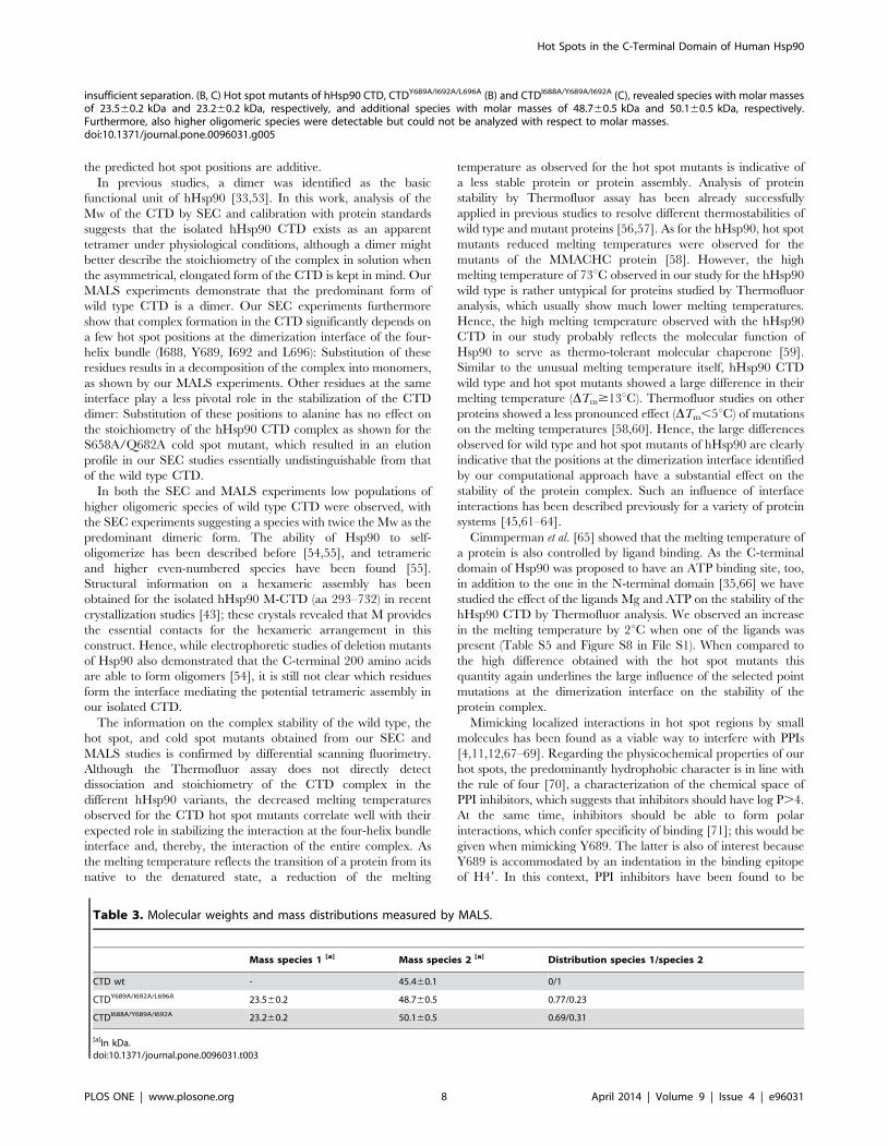
the predicted hot spot positions are additive.
In previous studies, a dimer was identified as the basic
functional unit of hHsp90 [33,53]. In this work, analysis of the
Mw of the CTD by SEC and calibration with protein standards
suggests that the isolated hHsp90 CTD exists as an apparent
tetramer under physiological conditions, although a dimer might
better describe the stoichiometry of the complex in solution when
the asymmetrical, elongated form of the CTD is kept in mind. Our
MALS experiments demonstrate that the predominant form of
wild type CTD is a dimer. Our SEC experiments furthermore
show that complex formation in the CTD significantly depends on
a few hot spot positions at the dimerization interface of the four-
helix bundle (I688, Y689, I692 and L696): Substitution of these
residues results in a decomposition of the complex into monomers,
as shown by our MALS experiments. Other residues at the same
interface play a less pivotal role in the stabilization of the CTD
dimer: Substitution of these positions to alanine has no effect on
the stoichiometry of the hHsp90 CTD complex as shown for the
S658A/Q682A cold spot mutant, which resulted in an elution
profile in our SEC studies essentially undistinguishable from that
of the wild type CTD.
In both the SEC and MALS experiments low populations of
higher oligomeric species of wild type CTD were observed, with
the SEC experiments suggesting a species with twice the Mw as the
predominant dimeric form. The ability of Hsp90 to self-
oligomerize has been described before [54,55], and tetrameric
and higher even-numbered species have been found [55].
Structural information on a hexameric assembly has been
obtained for the isolated hHsp90 M-CTD (aa 293–732) in recent
crystallization studies [43]; these crystals revealed that M provides
the essential contacts for the hexameric arrangement in this
construct. Hence, while electrophoretic studies of deletion mutants
of Hsp90 also demonstrated that the C-terminal 200 amino acids
are able to form oligomers [54], it is still not clear which residues
form the interface mediating the potential tetrameric assembly in
our isolated CTD.
The information on the complex stability of the wild type, the
hot spot, and cold spot mutants obtained from our SEC and
MALS studies is confirmed by differential scanning fluorimetry.
Although the Thermofluor assay does not directly detect
dissociation and stoichiometry of the CTD complex in the
different hHsp90 variants, the decreased melting temperatures
observed for the CTD hot spot mutants correlate well with their
expected role in stabilizing the interaction at the four-helix bundle
interface and, thereby, the interaction of the entire complex. As
the melting temperature reflects the transition of a protein from its
native to the denatured state, a reduction of the melting
temperature as observed for the hot spot mutants is indicative of
a less stable protein or protein assembly. Analysis of protein
stability by Thermofluor assay has been already successfully
applied in previous studies to resolve different thermostabilities of
wild type and mutant proteins [56,57]. As for the hHsp90, hot spot
mutants reduced melting temperatures were observed for the
mutants of the MMACHC protein [58]. However, the high
melting temperature of 73uC observed in our study for the hHsp90
wild type is rather untypical for proteins studied by Thermofluor
analysis, which usually show much lower melting temperatures.
Hence, the high melting temperature observed with the hHsp90
CTD in our study probably reflects the molecular function of
Hsp90 to serve as thermo-tolerant molecular chaperone [59].
Similar to the unusual melting temperature itself, hHsp90 CTD
wild type and hot spot mutants showed a large difference in their
melting temperature (DTm$13uC). Thermofluor studies on other
proteins showed a less pronounced effect (DTm,5uC) of mutations
on the melting temperatures [58,60]. Hence, the large differences
observed for wild type and hot spot mutants of hHsp90 are clearly
indicative that the positions at the dimerization interface identified
by our computational approach have a substantial effect on the
stability of the protein complex. Such an influence of interface
interactions has been described previously for a variety of protein
systems [45,61–64].
Cimmperman et al. [65] showed that the melting temperature of
a protein is also controlled by ligand binding. As the C-terminal
domain of Hsp90 was proposed to have an ATP binding site, too,
in addition to the one in the N-terminal domain [35,66] we have
studied the effect of the ligands Mg and ATP on the stability of the
hHsp90 CTD by Thermofluor analysis. We observed an increase
in the melting temperature by 2uC when one of the ligands was
present (Table S5 and Figure S8 in File S1). When compared to
the high difference obtained with the hot spot mutants this
quantity again underlines the large influence of the selected point
mutations at the dimerization interface on the stability of the
protein complex.
Mimicking localized interactions in hot spot regions by small
molecules has been found as a viable way to interfere with PPIs
[4,11,12,67–69]. Regarding the physicochemical properties of our
hot spots, the predominantly hydrophobic character is in line with
the rule of four [70], a characterization of the chemical space of
PPI inhibitors, which suggests that inhibitors should have log P.4.
At the same time, inhibitors should be able to form polar
interactions, which confer specificity of binding [71]; this would be
given when mimicking Y689. The latter is also of interest because
Y689 is accommodated by an indentation in the binding epitope
of H49. In this context, PPI inhibitors have been found to be
insufficient separation. (B, C) Hot spot mutants of hHsp90 CTD, CTDY689A/I692A/L696A (B) and CTDI688A/Y689A/I692A (C), revealed species with molar massesof 23.560.2 kDa and 23.260.2 kDa, respectively, and additional species with molar masses of 48.760.5 kDa and 50.160.5 kDa, respectively.Furthermore, also higher oligomeric species were detectable but could not be analyzed with respect to molar masses.doi:10.1371/journal.pone.0096031.g005
Table 3. Molecular weights and mass distributions measured by MALS.
Mass species 1 [a] Mass species 2 [a] Distribution species 1/species 2
CTD wt - 45.460.1 0/1
CTDY689A/I692A/L696A 23.560.2 48.760.5 0.77/0.23
CTDI688A/Y689A/I692A 23.260.2 50.160.5 0.69/0.31
[a]In kDa.doi:10.1371/journal.pone.0096031.t003
Hot Spots in the C-Terminal Domain of Human Hsp90
PLOS ONE | www.plosone.org 8 April 2014 | Volume 9 | Issue 4 | e96031

particularly effective when they bind to well-defined clefts or
grooves in the protein-protein interface [7,72]. Finally, the hot
spots in the main cluster show an i, i+4, i+8 pattern with respect to
sequence localization when starting with I688, which could be
mimicked by a novel class of non-peptidic a-helix mimetics
recently described [73]. In reverse order, an i, i+4, i+7 pattern
emerges, for which several scaffolds for a-helix mimetics have been
described [74,75]. Thus, it seems promising to use the hot spots of
the main cluster as a pharmacophoric template [12] for searching
and designing inhibitors that interfere with the dimerization of
hHsp90.
In summary, our computational results reveal the presence of
spatially clustered hot spot residues in the hHsp90 CTD interface,
which form a functional epitope and account for most of the
protein dimerization energy. The influence of these residues on the
stability and the oligomeric state of the CTD has been
demonstrated by experiments. These hot spots have favorable
properties with respect to using them as a pharmacophoric
template for identifying and designing small-molecule inhibitors of
hHsp90 dimerization. This opens up a new avenue for interfering
with hHsp90 function for treating cancer.
Materials and Methods
MaterialsChemicals and reagents were purchased from AppliChem,
Sigma-Aldrich, Carl Roth, VWR, Merck and Fluka at analytical
grade. Plasmids were derived from pET vectors from Merck/
Novagen (Darmstadt, Germany). SYPRO Orange was obtained
from Sigma-Aldrich (Steinheim, Germany).
Multiple sequence alignment and homology modelSequences of the Hsp90 CTD from S. cerevisiae and E. coli were
retrieved from the Protein Data Bank (PDB). Two sequences from
S. cerevisiae were used, corresponding to the PDB entries 2CG9 and
2CGE (UniProt code P02829, amino acids 540–677). One
sequence from E. coli was considered, corresponding to the PDB
entry 1SF8 (UniProt code P0A6Z3, amino acids 510–624). The
sequence of the hHsp90 a isoform CTD was retrieved from the
UniProt database (UniProt code P07900, amino acids 561–697). A
multiple sequence alignment was generated by means of
CLUSTALW [76,77].
Five homology models for the hHsp90 CTD were developed
using the ‘‘automodel’’ procedure and default parameters in
MODELLER 9.8 [41]. As templates, crystal structures from S.
cerevisiae (PDB codes 2CG9 and 2CGE) and E. coli (PDB code
1SF8) were used. The best model as evaluated from the
MODELLER objective function was chosen for the subsequent
work. The quality of this homology model was assessed by means
of PROCHEK [42] using default parameters.
Molecular dynamics simulationsAll the procedures described in the following were performed
with the Amber 11 software package [78], using the ff99SB force
field [79]. The homology model and a CTD dimer of a crystal
structure of an M-CTD construct of hHsp90 (PDB code: 3Q6M)
were initially placed in an octahedral periodical box of TIP3P
water molecules [80], where the distance between the edges of the
box and the closest atom of the solute is at least 11 A. Long-range
electrostatic interactions were treated using the Particle Mesh
Ewald (PME) method [81], and the SHAKE algorithm [82] was
employed to constrain bond lengths of heavy atoms to hydrogen
atoms. The time step for all MD simulations was set to 2 fs, with a
non-bonded cutoff of 8 A. The homology model and the CTD
dimer of the crystal structure, first, were geometry-optimized by 10
rounds of energy minimization; in each round 50 steps of steepest
descent minimization were followed by 450 steps of conjugate
gradient minimization, applying decreasing harmonic restraints on
the solute atoms (the force constant was 25 kcal mol21 A22 in the
first five rounds, and reduced to 5 kcal mol21 A22 in the
remaining). The systems were heated from 100 K to 300 K during
an MD simulation of 50 ps length performed in the canonical
(NVT) ensemble, applying harmonic restraints with force
constants of 5 kcal mol21 A22 to all solute atoms. Afterwards,
MD simulations of 250 ps length in the isothermal-isobaric
ensemble (NPT) were performed applying the same harmonic
restraints as in the previous step, in order to adjust the solvent
density. Finally, MD simulations of 100 ps length in the NVT
ensemble were performed, gradually reducing the force constants
of the harmonic restraints on the solute atoms to zero. Additional
100 ns of MD simulations at 300 K were performed, and the
coordinates were stored every 20 ps. These were used to extract
5000 snapshots for calculating the effective energy of dimerization
and the structural decomposition.
MM-GB/SA calculations, free energy decomposition andin silico alanine scanning
MM-GB/SA calculations were performed employing the
‘‘single trajectory approach’’ [83]. All counterions and water
molecules were stripped. For each snapshot, the gas-phase energy
(i.e., the sum of the internal energies plus electrostatic and van der
Waals energies) was calculated based on the ff99SB force field [79]
without applying a non-bonded cutoff. Polar contributions to the
solvation free energy were calculated using the ‘‘OBC’’ general-
ized Born model [84] together with mbondi2 radii, with a
dielectric constant of 1 for the solute and 80 for the solvent. The
polar contributions were computed at 100 mM ionic strength.
Nonpolar contributions to the solvation free energies were
calculated by a solvent-accessible surface area (SASA)-dependent
term using a surface tension of c= 0.0072 kcal mol21 A22.
Changes in the configurational entropy upon dimerization were
not considered. The contributions on a per-residue basis to the
overall effective energy (i.e., sum of gas-phase plus solvation free
energy) of dimerization were calculated employing the decompo-
sition scheme implemented in Amber 11 [78]. The same snapshots
were used to perform in silico alanine scanning using the
DrugScorePPI web server [38,39].
Cloning, expression and purificationSynthetic codon optimized DNA (GeneScript, Piscataway, NJ)
corresponding to the coding region of residues 563–732 of hHsp90
was cloned into expression vector pTEV21-a in E. coli BL21 (DE3)
(Agilent Technologies). Cultures were grown at 37uC in LB media
(5 g l21sodium chloride, 5 g l21 yeast extract and 10 g l21
peptone) with ampicillin to OD600 0.8–1.2. The production of
recombinant protein was induced by adding 1 mM isopropyl-1-
thio-b-D-galactopyranoside, and cells were grown for another 4 h
at 28uC. Cells were harvested by centrifugation, suspended in
binding buffer (40 mM HEPES, 20 mM KCl, 1 mM DTT, 1 mM
EDTA, 0.002% PMSF, pH 7.5) [85] and disrupted by sonifica-
tion. Recombinant proteins were purified via a C-terminal His6-
tag by immobilized metal ion affinity chromatography to
homogeneity (Figure S9 in File S1).
Alanine mutants and cold spot creationAlanines at positions 689, 692, and 696 as well as at positions
688, 689, and 692 for the hot spot mutants and at positions 658
Hot Spots in the C-Terminal Domain of Human Hsp90
PLOS ONE | www.plosone.org 9 April 2014 | Volume 9 | Issue 4 | e96031

and 682 for the cold spot mutant were introduced into the
constructs as described in the QuikChange Site-Directed Muta-
genesis Kit (Stratagene) using Pwo polymerase and designed
mutagenesis primers (Table 4). Additionally, alanine single
mutants were constructed with alanines at positions 688, 689,
692, and 696, respectively (Table S3 in File S1).
Stability assay by Fluorescence Thermal Shift Assay(Thermofluor)
Stability assay was performed in the real-time thermo-cycler
qTOWER 2.0 (Analytik Jena AG, Germany) with 0.15 mg/mL
protein and the fluorescent dye SYPRO Orange (1:1000) in 96
well PCR plates. The fluorescence signal of the SYPRO Orange
dye was measured at an initial start temperature of 25uC. Up to 12
different conditions were tested (Table 5) [86] with increasing the
temperature stepwise up to 95uC. When hydrophobic residues of
the protein become more accessible, this leads to binding of
SYPRO Orange. Fluorescence changes in the wells of the plate
were monitored simultaneously with Channel Photo Multiplier
(CPM). The wavelengths for excitation and emission were 490 nm
and 580 nm, respectively. The detected fluorescent signal corre-
sponds to the denaturation state of hHsp90. Melting points were
determined by the implemented software (qPCRsoft V2.0.37.0,
Analytik Jena AG, Germany) from the derivative of the
fluorescence data.
Size exclusion chromatographySize exclusion chromatography analyses were accomplished in
triplicates on a Superdex SD200 10/300 column on an
AKTAprime plus chromatography system (GE lifescience). The
experiments were performed at 4uC in HPLC buffer (10 mM
MES, 200 mM KCl, 1 mM EDTA, 1% Glycerol) with a flow rate
of 0.5 mL/min at pH 6. Samples were centrifuged before for
15 min at 14.000xg, and 110 mL of the purified CTD of hHsp90
was loaded on the column. Data were analyzed using the software
PrimeView Evaluation 5.0 (UNICORN, GE), and the maximal
absorbance was determined by peak integration.
Multi-angle light scatteringA size exclusion chromatography (SEC) column (Bio SEC-5,
150 A, Agilant Technologies) was equilibrated with the above
HPLC buffer without glycerol using a HPLC system (Agilant
Technologies) connected with a multi-angle light-scattering
detector (miniDAWN TREOS, Wyatt Technologies) and a
differential refractive-index detector (Optilab T-rEX, Wyatt
Technologies). Samples with concentrations of 2.4 mg/ml were
centrifuged at 74.000xg for 30 minutes at 4uC and loaded onto the
equilibrated SEC column and data were analyzed with the
ASTRA software (Wyatt Technologies).
CD spectroscopyCD measurements for wild type hHsp90 CTD as well as for all
mutants generated (CTDI688A/Y689A/I692A, CTDY689A/I692A/L696A,
CTDS658A/Q682A, CTDI688A, CTDY689A, CTDI692A, and
Table 4. Mutagenesis primers[a].
Cold spot mutant: CTDS658A/Q682A
Primer 1: TCG (Ser) R GCA (Ala): 45 nt (59-39)
Forw.: CAGAAGCTGATAAAAACGACAAAGCAGTGAAAGATCTGGTTATCC
Rev.: GGATAACCAGATCTTTCACTGCTTTGTCGTTTTTATCAGCTTCTG
Primer 2: CAG (Gln) R GCA (Ala); 34 nt (59-39)
Forw.: AGCCTGGAAGACCCGGCAACGCATGCCAACCGTA
Rev.: TACGGTTGGCATGCGTTGCCGGGTCTTCCAGGCT
Hot spot mutant I: CTDY689A/I692A/L696A
TAC (Tyr) R GCA (Ala); ATC (Ile) R GCA (Ala); CTG (Leu) R GCA (Ala): 44 nt (59-39)
Forw.: CAACCGTATTGCACGCATGGCAAAACTGGGCGCAGGTATTGATG
Rev.: CATCAATACCTGCGCCCAGTTTTGCCATGCGTGCAATACGGTTG
Hot spot mutant I: CTDI688A/Y689A/I692A
ATT (Ile) R GCA (Ala); TAC (Tyr) R GCA (Ala); ATC (Ile) R GCA (Ala): 37 nt (59-39)
Forw: GCATGCCAACCGTGCAGCACGCATGGCAAAACTGGGCC
Rev: GGCCCAGTTTTGCCATGCGTGCTGCACGGTTGGCATGC
[a]The primers were obtained by Sigma-Aldrich Chemie GmbH Steinheim, Germany. Bold nucleotides indicate the newly introduced alanines.doi:10.1371/journal.pone.0096031.t004
Table 5. pH values of screening buffers [86] for Thermofluorassay.
No. Buffer [100 mM] pH
1 Glycine 3.0
2 Citric acid 4.0
3 Sodium citrate 5.5
4 Sodium/potassium phosphate 6.0
5 MES 6.2
6 Bis-tris propane 6.5
7 Sodium/potassium phosphate 7.0
8 Tris 7.5
9 EPPS 8.0
10 Tris 8.5
11 CHES 9.0
12 CHAPS 10.0
doi:10.1371/journal.pone.0096031.t005
Hot Spots in the C-Terminal Domain of Human Hsp90
PLOS ONE | www.plosone.org 10 April 2014 | Volume 9 | Issue 4 | e96031

CTDL696A) were performed on a Jasco J-715 spectrometer, using
0.2 mg/ml protein solutions in 50 mM potassium phosphate
buffer, pH 7.6 and a cuvette with a path length of 1 mm. The
spectra were obtained by averaging five measurements for each
protein sample and correcting the signal by subtracting the buffer
signal. Data points were collected every 0.2 nm in the range from
195 to 260 nm.
Supporting Information
File S1 The file contains additional information to themanuscript explaining results in further details. It
consists of 14 pages, 5 tables and 9 figures.
(PDF)
Acknowledgments
H.G. is grateful to the ‘‘Zentrum fur Informations- und Medientechno-
logie’’ of the Heinrich-Heine-University (HHU), Dusseldorf, for compu-
tational support. We thank the Institute of Physical Biology at HHU for
providing us access to the CD spectrometer, Alexander Minges (Institute of
Biochemical Plant Physiology, HHU) for providing the Thermofluor
Analysis Software PyDSF, and Daniel Schlieper (Institute of Biochemical
Plant Physiology, HHU) for critical reading of the manuscript.
Author Contributions
Conceived and designed the experiments: LS GG HG. Performed the
experiments: EC JV SR SHJS. Analyzed the data: EC JV SR SHJS GG
HG. Wrote the paper: EC JV SR SHJS LS GG HG.
References
1. Zinzalla G, Thurston DE (2009) Targeting protein-protein interactions for
therapeutic intervention: a challenge for the future. Future Med Chem 1: 65–93.
2. Fischer PM (2005) Protein-protein Interactions in Drug Discovery. Drug Des
Rev—Online 2: 179–207.
3. Wells JA, McClendon CL (2007) Reaching for high-hanging fruit in drug
discovery at protein-protein interfaces. Nature 450: 1001–1009.
4. Blazer LL, Neubig RR (2009) Small molecule protein-protein interaction
inhibitors as CNS therapeutic agents: current progress and future hurdles.
Neuropsychopharmacology 34: 126–141.
5. Ryan DP, Matthews JM (2005) Protein-protein interactions in human disease.
Curr Opin Struct Biol 15: 441–446.
6. Gerrard JA, Hutton CA, Perugini MA (2007) Inhibiting protein-protein
interactions as an emerging paradigm for drug discovery. Mini Rev Med Chem
7: 151–157.
7. Chene P (2006) Drugs targeting protein-protein interactions. ChemMedChem 1:
400–411.
8. Clackson T, Wells JA (1995) A hot spot of binding energy in a hormone-receptor
interface. Science 267: 383–386.
9. Bogan AA, Thorn KS (1998) Anatomy of hot spots in protein interfaces. J Mol
Biol 280: 1–9.
10. Gohlke H, Kiel C, Case DA (2003) Insights into protein-protein binding by
binding free energy calculation and free energy decomposition for the Ras-Raf
and Ras-RalGDS complexes. J Mol Biol 330: 891–913.
11. Metz A, Pfleger C, Kopitz H, Pfeiffer-Marek S, Baringhaus KH, et al. (2012)
Hot spots and transient pockets: predicting the determinants of small-molecule
binding to a protein-protein interface. J Chem Inf Model 52: 120–133.
12. Metz A, Schanda J, Grez M, Wichmann C, Gohlke H (2013) From determinants
of RUNX1/ETO tetramerization to small-molecule protein-protein interaction
inhibitors targeting acute myeloid leukemia. J Chem Inf Model 53: 2197–2202.
13. Zerbe BS, Hall DR, Vajda S, Whitty A, Kozakov D (2012) Relationship between
hot spot residues and ligand binding hot spots in protein-protein interfaces.
J Chem Inf Model 52: 2236–2244.
14. Mayer MP, Prodromou C, Frydman J (2009) The Hsp90 mosaic: a picture
emerges. Nat Struct Mol Biol 16: 2–6.
15. Wiech H, Buchner J, Zimmermann R, Jakob U (1992) Hsp90 chaperones
protein folding in vitro. Nature 358: 169–170.
16. Young JC, Agashe VR, Siegers K, Hartl FU (2004) Pathways of chaperone-
mediated protein folding in the cytosol. Nat Rev Mol Cell Biol 5: 781–791.
17. Pearl LH, Prodromou C (2006) Structure and mechanism of the Hsp90
molecular chaperone machinery. Annu Rev Biochem 75: 271–294.
18. Wandinger SK, Richter K, Buchner J (2008) The Hsp90 chaperone machinery.
J Biol Chem 283: 18473–18477.
19. Nathan DF, Vos MH, Lindquist S (1997) In vivo functions of the Saccharomyces
cerevisiae Hsp90 chaperone. Proc Natl Acad Sci U S A 94: 12949–12956.
20. Bagatell R, Whitesell L (2004) Altered Hsp90 function in cancer: a unique
therapeutic opportunity. Mol Cancer Ther 3: 1021–1030.
21. Whitesell L, Lindquist SL (2005) HSP90 and the chaperoning of cancer. Nat
Rev Cancer 5: 761–772.
22. Young JC, Moarefi I, Hartl FU (2001) Hsp90: a specialized but essential protein-
folding tool. J Cell Biol 154: 267–273.
23. Pearl LH, Prodromou C (2000) Structure and in vivo function of Hsp90. Curr
Opin Struct Biol 10: 46–51.
24. Whitesell L, Mimnaugh EG, De Costa B, Myers CE, Neckers LM (1994)
Inhibition of heat shock protein HSP90-pp60v-src heteroprotein complex
formation by benzoquinone ansamycins: essential role for stress proteins in
oncogenic transformation. Proc Natl Acad Sci U S A 91: 8324–8328.
25. Sharma SV, Agatsuma T, Nakano H (1998) Targeting of the protein chaperone,
HSP90, by the transformation suppressing agent, radicicol. Oncogene 16: 2639–
2645.
26. Mahalingam D, Swords R, Carew JS, Nawrocki ST, Bhalla K, et al. (2009)
Targeting HSP90 for cancer therapy. Br J Cancer 100: 1523–1529.
27. Prodromou C, Roe SM, O’Brien R, Ladbury JE, Piper PW, et al. (1997)Identification and structural characterization of the ATP/ADP-binding site in
the Hsp90 molecular chaperone. Cell 90: 65–75.
28. Obermann WM, Sondermann H, Russo AA, Pavletich NP, Hartl FU (1998) Invivo function of Hsp90 is dependent on ATP binding and ATP hydrolysis. J Cell
Biol 143: 901–910.
29. Panaretou B, Prodromou C, Roe SM, O’Brien R, Ladbury JE, et al. (1998) ATPbinding and hydrolysis are essential to the function of the Hsp90 molecular
chaperone in vivo. EMBO J 17: 4829–4836.
30. Powers MV, Workman P (2007) Inhibitors of the heat shock response: biologyand pharmacology. FEBS Lett 581: 3758–3769.
31. Sharp S, Workman P (2006) Inhibitors of the HSP90 molecular chaperone:
current status. Adv Cancer Res 95: 323–348.
32. Minami Y, Kimura Y, Kawasaki H, Suzuki K, Yahara I (1994) The carboxy-
terminal region of mammalian HSP90 is required for its dimerization and
function in vivo. Mol Cell Biol 14: 1459–1464.
33. Harris SF, Shiau AK, Agard DA (2004) The crystal structure of the carboxy-
terminal dimerization domain of htpG, the Escherichia coli Hsp90, reveals a
potential substrate binding site. Structure 12: 1087–1097.
34. Ratzke C, Mickler M, Hellenkamp B, Buchner J, Hugel T (2010) Dynamics of
heat shock protein 90 C-terminal dimerization is an important part of its
conformational cycle. Proc Natl Acad Sci U S A 107: 16101–16106.
35. Marcu MG, Chadli A, Bouhouche I, Catelli M, Neckers LM (2000) The heat
shock protein 90 antagonist novobiocin interacts with a previously unrecognizedATP-binding domain in the carboxyl terminus of the chaperone. J Biol Chem
275: 37181–37186.
36. Yun BG, Huang W, Leach N, Hartson SD, Matts RL (2004) Novobiocininduces a distinct conformation of Hsp90 and alters Hsp90-cochaperone-client
interactions. Biochemistry 43: 8217–8229.
37. Massova I, Kollman PA (2000) Combined molecular mechanical and continuumsolvent approach (MM-PBSA/GBSA) to predict ligand binding. Perspect Drug
Discovery Des 18: 113–135.
38. Kruger DM, Gohlke H (2010) DrugScore(PPI) webserver: fast and accurate insilico alanine scanning for scoring protein-protein interactions. Nucleic Acids
Res 38: W480–W486.
39. Kruger DM, Gohlke H (2011) Protein-protein interactions, web-based analysis.Nachr Chem 59: 44–45.
40. Pantoliano MW, Petrella EC, Kwasnoski JD, Lobanov VS, Myslik J, et al. (2001)
High-density miniaturized thermal shift assays as a general strategy for drugdiscovery. J Biomol Screening 6: 429–440.
41. Sali A, Blundell TL (1993) Comparative protein modelling by satisfaction of
spatial restraints. J Mol Biol 234: 779–815.
42. Laskowski RA, Macarthur MW, Moss DS, Thornton JM (1993) Procheck - a
Program to Check the Stereochemical Quality of Protein Structures. J ApplCrystallogr 26: 283–291.
43. Lee CC, Lin TW, Ko TP, Wang AH (2011) The hexameric structures of human
heat shock protein 90. PLoS One 6: e19961.
44. Sgobba M, Degliesposti G, Ferrari AM, Rastelli G (2008) Structural models and
binding site prediction of the C-terminal domain of human Hsp90: a new target
for anticancer drugs. Chem Biol Drug Des 71: 420–433.
45. Wichmann C, Becker Y, Chen-Wichmann L, Vogel V, Vojtkova A, et al. (2010)
Dimer-tetramer transition controls RUNX1/ETO leukemogenic activity. Blood
116: 603–613.
46. Thorn KS, Bogan AA (2001) ASEdb: a database of alanine mutations and their
effects on the free energy of binding in protein interactions. Bioinformatics 17:
284–285.
47. Niesen FH, Berglund H, Vedadi M (2007) The use of differential scanning
fluorimetry to detect ligand interactions that promote protein stability. Nat
Protoc 2: 2212–2221.
48. Matulis D, Kranz JK, Salemme FR, Todd MJ (2005) Thermodynamic stability
of carbonic anhydrase: measurements of binding affinity and stoichiometry using
ThermoFluor. Biochemistry 44: 5258–5266.
Hot Spots in the C-Terminal Domain of Human Hsp90
PLOS ONE | www.plosone.org 11 April 2014 | Volume 9 | Issue 4 | e96031

49. Bullock AN, Henckel J, DeDecker BS, Johnson CM, Nikolova PV, et al. (1997)
Thermodynamic stability of wild-type and mutant p53 core domain. Proc NatlAcad Sci U S A 94: 14338–14342.
50. Erickson HP (2009) Size and shape of protein molecules at the nanometer level
determined by sedimentation, gel filtration, and electron microscopy. BiolProced Online 11: 32–51.
51. Archontis G, Simonson T, Karplus M (2001) Binding free energies and freeenergy components from molecular dynamics and Poisson-Boltzmann calcula-
tions. Application to amino acid recognition by aspartyl-tRNA synthetase. J Mol
Biol 306: 307–327.52. Moreira IS, Fernandes PA, Ramos MJ (2007) Hot spots—a review of the
protein-protein interface determinant amino-acid residues. Proteins 68: 803–812.
53. Retzlaff M, Stahl M, Eberl HC, Lagleder S, Beck J, et al. (2009) Hsp90 isregulated by a switch point in the C-terminal domain. EMBO Rep 10: 1147–
1153.
54. Nemoto T, Sato N (1998) Oligomeric forms of the 90-kDa heat shock protein.Biochemical Journal 330 (Pt 2): 989–995.
55. Moullintraffort L, Bruneaux M, Nazabal A, Allegro D, Giudice E, et al. (2010)Biochemical and biophysical characterization of the Mg2+-induced 90-kDa heat
shock protein oligomers. J Biol Chem 285: 15100–15110.
56. Lavinder JJ, Hari SB, Sullivan BJ, Magliery TJ (2009) High-throughput thermalscanning: a general, rapid dye-binding thermal shift screen for protein
engineering. J Am Chem Soc 131: 3794–3795.57. Mulepati S, Bailey S (2011) Structural and Biochemical Analysis of Nuclease
Domain of Clustered Regularly Interspaced Short Palindromic Repeat(CRISPR)-associated Protein 3 (Cas3). Journal of Biological Chemistry 286:
31896–31903.
58. Froese DS, Healy S, McDonald M, Kochan G, Oppermann U, et al. (2010)Thermolability of mutant MMACHC protein in the vitamin B12-responsive
cblC disorder. Mol Genet Metab 100: 29–36.59. Whitley D, Goldberg SP, Jordan WD (1999) Heat shock proteins: a review of the
molecular chaperones. J Vasc Surg 29: 748–751.
60. Kervinen J, Ma H, Bayoumy S, Schubert C, Milligan C, et al. (2006) Effect ofconstruct design on MAPKAP kinase-2 activity, thermodynamic stability and
ligand-binding affinity. Arch Biochem Biophys 449: 47–56.61. Bjork A, Mantzilas D, Sirevag R, Eijsink VG (2003) Electrostatic interactions
across the dimer-dimer interface contribute to the pH-dependent stability of atetrameric malate dehydrogenase. FEBS Lett 553: 423–426.
62. Baden EM, Owen BA, Peterson FC, Volkman BF, Ramirez-Alvarado M, et al.
(2008) Altered dimer interface decreases stability in an amyloidogenic protein.J Biol Chem 283: 15853–15860.
63. Mandelman D, Schwarz FP, Li H, Poulos TL (1998) The role of quaternaryinteractions on the stability and activity of ascorbate peroxidase. Protein Sci 7:
2089–2098.
64. Mateu MG, Fersht AR (1998) Nine hydrophobic side chains are keydeterminants of the thermodynamic stability and oligomerization status of
tumour suppressor p53 tetramerization domain. EMBO J 17: 2748–2758.65. Cimmperman P, Baranauskiene L, Jachimoviciute S, Jachno J, Torresan J, et al.
(2008) A quantitative model of thermal stabilization and destabilization ofproteins by ligands. Biophys J 95: 3222–3231.
66. Garnier C, Lafitte D, Tsvetkov PO, Barbier P, Leclerc-Devin J, et al. (2002)
Binding of ATP to heat shock protein 90: evidence for an ATP-binding site inthe C-terminal domain. J Biol Chem 277: 12208–12214.
67. Thanos CD, DeLano WL, Wells JA (2006) Hot-spot mimicry of a cytokinereceptor by a small molecule. Proc Natl Acad Sci U S A 103: 15422–15427.
68. Azzarito V, Long K, Murphy NS, Wilson AJ (2013) Inhibition of alpha-helix-
mediated protein-protein interactions using designed molecules. Nat Chem 5:
161–173.
69. Golden MS, Cote SM, Sayeg M, Zerbe BS, Villar EA, et al. (2013)
Comprehensive experimental and computational analysis of binding energy
hot spots at the NF-kappaB essential modulator/IKKbeta protein-protein
interface. J Am Chem Soc 135: 6242–6256.
70. Morelli X, Bourgeas R, Roche P (2011) Chemical and structural lessons from
recent successes in protein-protein interaction inhibition (2P2I). Curr Opin
Chem Biol 15: 475–481.
71. Gohlke H, Klebe G (2002) Approaches to the description and prediction of the
binding affinity of small-molecule ligands to macromolecular receptors. Angew
Chem Int Ed Engl 41: 2644–2676.
72. Metz A, Ciglia E, Gohlke H (2012) Modulating protein-protein interactions:
from structural determinants of binding to druggability prediction to application.
Curr Pharm Des 18: 4630–4647.
73. Spanier L, Ciglia E, Hansen FK, Kuna K, Frank W, et al. (2014) Design,
synthesis, and conformational analysis of trispyrimidonamides as a-helix
mimetics. J Org Chem DOI: 10.1021/jo402353z.
74. Cummings CG, Hamilton AD (2010) Disrupting protein-protein interactions
with non-peptidic, small molecule alpha-helix mimetics. Curr Opin Chem Biol
14: 341–346.
75. Davis JM, Tsou LK, Hamilton AD (2007) Synthetic non-peptide mimetics of
alpha-helices. Chem Soc Rev 36: 326–334.
76. Larkin MA, Blackshields G, Brown NP, Chenna R, McGettigan PA, et al. (2007)
Clustal W and Clustal X version 2.0. Bioinformatics 23: 2947–2948.
77. Goujon M, McWilliam H, Li W, Valentin F, Squizzato S, et al. (2010) A new
bioinformatics analysis tools framework at EMBL-EBI. Nucleic Acids Res 38:
W695–699.
78. Case DA, Darden TA, Cheatham TEI, Simmerling CL, Wang J, et al. (2010)
AMBER 11. University of California, San Francisco.
79. Hornak V, Abel R, Okur A, Strockbine B, Roitberg A, et al. (2006) Comparison
of multiple Amber force fields and development of improved protein backbone
parameters. Proteins 65: 712–725.
80. Jorgensen WL, Chandrasekhar J, Madura JD, Impey RW, Klein ML (1983)
Comparison of Simple Potential Functions for Simulating Liquid Water. J Chem
Phys 79: 926–935.
81. Darden T, York D, Pedersen L (1993) Particle Mesh Ewald - an N.Log(N)
Method for Ewald Sums in Large Systems. J Chem Phys 98: 10089–10092.
82. Ryckaert JP, Ciccotti G, Berendsen HJC (1977) Numerical-Integration of
Cartesian Equations of Motion of a System with Constraints - Molecular-
Dynamics of N-Alkanes. J Comput Phys 23: 327–341.
83. Homeyer N, Gohlke H (2012) Free Energy Calculations by the Molecular
Mechanics Poisson-Boltzmann Surface Area Method. Mol Inf 31: 114–122.
84. Onufriev A, Bashford D, Case DA (2004) Exploring protein native states and
large-scale conformational changes with a modified generalized born model.
Proteins 55: 383–394.
85. Richter K, Soroka J, Skalniak L, Leskovar A, Hessling M, et al. (2008)
Conserved conformational changes in the ATPase cycle of human Hsp90. J Biol
Chem 283: 17757–17765.
86. Jancarik J, Pufan R, Hong C, Kim SH, Kim R (2004) Optimum solubility (OS)
screening: an efficient method to optimize buffer conditions for homogeneity and
crystallization of proteins. Acta Crystallogr Sect D Biol Crystallogr 60: 1670–
1673.
Hot Spots in the C-Terminal Domain of Human Hsp90
PLOS ONE | www.plosone.org 12 April 2014 | Volume 9 | Issue 4 | e96031


![Identification of potential P. falciparum transketolase ... · modelling by satisfying spatial restraints in terms of probability density functions [16,18]. To this end, the homodimer](https://img.pdfslide.us/doc/110x75/603ca6db78468426fe376c56/identification-of-potential-p-falciparum-transketolase-modelling-by-satisfying.jpg)