Embed Size (px)
Citation preview
IntroductionBorrelia burgdorferi sensu lato is the causative agent ofLyme disease (LD). It is transmitted by infected ticksthat deposit a small number of organisms in the der-mis of a host animal during feeding, leading to a local-ized infection (1). The initial skin infection is usuallyaccompanied by a local rash (erythema migrans) andcan include other manifestations such as fever, musclepain, and headaches (2–4). Bacterial dissemination andcolonization of many different organs may follow. Latestages of LD can include neurological, ocular, cuta-neous, and cardiac disease in addition to arthritis (1–3).Early antibiotic therapy will usually resolve B. burgdor-feri infection. However, symptoms sometimes mayreappear, and up to 10% of patients with Lyme arthri-tis are classified as treatment resistant (1, 5, 6).
Microbial adhesion to and colonization of host tis-sues is an early, critical event in most infection process-es (7). Pathogenic bacteria often express several adhesinsthat can participate in parallel and independent attach-ment mechanisms that result in tissue colonization. InLD, bacterial adhesion to host tissue may be important
in determining both the fate of the initial challengeorganisms in the dermis and their ability to dissemi-nate. Spirochetes deposited in the dermis are foundassociated with collagen fibers in the extracellularmatrix (ECM). However, B. burgdorferi do not attachdirectly to collagen but to decorin, a small leucine-richproteoglycan (SLRP) that is associated with and “deco-rates” collagen fibers (8, 9). Decorin is distributedthroughout the mammalian body and could be a targetfor B. burgdorferi adherence during dissemination, asthese spirochetes express two decorin-binding proteins,DbpA and DbpA. However, alternate adhesion mecha-nisms also may be used by the spirochetes depending onthe tissue target and stage of dissemination (10, 11). Inaddition to the two decorin-binding microbial surfacecomponent–recognizing adhesive matrix molecules(MSCRAMMs), Borrelia also express a fibronectin-bind-ing MSCRAMM, BBK32, and a putative glycosamino-glycan-binding MSCRAMM (12–14). B. burgdorferi havebeen shown to attach to a variety of mammalian cells invitro, and the organisms can bind to the integrins αMβ2,αIIbβ3, α5β1, and αvβ3 (8, 9, 15–19).
The Journal of Clinical Investigation | April 2001 | Volume 107 | Number 7 845
Resistance to Lyme disease in decorin-deficient mice
Eric L. Brown,1 R. Mark Wooten,2 Barbara J.B. Johnson,3 Renato V. Iozzo,4
Amanda Smith,1 Marc C. Dolan,3 Betty P. Guo,1 Janis J. Weis,2 and Magnus Höök1
1The Center for Extracellular Matrix Biology, Texas A&M University System Health Science Center, Albert B. Alkek Institute of Biosciences and Technology, Houston, Texas, USA
2Division of Cell Biology and Immunology, Department of Pathology, University of Utah School of Medicine, Salt Lake City, Utah, USA
3Division of Vector-Borne Infectious Diseases, National Center for Infectious Diseases, Centers for Disease Control and Prevention, Fort Collins, Colorado, USA
4Department of Pathology, Anatomy, and Cell Biology, Thomas Jefferson University, Philadelphia, Pennsylvania, USA
Address correspondence to: Eric L. Brown, The Center for Extracellular Matrix Biology, Texas A&M University System Health Science Center, Albert B. Alkek Institute of Biosciences and Technology, 2121 West Holcombe Boulevard, Suite 603, Houston, Texas 77030, USA. Phone: (713) 677-7552; Fax: (713) 677-7576; E-mail: [email protected].
Betty P. Guo’s present address is: Harvard Medical School, Department of Microbiology and Molecular Genetics, Boston, Massachusetts, USA.
Received for publication November 2, 2000, and accepted in revised form February 12, 2001.
Microbial adhesion to the host tissue represents an early, critical step in the pathogenesis of mostinfectious diseases. Borrelia burgdorferi, the causative agent of Lyme disease (LD), expresses two sur-face-exposed decorin-binding adhesins, DbpA and DbpB. A decorin-deficient (Dcn–/–) mouse wasrecently developed and found to have a relatively mild phenotype. We have now examined the processof experimental LD in Dcn–/– mice using both needle inoculation and tick transmission of spirochetes.When exposed to low doses of the infective agent, Dcn–/– mice had fewer Borrelia-positive culturesfrom most tissues analyzed than did Dcn+/+ or Dcn+/– mice. When the infection dose was increased,similar differences were not observed in most tissues but were seen in bacterial colonization of jointsand the extent of Borrelia-induced arthritis. Quantitative PCR demonstrated that joints harvestedfrom Dcn–/– mice had diminished Borrelia numbers compared with issues harvested from Dcn+/+ con-trols. Histological examination also revealed a low incidence and severity of arthritis in Dcn–/– mice.Conversely, no differences in the numbers of Borrelia-positive skin cultures were observed among thedifferent genotypes regardless of the infection dose. These differences, which were observed regard-less of genetic background of the mice (BALB/c or C3H/HeN) or method of infection, demonstratethe importance of decorin in the pathogenesis of LD.
J. Clin. Invest. 107:845–852 (2001).
Recently, a decorin-deficient (Dcn–/–) mouse was devel-oped and characterized (20). The phenotype of thismouse was relatively mild and appeared to be restrictedto skin laxity and fragility, a consequence of irregular col-lagen fibers in the dermis (20). This result was somewhatsurprising given that decorin can have profound effectsin vitro. The proteoglycan can regulate the proliferationof cells, the activity of TGF-β and the complement sys-tem (C1q), as well as the adherence of mammalian cellsto fibronectin (21–27). The availability of a Dcn–/– mouseprovided an opportunity to assess the importance of thisproteoglycan in B. burgdorferi dissemination to variousorgans and tissue colonization in vivo. It has been previ-ously shown that the method of B. burgdorferi transmis-sion may affect the progression of LD and that differentstrains of mice can vary in the development and presen-tation of LD (28, 29). Therefore, we examined the role ofdecorin in the development of LD by infecting Dcn–/–
mice that have been backcrossed onto genetic back-grounds that exhibit either severe (C3H/Hen) or rela-tively mild arthritis (BALB/c). Although the ID50 forboth strains of mice is similar, spirochete disseminationand arthritis develop at different rates and intensities(28–30). Additionally, we examined the effect of decorinduring different infection processes by comparing dis-ease development in mice that had been infected usingeither cultured organisms administered by needle orarthropod-adapted spirochetes transmitted by ticks (31).
MethodsMice. Pathogen-free (MTV–) Dcn–/– mice (BL/Swiss ×129Sv) (20) were backcrossed for five and ten genera-tions into BALB/c or C3H/HeN backgrounds (HarlanSprague Dawley, Indianapolis, Indiana, USA). Mice weregenotyped for the decorin allele by PCR analysis ofmouse tail DNA as described previously (20). BALB/cand C3H/Hen mice used in all experiments were back-crossed to the fifth generation unless otherwise speci-fied. The animals were maintained in facilities approvedby the American Association for Accreditation of Labo-ratory Animal Care in accordance with current regula-tions and standards of the United States Department ofAgriculture, Department of Health and Human Ser-vices, and NIH. All animal procedures were approved bythe Institutional Animal Care and Use Committee ofTexas A&M University Health Science Center Instituteof Biosciences and Technology. Female Dcn–/–, Dcn+/–,and Dcn+/+ mice ranging in age from 8 to 10 weeks wereused at the start of the experiments.
Borrelia infections. Borrelia burgdorferi sensu stricto,strain B31, was obtained at in vitro passage 5 from S.J.Norris (The University of Texas Medical School atHouston, Houston, Texas, USA) (32) and inoculatedinto BSK-II medium supplemented with antibiotics (50 µg/ml rifampicin and 100 µg/ml phosphomycin)at 34°C as described previously (33). Borrelia used forinfections in all experiments described were culturedfrom aliquots of the same passage 5 frozen stock. Bac-terial cultures contained in screw-cap tubes were incu-
bated in a CO2-enriched atmosphere using a GasPakchamber (Becton Dickinson Microbiology Systems,Sparks, Maryland, USA) containing BBL GasPak Plusenvelopes and a GasPak anaerobic indicator (BectonDickinson, Cockeysville, Maryland, USA) until the cellsreached a density between 5 × 10⁷ and 1 × 10⁸/ml. Bor-relia were counted using dark-field microscopy and aPetroff-Hausser chamber. Tick- and needle-inoculationprocedures were used during the course of these exper-iments. Borrelia (101–104) were needle-inoculated intra-dermally into shaved dorsal skin at the base of the tail(100 µl/mouse). For tick transmission, B. burgdorferistrain B31-infected Ixodes scapularis nymphs wereallowed to feed to repletion. Mice were lightly anes-thetized with Metofane (methoxyflurane; Pitman-Moore, Mundelein, Illinois, USA) during the tickattachment period. Immediately after tick placement,mice were individually housed in raised wire-bottomcages (Lab Products, Maywood, New Jersey, USA) con-taining approximately 20 ml of water at their base forthe duration of the infection period (4–5 days). Allcages were monitored three times daily during the first3 days and four times daily during days 4 and 5, whenreplete ticks dropped from the mice (34).
Detection of Borrelia in replete ticks. Replete ticks fromeach mouse were collected and stored in a humidchamber for 12 days. At the end of this incubation peri-od, each tick was sterilized by incubating in 3% hydro-gen peroxide (3 minutes) followed by a 10- to 15-minute wash in 70% ethanol. Ticks were crushedindividually using flat-end tweezers in BSK II-filledtubes (6 ml). Tick-inoculated media was cultured for 2 weeks as already described here and were examinedfor the presence of Borrelia.
Culturing of blood and tissues. At various time pointsafter infection (days 3, 7, and 14) blood samples werecollected from the mice and cultured for the presenceof Borrelia. Tick-inoculated mice were maintained for28 days after infection, and blood was cultured for thepresence of Borrelia up to postinfection day 21. Theirtails were sterilized with a propidium-iodine swab (Pro-fessional Disposables Inc., Orangeburg, New York,USA) and bled under a laminar flow biosafety cabinet.Blood dilutions ranging from 1:100 to 1:10,000 (finalvolume of 5 ml) were made with BSK II and incubatedat 34°C. The cultures were checked 2 and 3 weeks laterfor the presence of viable Borrelia, using dark-fieldmicroscopy. Similarly, after sacrifice, joints, heart, blad-der, and ear samples were aseptically removed and cul-tured for the presence of Borrelia (days 14 and 28 forneedle- and tick-inoculated mice, respectively). Earbiopsy samples were collected by using a 3-mm Bakerbiopsy punch (Baker Norton Pharmaceuticals, Miami,Florida, USA). Care was taken to remove all skin fromisolated joints. All instruments were sterilized betweendissections (Dry Sterilizer IS-400; Inotech BiosystemsInternational, Lansing, Michigan, USA).
Arthritis assessment. Evidence of arthritis was deter-mined by histopathological examination of formalin-
846 The Journal of Clinical Investigation | April 2001 | Volume 107 | Number 7
fixed hind tibiotarsal joint samples. Tissues for histo-logical examination were embedded in paraffin andstained with hematoxylin and eosin. All sections wereexamined without knowledge of the infection or geno-type status of the mice. A positive/negative assessmentof arthritis was made initially. To determine the sever-ity of arthritis, joints were scored according to the lev-els of neutrophil infiltrate as follows: 0, no arthritis; 1,minimal or rare (≤ 10% tissue involvement); 2, mild(10–20%); frequent (20–50%); and 4, severe (>50%) (35).
Serum analysis (ELISA). Assays were performed onCorning Easy Wash modified flat-bottom 96-well plates(Corning-Costar Corp., Cambridge, Massachusetts,USA) as described previously (35). Briefly, the titer of theserum samples was determined and tested for reactivityagainst Borrelia using antibodies directed against eachmurine antibody class/subclass (IgG1, IgG2a, IgG2b,IgG3, IgE, IgA, and IgM) as described previously (35).
DNA preparation for PCR quantitation. A rear ankle joint(devoid of skin) and one ear were harvested from Dcn–/–
and Dcn+/+ C3H/HeN mice 4 weeks after infection. Indi-vidual tissues were incubated in 0.1% collagenase A at37°C overnight before addition of an equal volume of0.2 mg/ml proteinase K (Sigma Chemical Co., St. Louis,Missouri, USA) as described previously (36). After anovernight incubation at 55°C, DNA was recovered byphenol-chloroform extraction and ethanol precipita-tion (36). After digestion with DNase-free RNase(Sigma Chemical Co.) at 1 mg/ml, samples were againextracted and DNA was recovered by precipitation.This precipitate was resuspended in 1.5 ml of water,and the DNA content was determined by measuringthe absorbance at 260 nm (36).
Quantitation of Borrelia. PCR analyses were performedin a fluorescence temperature cycler (LightCycler LC24;Idaho Technology, Idaho Falls, Idaho, USA). Amplifi-cation was performed on 200 ng of sample DNA in a 10µl final volume containing 50 mM Tris (pH 8.3), 3 mMMgCl2, and 4.5 µg of BSA, 200 µM deoxynucleosidetriphosphates, a 1:30,000 dilution of SYBR Green I(Molecular Probes Inc., Eugene, Oregon, USA), 5 µM ofeach primer, 0.5 U of Taq polymerase (Life Technolo-gies Inc., Gaithersburg, Maryland, USA), and 110 ng ofTaqStart antibody (CLONTECH Laboratories Inc.,Palo Alto, California, USA). Amplification was per-formed for 40 cycles, with each cycle consisting of heat-ing at 20°C per second to 95°C with a 1-second hold,cooling at 20°C per second to 60°C with a 1-secondhold, and heating at 1°C per second to 84°C. This pro-cedure continuously monitored the cycle-by-cycle accu-mulation of fluorescently labeled product. The cycle atwhich the product was first detected was used as anindicator of the relative starting copy number presentin each sample. Copy numbers for the mouse nidogengene and B. burgdorferi recA were calculated by using theLightCycle software, and recA values were corrected bynormalization based on the nidogen gene copy number.The oligonucleotide primers used to detect murinenidogen were nidoF (5′-CCA GCC ACA GAA TCA CAT CC-
3′) and nidoR (5′-GGA CAT ACT CTG CTG CCA TC-3′).The oligonucleotide primers used to detect B. burgdor-feri recA were nTM17.F (5′-GTG GAT CTA TTG TAT TAGATG AGG CTC TCG-3′) and nTM17.R (5′-GCC AAA GTTCTG CAA CAT TAA CAC CTA AAG-3′) (36, 37).
ResultsExperimental Borrelia infection. The involvement of decorinin the establishment and dissemination of B. burgdorferiin mice was examined using Dcn–/–, Dcn+/–, and Dcn+/+
mice. Because the genetic background of an infectedmouse influences the severity of arthritis, the Dcn–/– micewere backcrossed to BALB/c and C3H/HeN animals.C3H/HeN mice are reported to develop severe arthritiswhen infected with B. burgdorferi, whereas BALB/c micedevelop a relatively mild arthritis that is influenced bythe dose of B. burgdorferi administered (28, 29, 38).
In initial experiments, BALB/c mice of each genotypewere needle inoculated with 103 or 104 B. burgdorferiB31. Blood was collected at days 3, 7, and 14 days afterinfection and cultured to detect the presence of Borre-lia (35). No spirochetes were detected in the blood 3 days after infection. Dcn–/– mice infected with either103 or 104 B. burgdorferi consistently had a lower per-centage of Borrelia-positive blood cultures 7 days afterinfection compared with Dcn+/+ and Dcn+/– mice at both1:100 and 1:1,000 blood dilutions in three separateexperiments (Table 1). These differences were not sta-tistically significant in individual experiments. Howev-er, if the data for experiments 1 and 2 were combined(both used an infectious dose of 104 spirochetes), thedifference in Borrelia-positive blood cultures betweenDcn–/– (2/14) and Dcn+/+ (12/20) was significant for the1:100 dilution (P < 0.05; Fisher’s exact test). Spirocheteswere not detected in blood dilutions of ≥ 1:10,000.Blood samples collected 14 days after infection had alow incidence of Borrelia-positive cultures (<20%), andno significant difference was observed between the dif-ferent Dcn genotypes (data not shown).
The Journal of Clinical Investigation | April 2001 | Volume 107 | Number 7 847
Table 1Blood cultured for the presence of B. burgdorferi at 7 days after infection
nA %Positive culturesB
Genotype 1:100C 1:1,000C
Experiment 1Dcn+/+ (104)D 10 60 (6/10) 30 (3/10)Dcn+/– (104)D 10 60 (6/10) 10 (1/10)Dcn–/– (104)D 6 16 (1/6) 0 (0/6)Experiment 2Dcn+/+ (104)D 10 60 (6/10) 20 (2/10)Dcn+/– (104)D 12 50 (6/12) 25 (3/12)Dcn–/– (104)D 8 12.5 (1/8) 12.5 (1/8)Experiment 3Dcn+/+ (103)D 5 100 (5/5) 100 (5/5)Dcn+/– (103)D 5 80 (4/5) 60 (3/5)Dcn–/– (103)D 5 40 (2/5) 40 (2/5)
ANumber of mice of each genotype used. BBlood was collected aseptically andcultured in BSK II medium for 2 weeks and examined for the presence of B.burgdorferi. CBlood dilution tested for the presence of B. burgdorferi. DB. burgdorferi infection dose.
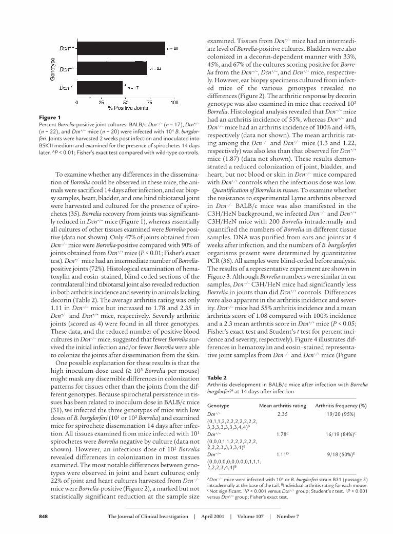
To examine whether any differences in the dissemina-tion of Borrelia could be observed in these mice, the ani-mals were sacrificed 14 days after infection, and ear biop-sy samples, heart, bladder, and one hind tibiotarsal jointwere harvested and cultured for the presence of spiro-chetes (35). Borrelia recovery from joints was significant-ly reduced in Dcn–/– mice (Figure 1), whereas essentiallyall cultures of other tissues examined were Borrelia-posi-tive (data not shown). Only 47% of joints obtained fromDcn–/– mice were Borrelia-positive compared with 90% ofjoints obtained from Dcn+/+ mice (P < 0.01; Fisher’s exacttest). Dcn+/– mice had an intermediate number of Borrelia-positive joints (72%). Histological examination of hema-toxylin and eosin–stained, blind-coded sections of thecontralateral hind tibiotarsal joint also revealed reductionin both arthritis incidence and severity in animals lackingdecorin (Table 2). The average arthritis rating was only1.11 in Dcn–/– mice but increased to 1.78 and 2.35 inDcn+/– and Dcn+/+ mice, respectively. Severely arthriticjoints (scored as 4) were found in all three genotypes.These data, and the reduced number of positive bloodcultures in Dcn–/– mice, suggested that fewer Borrelia sur-vived the initial infection and/or fewer Borrelia were ableto colonize the joints after dissemination from the skin.
One possible explanation for these results is that thehigh inoculum dose used (≥ 103 Borrelia per mouse)might mask any discernible differences in colonizationpatterns for tissues other than the joints from the dif-ferent genotypes. Because spirochetal persistence in tis-sues has been related to inoculum dose in BALB/c mice(31), we infected the three genotypes of mice with lowdoses of B. burgdorferi (101 or 102 Borrelia) and examinedmice for spirochete dissemination 14 days after infec-tion. All tissues examined from mice infected with 101
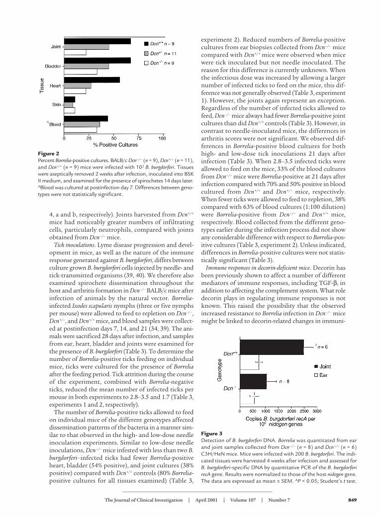
spirochetes were Borrelia negative by culture (data notshown). However, an infectious dose of 102 Borreliarevealed differences in colonization in most tissuesexamined. The most notable differences between geno-types were observed in joint and heart cultures; only22% of joint and heart cultures harvested from Dcn–/–
mice were Borrelia-positive (Figure 2), a marked but notstatistically significant reduction at the sample size
examined. Tissues from Dcn+/– mice had an intermedi-ate level of Borrelia-positive cultures. Bladders were alsocolonized in a decorin-dependent manner with 33%,45%, and 67% of the cultures scoring positive for Borre-lia from the Dcn–/–, Dcn+/–, and Dcn+/+ mice, respective-ly. However, ear biopsy specimens cultured from infect-ed mice of the various genotypes revealed nodifferences (Figure 2). The arthritic response by decoringenotype was also examined in mice that received 102
Borrelia. Histological analysis revealed that Dcn–/– micehad an arthritis incidence of 55%, whereas Dcn+/+ andDcn+/– mice had an arthritis incidence of 100% and 44%,respectively (data not shown). The mean arthritis rat-ing among the Dcn–/– and Dcn+/– mice (1.3 and 1.22,respectively) was also less than that observed for Dcn+/+
mice (1.87) (data not shown). These results demon-strated a reduced colonization of joint, bladder, andheart, but not blood or skin in Dcn–/– mice comparedwith Dcn+/+ controls when the infectious dose was low.
Quantification of Borrelia in tissues. To examine whetherthe resistance to experimental Lyme arthritis observedin Dcn–/– BALB/c mice was also manifested in theC3H/HeN background, we infected Dcn–/– and Dcn+/+
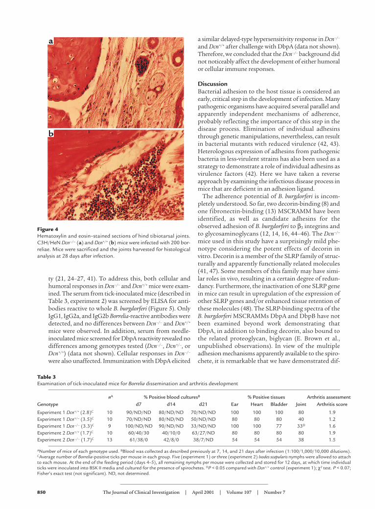
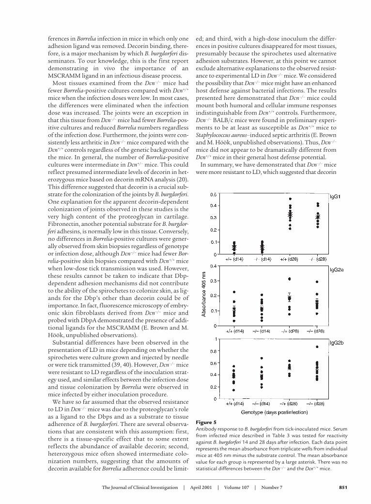
C3H/HeN mice with 200 Borrelia intradermally andquantified the numbers of Borrelia in different tissuesamples. DNA was purified from ears and joints at 4weeks after infection, and the numbers of B. burgdorferiorganisms present were determined by quantitativePCR (36). All samples were blind-coded before analysis.The results of a representative experiment are shown inFigure 3. Although Borrelia numbers were similar in earsamples, Dcn–/– C3H/HeN mice had significantly lessBorrelia in joints than did Dcn+/+ controls. Differenceswere also apparent in the arthritis incidence and sever-ity. Dcn–/– mice had 55% arthritis incidence and a meanarthritis score of 1.08 compared with 100% incidenceand a 2.3 mean arthritis score in Dcn+/+ mice (P < 0.05;Fisher’s exact test and Student’s t test for percent inci-dence and severity, respectively). Figure 4 illustrates dif-ferences in hematoxylin and eosin–stained representa-tive joint samples from Dcn–/– and Dcn+/+ mice (Figure
848 The Journal of Clinical Investigation | April 2001 | Volume 107 | Number 7
Figure 1Percent Borrelia-positive joint cultures. BALB/c Dcn–/– (n = 17), Dcn+/–
(n = 22), and Dcn+/+ mice (n = 20) were infected with 104 B. burgdor-feri. Joints were harvested 2 weeks post infection and inoculated intoBSK II medium and examined for the presence of spirochetes 14 dayslater. AP < 0.01; Fisher’s exact test compared with wild-type controls.
Table 2Arthritis development in BALB/c mice after infection with BorreliaburgdorferiA at 14 days after infection
Genotype Mean arthritis rating Arthritis frequency (%)
Dcn+/+ 2.35 19/20 (95%)(0,1,1,2,2,2,2,2,2,2,2,3,3,3,3,3,3,3,4,4)B
Dcn+/– 1.78C 16/19 (84%)C
(0,0,0,1,1,2,2,2,2,2,2,2,2,2,3,3,3,3,4)B
Dcn–/– 1.11D 9/18 (50%)E
(0,0,0,0,0,0,0,0,0,1,1,1,2,2,2,3,4,4)B
ADcn–/– mice were infected with 104 or B. burgdorferi strain B31 (passage 5)intradermally at the base of the tail. BIndividual arthritis rating for each mouse.CNot significant. DP < 0.001 versus Dcn+/+ group; Student’s t test. EP < 0.001versus Dcn+/+ group; Fisher’s exact test.
4, a and b, respectively). Joints harvested from Dcn+/+
mice had noticeably greater numbers of infiltratingcells, particularly neutrophils, compared with jointsobtained from Dcn–/– mice.
Tick inoculations. Lyme disease progression and devel-opment in mice, as well as the nature of the immuneresponse generated against B. burgdorferi, differs betweenculture grown B. burgdorferi cells injected by needle- andtick-transmitted organisms (39, 40). We therefore alsoexamined spirochete dissemination throughout thehost and arthritis formation in Dcn–/– BALB/c mice afterinfection of animals by the natural vector. Borrelia-infected Ixodes scapularis nymphs (three or five nymphsper mouse) were allowed to feed to repletion on Dcn–/–,Dcn+/–, and Dcn+/+ mice, and blood samples were collect-ed at postinfection days 7, 14, and 21 (34, 39). The ani-mals were sacrificed 28 days after infection, and samplesfrom ear, heart, bladder and joints were examined forthe presence of B. burgdorferi (Table 3). To determine thenumber of Borrelia-positive ticks feeding on individualmice, ticks were cultured for the presence of Borreliaafter the feeding period. Tick attrition during the courseof the experiment, combined with Borrelia-negativeticks, reduced the mean number of infected ticks permouse in both experiments to 2.8–3.5 and 1.7 (Table 3,experiments 1 and 2, respectively).
The number of Borrelia-positive ticks allowed to feedon individual mice of the different genotypes affecteddissemination patterns of the bacteria in a manner sim-ilar to that observed in the high- and low-dose needleinoculation experiments. Similar to low-dose needleinoculations, Dcn–/– mice infested with less than two B.burgdorferi–infected ticks had fewer Borrelia-positiveheart, bladder (54% positive), and joint cultures (38%positive) compared with Dcn+/+ controls (80% Borrelia-positive cultures for all tissues examined) (Table 3,
experiment 2). Reduced numbers of Borrelia-positivecultures from ear biopsies collected from Dcn–/– micecompared with Dcn+/+ mice were observed when micewere tick inoculated but not needle inoculated. Thereason for this difference is currently unknown. Whenthe infectious dose was increased by allowing a largernumber of infected ticks to feed on the mice, this dif-ference was not generally observed (Table 3, experiment1). However, the joints again represent an exception.Regardless of the number of infected ticks allowed tofeed, Dcn–/– mice always had fewer Borrelia-positive jointcultures than did Dcn+/+ controls (Table 3). However, incontrast to needle-inoculated mice, the differences inarthritis scores were not significant. We observed dif-ferences in Borrelia-positive blood cultures for bothhigh- and low-dose tick inoculations 21 days afterinfection (Table 3). When 2.8–3.5 infected ticks wereallowed to feed on the mice, 33% of the blood culturesfrom Dcn–/– mice were Borrelia-positive at 21 days afterinfection compared with 70% and 50% positive in bloodcultured from Dcn+/+ and Dcn+/– mice, respectively.When fewer ticks were allowed to feed to repletion, 38%compared with 63% of blood cultures (1:100 dilution)were Borrelia-positive from Dcn–/– and Dcn+/+ mice,respectively. Blood collected from the different geno-types earlier during the infection process did not showany considerable difference with respect to Borrelia-pos-itive cultures (Table 3, experiment 2). Unless indicated,differences in Borrelia-positive cultures were not statis-tically significant (Table 3).
Immune responses in decorin-deficient mice. Decorin hasbeen previously shown to affect a number of differentmediators of immune responses, including TGF-β, inaddition to affecting the complement system. What roledecorin plays in regulating immune responses is notknown. This raised the possibility that the observedincreased resistance to Borrelia infection in Dcn–/– micemight be linked to decorin-related changes in immuni-
The Journal of Clinical Investigation | April 2001 | Volume 107 | Number 7 849
Figure 2Percent Borrelia-positive cultures. BALB/c Dcn–/– (n = 9), Dcn+/– (n = 11),and Dcn+/+ (n = 9) mice were infected with 102 B. burgdorferi. Tissueswere aseptically removed 2 weeks after infection, inoculated into BSKII medium, and examined for the presence of spirochetes 14 days later.ABlood was cultured at postinfection day 7. Differences between geno-types were not statistically significant.
Figure 3Detection of B. burgdorferi DNA. Borrelia was quantitated from earand joint samples collected from Dcn–/– (n = 8) and Dcn+/+ (n = 6)C3H/HeN mice. Mice were infected with 200 B. burgdorferi. The indi-cated tissues were harvested 4 weeks after infection and assessed forB. burgdorferi-specific DNA by quantitative PCR of the B. burgdorferirecA gene. Results were normalized to those of the host nidogen gene.The data are expressed as mean ± SEM. AP < 0.05; Student’s t test.
ty (21, 24–27, 41). To address this, both cellular andhumoral responses in Dcn–/– and Dcn+/+ mice were exam-ined. The serum from tick-inoculated mice (described inTable 3, experiment 2) was screened by ELISA for anti-bodies reactive to whole B. burgdorferi (Figure 5). OnlyIgG1, IgG2a, and IgG2b Borrelia-reactive antibodies weredetected, and no differences between Dcn–/– and Dcn+/+
mice were observed. In addition, serum from needle-inoculated mice screened for DbpA reactivity revealed nodifferences among genotypes tested (Dcn–/–, Dcn+/–, orDcn+/+) (data not shown). Cellular responses in Dcn–/–
were also unaffected. Immunization with DbpA elicited
a similar delayed-type hypersensitivity response in Dcn–/–
and Dcn+/+ after challenge with DbpA (data not shown).Therefore, we concluded that the Dcn–/– background didnot noticeably affect the development of either humoralor cellular immune responses.
DiscussionBacterial adhesion to the host tissue is considered anearly, critical step in the development of infection. Manypathogenic organisms have acquired several parallel andapparently independent mechanisms of adherence,probably reflecting the importance of this step in thedisease process. Elimination of individual adhesinsthrough genetic manipulations, nevertheless, can resultin bacterial mutants with reduced virulence (42, 43).Heterologous expression of adhesins from pathogenicbacteria in less-virulent strains has also been used as astrategy to demonstrate a role of individual adhesins asvirulence factors (42). Here we have taken a reverseapproach by examining the infectious disease process inmice that are deficient in an adhesion ligand.
The adherence potential of B. burgdorferi is incom-pletely understood. So far, two decorin-binding (8) andone fibronectin-binding (13) MSCRAMM have beenidentified, as well as candidate adhesins for theobserved adhesion of B. burgdorferi to β3 integrins andto glycosaminoglycans (12, 14, 16, 44–46). The Dcn–/–
mice used in this study have a surprisingly mild phe-notype considering the potent effects of decorin invitro. Decorin is a member of the SLRP family of struc-turally and apparently functionally related molecules(41, 47). Some members of this family may have simi-lar roles in vivo, resulting in a certain degree of redun-dancy. Furthermore, the inactivation of one SLRP genein mice can result in upregulation of the expression ofother SLRP genes and/or enhanced tissue retention ofthese molecules (48). The SLRP-binding spectra of theB. burgdorferi MSCRAMMs DbpA and DbpB have notbeen examined beyond work demonstrating thatDbpA, in addition to binding decorin, also bound tothe related proteoglycan, biglycan (E. Brown et al.,unpublished observations). In view of the multipleadhesion mechanisms apparently available to the spiro-chete, it is remarkable that we have demonstrated dif-
850 The Journal of Clinical Investigation | April 2001 | Volume 107 | Number 7
Figure 4Hematoxylin and eosin–stained sections of hind tibiotarsal joints.C3H/HeN Dcn–/– (a) and Dcn+/+ (b) mice were infected with 200 bor-reliae. Mice were sacrificed and the joints harvested for histologicalanalysis at 28 days after infection.
Table 3Examination of tick-inoculated mice for Borrelia dissemination and arthritis development
nA % Positive blood culturesB % Positive tissues Arthritis assessmentGenotype d7 d14 d21 Ear Heart Bladder Joint Arthritis score
Experiment 1 Dcn+/+ (2.8)C 10 90/ND/ND 80/ND/ND 70/ND/ND 100 100 100 80 1.9Experiment 1 Dcn+/– (3.5)C 10 70/ND/ND 80/ND/ND 50/ND/ND 80 80 80 40 1.2Experiment 1 Dcn–/– (3.3)C 9 100/ND/ND 90/ND/ND 33/ND/ND 100 100 77 33D 1.6Experiment 2 Dcn+/+ (1.7)C 10 60/40/30 40/10/0 63/27/ND 80 80 80 80 1.9Experiment 2 Dcn–/– (1.7)C 13 61/38/0 42/8/0 38/7/ND 54 54 54 38 1.5
ANumber of mice of each genotype used. BBlood was collected as described previously at 7, 14, and 21 days after infection (1:100/1,000/10,000 dilutions).CAverage number of Borrelia-positive ticks per mouse in each group. Five (experiment 1) or three (experiment 2) Ixodes scapularis nymphs were allowed to attachto each mouse. At the end of the feeding period (days 4–5), all remaining nymphs per mouse were collected and stored for 12 days, at which time individualticks were inoculated into BSK II media and cultured for the presence of spirochetes. DP < 0.05 compared with Dcn+/+ control (experiment 1); χ2 test. P < 0.07;Fisher’s exact test (not significant). ND, not determined.
ferences in Borrelia infection in mice in which only oneadhesion ligand was removed. Decorin binding, there-fore, is a major mechanism by which B. burgdorferi dis-seminates. To our knowledge, this is the first reportdemonstrating in vivo the importance of anMSCRAMM ligand in an infectious disease process.
Most tissues examined from the Dcn–/– mice hadfewer Borrelia-positive cultures compared with Dcn+/+
mice when the infection doses were low. In most cases,the differences were eliminated when the infectiondose was increased. The joints were an exception inthat this tissue from Dcn–/– mice had fewer Borrelia-pos-itive cultures and reduced Borrelia numbers regardlessof the infection dose. Furthermore, the joints were con-sistently less arthritic in Dcn–/– mice compared with theDcn+/+ controls regardless of the genetic background ofthe mice. In general, the number of Borrelia-positivecultures were intermediate in Dcn+/– mice. This couldreflect presumed intermediate levels of decorin in het-erozygous mice based on decorin mRNA analysis (20).This difference suggested that decorin is a crucial sub-strate for the colonization of the joints by B. burgdorferi.One explanation for the apparent decorin-dependentcolonization of joints observed in these studies is thevery high content of the proteoglycan in cartilage.Fibronectin, another potential substrate for B. burgdor-feri adhesins, is normally low in this tissue. Conversely,no differences in Borrelia-positive cultures were gener-ally observed from skin biopsies regardless of genotypeor infection dose, although Dcn–/– mice had fewer Bor-relia-positive skin biopsies compared with Dcn+/+ micewhen low-dose tick transmission was used. However,these results cannot be taken to indicate that Dbp-dependent adhesion mechanisms did not contributeto the ability of the spirochetes to colonize skin, as lig-ands for the Dbp’s other than decorin could be ofimportance. In fact, fluorescence microscopy of embry-onic skin fibroblasts derived from Dcn–/– mice andprobed with DbpA demonstrated the presence of addi-tional ligands for the MSCRAMM (E. Brown and M.Höök, unpublished observations).
Substantial differences have been observed in thepresentation of LD in mice depending on whether thespirochetes were culture grown and injected by needleor were tick transmitted (39, 40). However, Dcn–/– micewere resistant to LD regardless of the inoculation strat-egy used, and similar effects between the infection doseand tissue colonization by Borrelia were observed inmice infected by either inoculation procedure.
We have so far assumed that the observed resistanceto LD in Dcn–/– mice was due to the proteoglycan’s roleas a ligand to the Dbps and as a substrate to tissueadherence of B. burgdorferi. There are several observa-tions that are consistent with this assumption: first,there is a tissue-specific effect that to some extentreflects the abundance of available decorin; second,heterozygous mice often showed intermediate colo-nization numbers, suggesting that the amounts ofdecorin available for Borrelia adherence could be limit-
ed; and third, with a high-dose inoculum the differ-ences in positive cultures disappeared for most tissues,presumably because the spirochetes used alternativeadhesion substrates. However, at this point we cannotexclude alternative explanations to the observed resist-ance to experimental LD in Dcn–/– mice. We consideredthe possibility that Dcn–/– mice might have an enhancedhost defense against bacterial infections. The resultspresented here demonstrated that Dcn–/– mice couldmount both humoral and cellular immune responsesindistinguishable from Dcn+/+ controls. Furthermore,Dcn–/– BALB/c mice were found in preliminary experi-ments to be at least as susceptible as Dcn+/+ mice toStaphylococcus aureus–induced septic arthritis (E. Brownand M. Höök, unpublished observations). Thus, Dcn–/–
mice did not appear to be dramatically different fromDcn+/+ mice in their general host defense potential.
In summary, we have demonstrated that Dcn–/– micewere more resistant to LD, which suggested that decorin
The Journal of Clinical Investigation | April 2001 | Volume 107 | Number 7 851
Figure 5Antibody response to B. burgdorferi from tick-inoculated mice. Serumfrom infected mice described in Table 3 was tested for reactivityagainst B. burgdorferi 14 and 28 days after infection. Each data pointrepresents the mean absorbance from triplicate wells from individualmice at 405 nm minus the substrate control. The mean absorbancevalue for each group is represented by a large asterisk. There was nostatistical differences between the Dcn–/– and the Dcn+/+ mice.

could be a limiting factor as a substrate for the adher-ence of B. burgdorferi. However, this resistance could beovercome for most tissues examined by increasing theinfection dose, suggesting that additional adhesion lig-ands could participate in the disease process in vivo.
AcknowledgmentsThis work was supported by a Department of Healthand Human Services cooperative agreement from theCenter for Disease Control (CCU614695 to E. Brownand M. Höök), a grant from the Neva Wesley WestFoundation (to M. Höök), an NIH grant AR43521 (toJ. Weis), an Arthritis Foundation Postdoctoral Fellow-ship (to R.M. Wooten), and grant 5P03-CA-42104 tothe University of Utah. Special thanks are extended toHowell Hankamer for excellent technical assistance inthe Program for Animal Resources.
1. Steere, A.C., et al. 1985. Successful parenteral penicillin therapy of estab-lished Lyme arthritis. N. Engl. J. Med. 312:869–874.
2. Steere, A.C. 1989. Lyme disease. N. Engl. J. Med. 321:586–596.3. Steere, A.C., et al. 1998. Vaccination against Lyme disease with recombinant
Borrelia burgdorferi outer-surface lipoprotein A with adjuvant. Lyme DiseaseVaccine Study Group. N. Engl. J. Med. 339:209–215.
4. Szczepanski, A., and Benach, J.L. 1991. Lyme borreliosis: host responses toBorrelia burgdorferi. Microbiol. Rev. 55:21–34.
5. Steere, A.C., et al. 1994. Treatment of Lyme arthritis. Arthritis Rheum.37:878–888.
6. Dattwyler, R.J., and Halperin, J.J. 1987. Failure of tetracycline therapy inearly Lyme disease. Arthritis Rheum. 30:448–450.
7. Patti, J.M., Allen, B.L., McGavin, M.J., and Höök, M. 1994. MSCRAMM-mediated adherence of microorganisms to host tissues. Annu. Rev. Microbi-ol. 48:585–617.
8. Guo, B.P., Brown, E.L., Dorward, D.W., Rosenberg, L.C., and Höök, M. 1998.Decorin-binding adhesins from Borrelia burgdorferi. Mol. Microbiol.30:711–723.
9. Guo, B.P., Norris, S.J., Rosenberg, L.C., and Höök, M. 1995. Adherence ofBorrelia burgdorferi to the proteoglycan decorin. Infect. Immun.63:3467–3472.
10. Duray, P.H. 1992. Target organs of Borrelia burgdorferi infections: function-al responses and histology. In Lyme disease: molecular and immunologicapproaches. S.E. Schutzer, editor. Cold Spring Harbor Laboratory Press. ColdSpring Harbor, New York, USA. 11–30.
11. Van Mierlo, P., Jacob, W., and Dockx, P. 1993. Erythema chronicummigrans: an electron-microscopic study. Dermatology. 186:306–310.
12. Magoun, L., et al. 2000. Variable small protein (Vsp)-dependent and Vsp-independent pathways for glycosaminoglycan recognition by relapsingfever spirochaetes. Mol. Microbiol. 36:886–897.
13. Probert, W.S., and Johnson, B.J. 1998. Identification of a 47 kDafibronectin-binding protein expressed by Borrelia burgdorferi isolate B31.Mol. Microbiol. 30:1003–1015.
14. Parveen, N., and Leong, J.M. 2000. Identification of a candidate gly-cosaminoglycan-binding adhesin of the Lyme disease spirochete Borreliaburgdorferi. Mol. Microbiol. 35:1220–1234.
15. Cinco, M., Murgia, R., Presani, G., and Perticarari, S. 1997. Integrin CR3mediates the binding of nonspecifically opsonized Borrelia burgdorferi tohuman phagocytes and mammalian cells. Infect. Immun. 65:4784–4789.
16. Coburn, J., Chege, W., Magoun, L., Bodary, S.C., and Leong, J.M. 1999. Char-acterization of a candidate Borrelia burgdorferi beta3-chain integrin ligandidentified using a phage display library. Mol. Microbiol. 34:926–940.
17. Isaacs, R.D. 1994. Borrelia burgdorferi bind to epithelial cell proteoglycans. J.Clin. Invest. 93:809–819.
18. Klempner, M.S., Noring, R., and Rogers, R.A. 1993. Invasion of human skinfibroblasts by the Lyme disease spirochete, Borrelia burgdorferi. J. Infect. Dis.167:1074–1081.
19. Szczepanski, A., Furie, M.B., Benach, J.L., Lane, B.P., and Fleit, H.B. 1990.Interaction between Borrelia burgdorferi and endothelium in vitro. J. Clin.Invest. 85:1637–1647.
20. Danielson, K.G., et al. 1997. Targeted disruption of decorin leads to abnor-mal collagen fibril morphology and skin fragility. J. Cell. Biol. 136:729–743.
21. De Luca, A., Santra, M., Baldi, A., Giordano, A., and Iozzo, R.V. 1996.Decorin-induced growth suppression is associated with up-regulation of
p21, an inhibitor of cyclin-dependent kinases. J. Biol. Chem.271:18961–18965.
22. Border, W.A., and Ruoslahti, E. 1990. Transforming growth factor-beta 1induces extracellular matrix formation in glomerulonephritis. Cell Differ.Dev. 32:425–431.
23. Breuer, B., Schmidt, G., and Kresse, H. 1990. Non-uniform influence oftransforming growth factor-beta on the biosynthesis of different forms ofsmall chondroitin sulphate/dermatan sulphate proteoglycan. Biochem. J.269:551–554.
24. Krumdieck, R., Höök, M., Rosenberg, L.C., and Volanakis, J.E. 1992. Theproteoglycan decorin binds C1q and inhibits the activity of the C1 com-plex. J. Immunol. 149:3695–3701.
25. Takagi, M., Yamada, T., Kamiya, N., Kumagai, T., and Yamaguchi, A. 1999.Effects of bone morphogenetic protein-2 and transforming growth factor-beta1 on gene expression of decorin and biglycan by cultured osteoblasticcells. Histochem. J. 31:403–409.
26. Winnemoller, M., Schmidt, G., and Kresse, H. 1991. Influence of decorinon fibroblast adhesion to fibronectin. Eur. J. Cell Biol. 54:10–17.
27. Winnemoller, M., Schon, P., Vischer, P., and Kresse, H. 1992. Interactionsbetween thrombospondin and the small proteoglycan decorin: interferencewith cell attachment. Eur. J. Cell. Biol. 59:47–55.
28. Barthold, S.W., Sidman, C.L., and Smith, A.L. 1992. Lyme borreliosis ingenetically resistant and susceptible mice with severe combined immun-odeficiency. Am. J. Trop. Med. Hyg. 47:605–613.
29. Barthold, S.W., Beck, D.S., Hansen, G.M., Terwilliger, G.A., and Moody,K.D. 1990. Lyme borreliosis in selected strains and ages of laboratory mice.J. Infect. Dis. 162:133–138.
30. Ma, Y., et al. 1998. Inhibition of collagen-induced arthritis in mice by viralIL-10 gene transfer. J. Immunol. 161:1516–1524.
31. Brown, C.R., and Reiner, S.L. 1999. Genetic control of experimental lymearthritis in the absence of specific immunity. Infect. Immun. 67:1967–1973.
32. Purser, J.E., and Norris, S.J. 2000. Correlation between plasmid content andinfectivity in Borrelia burgdorferi. Proc. Natl. Acad. Sci. USA. 97:13865–13870.
33. Barbour, A.G. 1984. Isolation and cultivation of Lyme disease spirochetes.Yale J. Biol. Med. 57:521–525.
34. Piesman, J. 1993. Standard system for infecting ticks (Acari: Ixodidae) withthe Lyme disease spirochete, Borrelia burgdorferi. J. Med. Entomol. 30:199–203.
35. Pride, M.W., et al. 1998. Specific Th1 cell lines that confer protective immu-nity against experimental Borrelia burgdorferi infection in mice. J. Leukoc. Biol.63:542–549.
36. Brown, J.P., Zachary, J.F., Teuscher, C., Weis, J.J., and Wooten, R.M. 1999.Dual role of interleukin-10 in murine Lyme disease: regulation of arthritisseverity and host defense. Infect. Immun. 67:5142–5150.
37. Morrison, T.B., Ma, Y., Weis, J.H., and Weis, J.J. 1999. Rapid and sensitivequantification of Borrelia burgdorferi-infected mouse tissues by continuousfluorescent monitoring of PCR. J. Clin. Microbiol. 37:987–992.
38. Ma, Y., et al. 1998. Distinct characteristics of resistance to Borrelia burgdor-feri-induced arthritis in C57BL/6N mice. Infect. Immun. 66:161–168.
39. Zeidner, N., et al. 1997. Effects of Ixodes scapularis and Borrelia burgdorferi onmodulation of the host immune response: induction of a TH2 cytokineresponse in Lyme disease-susceptible (C3H/HeJ) mice but not in disease-resistant (BALB/c) mice. Infect. Immun. 65:3100–3106.
40. Zeidner, N., Dreitz, M., Belasco, D., and Fish, D. 1996. Suppression of acuteIxodes scapularis-induced Borrelia burgdorferi infection using tumor necrosisfactor-alpha, interleukin-2, and interferon-gamma. J. Infect. Dis.173:187–195.
41. Iozzo, R.V. 1998. Matrix proteoglycans: from molecular design to cellularfunction. Annu. Rev. Biochem. 67:609–652.
42. Patti, J.M., et al. 1994. The Staphylococcus aureus collagen adhesin is a viru-lence determinant in experimental septic arthritis. Infect. Immun.62:152–161.
43. Vaudaux, P.E., et al. 1995. Use of adhesion-defective mutants of Staphylo-coccus aureus to define the role of specific plasma proteins in promoting bac-terial adhesion to canine arteriovenous shunts. Infect. Immun. 63:585–590.
44. Coburn, J., Leong, J.M., and Erban, J.K. 1993. Integrin alpha IIb beta 3 medi-ates binding of the Lyme disease agent Borrelia burgdorferi to humanplatelets. Proc. Natl. Acad. Sci. USA. 90:7059–7063.
45. Coburn, J., Barthold, S.W., and Leong, J.M. 1994. Diverse Lyme diseasespirochetes bind integrin alpha IIb beta 3 on human platelets. Infect. Immun.62:5559–5567.
46. Coburn, J., Magoun, L., Bodary, S.C., and Leong, J.M. 1998. Integrinsalpha(v)beta3 and alpha5beta1 mediate attachment of Lyme disease spiro-chetes to human cells. Infect. Immun. 66:1946–1952.
47. Iozzo, R.V. 1997. The family of small leucine-rich proteoglycans: key regu-lators of matrix assembly and cellular growth. Crit. Rev. Biochem. Mol. Biol.32:141–174.
48. Svensson, L., et al. 1999. Fibromodulin-null mice have abnormal collagenfibrils, tissue organization, and altered lumican deposition in tendon. J. Biol.Chem. 274:9636–9647.
852 The Journal of Clinical Investigation | April 2001 | Volume 107 | Number 7