Embed Size (px)
Citation preview
Resistance to Botrytis cinerea in sitiens, an AbscisicAcid-Deficient Tomato Mutant, Involves TimelyProduction of Hydrogen Peroxide and Cell WallModifications in the Epidermis1[C][W][OA]
Bob Asselbergh2, Katrien Curvers2, Soraya C. Francxa2, Kris Audenaert3, Marnik Vuylsteke,Frank Van Breusegem, and Monica Hofte*
Laboratory of Phytopathology (B.A., K.C., S.C.F., K.A., M.H.) and Department of Molecular Genetics(B.A., K.C., M.V., F.V.B.), Ghent University, B–9000 Ghent, Belgium; and Department of Plant SystemsBiology, Flanders Institute for Biotechnology, B–9052 Ghent, Belgium (B.A., K.C., M.V., F.V.B.)
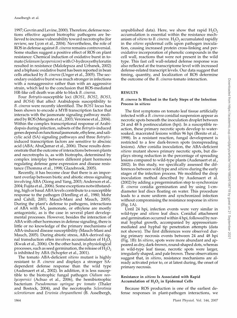
Plant defense mechanisms against necrotrophic pathogens, such as Botrytis cinerea, are considered to be complex and to differfrom those that are effective against biotrophs. In the abscisic acid-deficient sitiens tomato (Solanum lycopersicum) mutant, whichis highly resistant to B. cinerea, accumulation of hydrogen peroxide (H2O2) was earlier and stronger than in the susceptible wildtype at the site of infection. In sitiens, H2O2 accumulation was observed from 4 h postinoculation (hpi), specifically in the leafepidermal cell walls, where it caused modification by protein cross-linking and incorporation of phenolic compounds. In wild-type tomato plants, H2O2 started to accumulate 24 hpi in the mesophyll layer and was associated with spreading cell death.Transcript-profiling analysis using TOM1 microarrays revealed that defense-related transcript accumulation prior to infectionwas higher in sitiens than in wild type. Moreover, further elevation of sitiens defense gene expression was stronger than in wildtype 8 hpi both in number of genes and in their expression levels and confirmed a role for cell wall modification in the resistantreaction. Although, in general, plant defense-related reactive oxygen species formation facilitates necrotrophic colonization,these data indicate that timely hyperinduction of H2O2-dependent defenses in the epidermal cell wall can effectively blockearly development of B. cinerea.
Botrytis cinerea causes gray mold diseases in a broadrange of plant species and is one of the most com-prehensively studied necrotrophic plant pathogens.Necrotrophs kill their host cells by secreting toxiccompounds or lytic enzymes and, in addition, producean array of pathogenicity factors that can subdue hostdefenses (for review, see van Kan, 2006). Despite
elaborate research studies, the biochemical and geneticbasis of resistance to Botrytis is still not fully under-stood. The ability of the fungus to kill cells was pro-posed as a major determinant in host specificity ofdifferent Botrytis species (Mansfield and Hutson, 1980)and, similarly, plant resistance to Botrytis is supposed todepend on the balance between cell death and survival(van Baarlen et al., 2007). In addition, constitutive andinducible plant secondary metabolites determine hostspecificity of different Botrytis species and fungal col-onization in compatible interactions (for review, seevan Baarlen et al., 2004). Structural barriers and cellwall fortifications are also considered to be involved inarresting Botrytis, although the actual contribution tothe effective inhibition of infection is often unclear (vanBaarlen et al., 2004).
One of the most important plant defense responsesto pathogens is the production of reactive oxygenspecies (ROS), such as hydrogen peroxide (H2O2),during oxidative burst (Mehdy, 1994). ROS can func-tion in cell wall modification, defense signaling, thehypersensitive response (HR), or be directly toxic topathogens (Lamb and Dixon, 1997). There is evidence,however, that generation of ROS assists the coloniza-tion of plant tissue by necrotrophic pathogens: B. cinereainfection can be suppressed by spraying antioxidantson plants (Elad, 1992) and H2O2 is produced duringcommon bean (Phaseolus vulgaris) and Arabidopsis(Arabidopsis thaliana) colonization (von Tiedemann,
1 This work was supported by the Research Fund of GhentUniversity (Geconcerteerde Onderzoeksacties grant no. 12051403),the ‘‘Fonds voor Wetenschappelijk Onderzoek-Vlaanderen’’ (grantnos. G.0350.04 and G.0061.03), and the ‘‘Instituut voor de Aanmoe-diging van Innovatie door wetenschap en Technologie in Vlaande-ren’’ (predoctoral fellowship to K.C.).
2 These authors contributed equally to the article.3 Present address: Department of Biosciences and Landscape
Architecture, University College Ghent, Voskenslaan 270, B–9000Ghent, Belgium.
* Corresponding author; e-mail [email protected]; fax 32–9–264–62–38.
The author responsible for distribution of materials integral to thefindings presented in this article in accordance with the policydescribed in the Instructions for Authors (www.plantphysiol.org) is:Monica Hofte ([email protected]).
[C] Some figures in this article are displayed in color online but inblack and white in the print edition.
[W] The online version of this article contains Web-only data.[OA] Open Access articles can be viewed online without a sub-
scription.www.plantphysiol.org/cgi/doi/10.1104/pp.107.099226
Plant Physiology, August 2007, Vol. 144, pp. 1863–1877, www.plantphysiol.org � 2007 American Society of Plant Biologists 1863
Dow
nloaded from https://academ
ic.oup.com/plphys/article/144/4/1863/6106718 by guest on 21 Septem
ber 2021
1997; Govrin and Levine, 2000). Therefore, defense reac-tions effective against biotrophic pathogens are be-lieved to increase vulnerability toward necrotrophs (forreview, see Lyon et al., 2004). Nevertheless, the role ofROS in defense against B. cinerea remains controversial.Some studies suggest a positive effect of ROS on plantresistance: Chemical induction of oxidative burst in to-mato (Solanum lycopersicon) with O-hydroxyethylorutinresulted in resistance (Malolepsza and Urbanek, 2002)and a biphasic oxidative burst has been reported in beancells attacked by B. cinerea (Unger et al., 2005). The sec-ondary oxidative burst was much stronger in infectionswith a nonaggressive rather than with an aggressivestrain, which led to the conclusion that ROS-mediatedHR-like cell death was able to block B. cinerea.
Four Botrytis-susceptible loci (BOS1, BOS2, BOS3,and BOS4) that affect Arabidopsis susceptibility toB. cinerea were recently identified. The BOS1 locus hasbeen shown to encode a MYB transcription factor thatinteracts with the jasmonate signaling pathway medi-ated by ROS (Mengiste et al., 2003; Veronese et al., 2004).Within the complex transcriptional response of Arabi-dopsis during infection, subsets of the Botrytis-inducedgenesdependonfunctional jasmonate,ethylene,andsali-cylic acid (SA) signaling pathways and three Botrytis-induced transcription factors are sensitive to abscisicacid (ABA; AbuQamar et al., 2006). These results dem-onstrate that the outcome of interactions between plantsand necrotrophs is, as with biotrophs, determined bycomplex interplay between different plant hormonesregulating defense gene expression and disease resis-tance (Thomma et al., 1998; Glazebrook, 2005).
Recently, it has become clear that there is an impor-tant overlap between biotic and abiotic stress signalinginvolving ABA (Xiong and Yang, 2003; Anderson et al.,2004; Fujita et al., 2006). Some exceptions notwithstand-ing, high or basal ABA levels contribute to a susceptibleresponse to the pathogen (Henfling et al., 1980; Mohrand Cahill, 2001; Mauch-Mani and Mauch, 2005).During the plant’s defense to pathogens, interactionsof ABA with SA, jasmonate, or ethylene are mostlyantagonistic, as is the case in several plant develop-mental processes. However, besides the interaction ofABAwith other hormones in defense signaling, there islittle or no knowledge of the primary mechanisms ofABA-induced disease susceptibility (Mauch-Mani andMauch, 2005). During abiotic stress, ABA-derived sig-nal transduction often involves accumulation of H2O2(Kwak et al., 2006). On the other hand, in physiologicalprocesses, such as seed germination, the release of H2O2is inhibited by ABA (Schopfer et al., 2001).
The tomato ABA-deficient sitiens mutant is highlyresistant to B. cinerea and displays a stronger SA-dependent defense response than the wild type(Audenaert et al., 2002). In addition, it is less suscep-tible to the biotrophic fungal pathogen Oidium neo-lycopersici (Achuo et al., 2006), the hemibiotrophicbacterium Pseudomonas syringae pv tomato (Thalerand Bostock, 2004), and the necrotrophs Sclerotiniasclerotiorum and Erwinia chrysanthemi (B. Asselbergh,
unpublished data). Here, we show that rapid H2O2accumulation is essential within the resistance mech-anism of sitiens to B. cinerea. H2O2 accumulated rapidlyin the sitiens epidermal cells upon pathogen inocula-tion, causing increased protein cross-linking and per-oxidative incorporation of phenolic compounds in thecell wall, reactions that were not present in the wildtype. This fast cell wall-related defense response wasalso reflected at the transcriptome level with increaseddefense-related transcript levels. Our data suggest thattiming, quantity, and localization of ROS determinethe outcome of the B. cinerea-tomato interaction.
RESULTS
B. cinerea Is Blocked in the Early Steps of the InfectionProcess in sitiens
The first symptoms on tomato leaf tissue artificiallyinfected with a B. cinerea conidial suspension appear asnecrotic spots beneath the inoculation droplet between24 and 48 h postinoculation (hpi). In a susceptible re-action, these primary necrotic spots develop to water-soaked, macerated lesions within 96 hpi (Benito et al.,1998). In a resistant reaction, fungal development isrestricted to a few dark-brown spots (nonspreadinglesions). After conidia inoculation, the ABA-deficientsitiens mutant shows primary necrotic spots, but dis-plays strong reduction in the percentage of spreadinglesions compared to wild-type plants (Audenaert et al.,2002). In this study, we specifically assessed the dif-ferences between wild type and sitiens during the earlystages of the infection process. We modified the dropinoculation method described by Audenaert et al.(2002) by adding a pregermination step to synchronizeB. cinerea conidia germination and by using 1-cm-diameter leaf discs floating on water. This procedureleads to a more uniform and synchronized infectionwithout compromising the resistance response in sitiens(Fig. 1A).
Until 24 hpi, infection events were very similar inwild-type and sitiens leaf discs. Conidial attachmentand germination occurred within 4 hpi, followed by nor-mal hyphal growth, accompanied by appressorium-mediated and hyphal tip penetration attempts (datanot shown). The first differences were observed dur-ing primary necrosis events between 24 and 48 hpi(Fig. 1B): In sitiens, spots were more abundant and ap-peared as dry, dark-brown, round-shaped dots, whereasin wild-type leaf tissue, necrotic spots were larger,irregularly shaped, and pale brown. These observationssuggest that, in sitiens, resistance mechanisms are al-ready activated prior to, or at latest during, the onset ofprimary necrosis.
Resistance in sitiens Is Associated with RapidAccumulation of H2O2 in Epidermal Cells
Because ROS production is one of the earliest de-fense responses in plant-pathogen interactions, we
Asselbergh et al.
1864 Plant Physiol. Vol. 144, 2007
Dow
nloaded from https://academ
ic.oup.com/plphys/article/144/4/1863/6106718 by guest on 21 Septem
ber 2021

compared H2O2 accumulation at the inoculation siteby using 3,3#-diaminobenzidine (DAB) staining. Inthis protocol, brown precipitates are formed at thesites of H2O2 accumulation (Thordal-Christensen et al.,1997). Wild-type and sitiens leaf discs were inoculatedwith a droplet of a B. cinerea conidial suspension andfloated for 3 h in a solution containing 1 mg/mL DABbefore sampling 4, 8, 12, 24, 48, and 72 hpi. In mock-infected leaf discs, no DAB accumulation was observed.In wild type, DAB staining was macroscopically detect-able at 48 hpi and was associated with lesion progres-sion. In sitiens, staining became macroscopically visiblefrom 8 hpi and further intensified at later time points, butremained restricted to the area covered by the infectiondroplet (Fig. 2). When the DAB solution was supple-mented with ascorbic acid, staining was abolished,indicating that staining was due to H2O2 accumulation(data not shown).
Microscopic observations revealed that, in sitiens,DAB staining was detectable as early as 4 hpi inepidermal cell walls in close contact to the fungal germtubes (Fig. 3A). Between 4 and 8 hpi, H2O2 accumu-lation was detected also in the entire anticlinal wall ofepidermal cells in contact with the fungus and inneighboring epidermal cells. The spreading of H2O2accumulation from the sites of fungal contact resultedin general DAB staining of mainly the anticlinal wallsof the majority of epidermal cells beneath the infectiondroplet at 8 hpi (Fig. 3B). At later time points, extra-cellular H2O2 accumulation gradually decreased, butfrom 12 hpi, H2O2 was also clearly visible insidemultiple epidermal cells in sitiens (Fig. 3C). Epidermalcells with intracellular H2O2 accumulation displayedautofluorescence due to accumulation of phenolic com-pounds and cytoplasmic aggregation (Fig. 4), two fea-tures that are considered as hallmarks of a HR (Heath,2000). None of these reactions were visible in wild-
type cells at these early time points. Instead, the devel-oping lesion showed intense DAB staining in themesophyll layer that started between 24 and 48 hpi(Fig. 3; data not shown).
Nitroblue tetrazolium (NBT) was used to visualizeaccumulation of superoxide (Doke, 1983) in wild-typeand sitiens leaf tissue after inoculation with B. cinerea.NBT accumulated only at the leaf disc border and wasnot detected at the infection site at any of the examinedtime points (Supplemental Fig. S1).
H2O2 Burst Is Necessary for sitiens Resistance
To address whether the rapid H2O2 burst in sitiensis directly inferred from ABA deficiency, exogenousABA (100 mM) was applied before infection and DABstaining. Petiole feeding with exogenous ABA 24 hbefore infection was shown to restore B. cinerea sus-ceptibility of sitiens to wild-type levels (Audenaertet al., 2002). Repetitive spraying of ABA had the sameeffect (data not shown; see also Achuo et al., 2006) andsuppressed H2O2 accumulation (Fig. 5A). Microscopicassessment during the early stages of infection con-firmed that ABA supplementation decreased thenumber of DAB-positive epidermal cells toward theamount detectable in wild-type tissue (Fig. 3).
Artificial impairment of the rapid oxidative burstin sitiens also resulted in restoration of susceptibility.Floating of sitiens leaf discs on a solution supplementedwith 1,100 units/mL catalase or 5 mM ascorbate dur-ing conidia inoculation significantly increased or to-tally reestablished susceptibility, respectively (Fig. 5C).Whereas catalase treatment did not significantly affectsusceptibility of the wild type, ascorbate provokedpathogen lesions to spread earlier than in untreatedleaf discs and led to abundant superficial fungal growthand accelerated sporulation (Fig. 5C). Interestingly,
Figure 1. Disease symptoms onwild-type and sitiens tomato leafdiscs after infection with two 5-mLdrops of a B. cinerea conidial sus-pension. A, Macroscopic diseasesymptoms. In wild-type leaf discs,spreading gray disintegration of thehost tissue is visible and in sitiensdiscs necrotic spots at the site of theinfection droplet are observed. B,Close up of macroscopic diseasesymptoms. In sitiens, primary ne-crotic lesions contain a high num-ber of distinct dark spots at 48 and72 hpi. In wild type, primary ne-crotic spots at 48 hpi are typicallylarger, less numerous, have a palershade of brown, and more often de-velop into spreading water-soakedlesions.
H2O2 Burst in Resistance to B. cinerea in sitiens Tomato
Plant Physiol. Vol. 144, 2007 1865
Dow
nloaded from https://academ
ic.oup.com/plphys/article/144/4/1863/6106718 by guest on 21 Septem
ber 2021

only treatment with ascorbate completely eliminatedall detectable H2O2 from the sitiens leaf tissue. Thepartial nature of enzymatic H2O2 removal with cata-lase was observed both macroscopically (Fig. 5A) andmicroscopically: Catalase treatment resulted in faintDAB staining that covered only part of the epidermalanticlinal walls (Fig. 5B) without altering the numberof cells that displayed extracellular or intracellular H2O2accumulation (data not shown). Application of 50 mM
diphenilene iodonium (DPI), an inhibitor of the ROS-generating enzyme NADPH oxidase, restored B. cinereasusceptibility to wild-type levels (Fig. 5C). DPI inhib-ited the specific accumulation of H2O2 underneath theinoculation droplet, but also caused nonspecific back-ground staining covering the entire leaf disc surface(Fig. 5, A and B).
H2O2 Accumulation in sitiens Coincides with Elevated
Levels of Extracellular Peroxidase and Modification ofthe Epidermal Anticlinal Cell Walls
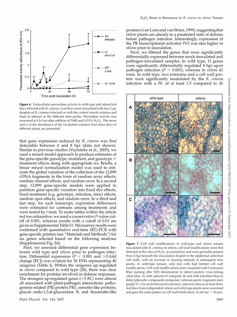
Because H2O2 production after pathogen attack canresult from increased peroxidase activity and peroxi-dases mediate many H2O2-related defense responses,we examined extracellular peroxidase activity with thetetramethylbenzoidine (TMB) assay described by RosBarcelo (1998). Leaf discs inoculated with B. cinereawere fixed in ethanol and incubated in a solution ofTMB and H2O2. Peroxidation of the TMB moleculeresulted in blue discoloration of both leaf tissue andincubation solution. The latter was used to quantifythe activity of extracellular peroxidases (Lucena et al.,2003). In wild-type leaf discs, a minor increase wasdetected 72 hpi, whereas in sitiens peroxidase activityincreased significantly between 4 and 24 hpi, followedby a drastic and sustained increase at 24 hpi (Fig. 6). Inmock-infected sitiens leaf discs, peroxidase levels alsoincreased at 48 and 72 hpi, but levels remained lowerthan in infected sitiens discs.
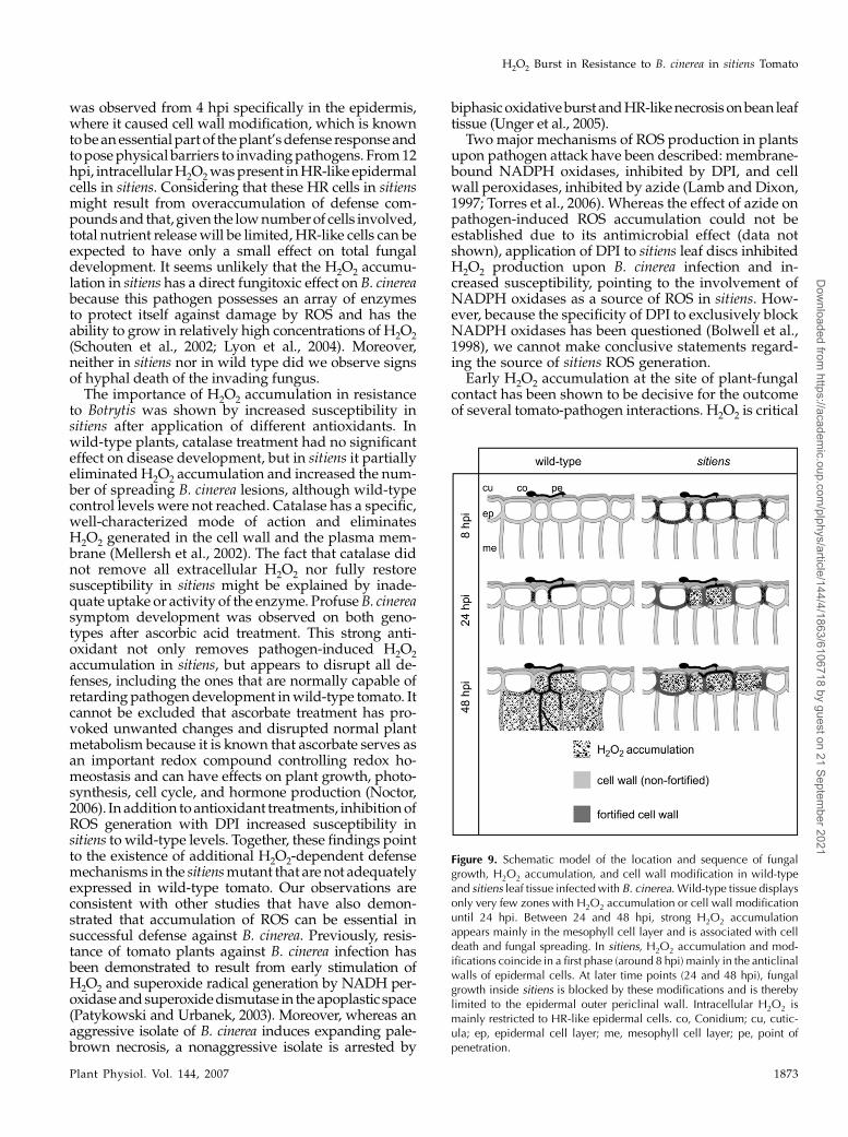
Because peroxidase-dependent defense responsesoften use H2O2 as a substrate to cross-link cell wallcomponents, we performed different staining proce-dures to visualize changes in the cell wall. Cross-linkingof cell wall proteins was detected with Coomassie Bluesubsequent to protein denaturation and free proteinremoval (Mellersh et al., 2002; Fig. 7A). In addition, weused safranin-O and toluidine blue to detect the per-oxidative incorporation of phenolic compounds in thecell wall, a fortification mechanism important duringlignification and suberization (Mellersh et al., 2002;Lucena et al., 2003; Fig. 7, B and C). For both genotypes,no staining was visible outside the infection droplets.Cell wall modification was more abundant and ap-peared earlier in sitiens than in the wild type. Moreover,accumulation of all three stains clearly coincided intiming and location with the presence of extracellularH2O2: Starting from 8 hpi, the anticlinal cell walls ofsitiens epidermal cells stained intensely during the timecourse (12, 16, 20, 24, 48, and 72 hpi; data not shown),whereas in wild-type tissue, only limited zones of the
Figure 2. Temporal evolution of H2O2 accumulation in wild-type andsitiens tomato after infection with B. cinerea. DAB staining of leaf discsinfected with two 5-mL drops of a conidial suspension was performed atdifferent time points post inoculation (4, 8, 12, 16, 20, 24, 48, and 72hpi). One representative disc out of three replicates is shown for eachtime point. [See online article for color version of this figure.]
Asselbergh et al.
1866 Plant Physiol. Vol. 144, 2007
Dow
nloaded from https://academ
ic.oup.com/plphys/article/144/4/1863/6106718 by guest on 21 Septem
ber 2021

Figure 3. Effect of ABA on H2O2 accumulation in epidermal cells after infection with B. cinerea. A, Association of DABaccumulation with B. cinerea conidia. In sitiens, H2O2 was located at the site of penetration and in some parts of the anticlinalwall of the penetrated cell, whereas in wild type and in sitiens supplemented with ABA, no H2O2 accumulation was detected.Germinating conidia were classified in two groups based on the presence or absence of associated DAB accumulation in theepidermal cells, whose percentage is shown. B, DAB accumulation in epidermal cell walls. In sitiens, DAB staining was generalin the entire outline of the anticlinal cell wall, whereas in wild type and sitiens supplemented with ABA, no H2O2 accumulationwas detected at 8 hpi (top). The restriction of sitiens DAB accumulation to the epidermal layer was confirmed on cross sections(bottom). Epidermal cells were classified in two groups based on the presence or absence of DAB accumulation in the anticlinalwalls, whose percentage is shown in the anticlinal walls at 8, 16, and 20 hpi. C, Intracellular DAB accumulation in epidermalcells showing an HR-like reaction. At 16 hpi, wild-type and ABA-supplemented sitiens epidermal cells did not accumulateintracellular DAB, whereas in sitiens, groups of HR-like cells with intracellular DAB accumulation were present near the site offungal penetration. Epidermal cells were classified in two groups based on the presence or absence of intracellular DABaccumulation and the percentage is shown at 12, 16, and 20 hpi. In all graphs, bars represent the means and the SDs of data fromsix inoculation droplets originating from three plants. In each inoculation droplet, at least 50 conidia (A) or 300 epidermal cells(B and C) originating from representative zones within each inoculation droplet were counted. Data from one experiment ispresented. The experiment was repeated with similar results. Scale bar 5 50 mm.
H2O2 Burst in Resistance to B. cinerea in sitiens Tomato
Plant Physiol. Vol. 144, 2007 1867
Dow
nloaded from https://academ
ic.oup.com/plphys/article/144/4/1863/6106718 by guest on 21 Septem
ber 2021

anticlinal cell walls of a few cells were positive and onlyafter 12 hpi (Fig. 7). Underneath the inoculation droplet,the number of epidermal cells displaying clear cell wallmodification in the anticlinal walls ranged between40% and 80% in sitiens, but was never higher than 20%in wild-type cell walls (data not shown).
H2O2 dependency of the cell wall modification insitiens was further confirmed on leaf discs treated withantioxidants. Whereas ascorbate treatment removedall sitiens cell wall modifications, treatment with cat-alase resulted in lower staining intensity (Fig. 8).
Transcriptome Profiling of the TomatoResponse to B. cinerea
To assess changes in gene expression, we comparedthe transcriptome of wild-type and sitiens plants in-fected with B. cinerea. Detached leaves were sprayinfected with a conidial suspension to ensure a stan-dardized sampling procedure because of a uniforminfection covering the entire leaf area. Disease scoringshowed that symptom development was similar tothat after drop inoculation, with sitiens being moreresistant than the wild type (Supplemental Fig. S2). Weperformed a genome-wide transcriptome analysis withTOM1 tomato cDNA arrays containing 12,899 ESTs,representing approximately 8,600 unigenes (Alba et al.,2004). A loop design was constructed, consisting of sevenreplicated dye-swap experiments in which two inde-pendent pools of four leaflets harvested from 5-week-old mock-sprayed and spray-infected plants at 0 and 8hpi were compared in an equal treatment replicationstructure on a total of 14 TOM1 cDNA microarrays(Supplemental Fig. S3). We chose to sample at 8 hpibecause, at that stage, no necrosis is visible yet, despiteobvious fungal penetration attempts and H2O2 accumu-lation. In addition, time-course analyses with cDNA-amplified fragment length polymorphism indicated
Figure 4. HR-like reaction of sitiens epidermal cells at 12 hpi. Cellsnear the site of fungal penetration with this reaction showed intracel-lular DAB accumulation (A), cytoplasmic aggregation (B), and green toyellow autofluorescence after UV excitation (C). Scale bar 5 50 mm.[See online article for color version of this figure.]
Figure 5. Effect of ABA, ascorbate, catalase, and DPI treatment onH2O2 accumulation and symptom development in wild-type andsitiens tomato leaf discs after inoculation with two 5-mL droplets ofB. cinerea conidial suspension. A, DAB staining at 20 hpi. Onerepresentative disc out of three replicates is presented. B, Micrographof sitiens H2O2 accumulation at 8 hpi. C, B. cinerea symptom devel-opment. For each treatment, at least 18 leaf discs from at least fiveplants were infected and the number of spreading lesions was evalu-ated at 4 dpi. Data were statistically analyzed using binary logisticregression. Bars with different letters are significantly different at P ,
0.05. Profuse pathogen development is visible on discs treated withascorbic acid at 4 dpi. All experiments were repeated with similarresults.
Asselbergh et al.
1868 Plant Physiol. Vol. 144, 2007
Dow
nloaded from https://academ
ic.oup.com/plphys/article/144/4/1863/6106718 by guest on 21 Septem
ber 2021
that gene expression induced by B. cinerea was firstdetectable between 4 and 8 hpi (data not shown).Similar to previous studies (Vuylsteke et al., 2005), weused a mixed-model approach to produce estimates ofthe gene-specific genotype, treatment, and genotype 3treatment effects along with appropriate SEs. Briefly, alinear mixed normalization model was used to esti-mate the global variation of the collection of the 12,899cDNA fragments in the form of random array effects,random channel effects, and random error. In a secondstep, 12,899 gene-specific models were applied topartition gene-specific variation into fixed dye effects,fixed treatment (e.g. genotype, infection, time) effects,random spot effects, and random error. In a third andlast step, for each transcript, expression differenceswere estimated for contrasts among treatments andwere tested by t tests. To make tables within the articlenot too exhaustive, we used a conservative P-value cut-off of 0.001, whereas results with a cutoff of 0.01 aregiven as Supplemental Table S1. Microarray results wereconfirmed with quantitative real-time (RT)-PCR withgene-specific primers (see ‘‘Materials and Methods’’) forsix genes selected based on the following analyses(Supplemental Fig. S4).
First, we assessed differential gene expression be-tween wild type and sitiens prior to pathogen infec-tion. Differential expression (P , 0.001 and .2-foldchange [FC]) was evident for 56 ESTs representing 40unigenes (Table I). Within the unigenes up-regulatedin sitiens compared to wild type (28), there was clearenrichment for proteins involved in defense responses.The strongest up-regulated genes (.5 FC) were almostall associated with plant-pathogen interactions: patho-genesis-related (PR) protein PR1, osmotin-like proteins,glucan endo-1,3-b-glucosidase B, and thaumatin-like
proteins (van Loon and van Strien, 1999), suggesting thatsitiens plants are already in a prealerted state of defensebefore pathogen infection. Interestingly, expression ofthe PR transcriptional activator Pti5 was also higher insitiens prior to inoculation.
Next, we filtered the genes that were significantlydifferentially expressed between mock-inoculated andpathogen-inoculated samples. In wild type, 11 geneswere significantly differentially regulated 8 hpi uponpathogen infection (P , 0.001), whereas in sitiens 41were. In wild type, two extensins and a cell wall pro-tein were significantly modulated by the B. cinereainfection with a FC of at least 1.5 compared to 26
Figure 6. Extracellular peroxidase activity in wild-type and sitiens leafdiscs infected with B. cinerea. Leaf discs were inoculated with two 5-mLdroplets of B. cinerea (infected) or with the control (mock) solution andfixed in ethanol at the different time points. Peroxidase activity wasmeasured at 654 nm after addition of TMB and 0.03% H2O2. The meanand SE of the absorbance of the incubation solution from three discs ofdifferent plants are presented.
Figure 7. Cell wall modifications in wild-type and sitiens tomatoinoculated with B. cinerea. In sitiens, cell wall modifications were firstdetected at the sites of H2O2 accumulation and were generally presentfrom 8 hpi beneath the inoculation droplet in the epidermal anticlinalcell walls, with an increase in staining intensity at subsequent timepoints. In wild-type tomato, only few cells had limited cell wallmodifications. Cell wall modifications were visualized with CoomassieBlue staining after SDS denaturation to detect protein cross-linking(dark blue; A) with safranin-O (red-pink; B) and with toluidine blue todetect phenolic compounds (turquoise), whereas pectic fragments stainpurple (C). For each time point and stain, infection sites on at least threeleaf discs from independent sitiens and wild-type plants were examinedand gave the same pattern of cell wall fortification. Scale bar 5 50 mm.
H2O2 Burst in Resistance to B. cinerea in sitiens Tomato
Plant Physiol. Vol. 144, 2007 1869
Dow
nloaded from https://academ
ic.oup.com/plphys/article/144/4/1863/6106718 by guest on 21 Septem
ber 2021

unigenes in sitiens (Table II; Supplemental Table S1;P , 0.01), indicating a stronger response to infection insitiens than in wild type at 8 hpi. Most prominentinductions were observed for PR-related genes, in-cluding PR-1A1, PR1, an extensin, a protease inhibitor,and a lipoxygenase. Because of the involvement of cellwall-related defense mechanisms in sitiens resistanceas indicated by the previous experiments, we assessedexpression patterns of cell wall-related genes uponinfection. Supplemental Table S2 presents the geneontology-annotated cell wall genes together with fivegenes that we manually annotated as cell wall-relatedgenes based on literature surveys, which were deregu-lated upon pathogen infection in sitiens with a FC of atleast 1.5. Most of these cell wall genes are involved incross-linking, supporting the evidence for rapid cellwall modification in the resistance of sitiens.
DISCUSSION
Here, we show that the very high level of resistanceto B. cinerea in ABA-deficient sitiens tomato plantscoincided with a prompt localized accumulation ofH2O2 that could be prevented by exogenous ABAapplication. H2O2 accumulation in sitiens was accom-panied by an increase of extracellular peroxidase ac-tivities and was located in the epidermal cell layer,where it caused both cell wall modification and anHR-like response. Although ROS have a dual role afterpathogen attack, acting as key defense compoundsagainst biotrophic pathogens on the one hand, butserving as the molecules by which necrotrophs exploitthese responses on the other hand, we demonstrate thattimely hyperinduction of H2O2-dependent defensescan be effective in arresting the necrotroph B. cinerea.
Transcript-profiling analysis revealed that defense-related transcript accumulation prior to infection ishigher in sitiens than in wild type. Moreover, furtherelevation of defense gene expression is stronger afterB. cinerea attack, both in number of genes and theirexpression levels. Our results indicate that lower basalABA levels result in a prealerted state of defense insitiens, allowing the mutant to respond earlier andmore strongly to pathogen challenge. Recently, Mohrand Cahill (2007) reported that addition of exogenousABA induced susceptibility in a normally incompatibleinteraction between Arabidopsis and P. syringae pvtomato by suppression of lignin and SA accumulation.Moreover, treatment with ABA suppressed the expres-sion of many defense-related genes (Mohr and Cahill,2007). Anderson et al. (2004) similarly observed higherbasal transcript levels of defense-related genes in ABA-deficient Arabidopsis mutants. Moreover, the abi2-1 mu-tant was more resistant against the necrotrophic fungusFusarium oxysporum. An obvious genetic or biochemicallink between ABA and the higher resistance level is notavailable. Pti5, a tomato pathogen-inducible ethyleneresponse element-binding protein-like transcription fac-tor, whose expression is higher in sitiens than in wild
Figure 8. Effect of ascorbate and catalase treatment on epidermal cellwall modification in sitiens tomato inoculated with B. cinerea. Thesitiens leaf discs that were floated in a solution of ascorbate or catalaseand infected with B. cinerea were fixed in ethanol at 16 hpi and stainedwith safranin-O. Ascorbate treatment completely removed the cell wallstaining, whereas catalase treatment resulted in fainter accumulation ofthe stain compared to the control treatment. For each treatment, at leastthree discs were examined and gave the same pattern of cell wallmodification. Scale bar 5 50 mm.
Asselbergh et al.
1870 Plant Physiol. Vol. 144, 2007
Dow
nloaded from https://academ
ic.oup.com/plphys/article/144/4/1863/6106718 by guest on 21 Septem
ber 2021

type prior to infection, can provide a link between ABAdeficiency and pathogen-induced gene activation. Pti5is expressed specifically during biotic but not abioticor hormonal stresses, suggesting a specific role for Pti5in plant defense against pathogens (Thara et al., 1999).Overexpression of Pti5 in tomato enhances resistanceagainst P. syringae pv tomato and primes for expression ofosmotin, b-glucanase, and catalase (He et al., 2001).Interestingly, an osmotin-like protein (SGN-U144488)and b-glucanase (SGN-U143416; Table I) are also highlyexpressed in sitiens prior to infection. In Arabidopsis,
SA-regulated PR protein genes were regulated by Pti5(Gu et al., 2002). Hence, high expression levels of PRproteins in sitiens before and after B. cinerea infectionmight be a result of Pti5-mediated regulation. Other-wise, the fact that the sitiens mutant can be considered topermanently suffer from drought stress because of thelack of ABA-mediated stomatal regulation (Nagel et al.,1994) might explain the higher basal expression ofdefense-related genes in the mutant because abioticstress can cause priming for pathogen defense (Conrathet al., 2002).
Table I. Nonredundant list of genes significantly differentially expressed in wild-type and sitiens plants prior to inoculation with B. cinerea(P , 0.001)
When more than one EST for the same gene is present in the data, the EST with the highest FC is presented. Unigene ID, Sol Genomics NetworkUnigene identifier; FC, fold change of expression in sitiens compared to wild type; GO, gene ontology based on Urbanczyk-Wochniak et al. (2006).
Unigene ID Gene Description ESTs FC GO Annotation
Genes with higher expression in sitiens than in wild typeSGN-U143838 PR protein PR-1 4 11.14 Biotic stressSGN-U144488 Osmotin-like protein (PA15) 1 10.67 Biotic stressSGN-U150295 Expressed protein 1 9.64 No ontologySGN-U143416 Glucan endo-1,3-b-glucosidase B 3 7.93 Miscellaneous b-1,3-glucan hydrolasesSGN-U160528 Thaumatin homolog NP24 1 7.23 Biotic stressSGN-U147967 No hits found 1 5.99 UnknownSGN-U143414 NP24 protein (P23) 7 5.61 Biotic stressSGN-U145000 Wound-induced protein 3 4.40 Abiotic stressSGN-U143337 Endochitinase 3 1 3.97 Biotic stressSGN-U149296 Protease inhibitor/seed storage/lipid transfer
protein family1 3.54 Miscellaneous protease inhibitor
SGN-U155388 Activator-like transposable element 1 3.34 DNA synthesisSGN-U144273 Expressed protein 1 3.33 No ontologySGN-U146585 Putative membrane protein 1 3.32 Hormone metabolismSGN-U145477 Peroxidase 1 2.98 Miscellaneous peroxidasesSGN-U147667 Expressed protein 1 2.72 UnknownSGN-U145711 Disease resistance response protein related/
dirigent protein related2 2.66 Biotic stress
SGN-U147854 PR transcriptional activator Pti5 1 2.61 Hormone metabolismSGN-U143283 Putative glutathione S-transferase 1 2.61 Miscellaneous glutathione S-transferasesSGN-U148968 Expressed protein 1 2.54 SignalingSGN-U145988 AAA-type ATPase family 2 2.49 Protein degradationSGN-U146340 9-cis-Epoxycarotenoid dioxygenase 4 1 2.37 Hormone metabolismSGN-U143678 Actin 1 2.32 Cellular organizationSGN-U144386 14-3-3 protein 7 1 2.14 SignalingSGN-U151205 Expressed protein 1 2.12 UnknownSGN-U146043 Putative PR protein 1 2.09 Biotic stressSGN-U157113 No hits found 1 2.04 UnknownSGN-U154156 Hypothetical protein 1 2.04 UnknownSGN-U147469 Acetyl-CoA C-acyltransferase 1 2.01 Lipid metabolism
Genes with lower expression in sitiens than in wild typeSGN-U143179 Lipid transfer protein 2 2 23.47 Lipid metabolismSGN-U144375 Monooxygenase 1 23.19 Miscellaneous oxidasesSGN-U151742 Small blue copper protein Bcp1 1 23.01 Miscellaneous plastocyanin likeSGN-U145371 Anthocyanidin 3-O-glucosyltransferase 1 22.60 Miscellaneous UDP glucosyl and
glucuronyl transferasesSGN-U144440 b-Amyrin synthase 1 22.27 Secondary metabolism isoprenoidsSGN-U144270 Probable cytochrome P450 1 22.16 Miscellaneous cytochrome P450SGN-U143901 Cytochrome P450 family 1 22.16 Miscellaneous cytochrome P450SGN-U148216 Receptor protein kinase-like protein 1 22.15 Posttranslational modificationSGN-U144995 No hits found 1 22.11 UnknownSGN-U151022 Chlorophyllase 1 22.03 Tetrapyrrole synthesisSGN-U144679 Annexin related 1 22.02 Cellular organizationSGN-U147643 Ca21/H1-exchanging protein 1 22.01 Transport
H2O2 Burst in Resistance to B. cinerea in sitiens Tomato
Plant Physiol. Vol. 144, 2007 1871
Dow
nloaded from https://academ
ic.oup.com/plphys/article/144/4/1863/6106718 by guest on 21 Septem
ber 2021

Consistent with Audenaert et al. (2002), we foundsome genes transcriptionally activated by B. cinereathat are involved in SA-dependent signaling (e.g. PR1protein) or the phenylpropanoid pathway (e.g. cin-namic acid 4-hydroxylase; Table II; SupplementalTable S1). It was shown that NahG tomato plants thatare unable to accumulate SA are slightly more suscep-tible to B. cinerea. Moreover, Phe ammonia lyase activ-ity, implicated in SA biosynthesis, was higher uponinfection in sitiens than in wild-type plants (Audenaertet al., 2002). Opposing results to these were found inArabidopsis, where NahG mutants showed no suscep-tibility to B. cinerea (Thomma et al., 1998; Veroneseet al., 2004) and mutants with induced or constitu-tively activated systemic acquired resistance (SAR)and SAR gene expression generally were more sus-ceptible to B. cinerea (Kachroo et al., 2001; Govrin andLevine, 2002). However, Govrin and Levine (2002)also reported that removal of basal SA accumulationby expression of the NahG gene or by infiltration of2-aminoindan-2-phosphonic acid increased B. cinerea
disease symptoms. In addition, SA accumulation wasshown to play a role in local resistance to B. cinereainfection in Arabidopsis mutants that constitutivelyexpress SAR, but do not have a cell death phenotype(Ferrari et al., 2003). Finally, it was shown before thatthe role of SA signaling in defense against necrotro-phic pathogens might differ according to the hostplant (Achuo et al., 2004).
Necrotrophic fungi, such as B. cinerea, feed upondead plant tissue, implying that the pathogen is able tokill host cells during infection. B. cinerea induces ROSformation in plants, resulting in hypersensitive celldeath that facilitates fungal colonization (Elad, 1992; vonTiedemann, 1997; Govrin and Levine, 2000; Schoutenet al., 2002; Govrin et al., 2006). We propose that timing,localization, and function of the increase in ROS arecrucial in its role on B. cinerea development. Under ourexperimental conditions, H2O2 in wild-type tomatostarted to accumulate after 24 h in mesophyll tissuecolonized by B. cinerea and was associated with spread-ing cell death. In sitiens, however, H2O2 accumulation
Table II. Nonredundant list of genes significantly differentially expressed in wild-type or sitiens plants after infection with B. cinerea(P , 0.001) with FC of at least 1.5
When more than one EST for the same gene is present in the data, the ESTwith the highest FC is presented. Genes are ranked according to the largestdifference in expression at 8 hpi between mock and pathogen infection. Unigene ID, Sol Genomics Network Unigene identifier; FC, fold change ofexpression 8 hpi compared to 0 hpi; GO, gene ontology based on Urbanczyk-Wochniak et al. (2006).
Unigene ID Gene Description ESTs FC Pathogen FC Mock GO Annotation
Genes differentially expressed in wild type after infection with B. cinereaSGN-U143866 Pro-rich protein EIG-I30, extensin 1 2.33 21.18 Cell wall relatedSGN-U161846 Gly-rich protein, extensin 1 4.64 1.87 Cell wall relatedSGN-U147913 Cell wall protein 1 3.27 1.6 Cell wall related
Genes differentially expressed in sitiens after infection with B. cinereaSGN-U143332 Proteinase inhibitor type II CEVI57 precursor 1 8.1 1.3 No ontologySGN-U144237 Hyp-rich glycoprotein 1 2.8 21.7 Cell wall relatedSGN-U147362 Ser-rich protein 1 4.9 1.2 No ontologySGN-U143866 Pro-rich protein EIG-I30, extensin 2 12.7 3.8 Cell wall relatedSGN-U144656 PR protein 1A1 3 32 10.2 Biotic stressSGN-U143838 PR protein PR-1 3 17.9 5.8 Biotic stressSGN-U143303 Lipoxygenase A 1 4.5 1.6 Hormone metabolismSGN-U145531 Expressed protein 3 3.5 1.2 Hormone metabolismSGN-U144553 Miraculin homolog 3 4.4 1.8 Biotic stressSGN-U154970 DnaJ domain-containing protein 1 4.9 2.1 Abiotic stressSGN-U144826 PR protein STH-2 1 2 21.2 Biotic stressSGN-U143809 Cinnamic acid 4-hydroxylase 1 2.1 21.1 Secondary metabolism,
lignin biosynthesisSGN-U144200 Cytochrome P450 76A2 1 3 1.3 Miscellaneous cytochrome P450SGN-U143841 Putative peroxidase 1 2.1 1 Miscellaneous peroxidasesSGN-U146275 Putative protein kinase 1 1.7 21 Posttranslational modificationSGN-U149410 GTP-binding protein Rab11e 1 1.5 21 SignalingSGN-U144528 Ethylene-responsive protein related 1 2 1.1 Hormone metabolismSGN-U145664 Suberization-associated anionic peroxidase 2 1 1.6 21.1 Miscellaneous peroxidasesSGN-U143930 Bifunctional Lys-ketoglutarate
reductase/saccharopine dehydrogenase2 3.7 2.2 Amino acid metabolism
SGN-U144114 Expressed protein 1 2.7 1.7 No ontologySGN-U151083 Unknown 1 1.6 1 Transcription regulationSGN-U148425 bHLH protein 1 1.8 1.2 Transcription regulationSGN-U156084 Phospholipid/glycerol acyltransferase 1 1.7 1.2 No ontologySGN-U144589 Ser/Thr specific protein phosphatase 2A B
regulatory subunit b
1 1.6 1.2 Protein degradation
SGN-U146465 Hsr201 protein 1 22.5 21.4 Biotic stressSGN-U145272 Wound-induced protein Sn 1 23.1 21.3 Abiotic stress
Asselbergh et al.
1872 Plant Physiol. Vol. 144, 2007
Dow
nloaded from https://academ
ic.oup.com/plphys/article/144/4/1863/6106718 by guest on 21 Septem
ber 2021
was observed from 4 hpi specifically in the epidermis,where it caused cell wall modification, which is knownto be an essential part of the plant’s defense response andto pose physical barriers to invading pathogens. From 12hpi, intracellular H2O2 was present in HR-like epidermalcells in sitiens. Considering that these HR cells in sitiensmight result from overaccumulation of defense com-pounds and that, given the low number of cells involved,total nutrient release will be limited, HR-like cells can beexpected to have only a small effect on total fungaldevelopment. It seems unlikely that the H2O2 accumu-lation in sitiens has a direct fungitoxic effect on B. cinereabecause this pathogen possesses an array of enzymesto protect itself against damage by ROS and has theability to grow in relatively high concentrations of H2O2(Schouten et al., 2002; Lyon et al., 2004). Moreover,neither in sitiens nor in wild type did we observe signsof hyphal death of the invading fungus.
The importance of H2O2 accumulation in resistanceto Botrytis was shown by increased susceptibility insitiens after application of different antioxidants. Inwild-type plants, catalase treatment had no significanteffect on disease development, but in sitiens it partiallyeliminated H2O2 accumulation and increased the num-ber of spreading B. cinerea lesions, although wild-typecontrol levels were not reached. Catalase has a specific,well-characterized mode of action and eliminatesH2O2 generated in the cell wall and the plasma mem-brane (Mellersh et al., 2002). The fact that catalase didnot remove all extracellular H2O2 nor fully restoresusceptibility in sitiens might be explained by inade-quate uptake or activity of the enzyme. Profuse B. cinereasymptom development was observed on both geno-types after ascorbic acid treatment. This strong anti-oxidant not only removes pathogen-induced H2O2accumulation in sitiens, but appears to disrupt all de-fenses, including the ones that are normally capable ofretarding pathogen development in wild-type tomato. Itcannot be excluded that ascorbate treatment has pro-voked unwanted changes and disrupted normal plantmetabolism because it is known that ascorbate serves asan important redox compound controlling redox ho-meostasis and can have effects on plant growth, photo-synthesis, cell cycle, and hormone production (Noctor,2006). In addition to antioxidant treatments, inhibition ofROS generation with DPI increased susceptibility insitiens to wild-type levels. Together, these findings pointto the existence of additional H2O2-dependent defensemechanisms in the sitiens mutant that are not adequatelyexpressed in wild-type tomato. Our observations areconsistent with other studies that have also demon-strated that accumulation of ROS can be essential insuccessful defense against B. cinerea. Previously, resis-tance of tomato plants against B. cinerea infection hasbeen demonstrated to result from early stimulation ofH2O2 and superoxide radical generation by NADH per-oxidase and superoxide dismutase in the apoplastic space(Patykowski and Urbanek, 2003). Moreover, whereas anaggressive isolate of B. cinerea induces expanding pale-brown necrosis, a nonaggressive isolate is arrested by
biphasic oxidative burst and HR-like necrosis on bean leaftissue (Unger et al., 2005).
Two major mechanisms of ROS production in plantsupon pathogen attack have been described: membrane-bound NADPH oxidases, inhibited by DPI, and cellwall peroxidases, inhibited by azide (Lamb and Dixon,1997; Torres et al., 2006). Whereas the effect of azide onpathogen-induced ROS accumulation could not beestablished due to its antimicrobial effect (data notshown), application of DPI to sitiens leaf discs inhibitedH2O2 production upon B. cinerea infection and in-creased susceptibility, pointing to the involvement ofNADPH oxidases as a source of ROS in sitiens. How-ever, because the specificity of DPI to exclusively blockNADPH oxidases has been questioned (Bolwell et al.,1998), we cannot make conclusive statements regard-ing the source of sitiens ROS generation.
Early H2O2 accumulation at the site of plant-fungalcontact has been shown to be decisive for the outcomeof several tomato-pathogen interactions. H2O2 is critical
Figure 9. Schematic model of the location and sequence of fungalgrowth, H2O2 accumulation, and cell wall modification in wild-typeand sitiens leaf tissue infected with B. cinerea. Wild-type tissue displaysonly very few zones with H2O2 accumulation or cell wall modificationuntil 24 hpi. Between 24 and 48 hpi, strong H2O2 accumulationappears mainly in the mesophyll cell layer and is associated with celldeath and fungal spreading. In sitiens, H2O2 accumulation and mod-ifications coincide in a first phase (around 8 hpi) mainly in the anticlinalwalls of epidermal cells. At later time points (24 and 48 hpi), fungalgrowth inside sitiens is blocked by these modifications and is therebylimited to the epidermal outer periclinal wall. Intracellular H2O2 ismainly restricted to HR-like epidermal cells. co, Conidium; cu, cutic-ula; ep, epidermal cell layer; me, mesophyll cell layer; pe, point ofpenetration.
H2O2 Burst in Resistance to B. cinerea in sitiens Tomato
Plant Physiol. Vol. 144, 2007 1873
Dow
nloaded from https://academ
ic.oup.com/plphys/article/144/4/1863/6106718 by guest on 21 Septem
ber 2021
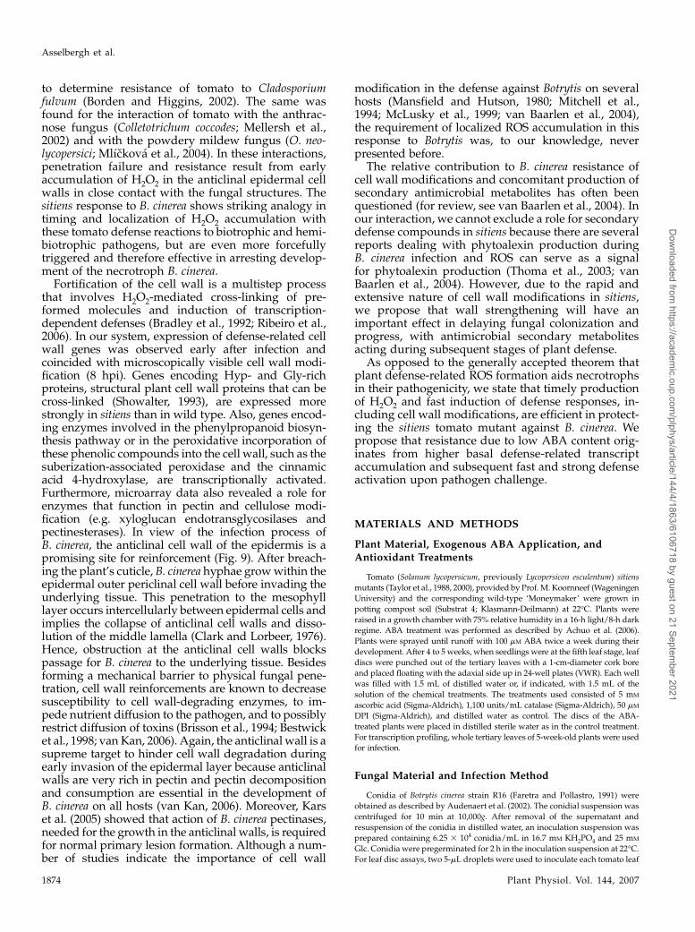
to determine resistance of tomato to Cladosporiumfulvum (Borden and Higgins, 2002). The same wasfound for the interaction of tomato with the anthrac-nose fungus (Colletotrichum coccodes; Mellersh et al.,2002) and with the powdery mildew fungus (O. neo-lycopersici; Mlıckova et al., 2004). In these interactions,penetration failure and resistance result from earlyaccumulation of H2O2 in the anticlinal epidermal cellwalls in close contact with the fungal structures. Thesitiens response to B. cinerea shows striking analogy intiming and localization of H2O2 accumulation withthese tomato defense reactions to biotrophic and hemi-biotrophic pathogens, but are even more forcefullytriggered and therefore effective in arresting develop-ment of the necrotroph B. cinerea.
Fortification of the cell wall is a multistep processthat involves H2O2-mediated cross-linking of pre-formed molecules and induction of transcription-dependent defenses (Bradley et al., 1992; Ribeiro et al.,2006). In our system, expression of defense-related cellwall genes was observed early after infection andcoincided with microscopically visible cell wall modi-fication (8 hpi). Genes encoding Hyp- and Gly-richproteins, structural plant cell wall proteins that can becross-linked (Showalter, 1993), are expressed morestrongly in sitiens than in wild type. Also, genes encod-ing enzymes involved in the phenylpropanoid biosyn-thesis pathway or in the peroxidative incorporation ofthese phenolic compounds into the cell wall, such as thesuberization-associated peroxidase and the cinnamicacid 4-hydroxylase, are transcriptionally activated.Furthermore, microarray data also revealed a role forenzymes that function in pectin and cellulose modi-fication (e.g. xyloglucan endotransglycosilases andpectinesterases). In view of the infection process ofB. cinerea, the anticlinal cell wall of the epidermis is apromising site for reinforcement (Fig. 9). After breach-ing the plant’s cuticle, B. cinerea hyphae grow within theepidermal outer periclinal cell wall before invading theunderlying tissue. This penetration to the mesophylllayer occurs intercellularly between epidermal cells andimplies the collapse of anticlinal cell walls and disso-lution of the middle lamella (Clark and Lorbeer, 1976).Hence, obstruction at the anticlinal cell walls blockspassage for B. cinerea to the underlying tissue. Besidesforming a mechanical barrier to physical fungal pene-tration, cell wall reinforcements are known to decreasesusceptibility to cell wall-degrading enzymes, to im-pede nutrient diffusion to the pathogen, and to possiblyrestrict diffusion of toxins (Brisson et al., 1994; Bestwicket al., 1998; van Kan, 2006). Again, the anticlinal wall is asupreme target to hinder cell wall degradation duringearly invasion of the epidermal layer because anticlinalwalls are very rich in pectin and pectin decompositionand consumption are essential in the development ofB. cinerea on all hosts (van Kan, 2006). Moreover, Karset al. (2005) showed that action of B. cinerea pectinases,needed for the growth in the anticlinal walls, is requiredfor normal primary lesion formation. Although a num-ber of studies indicate the importance of cell wall
modification in the defense against Botrytis on severalhosts (Mansfield and Hutson, 1980; Mitchell et al.,1994; McLusky et al., 1999; van Baarlen et al., 2004),the requirement of localized ROS accumulation in thisresponse to Botrytis was, to our knowledge, neverpresented before.
The relative contribution to B. cinerea resistance ofcell wall modifications and concomitant production ofsecondary antimicrobial metabolites has often beenquestioned (for review, see van Baarlen et al., 2004). Inour interaction, we cannot exclude a role for secondarydefense compounds in sitiens because there are severalreports dealing with phytoalexin production duringB. cinerea infection and ROS can serve as a signalfor phytoalexin production (Thoma et al., 2003; vanBaarlen et al., 2004). However, due to the rapid andextensive nature of cell wall modifications in sitiens,we propose that wall strengthening will have animportant effect in delaying fungal colonization andprogress, with antimicrobial secondary metabolitesacting during subsequent stages of plant defense.
As opposed to the generally accepted theorem thatplant defense-related ROS formation aids necrotrophsin their pathogenicity, we state that timely productionof H2O2 and fast induction of defense responses, in-cluding cell wall modifications, are efficient in protect-ing the sitiens tomato mutant against B. cinerea. Wepropose that resistance due to low ABA content orig-inates from higher basal defense-related transcriptaccumulation and subsequent fast and strong defenseactivation upon pathogen challenge.
MATERIALS AND METHODS
Plant Material, Exogenous ABA Application, and
Antioxidant Treatments
Tomato (Solanum lycopersicum, previously Lycopersicon esculentum) sitiens
mutants (Taylor et al., 1988, 2000), provided by Prof. M. Koornneef (Wageningen
University) and the corresponding wild-type ‘Moneymaker’ were grown in
potting compost soil (Substrat 4; Klasmann-Deilmann) at 22�C. Plants were
raised in a growth chamber with 75% relative humidity in a 16-h light/8-h dark
regime. ABA treatment was performed as described by Achuo et al. (2006).
Plants were sprayed until runoff with 100 mM ABA twice a week during their
development. After 4 to 5 weeks, when seedlings were at the fifth leaf stage, leaf
discs were punched out of the tertiary leaves with a 1-cm-diameter cork bore
and placed floating with the adaxial side up in 24-well plates (VWR). Each well
was filled with 1.5 mL of distilled water or, if indicated, with 1.5 mL of the
solution of the chemical treatments. The treatments used consisted of 5 mM
ascorbic acid (Sigma-Aldrich), 1,100 units/mL catalase (Sigma-Aldrich), 50 mM
DPI (Sigma-Aldrich), and distilled water as control. The discs of the ABA-
treated plants were placed in distilled sterile water as in the control treatment.
For transcription profiling, whole tertiary leaves of 5-week-old plants were used
for infection.
Fungal Material and Infection Method
Conidia of Botrytis cinerea strain R16 (Faretra and Pollastro, 1991) were
obtained as described by Audenaert et al. (2002). The conidial suspension was
centrifuged for 10 min at 10,000g. After removal of the supernatant and
resuspension of the conidia in distilled water, an inoculation suspension was
prepared containing 6.25 3 104 conidia/mL in 16.7 mM KH2PO4 and 25 mM
Glc. Conidia were pregerminated for 2 h in the inoculation suspension at 22�C.
For leaf disc assays, two 5-mL droplets were used to inoculate each tomato leaf
Asselbergh et al.
1874 Plant Physiol. Vol. 144, 2007
Dow
nloaded from https://academ
ic.oup.com/plphys/article/144/4/1863/6106718 by guest on 21 Septem
ber 2021

disc. The plates containing the leaf discs were incubated at 22�C under dark
conditions. Symptoms were evaluated after 4 d. Each inoculation droplet was
classified as a spreading or nonspreading lesion and the data were analyzed
with a binary logistic regression. At least 72 inoculation drops were evaluated
for each treatment. For RNA extraction, a spray inoculation was used to get a
uniform infection covering the total leaf area. Leaves were sprayed with an
inoculation solution containing 5 3 104 spores/mL, 0.01 M KH2PO4 (pH 5), and
6.67 mM Glc, and placed in plastic trays with high humidity according to
Audenaert et al. (2002) and incubated at 22�C under dark conditions. Mock
inoculation consisted of spraying plant leaves with a solution of 0.01 M
KH2PO4 (pH 5) and 6.67 mM Glc.
Visualization of Defense Responses
To compare the defense responses of sitiens and wild-type leaf tissue, different
staining techniques were used and evaluation was done macroscopically and
microscopically. Leaf discs were inoculated with B. cinerea as described above
and sampled at 4, 8, 12, 16, 20, 24, 48, and 72 hpi by clearing and fixing in 100%
ethanol. For each time point, at least three discs originating from different plants
of sitiens and wild type were used. For H2O2 accumulation, staining was
according to the protocol of Thordal-Christensen et al. (1997). Three hours before
each sampling time point, tomato leaf discs infected with B. cinerea were placed
under light conditions and floated in a solution of 1 mg/mL DAB-HCl (pH 4).
Polymerization of the DAB molecule at the site of H2O2 and peroxidase
accumulation results in a brown, reddish color that is macroscopically visible
and because of the high spatial and temporal distribution of the oxidized DAB
molecule, it can be visualized using bright-field microscopy. A subset of the
ethanol-fixed DAB-stained samples was embedded in Technovit 7100 histo-
embedding medium (Heraeus Kulzer) according to the manufacturer’s instruc-
tions and semithin cross sections (4 mm) were cut with a Leica RM2265 motorized
rotary microtome (Leica Microsystems). To detect superoxide, leaf discs were
floated in 0.05% NBT for 30 min before fixation in 100% ethanol, according to the
protocol of Doke (1983). For protein cross-linking, staining was performed as
described by Mellersh et al. (2002). Ethanol-fixed samples were placed in 1% SDS
at 80�C for 24 h and stained with 0.1% Coomassie Blue in 40% ethanol/10% acetic
acid for 15 min and subsequently washed in 40% ethanol/10% acetic acid. To
visualize cell wall modifications, safranin-O staining was according to Lucena
et al. (2003). Leaf discs were incubated in 0.01% safranin-O in 50% ethanol for
3 min. Accumulation of phenolics was detected by staining with 0.05% toluidine
blue in citrate/citric acid buffer (50 mM, pH 3.5; Mellersh et al., 2002). Fungal
structures were stained with 0.02% trypan blue in lactophenol. After staining,
leaf discs were mounted in 50% glycerol. Fluorescence and bright-field micros-
copy were performed with an Olympus BX-51 microscope and images were
captured with a ColorView III camera and edited with the software package
CELL-F (Olympus Soft Imaging Solutions).
Extracellular peroxidase activity was measured with the TMB assay based
on Ros Barcelo (1998). Leaf discs, inoculated with two drops of a B. cinerea
conidial suspension or with the appropriate mock solution, were fixed in
ethanol. After subsequent washing in distilled water, the discs were incubated
in 1.5 mL of 50 mM Tris-acetate buffer (pH 5.0) containing 0.1 mg/mL TMB and
0.03% H2O2 for 20 min. Peroxidase activity of the discs was determined by
measuring the absorbance of the incubation solution at 654 nm.
Sampling and RNA Preparation
Leaf samples were taken at 0 and 8 hpi. Two leaflets per leaf were excised
and immediately frozen in liquid nitrogen. Three days after inoculation,
infection levels on the three remaining leaflets were scored. For each geno-
type/treatment combination (i.e. wild-type mock, wild-type infected, sitiens
mock, and sitiens infected), four leaflets were sampled and pooled. Total RNA
was prepared from the sample pools with the RNeasy plant mini kit (Qiagen),
according to the manufacturer’s instructions. Total RNA quality was checked
on an agarose gel and concentrations were determined with a ND-1000
spectrophotometer (Nanodrop Technologies).
Microarrays
The tomato TOM1 microarray used was obtained from the Center for Gene
Expression Profiling of the Boyce Thompson Institute and consisted of 12,899
EST clones representing 8,600 independent tomato genes. The functional
annotation of the genes related to the spotted cDNAs can be viewed at http://
bti.cornell.edu/CGEP/CGEP.html.
Target Labeling and Hybridizations
Target labeling and hybridizations were carried out at the Flanders
Institute of Biotechnology MicroArray Facility (http://www.microarrays.be).
Total RNA (5 mg) of each sample was reverse transcribed to double-stranded
cDNA and further amplified according to a modified version of the protocol
described in Puskas et al. (2002; http://www.microarrays.be/service.htm).
Subsequent Cy5 and Cy3 (GE-Healthcare) labeling, hybridization, posthy-
bridization washing, and scanning were performed according to protocols
accessible through the Web site (http://www.microarrays.be/service.htm).
Experimental Design and Statistical Analysis of
Microarray Data
We constructed a loop design (Supplemental Fig. S3) consisting of 14 two-
dye TOM1 microarray experiments in which two independent pools of four
leaflets harvested from 5-week-old mock-sprayed and spray-infected plants at
0 and 8 hpi were compared. Expression data were analyzed in two steps: (1) a
within-slide analysis aimed at removing variation associated with differential
dye responses to binding and scanning, as noise; and (2) a between-slide
analysis aimed at estimating the mean differences between treatments (e.g.
genotypes and infection treatments) and their SE. To correct for dye intensity
differences, we used the robust scatter plot smoother LOWESS (Yang et al.,
2002) as implemented in Genstat (Payne and Lane, 2005), where the response
variable is the log2 ratio of the artifact-removed total foreground Cy3 and Cy5
fluorescence intensities measured at the 12,899 spots. The fraction of the data
used for estimating the local LOWESS fit was set at 20%.
For between-slide analysis, a two-step mixed-model ANOVA (Wolfinger
et al., 2001) was used and variance components were estimated by residual
maximum likelihood (REML), as implemented in Genstat (Payne and Lane,
2005) and described previously (Vuylsteke et al., 2005; Nettleton, 2006).
Following Wolfinger et al. (2001), the mixed-model analysis on the LOWESS
fit to the spot measurements consisted of two steps. First, array and channel
effects were removed from the expression responses by a normalization
ANOVA model of the form:
response 5 m 1 array 1 dye 1 ðarray 3 dyeÞ1 residual; ð1Þ
where the response variable represents the corrected log2-transformed Cy3
and Cy5 fluorescence intensity measurements of the 12,899 ESTs. Array
models the hybridization effects of each of the 14 microarrays, dye models the
effects of each of the two dyes, and dye by array models the 28-channel effects.
Array, dye, and dye by array were added as random terms.
In a second step, the residuals from model 1 were analyzed for each of the
12,899 ESTs separately by a mixed model of the following form:
residual 5 m 1 dye 1 replicate 1 genotype 1 treatment 1 ðgenotype
3 treatmentÞ1 ðgenotype 3 treatment 3 timeÞ1 array 1 error;
ð2Þ
partitioning gene-specific variation into gene-specific fixed dye effects, fixed
replicate effects, fixed genotype (wild type and sitiens) effects, fixed treatment
(mock and infected), fixed genotype-specific treatment effects, and fixed
genotype-specific treatment effects across time. The random array term
models the effects for each spot and equals the (EST 3 array) interaction
effect. These models were fitted by REML and Wald statistics were calculated
to assess significance of the fixed effects in the gene model. No further
adjustments for multiple testing were done. From REML analysis, we saved
the vector of estimated genotype, treatment, and interaction effects with the
corresponding estimated variance-covariance matrix for each gene. Test
statistics for contrasts were constructed from the parameter estimates divided
by their SEs. These ratios were supposed to follow approximately a t distri-
bution, with the degrees of freedom equal to those for the error term in the
gene-specific model. On the basis of the t approximation to the test statistics
for the contrasts, P values were calculated.
Quantitative RT-PCR Analysis
Poly(dT) cDNA was synthesized from 2 mg of total RNA with SuperScript
II reverse transcriptase (Invitrogen) and quantified on a LightCycler480 RT-
PCR detection system (Roche Diagnostics) with the SYBR Green I RT-PCR core
kit (Roche Diagnostics). PCR was carried out in triplicate. Gene-specific
primer pairs were designed with Beacon Designer 4.0 (Premier Biosoft
H2O2 Burst in Resistance to B. cinerea in sitiens Tomato
Plant Physiol. Vol. 144, 2007 1875
Dow
nloaded from https://academ
ic.oup.com/plphys/article/144/4/1863/6106718 by guest on 21 Septem
ber 2021

International): SGN-U143838 (FWD, AACCTAGCTGCCGCTTTCCC; REV,
TTACGCCACACCACCTGAGTATAG); SGN-U143866 (FWD, CATCAAA-
GGGTAAGTGCCCAAAGG; REV, AAGGCAAACAGCAGCATCAAGATC);
SGN-U134316 (FWD, TCGCCACCAACATTCACATAACAG; REV, AGAAT-
GTGATGGCAAGTTGTTCCC); SGN-U144656 (FWD, GGACGATGGTCTAG-
CAGCCTATG; REV, CAGCACCAGCAGCGTTTAGC); SGN-U144488 (FWD,
TTGTGGTGGAGTCCTGGATTGC; REV, TGGCTGTGCATTGAATTGGAT-
GAC); SGN-U147913 (FWD, CGGCGGTCGTGGTAGAGG; REV, AACAAC-
CATTGCGGCAGTACC).
Supplemental Data
The following materials are available in the online version of this article.
Supplemental Figure S1. Superoxide accumulation detected with NBT in
wild-type and sitiens tomato leaf discs inoculated with B. cinerea.
Supplemental Figure S2. Symptom development of wild-type and sitiens
tomato leaves 3 d after spray inoculation with 5 3 104 B. cinerea
conidia/mL.
Supplemental Figure S3. Experimental design consisting of 14 TOM1
cDNA arrays to examine transcript levels in RNA samples collected
from wild-type and sitiens plants inoculated either with an infection
solution or a mock solution at 0 and 8 hpi.
Supplemental Figure S4. Gene expression kinetics of osmotin-like protein
(SGN-U144488), endo-1,3-b-glucanase (SGN-U143416), and PR1 pro-
tein (SGN-U143838; Table I), PR1A1 protein (SGN-U144656), Pro-rich
protein EIG-I30 (SGN-U143866; Table II), and cell wall protein (SGN-
U147913; Supplemental Table S2) in wild-type and sitiens leaves spray-
inoculated with 5 3 104 B. cinerea conidia/mL.
Supplemental Table S1. Genes significantly differentially expressed in
wild-type and sitiens plants after infection with B. cinerea (P , 0.01)
with FC of at least 1.5.
Supplemental Table S2. Nonredundant list of cell wall-related genes
significantly differentially expressed upon B. cinerea infection at 8 hpi
(P , 0.05) with FC of at least 1.5 in sitiens plants.
ACKNOWLEDGMENTS
We thank Ilse Delaere, Greet De Backer, Carlos Puig, Michael Vandorpe,
Stijn Morsa, and Korneel Vandenbroucke for technical assistance, Sepp van
Rompaey for help with preparing figures, Paul Van Hummelen for the
Flanders Institute of Biotechnology Microarray Facility, and Martine De Cock
for help in preparing the manuscript.
Received March 12, 2007; accepted June 6, 2007; published June 15, 2007.
LITERATURE CITED
AbuQamar S, Chen X, Dhawan R, Bluhm B, Salmeron J, Lam S, Dietrich
RA, Mengiste T (2006) Expression profiling and mutant analysis reveals
complex regulatory networks involved in Arabidopsis response to
Botrytis infection. Plant J 48: 28–44
Achuo EA, Audenaert K, Meziane H, Hofte M (2004) The salicylic acid-
dependent defence pathway is effective against different pathogens in
tomato and tobacco. Plant Pathol 53: 65–72
Achuo EA, Prinsen E, Hofte M (2006) Influence of drought, salt stress and
abscisic acid on the resistance of tomato to Botrytis cinerea and Oidium
neolycopersici. Plant Pathol 55: 178–186
Alba R, Fei Z, Payton P, Liu Y, Moore SL, Debbie P, Cohn J, D’Ascenzo M,
Gordon JS, Rose JKC, et al (2004) ESTs, cDNA microarrays, and gene
expression profiling: tools for dissecting plant physiology and devel-
opment. Plant J 39: 697–714
Anderson JP, Badruzsaufari E, Schenk PM, Manners JM, Desmond OJ,
Ehlert C, Maclean DJ, Ebert PR, Kazan K (2004) Antagonistic interac-
tion between abscisic acid and jasmonate-ethylene signaling pathways
modulates defense gene expression and disease resistance in Arabidop-
sis. Plant Cell 16: 3460–3479
Audenaert K, De Meyer GB, Hofte MM (2002) Abscisic acid determines
basal susceptibility of tomato to Botrytis cinerea and suppresses salicylic
acid-dependent signaling mechanisms. Plant Physiol 128: 491–501
Benito EP, ten Have A, van’t Klooster JW, van Kan JAL (1998) Fungal and
plant gene expression during synchronized infection of tomato leaves
by Botrytis cinerea. Eur J Plant Pathol 104: 207–220
Bestwick CS, Brown IR, Mansfield JW (1998) Localized changes in
peroxidase activity accompany hydrogen peroxide generation during
the development of a nonhost hypersensitive reaction in lettuce. Plant
Physiol 118: 1067–1078
Bolwell GP, Davies DR, Gerrish C, Auh CK, Murphy TM (1998) Com-
parative histochemistry of the oxidative burst produced by rose and
French bean cells reveals two distinct mechanisms. Plant Physiol 116:
1379–1385
Borden S, Higgins VJ (2002) Hydrogen peroxide plays a critical role in the
defence response of tomato to Cladosporium fulvum. Physiol Mol Plant
Pathol 61: 227–236
Bradley DJ, Kjellbom P, Lamb CJ (1992) Elicitor- and wound-induced
oxidative cross-linking of a proline-rich plant cell wall protein: a novel,
rapid defense response. Cell 70: 21–30
Brisson LF, Tenhaken R, Lamb C (1994) Function of oxidative cross-linking
of cell wall structural proteins in plant disease resistance. Plant Cell 6:
1703–1712
Clark CA, Lorbeer JW (1976) Comparative histopathology of Botrytis
squamosa and B. cinerea on onion leaves. Phytopathology 66: 1279–1289
Conrath U, Pieterse CMJ, Mauch-Mani B (2002) Priming in plant-
pathogen interactions. Trends Plant Sci 7: 210–216
Doke N (1983) Involvement of superoxide anion generation in the hyper-
sensitive response of potato tuber tissues to infection with an incom-
patible race of Phytophthora infestans and to the hyphal wall components.
Physiol Plant Pathol 23: 345–357
Elad Y (1992) The use of antioxidants (free radical scavengers) to control
grey mould (Botrytis cinerea) and white mould (Sclerotinia sclerotiorum)
in various crops. Plant Pathol 41: 417–426
Faretra F, Pollastro S (1991) Genetic basis of resistance to benzimidazole
and dicarboximide fungicides in Botryotinia fuckeliana (Botrytis cinerea).
Mycol Res 95: 943–951
Ferrari S, Plotnikova JM, De Lorenzo G, Ausubel FM (2003) Arabidopsis
local resistance to Botrytis cinerea involves salicylic acid and camalexin
and requires EDS4 and PAD2, but not SID2, EDS5 or PAD4. Plant J 35:
193–205
Fujita M, Fujita Y, Noutoshi Y, Takahashi F, Narusaka Y, Yamaguchi-
Shinozaki K, Shinozaki K (2006) Crosstalk between abiotic and biotic
stress responses: a current view from the points of convergence in the
stress signaling networks. Curr Opin Plant Biol 9: 436–442
Glazebrook J (2005) Contrasting mechanisms of defense against biotrophic
and necrotrophic pathogens. Annu Rev Phytopathol 43: 205–227
Govrin EM, Levine A (2000) The hypersensitive response facilitates plant
infection by the necrotrophic pathogen Botrytis cinerea. Curr Biol 10:
751–757
Govrin EM, Levine A (2002) Infection of Arabidopsis with a necrotrophic
pathogen, Botrytis cinerea, elicits various defense responses but does not
induce systemic acquired resistance (SAR). Plant Mol Biol 48: 267–276
Govrin EM, Rachmilevitch S, Tiwari BS, Solomon M, Levine A (2006) An
elicitor from Botrytis cinerea induces the hypersensitive response in
Arabidopsis thaliana and other plants and promotes the gray mold
disease. Phytopathology 96: 299–307
Gu YQ, Wildermuth MC, Chakravarthy S, Loh YT, Yang C, He X, Han Y, Martin
GB (2002) Tomato transcription factors Pti4, Pti5, and Pti6 activate defense
responses when expressed in Arabidopsis. Plant Cell 14: 817–831
He P, Warren RF, Zhao T, Shan L, Zhu L, Tang X, Zhou JM (2001)
Overexpression of Pti5 in tomato potentiates pathogen-induced defense
gene expression and enhances disease resistance to Pseudomonas syrin-
gae pv tomato. Mol Plant Microbe Interact 14: 1453–1457
Heath MC (2000) Hypersensitive response-related death. Plant Mol Biol 44:
321–334
Henfling J, Bostock R, Kuc J (1980) Effect of abscisic acid on rishitin and
lubimin accumulation and resistance to Phytophthora infestans and
Cladosporium cucumerinum in potato tuber tissue slices. Phytopathology
70: 1074–1078
Kachroo P, Shanklin J, Shah J, Whittle EJ, Klessig DF (2001) A fatty acid
desaturase modulates the activation of defense signaling pathways in
plants. Proc Natl Acad Sci USA 98: 9448–9453
Kars I, Krooshof GH, Wagemakers L, Joosten R, Benen JAE, van Kan JAL
(2005) Necrotizing activity of five Botrytis cinerea endopolygalacturona-
ses produced in Pichia pastoris. Plant J 43: 213–225
Asselbergh et al.
1876 Plant Physiol. Vol. 144, 2007
Dow
nloaded from https://academ
ic.oup.com/plphys/article/144/4/1863/6106718 by guest on 21 Septem
ber 2021
Kwak JM, Nguyen V, Schroeder JI (2006) The role of reactive oxygen
species in hormonal responses. Plant Physiol 141: 323–329
Lamb C, Dixon RA (1997) The oxidative burst in plant disease resistance.
Annu Rev Plant Physiol Plant Mol Biol 48: 251–275
Lucena MA, Romero-Aranda R, Mercado JA, Cuartero J, Valpuesta V,
Quesada MA (2003) Structural and physiological changes in the roots of
tomato plants over-expressing a basic peroxidase. Physiol Plant 118:
422–429
Lyon GD, Goodman BA, Williamson B (2004) Botrytis cinerea perturbs
redox processes as an attack strategy in plants. In Y Elad, B Williamson,
P Tudzynski, N Delen, eds, Botrytis: Biology, Pathology and Control.
Kluwer Academic Publishers, Dordrecht, The Netherlands, pp 119–141
Malolepsza U, Urbanek H (2002) o-Hydroxyethylorutin-mediated en-
hancement of tomato resistance to Botrytis cinerea depends on a burst
of reactive oxygen species. J Phytopathol 150: 616–624
Mansfield JW, Hutson RA (1980) Microscopical studies on fungal devel-
opment and host responses in broad bean and tulip leaves inoculated
with five species of Botrytis. Physiol Plant Pathol 17: 131–143
Mauch-Mani B, Mauch F (2005) The role of abscisic acid in plant-pathogen
interactions. Curr Opin Plant Biol 8: 409–414
McLusky SR, Bennett MH, Beale MH, Lewis MJ, Gaskin P, Mansfield JW
(1999) Cell wall alterations and localized accumulation of feryloyl-3#-
methoxytyramine in onion epidermis at sites of attempted penetration
by Botrytis allii are associated with actin polarisation, peroxidase activ-
ity and suppression of flavonoid biosynthesis. Plant J 17: 523–534
Mehdy MC (1994) Active oxygen species in plant defense against patho-
gens. Plant Physiol 105: 467–472
Mellersh DG, Foulds IV, Higgins VJ, Heath MC (2002) H2O2 plays
different roles in determining penetration failure in three diverse
plant-fungal interactions. Plant J 29: 257–268
Mengiste T, Chen X, Salmeron J, Dietrich R (2003) The BOTRYTIS
SUSCEPTIBLE1 gene encodes an R2R3MYB transcription factor protein
that is required for biotic and abiotic stress responses in Arabidopsis.
Plant Cell 15: 2551–2565
Mitchell HJ, Hall JL, Barber MS (1994) Elicitor-induced cinnamyl alcohol
dehydrogenase activity in lignifying wheat (Triticum aestivum L.) leaves.
Plant Physiol 104: 551–556
Mlıckova K, Luhova L, Lebeda A, Mieslerova B, Pec P (2004) Reactive
oxygen species generation and peroxidase activity during Oidium neo-
lycopersici infection on Lycopersicon species. Plant Physiol Biochem 42:
753–761
Mohr PG, Cahill DM (2001) Relative roles of glyceollin, lignin and the
hypersensitive response and the influence of ABA in compatible and
incompatible interactions of soybeans with Phytophthora sojae. Physiol
Mol Plant Pathol 48: 31–41
Mohr PG, Cahill DM (2007) Suppression by ABA of salicylic acid and
lignin accumulation and the expression of multiple genes, in Arabi-
dopsis infected with Pseudomonas syringae pv tomato. Funct Integr
Genomics 7: 181–191
Nagel OW, Konings H, Lambers H (1994) Growth rate, plant development
and water relations of the ABA-deficient tomato mutant sitiens. Physiol
Plant 92: 102–108
Nettleton D (2006) A discussion of statistical methods for design and
analysis of microarray experiments for plant scientists. Plant Cell 18:
2112–2121
Noctor G (2006) Metabolic signaling in defence and stress: the central roles
of soluble redox couples. Plant Cell Environ 29: 409–425
Patykowski J, Urbanek H (2003) Activity of enzymes related to H2O2
generation and metabolism in leaf apoplastic fraction of tomato leaves
infected with Botrytis cinerea. J Phytopathol 151: 153–161
Payne RW, Lane PW (2005) GenStat� Release Reference Manual, Part 3:
Procedure Library PL16. VSN International, Oxford
Puskas LG, Zvara A, Hackler L Jr, Van Hummelen P (2002) RNA ampli-
fication results in reproducible microarray data with slight ratio bias.
Biotechniques 32: 1330–1340
Ribeiro JM, Pereira CS, Soares NC, Vieira AM, Feijo JA, Jackson PA
(2006) The contribution of extensin network formation to rapid, hydro-
gen peroxide-mediated increases in grapevine callus wall resistance to
fungal lytic enzymes. J Exp Bot 57: 2025–2035
Ros Barcelo A (1998) Hydrogen peroxide production is a general property
of the lignifying xylem from vascular plants. Ann Bot (Lond) 82: 97–103
Schopfer P, Plachy C, Frahry G (2001) Release of reactive oxygen inter-
mediates (superoxide radicals, hydrogen peroxide, and hydroxyl rad-
icals) and peroxidase in germinating radish seeds controlled by light,
gibberellin, and abscisic acid. Plant Physiol 125: 1591–1602
Schouten A, Tenberge KB, Vermeer J, Stewart J, Wagemakers L,
Williamson B, van Kan JAL (2002) Functional analysis of an extracel-
lular catalase of Botrytis cinerea. Mol Plant Pathol 3: 227–238
Showalter AM (1993) Structure and function of plant cell wall proteins.
Plant Cell 5: 9–23
Taylor IB, Burbidge A, Thompson AJ (2000) Control of abscisic acid
synthesis. J Exp Bot 51: 1563–1574
Taylor IB, Linforth RST, Al-Naieb RJ, Bowman WR, Marples BA (1988)
The wilty tomato mutants flacca and sitiens are impaired in the oxidation
of ABA-aldehyde to ABA. Plant Cell Environ 11: 739–745
Thaler JS, Bostock RM (2004) Interactions between abscisic-acid-mediated
responses and plant resistance to pathogens and insects. Ecology 85: 48–58
Thara VK, Tang X, Gu YQ, Martin GB, Zhou JM (1999) Pseudomonas
syringae pv tomato induces the expression of tomato EREBP-like genes
Pti4 and Pti5 independent of ethylene, salicylate and jasmonate. Plant J
20: 475–483
Thoma I, Loeffler C, Sinha AK, Gupta M, Krischke M, Steffan B, Roitsch
T, Mueller MJ (2003) Cyclopentenone isoprostanes induced by reactive
oxygen species trigger defense gene activation and phytoalexin accu-
mulation in plants. Plant J 34: 363–375
Thomma BPHJ, Eggermont K, Penninckx IAMA, Mauch-Mani B, Vogelsang
R, Cammue BPA, Broekaert WF (1998) Separate jasmonate-dependent
and salicylate-dependent defense-response pathways in Arabidopsis are
essential for resistance to distinct microbial pathogens. Proc Natl Acad
Sci USA 95: 15107–15111
Thordal-Christensen H, Zhang Z, Wei Y, Collinge DB (1997) Subcellular
localization of H2O2 in plants: H2O2 accumulation in papillae and
hypersensitive response during the barley-powdery mildew interac-
tion. Plant J 11: 1187–1194
Torres MA, Jones JDG, Dangl JL (2006) Reactive oxygen species signaling
in response to pathogens. Plant Physiol 141: 373–378
Unger C, Kleta S, Jandl G, von Tiedemann A (2005) Suppression of the
defence-related oxidative burst in bean leaf tissue and bean suspension cells
by the necrotrophic pathogen Botrytis cinerea. J Phytopathol 153: 15–26
Urbanczyk-Wochniak E, Usadel B, Thimm O, Nunes-Nesi A, Carrari F,
Davy M, Blasing O, Kowalczyk M, Weicht D, Polinceusz A, et al (2006)
Conversion of MapMan to allow the analysis of transcript data from
Solanaceous species: effects of genetic and environmental alterations in
energy metabolism in the leaf. Plant Mol Biol 60: 773–792
van Baarlen P, Staats M, van Kan JAL (2004) Induction of programmed cell
death in lily by the fungal pathogen Botrytis elliptica. Mol Plant Pathol
5: 559–574
van Baarlen P, Woltering EJ, Staats M, van Kan JAL (2007) Histochemical
and genetic analysis of host and non-host interactions of Arabidopsis
with three Botrytis species: an important role for cell death control. Mol
Plant Pathol 8: 41–54
van Kan JAL (2006) Licensed to kill: the lifestyle of a necrotrophic plant
pathogen. Trends Plant Sci 11: 247–253
van Loon LC, van Strien EA (1999) The families of pathogenesis-related
proteins, their activities, and comparative analysis of PR-1 type pro-
teins. Physiol Mol Plant Pathol 55: 85–97
Veronese P, Chen X, Bluhm B, Salmeron J, Dietrich R, Mengiste T (2004)
The BOS loci of Arabidopsis are required for resistance to Botrytis cinerea
infection. Plant J 40: 558–574
von Tiedemann A (1997) Evidence for a primary role of active oxygen
species in induction of host cell death during infection of bean leaves
with Botrytis cinerea. Physiol Mol Plant Pathol 50: 151–166
Vuylsteke M, van Eeuwijk F, Van Hummelen P, Kuiper M, Zabeau M
(2005) Genetic analysis of variation in gene expression in Arabidopsis
thaliana. Genetics 171: 1267–1275
Wolfinger RD, Gibson G, Wolfinger ED, Bennett L, Hamadeh H, Bushel P,
Afshari C, Paules RS (2001) Assessing gene significance from cDNA
microarray expression data via mixed models. J Comput Biol 8:
625–637
Xiong L, Yang Y (2003) Disease resistance and abiotic stress tolerance in
rice are inversely modulated by an abscisic acid-inducible mitogen-
activated protein kinase. Plant Cell 15: 745–759
Yang YH, Dudoit S, Luu P, Lin DM, Peng V, Ngai J, Speed TP (2002)
Normalization for cDNA microarray data: a robust composite method
addressing single and multiple slide systematic variation. Nucleic Acids
Res 30: e15
H2O2 Burst in Resistance to B. cinerea in sitiens Tomato
Plant Physiol. Vol. 144, 2007 1877
Dow
nloaded from https://academ
ic.oup.com/plphys/article/144/4/1863/6106718 by guest on 21 Septem
ber 2021