Embed Size (px)
DESCRIPTION
BEST
Citation preview
Clinical Case ReportsMittal et al., J Clin Case Rep 2013, 3:3
http://dx.doi.org/10.4172/2165-7920.1000262
Volume 3 • Issue 3 • 1000262J Clin Case RepISSN: 2165-7920 JCCR, an open access journal
Open AccessCase Report
Resin-bonded Bridge: Conservative Treatment Option for Single Tooth ReplacementSanjeev Mittal, Sandeep Garg, Navsharanjit Kaur Chhina and Monika Joshi* Department of Prosthodontics including Crown and Bridge, MM College of Dental Sciences and Research, Mullana, Ambala, India
*Corresponding author: Dr. Monika Joshi, BDS, Post-graduate student, Department of prosthodontics including crown and bridge, MM College of Dental Sciences and Research, Mullana, Ambala, Haryana, India, Tel: +91 9729451209; E-mail: [email protected]
Received January 16, 2013; Accepted March 15, 2013; Published March 18, 2013
Citation: Mittal S, Garg S, Chhina NK, Joshi M (2013) Resin-bonded Bridge: Conservative Treatment Option for Single Tooth Replacement. J Clin Case Rep 3: 262. doi:10.4172/2165-7920.1000262
Copyright: © 2013 Mittal S, et al. This is an open-access article distributed under the terms of the Creative Commons Attribution License, which permits unrestricted use, distribution, and reproduction in any medium, provided the original author and source are credited.
AbstractResin-Bonded Fixed Partial Dentures (RBFPD) was introduced to dentistry around 40 years ago. The typical
design of resin-bonded fixed partial denture is characterized by conservation of tooth structure of abutment compared to fixed treatment. This article presents a case report of a patient who reported with missing maxillary central incisor. Multiple treatment options are available for replacement of missing tooth. Use of conventional fixed partial denture in such a situation is criticized because modern dental practice revolves around the principle of preservation of tooth structure. So in such cases resin bonded fixed partial denture is best treatment option.
Keywords: Adhesive; Acid etching; Resin bonded fixed partial denture
IntroductionEra of adhesive dentistry dates back to 1955 when Dr. Buonocore
introduced enamel acid etching and resin bonding [1]. It not only improved the restorative dentistry but also opened new paths in preventive dentistry. Many enhancements followed this like direct restorations with resins, bondable brackets in orthodontics, and fissure sealants in prosthodontics.
In 1973, Dr. Rochette of France introduced the idea of bonding a cast metal bar to the lingual surfaces of periodontally involved anterior teeth for splinting purposes using the acid-etch technique and unfilled resin cement [2]. The cast metal splint had perforations made with sloping walls to permit attachment to the resin cement through mechanical interlocking. This idea was applied by Howe and Denehy in 1977 to a specially designed partial denture to the enamel of abutment teeth in the anterior segments of the mouth [3]. They used the perforated metal framework design and modified the technique by adding a pontic to replace a missing anterior tooth. This technique provided a Fixed Partial Denture (FPD) tooth replacement with minimal tooth preparation.
Thompson et al. refined the framework by electrolytically etching non-precious castings to produce a microscopically roughened surface for providing suitable mechanical retention to the tooth structure through an adhesive luting cement [4,5]. This etching process was first described by Dunn and Reisbick [6] and Tanaka et al. [7] (The resin provided mechanical linkage between the micropores of the alloy and the enamel).
Since that time, a number of significant modifications to this original design have improved its longevity in the oral environment, and currently Resin Bonded Fixed Partial Dentures (RBFPDs) are considered by many to be a viable alternative to conventional fixed partial dentures. Resin bonded bridges are a minimally invasive option for replacing missing teeth [8-10]. Preparation designs for RBFPDs are strictly limited to the enamel and may comprise palatal veneer preparations, proximal boxes, vertical grooves, guiding planes, or pinholes in the cingulum area [11].
This case report describes the use of RBFPD as a valuable treatment plan in restoring smile and oral functions with minimal biological cost.
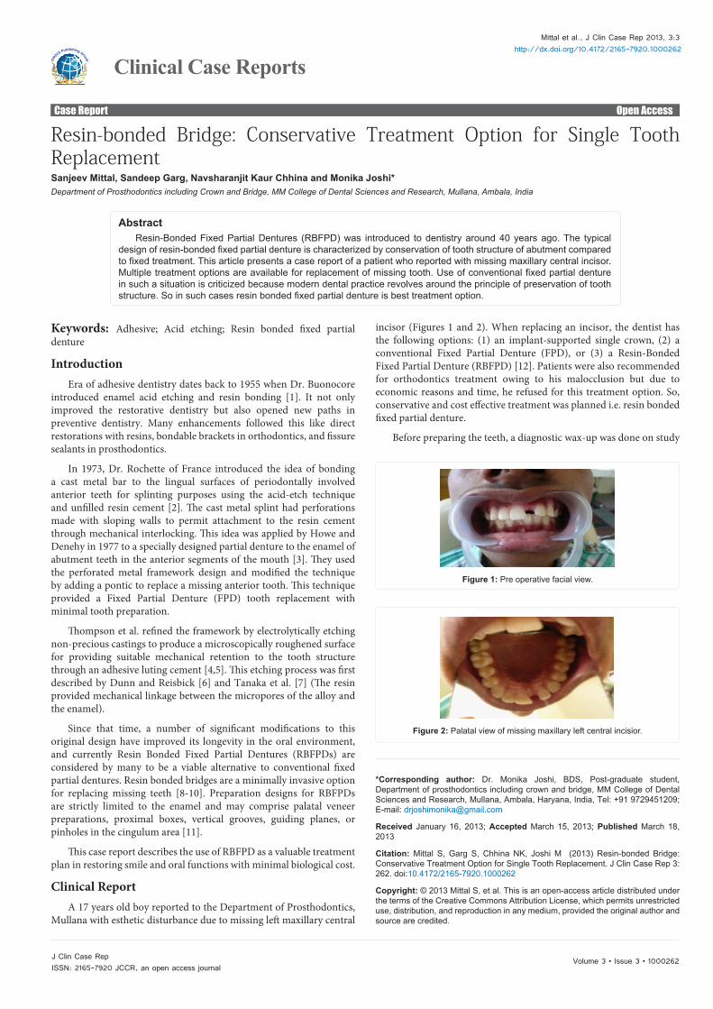
Clinical ReportA 17 years old boy reported to the Department of Prosthodontics,
Mullana with esthetic disturbance due to missing left maxillary central
incisor (Figures 1 and 2). When replacing an incisor, the dentist has the following options: (1) an implant-supported single crown, (2) a conventional Fixed Partial Denture (FPD), or (3) a Resin-Bonded Fixed Partial Denture (RBFPD) [12]. Patients were also recommended for orthodontics treatment owing to his malocclusion but due to economic reasons and time, he refused for this treatment option. So, conservative and cost effective treatment was planned i.e. resin bonded fixed partial denture.
Before preparing the teeth, a diagnostic wax-up was done on study
Figure 1: Pre operative facial view.
Figure 2: Palatal view of missing maxillary left central incisior.
Citation: Mittal S, Garg S, Chhina NK, Joshi M (2013) Resin-bonded Bridge: Conservative Treatment Option for Single Tooth Replacement. J Clin Case Rep 3: 262. doi:10.4172/2165-7920.1000262
Page 2 of 3
Volume 3 • Issue 3 • 1000262J Clin Case RepISSN: 2165-7920 JCCR, an open access journal
models. The classical principles for conservative supra-gingival tooth preparations for a resin-bonded fixed partial denture i.e. minimally invasive lingual preparation limited to enamel (0.5 mm), resistance form (lingual chamfer and proximal groove) and maximum covering of the lingual surface area were followed.
A complete final impression of the arch was made with double mix single step technique using polyvinyl siloxane impression material (putty and low viscosity, Aquasil, Dentsply/Caulk, Milford, DE) with metal stock tray. The impression was poured with die stone and cast was obtained. The stone cast was mounted on semi adjustable articulator along with opposing arch. The pattern of retainer and the pontic were fabricated using inlay wax (blue inlay wax, Bego). The pattern was invested and then casting was done. The framework so fabricated was checked on the mounted cast.
At the try-in appointment, the complete seating of the frameworks, marginal adaptation, pontics’ form and gingival pressure, esthetics and occlusion were assessed. The bridge was sent to the laboratory for final porcelain polishing and metal sandblasting.
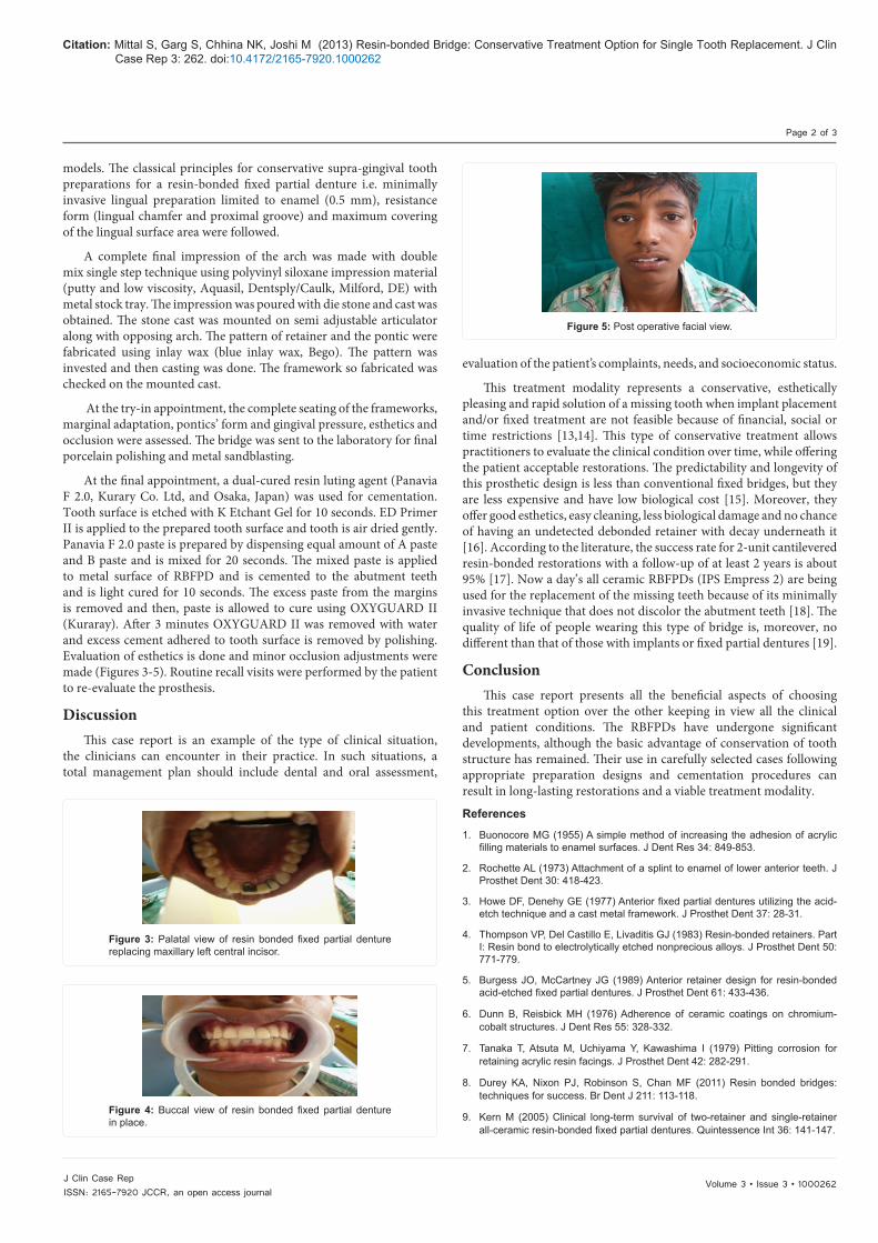
At the final appointment, a dual-cured resin luting agent (Panavia F 2.0, Kurary Co. Ltd, and Osaka, Japan) was used for cementation. Tooth surface is etched with K Etchant Gel for 10 seconds. ED Primer II is applied to the prepared tooth surface and tooth is air dried gently. Panavia F 2.0 paste is prepared by dispensing equal amount of A paste and B paste and is mixed for 20 seconds. The mixed paste is applied to metal surface of RBFPD and is cemented to the abutment teeth and is light cured for 10 seconds. The excess paste from the margins is removed and then, paste is allowed to cure using OXYGUARD II (Kuraray). After 3 minutes OXYGUARD II was removed with water and excess cement adhered to tooth surface is removed by polishing. Evaluation of esthetics is done and minor occlusion adjustments were made (Figures 3-5). Routine recall visits were performed by the patient to re-evaluate the prosthesis.
DiscussionThis case report is an example of the type of clinical situation,
the clinicians can encounter in their practice. In such situations, a total management plan should include dental and oral assessment,
evaluation of the patient’s complaints, needs, and socioeconomic status.
This treatment modality represents a conservative, esthetically pleasing and rapid solution of a missing tooth when implant placement and/or fixed treatment are not feasible because of financial, social or time restrictions [13,14]. This type of conservative treatment allows practitioners to evaluate the clinical condition over time, while offering the patient acceptable restorations. The predictability and longevity of this prosthetic design is less than conventional fixed bridges, but they are less expensive and have low biological cost [15]. Moreover, they offer good esthetics, easy cleaning, less biological damage and no chance of having an undetected debonded retainer with decay underneath it [16]. According to the literature, the success rate for 2-unit cantilevered resin-bonded restorations with a follow-up of at least 2 years is about 95% [17]. Now a day’s all ceramic RBFPDs (IPS Empress 2) are being used for the replacement of the missing teeth because of its minimally invasive technique that does not discolor the abutment teeth [18]. The quality of life of people wearing this type of bridge is, moreover, no different than that of those with implants or fixed partial dentures [19].
ConclusionThis case report presents all the beneficial aspects of choosing
this treatment option over the other keeping in view all the clinical and patient conditions. The RBFPDs have undergone significant developments, although the basic advantage of conservation of tooth structure has remained. Their use in carefully selected cases following appropriate preparation designs and cementation procedures can result in long-lasting restorations and a viable treatment modality.
References
1. Buonocore MG (1955) A simple method of increasing the adhesion of acrylic filling materials to enamel surfaces. J Dent Res 34: 849-853.
2. Rochette AL (1973) Attachment of a splint to enamel of lower anterior teeth. J Prosthet Dent 30: 418-423.
3. Howe DF, Denehy GE (1977) Anterior fixed partial dentures utilizing the acid-etch technique and a cast metal framework. J Prosthet Dent 37: 28-31.
4. Thompson VP, Del Castillo E, Livaditis GJ (1983) Resin-bonded retainers. Part I: Resin bond to electrolytically etched nonprecious alloys. J Prosthet Dent 50: 771-779.
5. Burgess JO, McCartney JG (1989) Anterior retainer design for resin-bonded acid-etched fixed partial dentures. J Prosthet Dent 61: 433-436.
6. Dunn B, Reisbick MH (1976) Adherence of ceramic coatings on chromium-cobalt structures. J Dent Res 55: 328-332.
7. Tanaka T, Atsuta M, Uchiyama Y, Kawashima I (1979) Pitting corrosion for retaining acrylic resin facings. J Prosthet Dent 42: 282-291.
8. Durey KA, Nixon PJ, Robinson S, Chan MF (2011) Resin bonded bridges: techniques for success. Br Dent J 211: 113-118.
9. Kern M (2005) Clinical long-term survival of two-retainer and single-retainer all-ceramic resin-bonded fixed partial dentures. Quintessence Int 36: 141-147.
Figure 3: Palatal view of resin bonded fixed partial denture replacing maxillary left central incisor.
Figure 4: Buccal view of resin bonded fixed partial denture in place.
Figure 5: Post operative facial view.
Citation: Mittal S, Garg S, Chhina NK, Joshi M (2013) Resin-bonded Bridge: Conservative Treatment Option for Single Tooth Replacement. J Clin Case Rep 3: 262. doi:10.4172/2165-7920.1000262
Page 3 of 3
Volume 3 • Issue 3 • 1000262J Clin Case RepISSN: 2165-7920 JCCR, an open access journal
10. Ries S, Wolz J, Richter EJ (2006) Effect of design of all-ceramic resin-bonded fixed partial dentures on clinical survival rate. Int J Periodontics Restorative Dent 26: 143-149.
11. Boening KW, Ullmann K (2012) A retrospective study of the clinical performance of porcelain-fused-to-metal resin-bonded fixed partial dentures. Int J Prosthodont 25: 265-269.
12. Cakan U, Demiralp B, Aksu M, Taner T (2009) Clinical showcase. Replacement of congenitally missing lateral incisor using a metal-free, resin-bonded fixed partial denture: case report. J Can Dent Assoc 75: 509-512.
13. Hagiwara Y, Matsumura H, Tanaka S, Woelfel JB (2004) Single tooth replacement using a modified metal-ceramic resin-bonded fixed partial denture: a clinical report. J Prosthet Dent 91: 414-417.
14. Balkaya MC, Gur H, Pamuk S (2005) The use of a resin-bonded prosthesis while maintaining the diastemata: a clinical report. J Prosthet Dent 94: 507-510.
15. Wyatt CC (2007) Resin-bonded fixed partial dentures: what’s new? J Can Dent Assoc 73: 933-938.
16. Briggs P, Dunne S, Bishop K (1996) The single unit, single retainer, cantilever resin-bonded bridge. Br Dent J 181: 373-379.
17. van Dalen A, Feilzer AJ, Kleverlaan CJ (2004) A literature review of two-unit cantilevered FPDs. Int J Prosthodont 17: 281-284.
18. Kara HB, Aykent F (2012) Single tooth replacement using a ceramic resin bonded fixed partial denture: A case report. Eur J Dent 6: 101-104.
19. Sonoyama W, Kuboki T, Okamoto S, Suzuki H, Arakawa H, et al. (2002) Quality of life assessment in patients with implant-supported and resin-bonded fixed prosthesis for bounded edentulous spaces. Clin Oral Implants Res 13: 359-364.
Citation: Mittal S, Garg S, Chhina NK, Joshi M (2013) Resin-bonded Bridge: Conservative Treatment Option for Single Tooth Replacement. J Clin Case Rep 3: 262. doi:10.4172/2165-7920.1000262
Submit your next manuscript and get advantages of OMICS Group submissionsUnique features:
• Userfriendly/feasiblewebsite-translationofyourpaperto50world’sleadinglanguages• AudioVersionofpublishedpaper• Digitalarticlestoshareandexplore
Special features:
• 250OpenAccessJournals• 20,000editorialteam• 21daysrapidreviewprocess• Qualityandquickeditorial,reviewandpublicationprocessing• IndexingatPubMed(partial),Scopus,EBSCO,IndexCopernicusandGoogleScholaretc• SharingOption:SocialNetworkingEnabled• Authors,ReviewersandEditorsrewardedwithonlineScientificCredits• Betterdiscountforyoursubsequentarticles
Submityourmanuscriptat:http://www.omicsonline.org/submission