Embed Size (px)
Citation preview

MOLECULAR AND CELLULAR BIOLOGY, July 2008, p. 4424–4433 Vol. 28, No. 130270-7306/08/$08.00!0 doi:10.1128/MCB.00007-08Copyright © 2008, American Society for Microbiology. All Rights Reserved.
Residues of Tim44 Involved in both Association with the Transloconof the Inner Mitochondrial Membrane and Regulation of
Mitochondrial Hsp70 Tethering!
Dirk Schiller, Yu Chin Cheng,† Qinglian Liu,‡ William Walter, and Elizabeth A. Craig*Department of Biochemistry, University of Wisconsin—Madison, Madison, Wisconsin 53706
Received 3 January 2008/Returned for modification 5 March 2008/Accepted 10 April 2008
Translocation of proteins from the cytosol across the mitochondrial inner membrane is driven by the actionof the import motor, which is associated with the translocon on the matrix side of the membrane. It is wellestablished that an essential peripheral membrane protein, Tim44, tethers mitochondrial Hsp70 (mtHsp70),the core of the import motor, to the translocon. This Tim44-mtHsp70 interaction, which can be recapitulatedin vitro, is destabilized by binding of mtHsp70 to a substrate polypeptide. Here we report that the N-terminal167-amino-acid segment of mature Tim44 is sufficient for both interaction with mtHsp70 and destabilizationof a Tim44-mtHsp70 complex caused by client protein binding. Amino acid alterations within a 30-amino-acidsegment affected both the release of mtHsp70 upon peptide binding and the interaction of Tim44 with thetranslocon. Our results support the idea that Tim44 plays multiple roles in mitochondrial protein import byrecruiting Ssc1 and its J protein cochaperone to the translocon and coordinating their interactions to promoteefficient protein translocation in vivo.
On the order of 99% of proteins residing in mitochondriaare encoded by nuclear DNA and synthesized on cytosolicribosomes (38). Thus, mitochondrial biogenesis and functionare dependent on efficient protein import mechanisms. Pro-teins destined for the mitochondrial matrix are first recognizedby receptors on the outer mitochondrial membrane and thenimported and sorted by the action of multiprotein complexes,the TOM and TIM23 complexes, in the inner and outer mem-branes, respectively (3, 6, 13, 14, 19, 28, 30, 33, 36). Mostmatrix-bound proteins are synthesized as preproteins withpositively charged N-terminal targeting sequences, whose in-sertion into the TIM23 complex is strictly dependent on amembrane potential across the inner membrane (2, 26). Trans-location of the remainder of the preprotein requires thepresequence translocase-associated motor complex (PAM)(32, 34).
PAM acts as a functional unit to couple the action of mito-chondrial Hsp70 (mtHsp70) chaperone (Ssc1 in Saccharomycescerevisiae) to the movement of preproteins through the Tim23translocon into the mitochondrial matrix. The core of the im-port motor is mtHsp70 (12, 18). The essential, 44-kDa periph-eral membrane protein Tim44 recruits Ssc1 to the translocon,where Ssc1’s interaction with incoming preproteins is initiated(15, 22, 25, 35, 37, 43). Like other Hsp70s, Ssc1 rapidly inter-acts with client proteins when it is in the ATP-bound state. Theinteraction is stabilized by ATP hydrolysis, triggered by inter-
action with a J protein partner. Pam18 (also known as Tim14),which forms a stable heterodimer with the related proteinPam16 (also known as Tim16), serves this function in the caseof import (8, 10, 31, 41). In vitro, the Tim44-Ssc1 complex isdestabilized upon binding of peptide substrate, leading to theidea that release of preprotein-bound Ssc1 allows anotherSsc1(ATP) to be recruited to the import channel, facilitatingfurther import (7, 23, 40). In addition, Tim44 is involved intethering the Pam18-Pam16 heterodimer to the translocon, asdepletion leads to reduced association (20, 31).
Tim44 plays a central role in protein translocation. Besidesacting as a tether that regulates the interaction of Ssc1 with thetranslocon (7), it is also important for the association of thePam16-Pam18 heterodimer with the TIM23 translocase (20,29, 31). Despite these critical roles, little functional informa-tion about Tim44 is available. We undertook a comprehensivemutational analysis of the portion of TIM44 encoding the N-terminal 18 kDa of the mature protein, a segment we found tobe sufficient for interaction with Ssc1 and for which there is nostructural information available (17). Residues critical for as-sociation of Tim44 with the TIM23 translocase and for regu-lation of the stability of the interaction with Ssc1 were identi-fied. Our results are consistent with the existence of a dynamicinteraction between Tim44 and both Ssc1 and the translocon,mediated by a small segment of Tim44.
MATERIALS AND METHODS
Site-directed mutagenesis of TIM44. TIM44 truncations were constructed us-ing a two-step PCR procedure. In the case of tim4443-209, the DNA sequenceencoding amino acids 1 to 209 and the 3"-untranslated region of TIM44 wereamplified first and then fused in a second round of PCR. For creation oftim44210-431, DNAs encoding amino acids 210 to 431 and 1 to 48, which containthe cleavable mitochondrial targeting sequence (residues 1 to 42) and the pro-cessing site (C terminus of amino acid 42), were fused by PCR and cloned intopRS314-TIM44 (23) by replacing the open reading frame of TIM44. TIM44mutants encoding internal deletions within the N-terminal segment of Tim44were constructed using a two-step PCR procedure. DNA sequences carrying the
* Corresponding author. Mailing address: Department of Biochem-istry, 433 Babcock Drive, University of Wisconsin—Madison, Madi-son, WI 53726. Phone: (608) 262-1358. Fax: (608) 262-3453. E-mail:[email protected].
† Present address: Abbott Laboratories, 100 Abbott Park Road,Abbott Park, IL 60064.
‡ Present address: Department of Biochemistry & Molecular Bio-physics, Columbia University, 650 W. 168th St., New York, NY 10032.
! Published ahead of print on 21 April 2008.
4424
at Univ of Wisconsin - M
ad on August 21, 2008 m
cb.asm.org
Downloaded from

5"-untranslated region and 3"-untranslated region of TIM44, as well as the codingsequences flanking the desired deletions, were amplified first and then fused ina second-round PCR, resulting in an EcoRI restriction site replacing the deletedDNA stretch. The following primers were used: tim44(#68-82) sense, 5" TGGGAG AAG TCT CAG GAA GAA TTC GGC GAG TCT; tim44(#68-82) anti-sense, 5" AGA GTC CTT GAA TTC CCG CTC AGA CTC CGG ATG;tim44(#85-99) sense, 5" TTA GGC GAG GAA TTC AGA GGC TCC ACAATT GTG; tim44(#85-99) antisense, 5" GTG TTA ACA CCT CGG AGA CTTAAG GAG CGG ATT; tim44(#103-118) sense, 5" AGT GGC TAT GAA TTCGGA GCC TCT CTG AGC TTT; tim44(#103-118) antisense, 5" AGA GGCTCC GAA TTC ATA GCC ACT AAG GCC TGG; tim44(#115-127) sense, 5"ACC GGT GAG GAA TTC CTC GGT AAG AAC ACA AGA; tim44(#115-127) antisense, 5" CTT ACC GAG GAA TTC CTC ACC GGT CTT TTT CAA;tim44(#131-147) sense, 5" CAC TGG CTC GAA TTC CTT ACC GAG CTCGGA C; tim44(#131-147) antisense, 5" CTC GGT AAG GAA TTC GAG CCAGTG AGA CAG AC; tim44(#150-164) sense, 5" TTT GAG CCA GAA TTCGAT GGG GAG AGT TCC C; tim44(#150-164) antisense, 5" TCT CCC CATCGA ATT CTG GCT CAA AAC TCT CAT C; tim44(#170-185) sense, 5" GAGAGT TCC GAA TTC AGA GAC CTG GCC TCT GG; tim44(#170-185) anti-sense, 5" CCA GGT CTC TGA ATT CGG AAC TCT CCC CAT CGT C;tim44(#185-202) sense, 5" GAG ACT TAA ACG TGC AGG AAC AGC AGTGG; and tim44(#185-202) antisense, 5" CTG CTG TTC CTG CAC GTT TAAGTC TCC TTT GC. For alanine scanning mutagenesis of the segment of full-length TIM44 encoding amino acids 126 to 210, a site-directed mutagenesis kit(Stratagene, Heidelberg, Germany) was used, starting with pRS314-TIM44 as atemplate. Primers were designed and mutagenesis was carried out according tothe manufacturer’s instructions. For creation of tim44 mutants, the followingoligonucleotides were used: tim44(150AAAA) sense, 5" GAG AGT TTT GAGCCA GCG GCA GCG GCG AAG ATT TAC AAG GAA; tim44(150AAAA)
antisense, 5" TTC CTT GTA AAT CTT CGC CGC TGC CGC TGG CTC AAAACT CTC; tim44(154AAAA) sense, 5" CCA GTG AGA CAG ACG GCG GCTGCC GCG GAA GTC TCA GAA GTC; tim44(154AAAA) antisense, 5" GACTTC TGA GAC TTC CGC GGC AGC CGC CGT CTG TCT CAC TGG;tim44(170AAAA) sense, 5" GAT GGG GAG AGT TCC GCA GCC GCT GCGTTT ATC ACG AAA GAG; tim44(170AAAA) antisense, 5" CTC TTT CGT GATAAA CGC AGC GGC TGC GGA ACT CTC CCC ATC; tim44(174AAAA) sense,5" TCC CGA TAC GGT GGG GCT GCC GCG GCA GAG CAA AGG AGACTT; tim44(174AAAA) antisense, 5" AAG TCT CCT TTG CTC TGC CGC GGCAGC CCC ACC GTA TCG GGA; tim44(178AAAA) sense, 5" GGG TTT ATCACG AAA GCG GCA GCG GCA CTT AAA CGT GAG AGA;
tim44(178AAAA) antisense, 5" TCT CTC ACG TTT AAG TGC CGC TGC CGCTTT CGT GAT AAA CCC; tim44(R180A) sense, 5" ATC ACG AAA GAG CAAGCG AGA CTT AAA CGT GAG; tim44(R180A) antisense, 5" CTC ACG TTTAAG TCT CGC TTG CTC TTT CGT GAT; tim44(R180K) sense, 5" ATC ACGAAA GAG CAA AAG AGA CTT AAA CGT GAG; and tim44(R180K) anti-sense, 5" CTC ACG TTT AAG TCT CTT TTG CTC TTT CGT GAT. Themutated TIM44 DNA sequences were then subcloned into pRS314-TIM44, usingintrinsic restriction sites for PstI and BglII within TIM44. The presence of theexpected mutations was verified by sequencing of the respective mutagenizedopen reading frames of TIM44.
Protein purification and size-exclusion chromatography. N-terminally six-His-tagged wild-type Tim44, Tim44 variants, and terminally truncated Tim44 pro-teins were purified from the yeast strain BJ3497 (pep4::HIS3 ura3-52 his3#200),using the expression plasmid pYES 2.0 (Invitrogen Corp., Carlsbad, CA), asdescribed previously (16, 23). Purification of His-tagged Ssc1 was carried out asdescribed previously (24).
To assess the protein-protein interaction in vitro, purified Tim44 and Ssc1were incubated at a molar ratio of 2:1 (Tim44 to Ssc1) in the presence of ATPfor 60 min at 23°C. The reaction mixture was subjected to chromatography on aSuperdex HiLoad 16/60 column (GE Healthcare BioSciences AB, Uppsala,Sweden) run at 1 ml/min with column buffer (25 mM HEPES-KOH, pH 7.4, 80mM KCl, 10 mM MgCl2, 10% glycerol, and 0.05% Triton X-100) at 4°C. Five-hundred-microliter fractions were collected, and aliquots from alternate frac-tions were analyzed by sodium dodecyl sulfate-polyacrylamide gel electrophore-sis (SDS-PAGE) and immunoblotting using Ssc1- and Tim44-specific antibodies.Signals were quantitated by densitometry using MacBAS 2.0 (Fuji) software.
Testing of in vivo phenotypes and Tim44 expression. To test the ability ofTIM44 mutants to rescue the lethal phenotype of a chromosomal deletion mu-tant of TIM44, yeast strain Y1182 (his3 leu2 lys2-801 trp1 ura3-1 ade2-1 met2tim44::LYS2) harboring plasmid ycplac33-TIM44 (35) was transformed withpRS314 plasmids encoding wild-type or the indicated mutant TIM44 under thecontrol of the TIM44 promoter (see Table 1 for all plasmids used). Cells havinglost ycplac33-TIM44 were selected on 5-fluoroorotic acid (5-FOA), which inhibitsgrowth of cells retaining the URA3-containing plasmid ycplac33-TIM44, since thefunctional expression of the URA3 gene (encoding orotidine-5"-monophosphatedecarboxylase and critical for a step in the biosynthesis of uracil) converts theotherwise nontoxic form of 5-FOA to 5-fluorouracil, which is toxic (4). Fortesting of the level of expression of Tim44 variants that could not maintainviability, cell extracts were prepared from initial transformants harboring boththe plasmid encoding wild-type Tim44 and a plasmid encoding a Tim44 variant.
TABLE 1. Plasmids used in this study
Plasmid Description Reference
YCplac33-TIM44 YCplac33(URA3) carrying complete TIM44 gene 35pRS314 pBluescript; TRP1 CEN6 ARSH4 yeast shuttle vector 39pRS314-Tim44 Encodes full-length Tim44 under control of native TIM44 promoter 23pRS314-Tim4443-209 Encodes N-terminal 209 amino acids under control of native TIM44 promoter This studypRS314-Tim44210-431 Encodes Tim441-48 and Tim44210-431 under control of native TIM44 promoter This studypRS314-Tim44#68-82 Tim44 with internal deletion of codons 68 to 82 This studypRS314-Tim44#85-99 Tim44 with internal deletion of codons 85 to 99 This studypRS314-Tim44#103-118 Tim44 with internal deletion of codons 103 to 118 This studypRS314-Tim44#115-127 Tim44 with internal deletion of codons 115 to 127 This studypRS314-Tim44#131-147 Tim44 with internal deletion of codons 131 to 147 This studypRS314-Tim44#150-164 Tim44 with internal deletion of codons 150 to 164 This studypRS314-Tim44#170-185 Tim44 with internal deletion of codons 170 to 185 This studypRS314-Tim44#185-202 Tim44 with internal deletion of codons 185 to 202 This studypRS314-Tim44150AAAA Tim44 with V150A, R151A, Q152A, and T153A substitutions This studypRS314-Tim44154AAAA Tim44 with K154A, I155A, Y156A, and K157A substitutions This studypRS314-Tim44170AAAA Tim44 with R170A, Y171A, G172A, and G173A substitutions This studypRS314-Tim44174AAAA Tim44 with F174A, I175A, T176A, and K177A substitutions This studypRS314-Tim44178AAAA Tim44 with E178A, Q179A, R180A, and R181A substitutions This studypRS314-Tim44R180A Tim44 with R180A substitution This studypRS314-Tim44R180K Tim44 with R180K substitution This studypYES Yeast protein expression vector with the GAL1 promoter InvitrogenpYES-Tim44-His tag Tim44 with C-terminal His tag 23pYES-Tim4443-209-His tag Tim4443-209 with C-terminal His tag This studypYES-Tim44210-431-His tag Tim44210-431 with C-terminal His tag This studypYES-Tim44174AAAA-His tag Tim44174AAAA with C-terminal His tag This studypYES-Tim44R180A-His tag Tim44R180A with C-terminal His tag This study
VOL. 28, 2008 Tim44 ASSOCIATION WITH TRANSLOCON AND Hsp70 TETHERING 4425
at Univ of Wisconsin - M
ad on August 21, 2008 m
cb.asm.org
Downloaded from

Cell extracts were prepared by incubating a cell pellet equivalent to an opticaldensity at 600 nm of 0.15 in 1 M NaOH for 5 min at 23°C. Cells were sedimentedand resuspended in 2$ SDS-PAGE sample buffer containing 1 mM phenylmeth-ylsulfonyl fluoride (PMSF). After being mixed vigorously for 5 min at 23°C, thecells were boiled for 3 min. After a clarifying spin, the supernatant was subjectedto SDS-PAGE and immunoblot analysis using Tim44-specific antibodies.
To test for the accumulation of precursors in vivo, tim44 strains were grown inminimal medium at 23°C to mid-logarithmic phase and then shifted to 37°C for6 h. Cell extracts (optical density at 600 nm, 0.2) were prepared and analyzed bySDS-PAGE and immunoblotting using antibodies specific for the mitochondrialmatrix protein Hsp60.
Isolation and analysis of mitochondria. Mitochondria were isolated as de-scribed previously (24). For submitochondrial protein localization studies, mito-chondria were diluted to 0.1 mg/ml in sonication buffer (25 mM HEPES-KOH,pH 7.4, 50 mM KCl, 10 mM magnesium acetate, and 1 mM PMSF) and subjectedto sonication five times for 5 seconds each on ice at output level 4 (W-220sonicator; Heat Systems-Ultrasonics, Farmingdale, NY). After a clarifying spin(3,000 $ g for 5 min), mitochondria were separated into membrane and solublefractions at 250,000 $ g for 20 min. The membrane fraction was washed oncewith sonication buffer. Proteins in the soluble fraction were precipitated withtrichloroacetic acid, which was added to a final concentration of 15% (wt/vol).After 30 min on ice, the supernatant was centrifuged at 16,000 $ g at 4°C for 15min. The sediment was washed twice with ice-cold acetone. The dried pellet wasresuspended in SDS-PAGE sample buffer (soluble fraction). Total, soluble, andmembrane fractions were analyzed by SDS-PAGE and immunoblotting usingantibodies specific for Tim44, Mge1, and Tim23. To test whether Tim44 variantswere prone to aggregation at elevated temperatures, energized mitochondriawere incubated at 37°C or, as a control, on ice for 30 min. Subsequently, mito-chondria were lysed with Triton X-100 for 10 min at 4°C. Lysates were subjectedto ultracentrifugation at 106,000 $ g for 1 h at 4°C, as described previously (5).Solubilized and insoluble fractions were analyzed by SDS-PAGE and immuno-blotting.
In vitro import of precursor proteins was carried out as described previously(21, 42). Radiolabeled cytochrome b2(220)#19-dihydrofolate reductase [cyto-chrome b2(220)#19-DHFR] was synthesized by in vitro transcription and trans-lation of the precursor from an SP6 promoter in rabbit reticulocyte lysate in thepresence of [35S]methionine, using a TNT coupled reticulocyte lysate system(Promega, Madison, WI). The precursor was partially purified by precipitationwith ammonium sulfate and ultracentrifugation and incubated with energizedmitochondria. Import reactions were stopped by the addition of 1 %M valino-mycin and cooling on ice. Following protease treatment with proteinase K (ratioof mitochondrial protein to protease mass, 10:1) for 20 min on ice to removeunimported precursor, protease-resistant Cytb2(220)#19-DHFR was detected bySDS-PAGE and autoradiography.
Coimmunoprecipitation in mitochondrial lysates. To analyze the interactionof Tim44 with Ssc1 in mitochondrial lysates, mitochondria (100 %g protein) werelysed in lysis buffer (20 mM morpholinepropanesulfonic acid [MOPS]-KOH, pH7.2, 250 mM sucrose, 80 mM KCl, 0.2% [vol/vol] Triton X-100, and 1 mM PMSF)in the presence of either 1 mM ATP and 10 mM magnesium acetate or 5 mMEDTA for 20 min on ice. Nonsolubilized material was sedimented at 20,000 $ gfor 15 min. The supernatant was incubated with antibodies against Tim44 cross-linked to protein A beads for 1 h at 4°C, as described previously (23). Tim44,Mge1, and Ssc1 bound to the beads were detected by immunoblot analysis usingTim44-, Ssc1-, and Mge1-specific antibodies.
For analysis of the association of Tim44, Pam16, and Pam18 with the TIM23translocase, mitochondria were lysed in lysis buffer (25 mM Tris-Cl, pH 7.5, 80mM KCl, 5 mM EDTA, 10% [vol/vol] glycerol, 1% [wt/vol] digitonin, 1 mMPMSF) for 1 h at 4°C with gentle agitation. Insoluble material was sedimented at20,000 $ g for 20 min. The supernatant was incubated with affinity-purifiedTim23-specific antibodies cross-linked to protein A beads for 1.5 h at 4°C. Thebeads were sedimented and washed three times with lysis buffer. Proteins boundto the beads were eluted with SDS-PAGE sample buffer and detected by immu-noblot analysis. As a control for unspecific binding of PAM components, pre-immune serum and purified Pam16, Pam18, or Tim44 were added to anti-Tim23beads. No unspecific binding of these proteins to antibodies against Tim23cross-linked to protein A beads was detected.
Miscellaneous. Affinity purification of antibodies against Tim23, Tim44, andSsc1 and cross-linking of the purified antibodies to protein A beads by use ofdimethylpimelimidate dihydrochloride were carried out as described previously(9, 23). Immunoblot analysis was carried out using an ECL system (AmershamPharmacia Biotech) according to the manufacturer’s instructions, using poly-clonal antibodies specific for Tim17, Tim23, Tim50, Tim44, Pam16, and Pam18
(9). All results presented were obtained in a minimum of two independentexperiments.
RESULTS
The N-terminal segment of Tim44 interacts with Ssc1. As afirst step in our analysis, we set out to find a fragment of Tim44that retained the ability to bind Ssc1. During purification ofTim44, we observed a stable degradation product that encom-passed amino acids 210 to 431 (Fig. 1A). Thus, as an initial test,we proceeded to purify Tim4443-209, providing us with twofragments that encompassed the entirety of the mature form ofTim44. Since the N-terminal 42 amino acids of the Tim44preprotein are removed upon translocation into the matrix,Tim4443-209 represents the N-terminal segment of functionalTim44. The interaction of the purified truncated Tim44 pro-teins with Ssc1 was assessed by size-exclusion chromatography.Ssc1 with ATP bound [Ssc1(ATP)] was incubated with theTim44 fragments prior to being loaded on the column. Similarto what had been observed with full-length proteins (7, 23), aportion of Tim4443-209 coeluted with Ssc1 (Fig. 1B, top panel).However, no interaction between Ssc1 and Tim44210-431 wasobserved, as there was no significant change in migration ofTim44210-431 in the presence of Ssc1 (Fig. 1C and data notshown).
As previously shown (7, 23), the Tim44-Ssc1(ATP) complexis destabilized upon addition of a peptide substrate, such as aportion of the matrix-targeting sequence of chicken aspartateaminotransferase (CALLLSAPRR), called P5, in vitro. To testwhether this peptide substrate-induced destabilization couldalso be observed with the N-terminal fragment of Tim44, weadded peptide P5 to a preformed Tim4443-209-Ssc1(ATP) com-plex prior to loading it on the size-exclusion column. Theelution profiles of both proteins were shifted to positions cor-responding to those of the individual proteins alone (Fig. 1B;data not shown). Thus, the peptide substrate efficiently facili-tated the destabilization of the Tim4443-209-Ssc1(ATP) com-plex, indicating that the N-terminal segment of mature Tim44(residues 43 to 209) is sufficient for physical interaction withSsc1 and for responding to peptide binding by Ssc1.
Mutational analysis of the N-terminal segment of Tim44.We first asked if either the N- or C-terminal fragment of Tim44was capable of substituting for the full-length protein in vivo.TIM44 mutant plasmids were transformed into a tim44# strainharboring a plasmid carrying a wild-type copy of TIM44 as wellas the URA3 gene. Plasmids harboring the mutant genesdesignated tim44#210-431 and tim44#43-209, thus producingTim4443-209 and Tim44210-431, respectively, upon removal ofthe presequence (amino acids 1 to 42) by the matrix processingprotease, were used for this analysis. The presence of theURA3 gene allowed for selection of cells that had lost theplasmid carrying the wild-type TIM44 gene, as 5-FOA is toxicto cells having a functional uracil biosynthetic pathway but notto cells lacking a functional URA3 gene (see Materials andMethods). No colonies were obtained after incubating thetransformants on 5-FOA-containing plates at 23, 30, or 37°C.We concluded that neither Tim4443-209 nor Tim44210-431 iscapable of substituting for Tim44 in vivo, even though they areexpressed at levels similar to that of wild-type protein andimported into the mitochondrial matrix (data not shown). We
4426 SCHILLER ET AL. MOL. CELL. BIOL.
at Univ of Wisconsin - M
ad on August 21, 2008 m
cb.asm.org
Downloaded from

concluded that both segments of Tim44 play important roles invivo. As described below, we focused our analysis on the N-terminal fragment because of its ability to interact with Ssc1.
As a first step, we constructed eight mutant TIM44 genes,each with a deletion of 12 to 17 codons predicted to encode anentire alpha helix (Fig. 2A and data not shown). We alsoincluded in our analysis tim44#185-202, a previously reportednull mutant (27). All TIM44 genes were under the control ofthe native TIM44 promoter. To test the ability of the genes tofunction in vivo, we utilized the tester strain as described above(Fig. 2B). tim44#170-185 and tim44#185-202 cells were not recov-ered on 5-FOA plates. The other six strains expressing only theTim44 variant were viable. The four strains with Tim44 dele-tions within the interval of amino acids 68 to 127 grew as wellas the wild type at a variety of temperatures on several differ-ent carbon sources (Fig. 2B and data not shown). However, allstrains with deletions within the region of amino acids 131 to202 had severe consequences. The tim44#131-147 mutant grewvery slowly at all temperatures tested; the tim44#150-164 mutantgrew robustly at 23 and 30°C but poorly at 37°C. To determinewhether any phenotypic effects were due to expression levels,extracts from tester strains harboring both plasmids, the onecarrying wild-type TIM44 and one carrying a mutant TIM44gene under the control of the native TIM44 promoter, weresubjected to immunoblotting using Tim44-specific antibodies.Wild-type Tim44 migrated slightly more slowly than the mu-tant proteins did, allowing comparison of protein levels. Sim-ilar levels of mutant and wild-type protein were expressed ineach case (Fig. 2C), indicating that amino acids within residues131 to 202 of full-length Tim44 are critical for normal cellgrowth.
Deletion mutations can cause phenotypic effects due to dras-tic effects on overall protein conformation. Therefore, alaninescanning mutagenesis was performed with the segment ofTIM44 encoding amino acids 126 to 210, creating 21 mutants,with substitutions of four consecutive native residues in eachcase. Sixteen mutants displayed no obvious growth defect oneither fermentable or nonfermentable carbon sources at ourthree test temperatures (data not shown). These included themutants carrying substitutions within the segments deleted inthe tim44#131-147 and tim44#185-202 mutants, suggesting that theeffects caused by the deletions were indirect, likely affecting theoverall protein conformation. On the other hand, the mutantencoding alanines at positions 178 to 181, referred to astim44178AAAA, was inviable. tim44150AAAA, tim44154AAAA,tim44170AAAA, and tim44174AAAA mutants displayed differingseverities of temperature-sensitive growth defects (Fig. 2D).All grew similarly to a wild-type strain at 23°C, with thetim44154AAAA and tim44174AAAA mutants displaying strong de-fects at 30°C as well as 37°C.
Single alanine substitution mutations were made in the re-gions encoding segments that displayed the most severe phe-notypes when four amino acids were altered, i.e., amino acids154 to 157, 174 to 177, and 178 to 181. Only very mild or nogrowth defects were observed for the single alanine substitu-tion mutants in these intervals, with the sole exception beingthe R180 substitution. The tim44R180A mutant grew robustly at23°C but not at 30 and 37°C; the conservative substitutionR180K had a more severe effect, as the tim44R180K mutant wasunable to form colonies at 30°C and grew only poorly at 23°C
FIG. 1. Interaction of Tim44 fragments with Ssc1. (A) Diagram ofTim44 and two fragments used in this study. Tim4443-209, N terminus ofmature Tim44; Tim44210-431, C terminus. The arrow indicates the siteof cleavage by the matrix processing protease (MPP) to generatemature Tim44. (B) (Top) Tim4443-209 and full-length Ssc1 were incu-bated together at a molar ratio of 2:1 (Tim44 to Ssc1) for 60 min in thepresence of ATP. (Bottom) Same as top panel, except that theTim4443-209-Ssc1 mixture was incubated for an additional 5 min withsubstrate peptide P5 prior to analysis. The mixtures were subjected tochromatography, and eluted fractions were analyzed by SDS-PAGEand immunoblotting using Tim44- or Ssc1-specific antibodies. Migra-tion of Ssc1 alone (short dashes) and Tim4443-209 alone (long dashes)is indicated by vertical lines. (C) Tim44210-431 and full-length Ssc1 wereincubated together in the presence of ATP (!Ssc1). As a control,Tim44210-431 alone was subjected to chromatography (&Ssc1). Analysisof samples was done as described for panel B.
VOL. 28, 2008 Tim44 ASSOCIATION WITH TRANSLOCON AND Hsp70 TETHERING 4427
at Univ of Wisconsin - M
ad on August 21, 2008 m
cb.asm.org
Downloaded from

(Fig. 2D). Thus, R180 is a critical residue, with additionalchanges in adjacent residues leading to the inviability of thetim44178AAAA mutant. In the case of the tim44174AAAA andtim44154AAAA mutants, the phenotypic effect was probably dueto cumulative effects of the individual alterations. For furtheranalysis, we focused on amino acid substitution mutants thatshowed significant in vivo phenotypes, choosing thetim44R180A, tim44R180K, tim44174AAAA, and tim44154AAAA mu-tants.
Substitutions in Tim44’s N-terminal region cause proteinimport defects. As a first step in the analysis of the four TIM44mutants, we asked whether the observed growth phenotypescorrelated with defects in the import of mitochondrial prepro-teins in vivo. Import of the chaperonin Hsp60 into mitochon-dria was assessed by monitoring the removal of the N-terminaltargeting sequence, which is cleaved off by the matrix process-
ing peptidase. Wild-type and mutant cells were grown at 23°Cand then shifted to 37°C, the temperature at which all themutant strains showed a growth defect (Fig. 2D). In wild-typecells, cleavage of the presequence occurs rapidly (12, 18); thus,the Hsp60 preprotein was undetectable (Fig. 3A). However,the tim44 mutant strains accumulated the precursor form ofHsp60 at the nonpermissive temperature of 37°C, indicating animport defect in vivo. The tim44R180K mutant also accumulatedprecursor at 23°C, consistent with its more severe growth de-fect (Fig. 2D).
Import of a recombinant precursor protein into mitochon-dria isolated from the respective tim44 mutant strains was alsotested. We utilized cytochrome b2(220)#19-DHFR, a precursorprotein composed of mouse DHFR fused to the N-terminal220 amino acids of cytochrome b2 with a deletion of 19 aminoacids within the inner membrane sorting signal to ensure sort-
FIG. 2. Mutational analysis of TIM44. (A) Overview of the phenotypes of tim44 mutants. (Top) Eight deletion mutants lacking 12 to 17 aminoacids within the interval of residues 68 to 202 of Tim44 (dotted rectangles). Deletion mutations causing phenotypic effects are underlined (see panelB). (Bottom) Alanine scanning of the segment containing residues 126 to 209. Twenty-one quadruple mutants substituting four alanines for fourconsecutive native residues were constructed. Amino acid substitutions leading to growth defects are underlined. Residue R180, which is criticalfor Tim44’s in vivo function, is labeled with an asterisk. ts: temperature sensitive. (B) Growth phenotypes of tim44# strains carrying deletions withinthe coding sequence for the N-terminal segment of Tim44. Tenfold serial dilutions of cells were plated on the indicated medium. (Left) Mediumcontaining 5-FOA. tim44# strains expressing the indicated plasmid-encoded mutant and wild-type (WT) TIM44 genes were expressed at 23°C for4 days. (Right) Strains recovered from plating on 5-FOA were spotted onto rich glucose-based medium (rich) and incubated for 3 days (23°C) or2 days (30°C and 37°C). (C) Protein expression. Cell extracts of tim44# strains expressing both the indicated mutant and WT TIM44 genes wereanalyzed by SDS-PAGE and immunoblotting using Tim44-specific antibodies. Cells harboring both WT and mutant TIM44 genes on differentplasmids were grown at 23°C in minimal medium to select for the presence of both plasmids. See Materials and Methods for details. (D) Growthphenotypes of TIM44 amino acid substitution mutants. Cells were plated onto rich medium and incubated at the indicated temperatures for 3 days.
4428 SCHILLER ET AL. MOL. CELL. BIOL.
at Univ of Wisconsin - M
ad on August 21, 2008 m
cb.asm.org
Downloaded from

ing to the matrix rather than to the inner membrane (42).Radiolabeled cytochrome b2(220)#19-DHFR was incubatedwith isolated wild-type and tim44 mitochondria. After 10 min,approximately 30% of the precursor offered had been im-ported into wild-type mitochondria (Fig. 3B). In contrast, only5 to 10% was imported into tim44 mitochondria in the sameperiod. To test whether the observed import defects were dueto altered expression of either the mutant Tim44 proteins orcomponents of the import motor and the translocon, we mon-itored the steady-state levels of components of the PAM im-port motor and the TIM23 translocase. No apparent differ-ences in the protein levels in the mutants were detectedcompared to the wild type (Fig. 3C). We therefore concludedthat the four tim44 mutants are defective in translocation ofpreproteins into the mitochondrial matrix.
Next, we performed a series of tests to generally assessproper membrane association of the mutant Tim44 proteins.
To test the possibility that the protein import defect might bedue to a mislocalization of the Tim44 variants within submito-chondrial compartments, we carried out fractionation experi-ments by sonication of isolated mitochondria and immunoblotanalyses of soluble and membrane fractions (Fig. 3D). Likewild-type Tim44, the majority of the mutant Tim44 proteinswere localized to the insoluble membrane fraction. Further-more, the Tim44 mutant proteins could be extracted from themembrane by alkaline carbonate extraction, as is characteristicof membrane-associated proteins (data not shown). Also, sincethe temperature sensitivity of a TIM44 mutant, tim44-8, wasreported to be due to aggregation of the mutant protein atelevated temperatures (5), we tested the solubility of the mu-tant Tim44 proteins as in that study. Energized mitochondriafrom wild-type and mutant strains were incubated at 0°C or37°C, lysed with detergent, and separated into soluble andinsoluble fractions by ultracentrifugation. Tim23 and Tim44
FIG. 3. Translocation defect of TIM44 mutants. (A) Accumulation of a preprotein in vivo. Cell extracts from tim44 mutant strains grown at 23°Cand subsequently shifted to 37°C for 6 h were analyzed by SDS-PAGE and immunoblotting using Hsp60-specific antibodies. p, premature Hsp60;m, mature Hsp60. (B) Import of the recombinant precursor cytochrome b2(220)#19-DHFR into tim44 mitochondria. Radiolabeled cytochromeb2(220)#19-DHFR was incubated with energized mitochondria for the indicated times. Following protease treatment to remove unimportedprecursor, cytochrome b2(220)#19-DHFR was detected by SDS-PAGE and autoradiography. (C) Steady-state protein levels in tim44 mitochondria.Five and 10 %g of total mitochondrial protein was subjected to SDS-PAGE and immunoblotting using antibodies against the indicated componentsof the TIM23 translocase and the PAM. (D) Fractionation of tim44 mitochondria. Purified mitochondria were fractionated by sonication followedby ultracentrifugation. The distributions of the respective Tim44 variants in the total (T) and soluble (S) fractions and the pellet containing themembranes (P) were monitored using Tim44-specific antibodies. As controls, the presence of the matrix-soluble Mge1 and the inner membraneprotein Tim23 was also assessed. (E) Solubility of mutant Tim44 proteins after temperature upshift. Energized mitochondria were incubated for30 min at the indicated temperatures. After solubilization with detergent, mitochondrial lysates were subjected to ultracentrifugation to separatethem into supernatant (S) and pellet (P) fractions that were subsequently subjected to SDS-PAGE and immunoblotting using antibodies againstTim44, Mge1, and Tim23.
VOL. 28, 2008 Tim44 ASSOCIATION WITH TRANSLOCON AND Hsp70 TETHERING 4429
at Univ of Wisconsin - M
ad on August 21, 2008 m
cb.asm.org
Downloaded from
were found exclusively in the soluble fraction, as was the ma-trix-soluble nucleotide exchange factor Mge1 (Fig. 3E), indi-cating that the observed phenotypes of the tim44 strains arenot caused by depletion of functional Tim44 caused by proteinaggregation. In sum, our results support the idea that the al-terations present in Tim44R180A, Tim44R180K, Tim44174AAAA,and Tim44154AAAA directly affect the ability of Tim44 to carryout its in vivo functions.
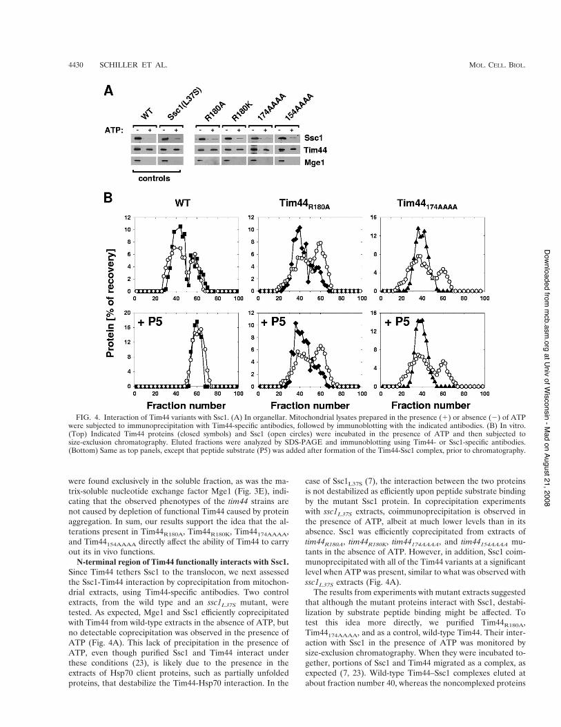
N-terminal region of Tim44 functionally interacts with Ssc1.Since Tim44 tethers Ssc1 to the translocon, we next assessedthe Ssc1-Tim44 interaction by coprecipitation from mitochon-drial extracts, using Tim44-specific antibodies. Two controlextracts, from the wild type and an ssc1L37S mutant, weretested. As expected, Mge1 and Ssc1 efficiently coprecipitatedwith Tim44 from wild-type extracts in the absence of ATP, butno detectable coprecipitation was observed in the presence ofATP (Fig. 4A). This lack of precipitation in the presence ofATP, even though purified Ssc1 and Tim44 interact underthese conditions (23), is likely due to the presence in theextracts of Hsp70 client proteins, such as partially unfoldedproteins, that destabilize the Tim44-Hsp70 interaction. In the
case of Ssc1L37S (7), the interaction between the two proteinsis not destabilized as efficiently upon peptide substrate bindingby the mutant Ssc1 protein. In coprecipitation experimentswith ssc1L37S extracts, coimmunoprecipitation is observed inthe presence of ATP, albeit at much lower levels than in itsabsence. Ssc1 was efficiently coprecipitated from extracts oftim44R180A, tim44R180K, tim44174AAAA, and tim44154AAAA mu-tants in the absence of ATP. However, in addition, Ssc1 coim-munoprecipitated with all of the Tim44 variants at a significantlevel when ATP was present, similar to what was observed withssc1L37S extracts (Fig. 4A).
The results from experiments with mutant extracts suggestedthat although the mutant proteins interact with Ssc1, destabi-lization by substrate peptide binding might be affected. Totest this idea more directly, we purified Tim44R180A,Tim44174AAAA, and as a control, wild-type Tim44. Their inter-action with Ssc1 in the presence of ATP was monitored bysize-exclusion chromatography. When they were incubated to-gether, portions of Ssc1 and Tim44 migrated as a complex, asexpected (7, 23). Wild-type Tim44–Ssc1 complexes eluted atabout fraction number 40, whereas the noncomplexed proteins
FIG. 4. Interaction of Tim44 variants with Ssc1. (A) In organellar. Mitochondrial lysates prepared in the presence (!) or absence (&) of ATPwere subjected to immunoprecipitation with Tim44-specific antibodies, followed by immunoblotting with the indicated antibodies. (B) In vitro.(Top) Indicated Tim44 proteins (closed symbols) and Ssc1 (open circles) were incubated in the presence of ATP and then subjected tosize-exclusion chromatography. Eluted fractions were analyzed by SDS-PAGE and immunoblotting using Tim44- or Ssc1-specific antibodies.(Bottom) Same as top panels, except that peptide substrate (P5) was added after formation of the Tim44-Ssc1 complex, prior to chromatography.
4430 SCHILLER ET AL. MOL. CELL. BIOL.
at Univ of Wisconsin - M
ad on August 21, 2008 m
cb.asm.org
Downloaded from
eluted at fractions around fraction 60. Both Tim44 variantsformed a complex with Ssc1 in the presence of ATP, showingprofiles similar to that of the wild-type control (Fig. 4B, toppanels; data not shown). To address the effect of peptide onmutant Tim44-Ssc1 interactions, we added peptide to pre-formed Ssc1(ATP)-Tim44 complexes and subsequently moni-tored the effect on complex stability by chromatography. Con-sistent with the observations in mitochondrial lysates (Fig. 4A),the wild-type Tim44–Ssc1 complex was efficiently dissociatedupon addition of peptide substrate, as no complex was ob-served (Fig. 4B, bottom left panel). In contrast, preformedcomplexes of Tim44R180A or Tim44174AAAA with Ssc1(ATP)remained intact during the course of the experiment. Likewild-type protein, the mutant proteins formed a complex withSsc1(ADP) which was not destabilized upon peptide addition(data not shown). Together, these results indicate that theamino acid substitutions within Tim44 alter the structure of theprotein such that a Tim44-Ssc1 complex is not rapidly desta-bilized upon addition of peptide substrate.
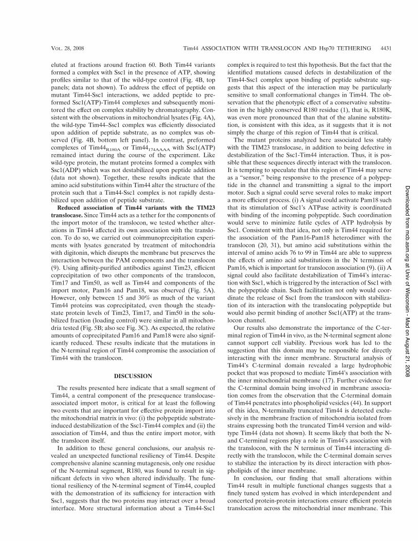
Reduced association of Tim44 variants with the TIM23translocase. Since Tim44 acts as a tether for the components ofthe import motor of the translocon, we tested whether alter-ations in Tim44 affected its own association with the translo-con. To do so, we carried out coimmunoprecipitation experi-ments with lysates generated by treatment of mitochondriawith digitonin, which disrupts the membrane but preserves theinteraction between the PAM components and the translocon(9). Using affinity-purified antibodies against Tim23, efficientcoprecipitation of two other components of the translocon,Tim17 and Tim50, as well as Tim44 and components of theimport motor, Pam16 and Pam18, was observed (Fig. 5A).However, only between 15 and 30% as much of the variantTim44 proteins was coprecipitated, even though the steady-state protein levels of Tim23, Tim17, and Tim50 in the solu-bilized fraction (loading control) were similar in all mitochon-dria tested (Fig. 5B; also see Fig. 3C). As expected, the relativeamounts of coprecipitated Pam16 and Pam18 were also signif-icantly reduced. These results indicate that the mutations inthe N-terminal region of Tim44 compromise the association ofTim44 with the translocon.
DISCUSSION
The results presented here indicate that a small segment ofTim44, a central component of the presequence translocase-associated import motor, is critical for at least the followingtwo events that are important for effective protein import intothe mitochondrial matrix in vivo: (i) the polypeptide substrate-induced destabilization of the Ssc1-Tim44 complex and (ii) theassociation of Tim44, and thus the entire import motor, withthe translocon itself.
In addition to these general conclusions, our analysis re-vealed an unexpected functional resiliency of Tim44. Despitecomprehensive alanine scanning mutagenesis, only one residueof the N-terminal segment, R180, was found to result in sig-nificant defects in vivo when altered individually. The func-tional resiliency of the N-terminal segment of Tim44, coupledwith the demonstration of its sufficiency for interaction withSsc1, suggests that the two proteins may interact over a broadinterface. More structural information about a Tim44-Ssc1
complex is required to test this hypothesis. But the fact that theidentified mutations caused defects in destabilization of theTim44-Ssc1 complex upon binding of peptide substrate sug-gests that this aspect of the interaction may be particularlysensitive to small conformational changes in Tim44. The ob-servation that the phenotypic effect of a conservative substitu-tion in the highly conserved R180 residue (1), that is, R180K,was even more pronounced than that of the alanine substitu-tion, is consistent with this idea, as it suggests that it is notsimply the charge of this region of Tim44 that is critical.
The mutant proteins analyzed here associated less stablywith the TIM23 translocase, in addition to being defective indestabilization of the Ssc1-Tim44 interaction. Thus, it is pos-sible that these sequences directly interact with the translocon.It is tempting to speculate that this region of Tim44 may serveas a “sensor,” being responsive to the presence of a polypep-tide in the channel and transmitting a signal to the importmotor. Such a signal could serve several roles to make importa more efficient process. (i) A signal could activate Pam18 suchthat its stimulation of Ssc1’s ATPase activity is coordinatedwith binding of the incoming polypeptide. Such coordinationwould serve to minimize futile cycles of ATP hydrolysis bySsc1. Consistent with that idea, not only is Tim44 required forthe association of the Pam16-Pam18 heterodimer with thetranslocon (20, 31), but amino acid substitutions within theinterval of amino acids 76 to 99 in Tim44 are able to suppressthe effects of amino acid substitutions in the N terminus ofPam16, which is important for translocon association (9). (ii) Asignal could also facilitate destabilization of Tim44’s interac-tion with Ssc1, which is triggered by the interaction of Ssc1 withthe polypeptide chain. Such facilitation not only would coor-dinate the release of Ssc1 from the translocon with stabiliza-tion of its interaction with the translocating polypeptide butwould also permit binding of another Ssc1(ATP) at the trans-locon channel.
Our results also demonstrate the importance of the C-ter-minal region of Tim44 in vivo, as the N-terminal segment alonecannot support cell viability. Previous work has led to thesuggestion that this domain may be responsible for directlyinteracting with the inner membrane. Structural analysis ofTim44’s C-terminal domain revealed a large hydrophobicpocket that was proposed to mediate Tim44’s association withthe inner mitochondrial membrane (17). Further evidence forthe C-terminal domain being involved in membrane associa-tion comes from the observation that the C-terminal domainof Tim44 penetrates into phospholipid vesicles (44). In supportof this idea, N-terminally truncated Tim44 is detected exclu-sively in the membrane fraction of mitochondria isolated fromstrains expressing both the truncated Tim44 version and wild-type Tim44 (data not shown). It seems likely that both the N-and C-terminal regions play a role in Tim44’s association withthe translocon, with the N terminus of Tim44 interacting di-rectly with the translocon, while the C-terminal domain servesto stabilize the interaction by its direct interaction with phos-pholipids of the inner membrane.
In conclusion, our finding that small alterations withinTim44 result in multiple functional changes suggests that afinely tuned system has evolved in which interdependent andconcerted protein-protein interactions ensure efficient proteintranslocation across the mitochondrial inner membrane. This
VOL. 28, 2008 Tim44 ASSOCIATION WITH TRANSLOCON AND Hsp70 TETHERING 4431
at Univ of Wisconsin - M
ad on August 21, 2008 m
cb.asm.org
Downloaded from

is consistent with the fact that, overall, the mitochondrial pro-tein import machinery is one of the most complex proteintranslocation systems known. A mechanistic understanding ofthe function of this system at the molecular level is just begin-ning to emerge.
ACKNOWLEDGMENTS
We thank Robert Jensen for Tim17 antibody and Justin Hines,Thomas Lee, and Amy Prunuske for helpful comments on the manu-script.
This work was supported by National Institutes of Health grantGM278709 (to E.A.C.) and by a research fellowship of the DeutscheForschungsgemeinschaft (to D.S.).
REFERENCES1. Bauer, M. F., K. Gempel, A. S. Reichert, G. A. Rappold, P. Lichtner, K. D.
Gerbitz, W. Neupert, M. Brunner, and S. Hofmann. 1999. Genetic and
structural characterization of the human mitochondrial inner membranetranslocase. J. Mol. Biol. 289:69–82.
2. Bauer, M. F., C. Sirrenberg, W. Neupert, and M. Brunner. 1996. Role ofTim23 as voltage sensor and presequence receptor in protein import intomitochondria. Cell 87:33–41.
3. Becker, L., M. Bannwarth, C. Meisinger, K. Hill, K. Model, T. Krimmer, R.Casadio, K. N. Truscott, G. E. Schulz, N. Pfanner, and R. Wagner. 2005.Preprotein translocase of the outer mitochondrial membrane: reconstitutedTom40 forms a characteristic TOM pore. J. Mol. Biol. 353:1011–1020.
4. Boeke, J. D., F. LaCroute, and G. R. Fink. 1984. A positive selection formutants lacking orotidine-5"-phosphate decarboxylase activity in yeast:5-fluoro-orotic acid resistance. Mol. Gen. Genet. 197:345–346.
5. Bomer, U., A. C. Maarse, F. Martin, A. Geissler, A. Merlin, B. Schonfisch,M. Meijer, N. Pfanner, and J. Rassow. 1998. Separation of structural anddynamic functions of the mitochondrial translocase: Tim44 is crucial for theinner membrane import sites in translocation of tightly folded domains, butnot of loosely folded preproteins. EMBO J. 17:4226–4237.
6. Donzeau, M., K. Kaldi, A. Adam, S. Paschen, G. Wanner, B. Guiard, M. F.Bauer, W. Neupert, and M. Brunner. 2000. Tim23 links the inner and outermitochondrial membranes. Cell 101:401–412.
FIG. 5. Association of the import motor with the TIM23 translocase. (A) Mitochondria were solubilized by treatment with digitonin.Solubilized material was subjected to immunoprecipitation using Tim23-specific antibodies cross-linked to protein A beads. Precipitates wereanalyzed by SDS-PAGE and immunoblotting using antibodies specific for the indicated proteins (IP). A total of 4% solubilized material was usedas a loading control (load). (B) Signals were quantified by densitometry and plotted as percentages of the wild-type control reaction value.
4432 SCHILLER ET AL. MOL. CELL. BIOL.
at Univ of Wisconsin - M
ad on August 21, 2008 m
cb.asm.org
Downloaded from

7. D’Silva, P., Q. Liu, W. Walter, and E. A. Craig. 2004. Regulated interactionsof mtHsp70 with Tim44 at the translocon in the mitochondrial inner mem-brane. Nat. Struct. Mol. Biol. 11:1084–1091.
8. D’Silva, P. D., B. Schilke, W. Walter, A. Andrew, and E. A. Craig. 2003. Jprotein cochaperone of the mitochondrial inner membrane required forprotein import into the mitochondrial matrix. Proc. Natl. Acad. Sci. USA100:13839–13844.
9. D’Silva, P. R., B. Schilke, M. Hayashi, and E. A. Craig. 2008. Interaction ofthe J-protein heterodimer, Pam18/Pam16, of the mitochondrial import mo-tor with the translocon of the inner membrane. Mol. Biol. Cell 19:424–432.
10. D’Silva, P. R., B. Schilke, W. Walter, and E. A. Craig. 2005. Role of Pam16’sdegenerate J domain in protein import across the mitochondrial inner mem-brane. Proc. Natl. Acad. Sci. USA 102:12419–12424.
11. Frazier, A. E., J. Dudek, B. Guiard, W. Voos, Y. Li, M. Lind, C. Meisinger,A. Geissler, A. Sickmann, H. E. Meyer, V. Bilanchone, M. G. Cumsky, K. N.Truscott, N. Pfanner, and P. Rehling. 2004. Pam16 has an essential role inthe mitochondrial protein import motor. Nat. Struct. Mol. Biol. 11:226–233.
12. Gambill, B. D., W. Voos, P. J. Kang, B. Miao, T. Langer, E. A. Craig, and N.Pfanner. 1993. A dual role for mitochondrial heat shock protein 70 inmembrane translocation of preproteins. J. Cell Biol. 123:109–117.
13. Geissler, A., A. Chacinska, K. N. Truscott, N. Wiedemann, K. Brandner, A.Sickmann, H. E. Meyer, C. Meisinger, N. Pfanner, and P. Rehling. 2002. Themitochondrial presequence translocase: an essential role of Tim50 in direct-ing preproteins to the import channel. Cell 111:507–518.
14. Hill, K., K. Model, M. T. Ryan, K. Dietmeier, F. Martin, R. Wagner, and N.Pfanner. 1998. Tom40 forms the hydrophilic channel of the mitochondrialimport pore for preproteins. Nature 395:516–521.
15. Horst, M., P. Jeno, N. G. Kronidou, L. Bolliger, W. Oppliger, P. Scherer, U.Manning-Krieg, T. Jascur, and G. Schatz. 1993. Protein import into yeastmitochondria: the inner membrane import site protein ISP45 is the MPI1gene product. EMBO J. 12:3035–3041.
16. Jones, E. W. 1991. Three proteolytic systems in the yeast Saccharomycescerevisiae. J. Biol. Chem. 266:7963–7966.
17. Josyula, R., Z. Jin, Z. Fu, and B. Sha. 2006. Crystal structure of yeastmitochondrial peripheral membrane protein Tim44p C-terminal domain. J.Mol. Biol. 359:798–804.
18. Kang, P. J., J. Ostermann, J. Shilling, W. Neupert, E. A. Craig, and N.Pfanner. 1990. Requirement for hsp70 in the mitochondrial matrix for trans-location and folding of precursor proteins. Nature 348:137–143.
19. Koehler, C. M. 2004. New developments in mitochondrial assembly. Annu.Rev. Cell Dev. Biol. 20:309–335.
20. Kozany, C., D. Mokranjac, M. Sichting, W. Neupert, and K. Hell. 2004. TheJ domain-related cochaperone Tim16 is a constituent of the mitochondrialTIM23 preprotein translocase. Nat. Struct. Mol. Biol. 11:234–241.
21. Krayl, M., J. H. Lim, F. Martin, B. Guiard, and W. Voos. 2007. A cooper-ative action of the ATP-dependent import motor complex and the innermembrane potential drives mitochondrial preprotein import. Mol. Cell. Biol.27:411–425.
22. Kronidou, N. G., W. Oppliger, L. Bolliger, K. Hannavy, B. S. Glick, G.Schatz, and M. Horst. 1994. Dynamic interaction between Isp45 and mito-chondrial hsp70 in the protein import system of the yeast mitochondrialinner membrane. Proc. Natl. Acad. Sci. USA 91:12818–12822.
23. Liu, Q., P. D’Silva, W. Walter, J. Marszalek, and E. A. Craig. 2003. Regu-lated cycling of mitochondrial Hsp70 at the protein import channel. Science300:139–141.
24. Liu, Q., J. Krzewska, K. Liberek, and E. A. Craig. 2001. MitochondrialHsp70 Ssc1: role in protein folding. J. Biol. Chem. 276:6112–6118.
25. Maarse, A. C., J. Blom, L. A. Grivell, and M. Meijer. 1992. MPI1, anessential gene encoding a mitochondrial membrane protein, is possibly in-volved in protein import into yeast mitochondria. EMBO J. 11:3619–3628.
26. Martin, J., K. Mahlke, and N. Pfanner. 1991. Role of an energized inner
membrane in mitochondrial protein import. Delta psi drives the movementof presequences. J. Biol. Chem. 266:18051–18057.
27. Merlin, A., W. Voos, A. C. Maarse, M. Meijer, N. Pfanner, and J. Rassow.1999. The J-related segment of Tim44 is essential for cell viability: a mutantTim44 remains in the mitochondrial import site, but inefficiently recruitsmtHsp70 and impairs protein translocation. J. Cell Biol. 145:961–972.
28. Milisav, I., F. Moro, W. Neupert, and M. Brunner. 2001. Modular structureof the TIM23 preprotein translocase of mitochondria. J. Biol. Chem. 276:25856–25861.
29. Mokranjac, D., A. Berg, A. Adam, W. Neupert, and K. Hell. 2007. Associa-tion of the Tim14.Tim16 subcomplex with the TIM23 translocase is crucialfor function of the mitochondrial protein import motor. J. Biol. Chem.282:18037–18045.
30. Mokranjac, D., S. A. Paschen, C. Kozany, H. Prokisch, S. C. Hoppins, F. E.Nargang, W. Neupert, and K. Hell. 2003. Tim50, a novel component of theTIM23 preprotein translocase of mitochondria. EMBO J. 22:816–825.
31. Mokranjac, D., M. Sichting, W. Neupert, and K. Hell. 2003. Tim14, a novelkey component of the import motor of the TIM23 protein translocase ofmitochondria. EMBO J. 22:4945–4956.
32. Neupert, W., and M. Brunner. 2002. The protein import motor of mitochon-dria. Nat. Rev. Mol. Cell Biol. 3:555–565.
33. Neupert, W., and J. M. Herrmann. 2007. Translocation of proteins intomitochondria. Annu. Rev. Biochem. 76:723–749.
34. Pfanner, N., and K. N. Truscott. 2002. Powering mitochondrial proteinimport. Nat. Struct. Biol. 9:234–236.
35. Rassow, J., A. C. Maarse, E. Krainer, M. Kubrich, H. Muller, M. Meijer,E. A. Craig, and N. Pfanner. 1994. Mitochondrial protein import: biochem-ical and genetic evidence for interaction of matrix hsp70 and the innermembrane protein MIM44. J. Cell Biol. 127:1547–1556.
36. Rehling, P., N. Wiedemann, N. Pfanner, and K. N. Truscott. 2001. Themitochondrial import machinery for preproteins. Crit. Rev. Biochem. Mol.Biol. 36:291–336.
37. Schneider, H. C., J. Berthold, M. F. Bauer, K. Dietmeier, B. Guiard, M.Brunner, and W. Neupert. 1994. Mitochondrial Hsp70/MIM44 complex fa-cilitates protein import. Nature 371:768–774.
38. Sickmann, A., J. Reinders, Y. Wagner, C. Joppich, R. Zahedi, H. E. Meyer,B. Schonfisch, I. Perschil, A. Chacinska, B. Guiard, P. Rehling, N. Pfanner,and C. Meisinger. 2003. The proteome of Saccharomyces cerevisiae mito-chondria. Proc. Natl. Acad. Sci. USA 100:13207–13212.
39. Sikorski, R. S., and P. Hieter. 1989. A system of shuttle vectors and yeasthost strains designed for efficient manipulation of DNA in Saccharomycescerevisiae. Genetics 122:19–27.
40. Slutsky-Leiderman, O., M. Marom, O. Iosefson, R. Levy, S. Maoz, and A.Azem. 2007. The interplay between components of the mitochondrial proteintranslocation motor studied using purified components. J. Biol. Chem. 282:33935–33942.
41. Truscott, K. N., W. Voos, A. E. Frazier, M. Lind, Y. Li, A. Geissler, J. Dudek,H. Muller, A. Sickmann, H. E. Meyer, C. Meisinger, B. Guiard, P. Rehling,and N. Pfanner. 2003. A J-protein is an essential subunit of the presequencetranslocase-associated protein import motor of mitochondria. J. Cell Biol.163:707–713.
42. Voisine, C., E. A. Craig, N. Zufall, O. von Ahsen, N. Pfanner, and W. Voos.1999. The protein import motor of mitochondria: unfolding and trapping ofpreproteins are distinct and separable functions of matrix Hsp70. Cell 97:565–574.
43. Voos, W., O. von Ahsen, H. Muller, B. Guiard, J. Rassow, and N. Pfanner.1996. Differential requirement for the mitochondrial Hsp70-Tim44 complexin unfolding and translocation of preproteins. EMBO J. 15:2668–2677.
44. Weiss, C., W. Oppliger, G. Vergeres, R. Demel, P. Jeno, M. Horst, B. deKruijff, G. Schatz, and A. Azem. 1999. Domain structure and lipid interac-tion of recombinant yeast Tim44. Proc. Natl. Acad. Sci. USA 96:8890–8894.
VOL. 28, 2008 Tim44 ASSOCIATION WITH TRANSLOCON AND Hsp70 TETHERING 4433
at Univ of Wisconsin - M
ad on August 21, 2008 m
cb.asm.org
Downloaded from