Embed Size (px)
Citation preview

RESEARCH ARTICLE
Muscle Regeneration with IntermuscularAdipose Tissue (IMAT) Accumulation IsModulated by Mechanical ConstraintsAllan F. Pagano1☯, Rémi Demangel1☯, Thomas Brioche1, Elodie Jublanc1,Christelle Bertrand-Gaday1, Robin Candau1, Claude A. Dechesne2, Christian Dani2,Anne Bonnieu1, Guillaume Py1‡, Angèle Chopard1‡*
1 Université de Montpellier, INRA, UMR866 Dynamique Musculaire et Métabolisme, F-34060, Montpellier,France, 2 Université Nice-Sophia Antipolis, iBV, CNRS UMR7277, INSERMU1091, 06107, Nice, France
☯ These authors contributed equally to this work.‡ These authors also contributed equally to this work.* [email protected]
AbstractSports trauma are able to induce muscle injury with fibrosis and accumulation of intermus-
cular adipose tissue (IMAT), which affect muscle function. This study was designed to
investigate whether hypoactivity would influence IMAT accumulation in regenerating mouse
skeletal muscle using the glycerol model of muscle regeneration. The animals were immedi-
ately hindlimb unloaded for 21 days after glycerol injection into the tibialis anterior (TA) mus-
cle. Muscle fiber and adipocyte cross-sectional area (CSA) and IMAT accumulation were
determined by histomorphometric analysis. Adipogenesis during regenerative processes
was examined using RT-qPCR andWestern blot quantification. Twenty-one days of hin-
dlimb unloading resulted in decreases of 38% and 50.6% in the muscle weight/body weight
ratio and CSA, respectively, in soleus muscle. Glycerol injection into TA induced IMAT
accumulation, reaching 3% of control normal-loading muscle area. This IMAT accumulation
was largely inhibited in unloading conditions (0.09%) and concomitant with a marked reduc-
tion in perilipin and FABP4 protein content, two key markers of mature adipocytes. Induction
of PPARγ and C/EBPαmRNA, two markers of adipogenesis, was also decreased. Further-
more, the protein expression of PDGFRα, a cell surface marker of fibro/adipogenic progeni-
tors, was much lower in regenerating TA from the unloaded group. Exposure of
regenerating muscle to hypoactivity severely reduces IMAT development and accumula-
tion. These results provide new insight into the mechanisms regulating IMAT development
in skeletal muscle and highlight the importance of taking into account the level of mechani-
cal constraint imposed on skeletal muscle during the regeneration processes.
PLOS ONE | DOI:10.1371/journal.pone.0144230 December 2, 2015 1 / 17
a11111
OPEN ACCESS
Citation: Pagano AF, Demangel R, Brioche T,Jublanc E, Bertrand-Gaday C, Candau R, et al.(2015) Muscle Regeneration with IntermuscularAdipose Tissue (IMAT) Accumulation Is Modulated byMechanical Constraints. PLoS ONE 10(12):e0144230. doi:10.1371/journal.pone.0144230
Editor: Stephen E Alway, West Virginia UniversitySchool of Medicine, UNITED STATES
Received: May 21, 2015
Accepted: November 16, 2015
Published: December 2, 2015
Copyright: © 2015 Pagano et al. This is an openaccess article distributed under the terms of theCreative Commons Attribution License, which permitsunrestricted use, distribution, and reproduction in anymedium, provided the original author and source arecredited.
Data Availability Statement: All relevant data arewithin the paper and its Supporting Information files.
Funding: This work was supported by the CentreNational d’Etudes Spatiales (CNES).
Competing Interests: The authors have declaredthat no competing interests exist.

IntroductionThe capacity of skeletal muscle to regenerate is a key parameter of its plasticity. A wide varietyof stress can induce muscle injuries, including sport traumas, prolonged blood flow disruptionor even muscle diseases. After injury, skeletal muscle is able to regenerate through various andhigh coordinated stages including degeneration, inflammation, and regeneration process [1].These steps include recruitment of satellite cells (SCs), which are localized between the sarco-lemma and the basal lamina of myofibers [2]. Indeed, it is now well known that quiescent satel-lite cells proliferate, migrate and differentiate into mature myofibers to regenerate injuredmuscle tissue [3–5]. Numerous studies have already shown that hindlimb unloading (HU),commonly used to mimic hypoactivity and also microgravity [6], induces a loss in SC contentand mitotic activity, which disturbs muscle regeneration by reducing growth of the newlyformed myofibers [7–9].
The research literature also indicates that abnormal fibrosis and intermuscular adipose tis-sue (IMAT) accumulation occur, particularly when early regeneration processes are altered,and that this in turn alters muscle function. IMAT is defined as adipocyte accumulationbetween muscle cells and beneath the muscle fascia, and it should not be confused with intra-myocellular triglyceride accumulation [10]. Studies have shown that impaired macrophagefunction is linked to poor muscle regeneration and IMAT accumulation after freeze-induced[11], ischemic [12, 13], notexin-induced [14] and cardiotoxin-induced [15] injury.
In these regeneration models, little or no IMAT accumulation is naturally observed.Although IMAT does not occur naturally in rodent skeletal muscles, a skeletal muscle regener-ation model with IMAT accumulation was developed in rabbit by Kawai et al. [16] and waslater used in mice in several studies [17–20]. This regeneration model consists of injecting glyc-erol into skeletal muscle, and Dani’s group was the first to present a detailed characterizationof the “glycerol approach” [21]. The model has been used in several studies to investigateIMAT development and its related adipogenic processes and, more recently, to better charac-terize muscle-resident adipocyte precursors [19, 20, 22]. To our knowledge, the study of Lukja-nenko et al. [22] has been the only one to provide a detailed characterization of some of thecellular responses related to this regeneration model in comparison with the more classic cardi-otoxin model. Their study clearly showed that the two models induced similar kinetics of skele-tal muscle degeneration and regeneration, but they differed with regard to the adipogenicresponse amplitude. The glycerol model was therefore associated with more mature adipocytesaccumulation.
Recently, studies have highlighted the growing importance of muscle-resident mesenchymalstem cells in the regeneration process of skeletal muscle [23, 24]. In particular, fibro/adipogenicprogenitors (FAPs), which are mainly positive for the cell surface marker platelet-derivedgrowth factor receptor alpha (PDGFRα or CD140a), play an important role in efficient regen-eration. In a healthy but damaged muscle, FAPs proliferate, phagocytize necrotic debris, andincrease the proliferation of SCs without differentiating into adipocytes [19, 25]. In muscle dis-use or pathological conditions, such as Duchenne muscular dystrophy, FAPs proliferate anddifferentiate into adipose and/or fibrous tissue [26, 27]. Parallel to the decrease in SC content[28, 29], FAPs in this case lead to an accumulation of IMAT [10, 20].
Although it is clear that disturbed regeneration promotes IMAT accumulation and impairedskeletal muscle function [30], there is a lack of data evaluating the effects of prolonged hypoac-tivity on IMAT accumulation during regeneration processes. In a closed context, the study ofJarvinen and Lehto [31] conducted in rats showed that immobilization following contusioninjury limited the size of the connective tissue area formed within the injury site. One mighttherefore ask whether a period of hypoactivity would modulate IMAT development in a
Mechanical Constraints Modulate IMAT Associated to Muscle Regeneration
PLOS ONE | DOI:10.1371/journal.pone.0144230 December 2, 2015 2 / 17

regeneration model characterized by IMAT development. Our study was designed to investi-gate and characterize the effects of prolonged hypoactivity induced by hindlimb unloading onIMAT development and accumulation in the glycerol model of muscle regeneration. RodentHU was set up immediately after glycerol injection to ensure hypoactivity during the entireperiod of the regeneration process. Our results clearly showed that IMAT accumulation inregenerating muscle was substantially lowered with unloading conditions, with a subsequentdecrease in FAP recruitment.
Materials and Methods
Ethics statementThis study was approved by the Committee on the Ethics of Animal Experiments of LanguedocRoussillon in accordance with the guidelines from the French National Research Council forthe Care and Use of Laboratory Animals (CEEA-LR-14002). All efforts were made to minimizeanimal suffering.
AnimalsExperiments were carried out on 6-month-old C57BL6J/CBA female mice (n = 36; mean bodymass = 25.4g ±0.44) from our own stock. Animals were maintained on a 12h/12h light–darkcycle and provided with food and water ad libitum. Experiments were performed at 22°C.
Experimental procedures and muscle samplingExperimental procedures were performed under anesthesia using isoflurane inhalation. Inaccordance with the study of Pisani et al. [21], the mice were injected with 25μl of 50% v/v glyc-erol in the right tibialis anterior (TAg) and with saline solution in the contralateral tibialis ante-rior (TA). Two experimental groups were formed: hindlimb-unloaded (HU, n = 6) and control(CTL, n = 6), for 21 days. Tail-suspended experiments were conducted with suspension cagesand the protocol used in other studies [32, 33]. At the end of the 21 days of unloading or con-trol conditions, soleus (SOL) and extensor digitorum longus (EDL) hindlimb muscles were rap-idly dissected out and immediately frozen in isopentane cooled with liquid nitrogen and thenstored at -80°C. For the first set of experiments, TAg and TA muscles were also rapidly dis-sected out and immediately fixed overnight in 4% paraformaldehyde solution at room temper-ature and then paraffin-embedded. For the second and third set of experiments, TAg and TAfrom CTL and HU animals were dissected out and rapidly frozen in liquid nitrogen for quanti-fication of mRNA and protein content, as described elsewhere.
HistologyTransverse serial sections of SOL and EDL muscles (9μm thick) were obtained at -20°C using acryostat and sections were subjected to hematoxylin-eosin staining for subsequent cross-sec-tional area (CSA) measurements.
TA and TAg muscles were fixed in 4% neutral-buffered formalin (24h) and paraffin-embed-ded. The paraffin-embedded tissues were sectioned (3μm thick) every 50μm over the entiremuscle depth, and sections were processed by hematoxylin/eosin/saffron (H/E/S) staining.Stained slides were digitalized with the NanoZoomer slide scanner with a x40 objective (Hama-matsu) for subsequent muscle fibers and adipocytes CSA evaluation. Images were analyzedwith ImageJ1 (1.46r version) software to measure the CSA.
Mechanical Constraints Modulate IMAT Associated to Muscle Regeneration
PLOS ONE | DOI:10.1371/journal.pone.0144230 December 2, 2015 3 / 17

ImmunohistochemistryThe immunohistochemistry protocol was globally performed as previously described [34].Briefly, mouse colon (positive control) and TAg muscle sections were deparaffinized, rehy-drated, and incubated for antigen retrieval in EDTA buffer at 100°C for 10 min. Sections wereincubated in 0.3% H2O2 for 20 min and endogenous biotins were blocked using the Avidin-Biotin Blocking kit (Vector Laboratories, CliniSciences). Nonspecific antibody binding wasblocked by incubation with TBS containing 20% normal goat serum for 30 min at RT. Sectionswere then incubated ON at 4°C with rabbit anti-PDGFRα diluted at 1:250 or non-specific rab-bit IgG (Vector Laboratories, CliniSciences) at the same concentration. Antibody binding wasrevealed by the streptavidin/biotin-peroxydase complex method using ABC Vectastain kit andthe peroxidase substrate DAB (Vector Laboratories, Clinisciences).
RNA extraction and real-time polymerase chain reaction (RT-qPCR)Total RNAs were isolated from homogenate muscle samples using the RNeasy Fibrous TissueMini Kit following the manufacturer’s instructions (Qiagen). RNA concentration was deter-mined by spectrophotometric analysis (Eppendorf AG, Hamburg, Germany), and integrity waschecked by the OD260nm/OD280nm absorption ratio (>1.7). Reverse transcription reaction wasperformed with 2μg of total RNA using the RevertAid First Strand cDNA Synthesis kit(Thermo Scientific) according to the manufacturer’s instructions. qPCR analysis was per-formed in a MiniOpticon detection system (Bio-Rad, Hercules, CA) with 10μL of KAPA SYBRFast Universal Readymix (CliniSciences), 300nM of both forward and reverse primers, 2μL ofdiluted cDNA template and water to a final volume of 20μL. The forward and reverse primersused to amplify genes are listed in Table 1. All PCRs were performed in duplicate using the fol-lowing cycle parameters: 30s at 98°C, 40 cycles of 1s at 95°C and 15s at 60°C. Relative mRNAlevels were normalized to ribosomal protein S9 (rpS9) and cyclophilin A housekeeping genelevels, which were unaffected by treatments. Results are expressed using the comparative cyclethreshold (CT). The relative changes in the level of a specific gene were calculated with theΔΔCT formula.
Protein isolation andWestern blottingTheWestern blot protocol was performed as previously described [35]. Briefly, muscle sampleswere homogenized in 10 volumes of lysis buffer (50mM Tris–HCl pH7.5, 150mMNaCl, 1mMEGTA, 100mMNaF, 5mMNa3VO4, 1% Triton X-100, 40mM β-glycerophosphate and prote-ase inhibitor mixture (P8340; Sigma-Aldrich)) and centrifuged at 10.000g for 10 min (4°C).60μg of protein extracts were loaded on SDS–polyacrylamide gels before electrophoretic trans-fer onto a nitrocellulose membrane (Bio-Rad). After transfer, membranes were blocked with50mM Tris-HCl pH7.5, 150mMNaCl, and 0.1% Tween 20 (TBS-T) containing 5% skimmedmilk or BSA and incubated overnight at 4°C with primary antibodies. Membranes were incu-bated for 1 h with a peroxidase-conjugated secondary antibody. Immunoblots were revealedusing a Pierce ECL kit (32106; Thermo Scientific) and proteins were visualized by enhancedchemiluminescence and quantified with ImageJ1 (1.46r version) software. β-actin was used asa loading control.
AntibodiesAnti-PDGFRα (#3174), anti-perilipin (#9349), anti-FABP4 (#3544) and anti-adiponectin(#2789) primary antibodies were purchased from Cell Signaling and used at 1:500. Anti-β-actin (sc-81178) primary antibody was purchased from Santa Cruz and used at 1:2000. Anti-
Mechanical Constraints Modulate IMAT Associated to Muscle Regeneration
PLOS ONE | DOI:10.1371/journal.pone.0144230 December 2, 2015 4 / 17

mouse (NA931V) and anti-rabbit (NA934V) HRP-conjugated secondary antibodies were pur-chased from GE Healthcare Life Science and used at 1:3000.
StatisticsAll values are expressed as mean ± SEM and the significance level was set as p<0.05. Differ-ences between the two groups were evaluated for significance using the unpaired Student t-testor the Mann-Whitney test when data deviated from a normal distribution. When more thantwo simultaneous comparisons were made, a two-way ANOVA was employed to compare data(unloading and glycerol-injection factors). When a significant effect was indicated, a Fisher sig-nificant difference post hoc test was performed.
Results
Body mass, muscle mass, and fiber cross-sectional areas following 21days of hindlimb unloadingBody mass was significantly changed after 21 days of HU (24.57g at day 0 and 22.81g at day 21,p<0.001), whereas no difference was found in control conditions (26.13g at day 0 and 26.47gat day 21). SOL muscle mass, expressed in either absolute values or values normalized by bodymass, decreased significantly after HU (-50.8% and -38% respectively, p<0.001, Fig 1a and 1b)compared with CTL. EDL and TA muscle mass decreased significantly after HU, but onlywhen expressed in absolute values (-15.1%, p<0.05, and -11.5%, p<0.001, respectively, Fig 1aand 1b). The mean CSA of SOL fibers was significantly reduced after HU compared with CTL(-50.6%, p<0.001, Fig 1c). However, no significant differences were detected for the mean CSAof EDL and TA muscle fibers (Fig 1c). Thus, our results are in agreement with the literature,which has shown major atrophy in SOL, a postural muscle, following 21 days of HU [6, 36].
Hindlimb unloading disturbs regeneration in glycerol-injected tibialisanteriorGlycerol-injected tibialis anterior (TAg) muscle mass, expressed in either absolute values orvalues normalized by body mass, decreased significantly after HU (-27.87% and -17.1% respec-tively, p<0.001, Fig 2a and 2b) compared with TAg-CTL. In parallel, the mean CSA of TAg-HU fibers versus TAg-CTL was significantly reduced (-25.1%, p<0.001, Fig 2c and 2d). Thissuggests that hindlimb unloading affects skeletal muscle regeneration and thereby reducesmyofiber size and mass recovery. We further examined the mRNA induction of key genesimplicated in the muscle regeneration processes and myogenesis in TA and TAg of the CTL
Table 1. Real-time PCR primers.
Gene Forward Reverse Amplicon size
C/EBPα GACCAGAAAGCTGAGTTGTGAG CCACAAAGCCCAGAAACCTA 69 bp
C/EBPβ CTCCAGGTAGGGGCTGAAGT TTTAGACCCATGGAAGTGGC 150 bp
Cyclophilin A TTCCTCCTTTCACAGAATTATTCCA CCGCCAGTGCCATTATGG 75 bp
MyoD AGCACTACAGTGGCGACTCA GGCCGCTGTAATCCATCAT 75 bp
myogenin ACAGGCCTTGCTCAGCTC CGCTGTGGGAGTTGCATT 102 bp
Pax7 GCTACCAGTACAGCCAGTATG GTCACTAAGCATGGGTAGATG 328 bp
PDGFRα AAGACCTGGGCAAGAGGAAC GAACCTGTCTCGATGGCACT 67 bp
PPARγ GTGCCAGTTTCGATCCGTAGA GGCCAGCATCGTGTAGATGA 142 bp
rpS9 CGGCCCGGGAGCTGTTGACG CTGCTTGCGGACCCTAATGT 247 bp
doi:10.1371/journal.pone.0144230.t001
Mechanical Constraints Modulate IMAT Associated to Muscle Regeneration
PLOS ONE | DOI:10.1371/journal.pone.0144230 December 2, 2015 5 / 17

and HU groups. mRNA induction of Pax7, a marker of quiescent and proliferative SCs in adultmice, decreased only in TAg-HU compared with TAg-CTL (-46.9%, p<0.05, Fig 2e). Levels ofMyoD and myogenin mRNA, two markers of activated and differentiation-engaged myoblasts,respectively, were reduced significantly in TA-HU versus TA-CTL (-43.7% and -43.5% respec-tively, p<0.05, Fig 2f and 2g). A similar decrease in magnitude was also observed in TAg-HUversus TAg-CTL (-45.7%, p<0.01 and -44.85%, p<0.05, respectively, for MyoD and myogenin,Fig 2f and 2g). Taken together, these results based on mRNA analysis could indicate that hin-dlimb unloading alone did not alter the number of SCs but did decrease their mitotic activity.
Fig 1. Effects of hindlimb unloading onmuscle mass and fiber cross-sectional areas. Soleus (SOL),extensor digitorum longus (EDL), tibialis anterior (TA) skeletal muscle mass from control (CTL) and hindlimbunloading (HU) mice (a). SOL, EDL, and TA mass normalized by body mass from CTL and HUmice (b).Myofiber cross-sectional area (CSA) in μm² of SOL, EDL, and TA skeletal muscles from CTL and HUmice(c). * p<0.05 vs control group, and *** p<0.001 vs control group.
doi:10.1371/journal.pone.0144230.g001
Mechanical Constraints Modulate IMAT Associated to Muscle Regeneration
PLOS ONE | DOI:10.1371/journal.pone.0144230 December 2, 2015 6 / 17

However, when HU was associated with a regenerating context, we found a decrease in the SCpool and mitotic activity, which thus decreased the subsequent regrowth and myofiber sizerecovery.
Hypoactivity inhibits IMAT accumulation in regenerating TA muscleIt is well known that the glycerol model of muscle regeneration induces IMAT developmentduring muscle regeneration. In our study, we confirmed the presence of IMAT in the TAinjected with glycerol of normal-loaded animals (TAg-CTL). The area occupied by IMAT 21days after glycerol injection reached a mean 2.83% of the total muscle CSA, whereas TA-CTLshowed no histological sign of IMAT (Fig 3a and 3b). Thus, as previously described in thestudy of Dani’s group [21], it seems that the muscle microenvironment created by glycerolinjection may favor adipogenesis from resident and/or recruited precursors, unlike what occursin other regeneration models.
Conversely, unloading almost completely prevented IMAT development in TAg: the areaoccupied by IMAT was almost nil (0.09%, p<0.05) in TAg-HU (Fig 3a and 3b). Moreover,CSA morphometric analysis of the adipocytes from IMAT showed a decrease in TAg-HU com-pared with TAg-CTL (130μm2 and 510μm2, respectively, p<0.05, Fig 3c and 3d).
Fig 2. Effects of hindlimb unloading and glycerol injection on tibialis anterior muscle.Muscle mass (a), muscle mass/body mass (b) and cross-sectional area (CSA) (c) of glycerol-injected tibialis anterior (TAg) from control (CTL) and hindlimb unloading (HU) mice. Representative histologicaltransverse paraffin-embeddedmuscle sections, stained with hematoxylin-eosin-saffron from TAg of each experimental group (CTL and HU) (d). Pax7 (e),MyoD (f) and myogenin (g)mRNA levels of TA and TAg from CTL and HUmice. * p<0.05 vs control group, ** p<0.01 vs control group, *** p<0.001 vscontrol group (unloading effect).
doi:10.1371/journal.pone.0144230.g002
Mechanical Constraints Modulate IMAT Associated to Muscle Regeneration
PLOS ONE | DOI:10.1371/journal.pone.0144230 December 2, 2015 7 / 17

To further confirm the effects of hypoactivity on IMAT development, we quantified theexpression of perilipin, fatty acid binding protein 4 (FABP4) and adiponectin, three markers ofmature adipocytes. Concerning perilipin, the adipocyte lipid droplet protein marker, no signalwas detected for the saline-injected TA, whereas its expression was clear in TAg, as expected(Fig 4a). In line with our previous results, perilipin expression in TAg was considerablydecreased by HU (-87.7%, p<0.001, Fig 4a). The expression level of FABP4, a cytoplasmic fattyacid chaperone, increased in TAg-CTL compared with TA-CTL (+246.7%, p<0.001) andreturned to baseline level in TAg-HU (p<0.01, Fig 4b). The expression level of adiponectin, anadipokine, increased in TAg-CTL compared with TA-CTL (+228.2%, p<0.05), whereas thisdifference was not observed between TAg-HU and TA-HU (Fig 4c). All together, these resultswere in agreement with our histological findings and confirmed that HU reduces IMAT accu-mulation in this model of muscle regeneration.
Hypoactivity decreases the adipogenesis processes in regeneratingmusclesAfter determining that unloading was able to inhibit IMAT development in the glycerol regen-eration model, we investigated the underlying mechanisms that led to the decrease in IMATdevelopment in HU conditions. We first analyzed the mRNA induction and protein expression
Fig 3. Effects of hindlimb unloading on IMAT development and accumulation. Intermuscular adipose tissue (IMAT) area in percentage of total musclesection area (a) and IMAT adipocyte cross-sectional area (CSA) in μm² (c) of saline-injected (TA) and glycerol-injected (TAg) tibialis anterior from control(CTL) and hindlimb unloading (HU) mice. * p<0.05 vs control group. Representative histological longitudinal paraffin-embedded muscle sections, stainedwith hematoxylin-eosin-saffron from TAg of each experimental group (CTL and HU) (b, d).
doi:10.1371/journal.pone.0144230.g003
Mechanical Constraints Modulate IMAT Associated to Muscle Regeneration
PLOS ONE | DOI:10.1371/journal.pone.0144230 December 2, 2015 8 / 17

Fig 4. Protein expression levels of mature adipocyte markers. Perilipin (a), FABP4 (b) and adiponectin(c) protein content of saline-injected (TA) and glycerol-injected (TAg) tibialis anterior from control (CTL) andhindlimb unloading (HU) mice. ** p<0.01 vs control group, *** p<0.001 vs control group (unloading effect). #p<0.05 vs TA-CTL, ### p<0.001 vs TA-CTL (glycerol-injection effect).
doi:10.1371/journal.pone.0144230.g004
Mechanical Constraints Modulate IMAT Associated to Muscle Regeneration
PLOS ONE | DOI:10.1371/journal.pone.0144230 December 2, 2015 9 / 17
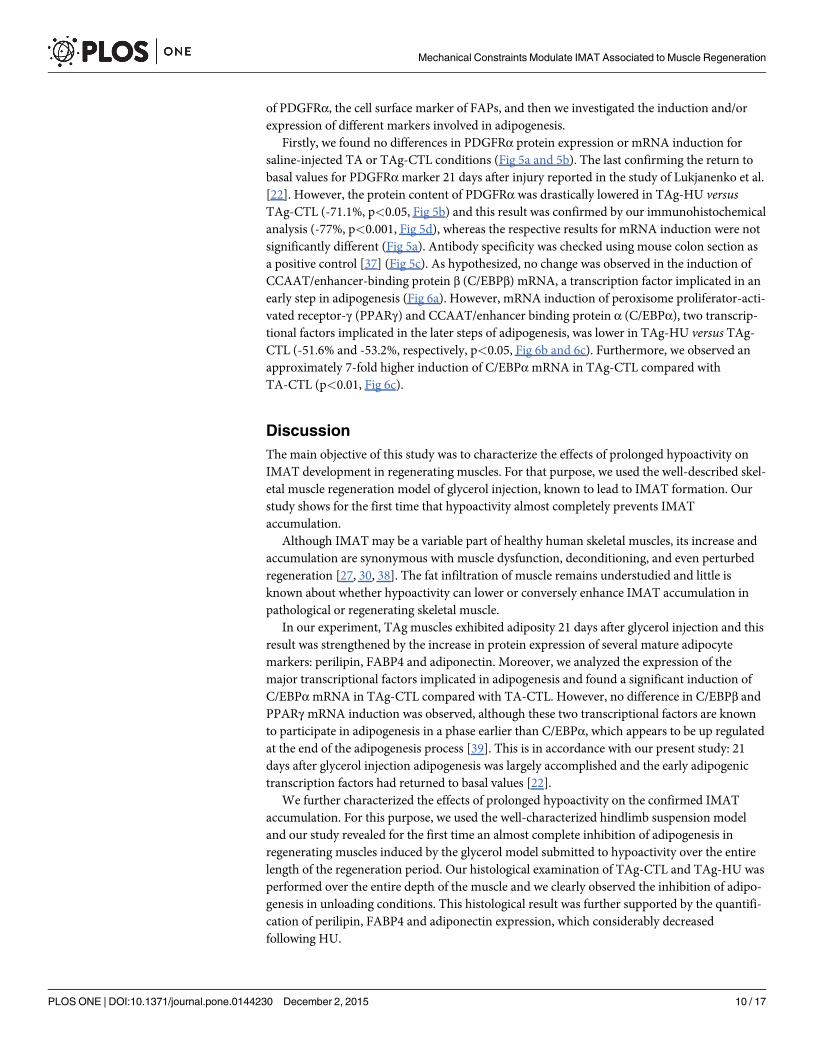
of PDGFRα, the cell surface marker of FAPs, and then we investigated the induction and/orexpression of different markers involved in adipogenesis.
Firstly, we found no differences in PDGFRα protein expression or mRNA induction forsaline-injected TA or TAg-CTL conditions (Fig 5a and 5b). The last confirming the return tobasal values for PDGFRαmarker 21 days after injury reported in the study of Lukjanenko et al.[22]. However, the protein content of PDGFRα was drastically lowered in TAg-HU versusTAg-CTL (-71.1%, p<0.05, Fig 5b) and this result was confirmed by our immunohistochemicalanalysis (-77%, p<0.001, Fig 5d), whereas the respective results for mRNA induction were notsignificantly different (Fig 5a). Antibody specificity was checked using mouse colon section asa positive control [37] (Fig 5c). As hypothesized, no change was observed in the induction ofCCAAT/enhancer-binding protein β (C/EBPβ) mRNA, a transcription factor implicated in anearly step in adipogenesis (Fig 6a). However, mRNA induction of peroxisome proliferator-acti-vated receptor-γ (PPARγ) and CCAAT/enhancer binding protein α (C/EBPα), two transcrip-tional factors implicated in the later steps of adipogenesis, was lower in TAg-HU versus TAg-CTL (-51.6% and -53.2%, respectively, p<0.05, Fig 6b and 6c). Furthermore, we observed anapproximately 7-fold higher induction of C/EBPαmRNA in TAg-CTL compared withTA-CTL (p<0.01, Fig 6c).
DiscussionThe main objective of this study was to characterize the effects of prolonged hypoactivity onIMAT development in regenerating muscles. For that purpose, we used the well-described skel-etal muscle regeneration model of glycerol injection, known to lead to IMAT formation. Ourstudy shows for the first time that hypoactivity almost completely prevents IMATaccumulation.
Although IMAT may be a variable part of healthy human skeletal muscles, its increase andaccumulation are synonymous with muscle dysfunction, deconditioning, and even perturbedregeneration [27, 30, 38]. The fat infiltration of muscle remains understudied and little isknown about whether hypoactivity can lower or conversely enhance IMAT accumulation inpathological or regenerating skeletal muscle.
In our experiment, TAg muscles exhibited adiposity 21 days after glycerol injection and thisresult was strengthened by the increase in protein expression of several mature adipocytemarkers: perilipin, FABP4 and adiponectin. Moreover, we analyzed the expression of themajor transcriptional factors implicated in adipogenesis and found a significant induction ofC/EBPαmRNA in TAg-CTL compared with TA-CTL. However, no difference in C/EBPβ andPPARγmRNA induction was observed, although these two transcriptional factors are knownto participate in adipogenesis in a phase earlier than C/EBPα, which appears to be up regulatedat the end of the adipogenesis process [39]. This is in accordance with our present study: 21days after glycerol injection adipogenesis was largely accomplished and the early adipogenictranscription factors had returned to basal values [22].
We further characterized the effects of prolonged hypoactivity on the confirmed IMATaccumulation. For this purpose, we used the well-characterized hindlimb suspension modeland our study revealed for the first time an almost complete inhibition of adipogenesis inregenerating muscles induced by the glycerol model submitted to hypoactivity over the entirelength of the regeneration period. Our histological examination of TAg-CTL and TAg-HU wasperformed over the entire depth of the muscle and we clearly observed the inhibition of adipo-genesis in unloading conditions. This histological result was further supported by the quantifi-cation of perilipin, FABP4 and adiponectin expression, which considerably decreasedfollowing HU.
Mechanical Constraints Modulate IMAT Associated to Muscle Regeneration
PLOS ONE | DOI:10.1371/journal.pone.0144230 December 2, 2015 10 / 17

Even though the precise mechanisms leading to IMAT formation in the glycerol model arestill unclear, previous studies have described the respective roles of multipotent stem cells suchas muscle-derived FAPs, pericytes, and side population cells giving rise to adipogenic precur-sors [26, 40–43]. Recently, a few groups have identified muscle mesenchymal progenitors withthe immunophenotype CD31-CD45-SM/C-2.6-PDGFRα+, which contribute to fat cell forma-tion in skeletal muscle [20, 43, 44]. PDGFRα has also been used very recently in human to iso-late muscle mesenchymal progenitors, which are equivalent to the mouse FAPs [27, 45]. We
Fig 5. Expression levels of the FAP cell surface marker PDGFRα. PDGFRαmRNA (a) and protein (b) content of saline-injected (TA) and glycerol-injected (TAg) tibialis anterior from control (CTL) and hindlimb unloading (HU) mice. * p<0.05 vs control group (unloading effect). Representative images (c,d) and quantification (e) of immunohistochemical analysis of PDGFRα-positive cells in mouse colon positive control (c) and glycerol-injected (TAg) tibialisanterior from control (CTL) and hindlimb unloading (HU) mice (d). *** p<0.001 vs control group.
doi:10.1371/journal.pone.0144230.g005
Mechanical Constraints Modulate IMAT Associated to Muscle Regeneration
PLOS ONE | DOI:10.1371/journal.pone.0144230 December 2, 2015 11 / 17

used this FAP cell surface marker in the present study and found a marked decrease in its pro-tein expression following HU. Our study thus indicates that hypoactivity is able to decreasePDGFRα-positive FAPs, which represent 98% of PDGFRα-positive cells in regenerating mus-cle [19]. Moreover, as reported by Uezumi et al. [20], only the PDGFRα-positive cells can dif-ferentiate into adipocytes in glycerol-injected muscles. We further observed reduced levels of
Fig 6. Changes in mRNA induction of adipogenesis markers. C/EBPβ (a), PPARγ (b) and C/EBPα (c)mRNA levels of saline-injected (TA) and glycerol-injected (TAg) tibialis anterior from control (CTL) andhindlimb unloading (HU) mice. * p<0.05 vs control group (unloading effect). ## p<0.01 vs TA-CTL (glycerol-injection effect).
doi:10.1371/journal.pone.0144230.g006
Mechanical Constraints Modulate IMAT Associated to Muscle Regeneration
PLOS ONE | DOI:10.1371/journal.pone.0144230 December 2, 2015 12 / 17
the later adipogenic transcription factors PPARγ and C/EBPα. We did not detect any differ-ences for C/EBPβ, certainly due to its earlier implication in adipogenesis [39].
Interestingly, FAPs are also known to promote skeletal muscle regeneration after injury inhealthy muscle [19, 25]. Heredia et al. [25] showed that FAP proliferation and macrophage-like activity are essential to the regeneration process. In this context, decreased FAP prolifera-tion could result in a decreased number of adipocytes, as well as impair the regenerative kinet-ics [25]. Skeletal muscle regenerative capacity is mainly dependent on the activation of SCs,which are finely controlled by the myogenic regulatory factors. Quiescent and proliferative SCsexpress the paired box transcription factor (Pax7) and its inactivation leads to a severe deple-tion of these muscle myogenic stem cells [46]. In regenerating muscle, the proliferating processis triggered by the expression of myoblast determination protein 1 (MyoD) and myogenic fac-tor 5 (Myf5) [47, 48]. Once differentiation is initiated, myogenin appears to be implicated first,and then muscle-specific regulatory factor 4 (Mrf4) is activated during the maturation phase[49, 50]. In our study, we first described a decrease in MyoD and myogenin mRNA inductionin TA-HU concomitant with no significant effect on Pax7. These results are in accordance withstudies demonstrating that HU does not necessarily alter the number of SCs in fast-twitch skel-etal muscle, probably indicating no SC loss by apoptosis [51], but that it does alter SC mitoticactivity [7, 9, 51, 52]. We then observed a decrease in Pax7, MyoD, and myogenin mRNAinduction, parallel to the decrease in muscle mass and CSA in TAg-HU. Taken together, theseresults confirm that reducing mechanical constraints throughout muscle regeneration disturbsSC-mediated regeneration and thus delays myofiber size recovery, as indicated in the study ofMatsuba et al. [53]. Nevertheless, the study of Mozdziak et al. [8] showed that the hindlimbunloading condition does not decrease but could even enhance the mitotic activity of SCs andprobably that of non-muscle cells in the earlier stages of regeneration. However, once newmyofibers are formed, their growth capacity is altered. Our results suggest that HU may alterregrowth after regeneration, but we cannot exclude the hypothesis that HU may enhance earlyregeneration processes, thereby inhibiting IMAT occurrence and further disturbing muscleregrowth. Clearly, additional studies are warranted to elucidate the early events related to mes-enchymal non-muscle cells and their implication in both the regeneration process and adipo-genesis during unloading conditions.
Currently, we do not know the exact underlying mechanisms leading to the inhibition ofmuscle adipogenesis in regenerating muscles under unloading conditions. As highlighted inthe literature, hindlimb unloading appears to be a proinflammatory situation with macrophageinfiltration [54–58]. Interestingly, recent studies have reported the critical role of inflammationand the immune system in muscle regeneration [59–62], and it appears that both macrophageshift and activity are essentials in this process. In addition, the study of Lukjanenko et al. [22]revealed that the glycerol injury model exhibits a disrupted inflammatory response comparedwith the cardiotoxin-induced injury model. Further studies are needed to elucidate the under-lying mechanisms of unloading-induced inhibition of skeletal muscle IMAT development andaccumulation and especially the effects of an inflammatory response on mesenchymal stemcells.
In conclusion, our study reports for the first time an almost complete inhibition of IMATdevelopment in regenerating muscles under hypoactivity conditions. We found a decreasedresponse of mesenchymal-derived precursor FAPs (PDGFRα+), which could explain thedecrease in IMAT development in the present model. Hypoactivity seems to locally create afavorable environment leading to a decrease in PDGFRα positive cells.
These observations shed new light on the mechanisms that regulate IMAT development inskeletal muscle and highlight the importance of taking into account the level of mechanicalconstraint imposed on skeletal muscle during regeneration processes. Our findings point in the
Mechanical Constraints Modulate IMAT Associated to Muscle Regeneration
PLOS ONE | DOI:10.1371/journal.pone.0144230 December 2, 2015 13 / 17

same direction as those reported by Jarvinen and Lehto [31] concerning immobilization, whichwas found to mediate a decrease in fibrotic area after gastrocnemius injury in rats.
In the one hand, our results suggest that a rest period with reduced mechanical constraintsmight be needed immediately after injury to prevent IMAT accumulation. However, our studyalso shows that the regrowth of skeletal muscle fibers is impaired under hypoactivity, whichhighlights the importance of applying mechanical constraints as soon as possible after the restperiod for the recovery of fiber size. Our present and future studies should contribute to a fullerunderstanding of IMAT accumulation and the establishment of rehabilitation guidelines forhuman muscle injuries.
Supporting InformationS1 Checklist. ARRIVE Checklist.(PDF)
AcknowledgmentsWe particularly thank Laurence Vico for providing the materials used in the tail-suspendedexperiments and Catherine Stott for the English correction of the manuscript. The authorsgreatly acknowledge the “Réseau d’Histologie Expérimentale de Montpellier” (RHEM) plate-form for histology core facilities and paraffin processing of the tibialis anterior muscles, andespecially Nelly Pirot, Charlène Berthet, and Yohan Noël. We also thank the animal staff fromour METAMUS platform facility, which belongs to the “Montpellier animal facilities network”(RAM), as well as the “Montpellier RIO Imaging” (MRI) platform for the use of theNanozoomer.
Author ContributionsConceived and designed the experiments: AFP RD AC TB GP CD CAD. Performed the experi-ments: AFP RD GP TB. Analyzed the data: AFP RD GP AC AB. Contributed reagents/materi-als/analysis tools: AFP RD AC GP EJ CBG. Wrote the paper: AFP GP AC. Manuscript revisionand feedback: AFP AC GP CD CAD RC AB.
References1. Huard J, Li Y, Fu FH. Muscle injuries and repair: current trends in research. J Bone Joint Surg Am.
2002; 84-A(5):822–832. PMID: 12004029
2. Mauro A. Satellite cell of skeletal muscle fibers. The Journal of biophysical and biochemical cytology.1961; 9:493–495. PMID: 13768451
3. Zammit PS, Heslop L, Hudon V, Rosenblatt JD, Tajbakhsh S, BuckinghamME, et al. Kinetics of myo-blast proliferation show that resident satellite cells are competent to fully regenerate skeletal musclefibers. Experimental cell research. 2002; 281(1):39–49. PMID: 12441128
4. Sambasivan R, Yao R, Kissenpfennig A, VanWittenberghe L, Paldi A, Gayraud-Morel B, et al. Pax7-expressing satellite cells are indispensable for adult skeletal muscle regeneration. Development. 2011;138(17):3647–3656. doi: 10.1242/dev.067587 PMID: 21828093
5. Relaix F, Zammit PS. Satellite cells are essential for skeletal muscle regeneration: the cell on the edgereturns centre stage. Development. 2012; 139(16):2845–2856. doi: 10.1242/dev.069088 PMID:22833472
6. Baldwin KM, Haddad F, Pandorf CE, Roy RR, Edgerton VR. Alterations in muscle mass and contractilephenotype in response to unloading models: role of transcriptional/pretranslational mechanisms. Fron-tiers in physiology. 2013; 4:284. doi: 10.3389/fphys.2013.00284 PMID: 24130531
7. Darr KC, Schultz E. Hindlimb suspension suppresses muscle growth and satellite cell proliferation. JAppl Physiol (1985). 1989; 67(5):1827–1834.
Mechanical Constraints Modulate IMAT Associated to Muscle Regeneration
PLOS ONE | DOI:10.1371/journal.pone.0144230 December 2, 2015 14 / 17

8. Mozdziak PE, Truong Q, Macius A, Schultz E. Hindlimb suspension reduces muscle regeneration.European journal of applied physiology and occupational physiology. 1998; 78(2):136–140. doi: 10.1007/s004210050398 PMID: 9694312
9. Wang XD, Kawano F, Matsuoka Y, Fukunaga K, Terada M, Sudoh M, et al. Mechanical load-dependentregulation of satellite cell and fiber size in rat soleus muscle. American journal of physiology Cell physi-ology. 2006; 290(4):C981–989. doi: 10.1152/ajpcell.00298.2005 PMID: 16291821
10. Vettor R, Milan G, Franzin C, Sanna M, De Coppi P, Rizzuto R, et al. The origin of intermuscular adi-pose tissue and its pathophysiological implications. American journal of physiology Endocrinology andmetabolism. 2009; 297(5):E987–998. doi: 10.1152/ajpendo.00229.2009 PMID: 19738037
11. Summan M,Warren GL, Mercer RR, Chapman R, Hulderman T, Van Rooijen N, et al. Macrophagesand skeletal muscle regeneration: a clodronate-containing liposome depletion study. American journalof physiology Regulatory, integrative and comparative physiology. 2006; 290(6):R1488–1495. doi: 10.1152/ajpregu.00465.2005 PMID: 16424086
12. Contreras-Shannon V, Ochoa O, Reyes-Reyna SM, Sun D, Michalek JE, Kuziel WA, et al. Fat accumu-lation with altered inflammation and regeneration in skeletal muscle of CCR2-/- mice following ischemicinjury. American journal of physiology Cell physiology. 2007; 292(2):C953–967. doi: 10.1152/ajpcell.00154.2006 PMID: 17020936
13. Shireman PK, Contreras-Shannon V, Ochoa O, Karia BP, Michalek JE, McManus LM. MCP-1 defi-ciency causes altered inflammation with impaired skeletal muscle regeneration. Journal of leukocytebiology. 2007; 81(3):775–785. doi: 10.1189/jlb.0506356 PMID: 17135576
14. Arnold L, Henry A, Poron F, Baba-Amer Y, van Rooijen N, Plonquet A, et al. Inflammatory monocytesrecruited after skeletal muscle injury switch into antiinflammatory macrophages to support myogenesis.The Journal of experimental medicine. 2007; 204(5):1057–1069. doi: 10.1084/jem.20070075 PMID:17485518
15. Segawa M, Fukada S, Yamamoto Y, Yahagi H, Kanematsu M, Sato M, et al. Suppression of macro-phage functions impairs skeletal muscle regeneration with severe fibrosis. Experimental cell research.2008; 314(17):3232–3244. doi: 10.1016/j.yexcr.2008.08.008 PMID: 18775697
16. Kawai H, Nishino H, Kusaka K, Naruo T, Tamaki Y, Iwasa M. Experimental glycerol myopathy: a histo-logical study. Acta neuropathologica. 1990; 80(2):192–197. PMID: 2389683
17. Arsic N, Zacchigna S, Zentilin L, Ramirez-Correa G, Pattarini L, Salvi A, et al. Vascular endothelialgrowth factor stimulates skeletal muscle regeneration in vivo. Molecular therapy: the journal of theAmerican Society of Gene Therapy. 2004; 10(5):844–854. doi: 10.1016/j.ymthe.2004.08.007
18. Abraham ST, Shaw C. Increased expression of deltaCaMKII isoforms in skeletal muscle regeneration:Implications in dystrophic muscle disease. J Cell Biochem. 2006; 97(3):621–632. doi: 10.1002/jcb.20669 PMID: 16215994
19. Joe AW, Yi L, Natarajan A, Le Grand F, So L, Wang J, et al. Muscle injury activates resident fibro/adipo-genic progenitors that facilitate myogenesis. Nature cell biology. 2010; 12(2):153–163. doi: 10.1038/ncb2015 PMID: 20081841
20. Uezumi A, Fukada S, Yamamoto N, Takeda S, Tsuchida K. Mesenchymal progenitors distinct from sat-ellite cells contribute to ectopic fat cell formation in skeletal muscle. Nature cell biology. 2010; 12(2):143–152. doi: 10.1038/ncb2014 PMID: 20081842
21. Pisani DF, Bottema CD, Butori C, Dani C, Dechesne CA. Mouse model of skeletal muscle adiposity: aglycerol treatment approach. Biochemical and biophysical research communications. 2010; 396(3):767–773. doi: 10.1016/j.bbrc.2010.05.021 PMID: 20457129
22. Lukjanenko L, Brachat S, Pierrel E, Lach-Trifilieff E, Feige JN. Genomic profiling reveals that transientadipogenic activation is a hallmark of mouse models of skeletal muscle regeneration. PloS one. 2013;8(8):e71084. doi: 10.1371/journal.pone.0071084 PMID: 23976982
23. Motohashi N, Uezumi A, Yada E, Fukada S, Fukushima K, Imaizumi K, et al. Muscle CD31(-) CD45(-)side population cells promote muscle regeneration by stimulating proliferation and migration of myo-blasts. The American journal of pathology. 2008; 173(3):781–791. doi: 10.2353/ajpath.2008.070902PMID: 18669618
24. Takegahara Y, Yamanouchi K, Nakamura K, Nakano S, Nishihara M. Myotube formation is affected byadipogenic lineage cells in a cell-to-cell contact-independent manner. Experimental cell research.2014; 324(1):105–114. doi: 10.1016/j.yexcr.2014.03.021 PMID: 24720912
25. Heredia JE, Mukundan L, Chen FM, Mueller AA, Deo RC, Locksley RM, et al. Type 2 innate signalsstimulate fibro/adipogenic progenitors to facilitate muscle regeneration. Cell. 2013; 153(2):376–388.doi: 10.1016/j.cell.2013.02.053 PMID: 23582327
26. Uezumi A, Ito T, Morikawa D, Shimizu N, Yoneda T, Segawa M, et al. Fibrosis and adipogenesis origi-nate from a commonmesenchymal progenitor in skeletal muscle. Journal of cell science. 2011; 124(Pt21):3654–3664. doi: 10.1242/jcs.086629 PMID: 22045730
Mechanical Constraints Modulate IMAT Associated to Muscle Regeneration
PLOS ONE | DOI:10.1371/journal.pone.0144230 December 2, 2015 15 / 17

27. Uezumi A, Fukada S, Yamamoto N, Ikemoto-Uezumi M, Nakatani M, Morita M, et al. Identification andcharacterization of PDGFRalpha(+) mesenchymal progenitors in human skeletal muscle. Cell death &disease. 2014; 5:e1186. doi: 10.1038/cddis.2014.161
28. Blau HM, Webster C, Pavlath GK. Defective myoblasts identified in Duchenne muscular dystrophy.Proceedings of the National Academy of Sciences of the United States of America. 1983; 80(15):4856–4860. PMID: 6576361
29. Heslop L, Morgan JE, Partridge TA. Evidence for a myogenic stem cell that is exhausted in dystrophicmuscle. Journal of cell science. 2000; 113:2299–2308. PMID: 10825301
30. Sciorati C, Clementi E, Manfredi AA, Rovere-Querini P. Fat deposition and accumulation in the dam-aged and inflamed skeletal muscle: cellular and molecular players. Cellular and molecular life sciences:CMLS. 2015. doi: 10.1007/s00018-015-1857-7
31. Jarvinen MJ, Lehto MU. The effects of early mobilisation and immobilisation on the healing process fol-lowing muscle injuries. Sports Med. 1993; 15(2):78–89. PMID: 8446826
32. Amblard D, Lafage-Proust MH, Laib A, Thomas T, Ruegsegger P, Alexandre C, et al. Tail suspensioninduces bone loss in skeletally mature mice in the C57BL/6J strain but not in the C3H/HeJ strain. Jour-nal of bone and mineral research: the official journal of the American Society for Bone and MineralResearch. 2003; 18(3):561–569. doi: 10.1359/jbmr.2003.18.3.561
33. Malaval L, Wade-Gueye NM, Boudiffa M, Fei J, Zirngibl R, Chen F, et al. Bone sialoprotein plays a func-tional role in bone formation and osteoclastogenesis. The Journal of experimental medicine. 2008; 205(5):1145–1153. doi: 10.1084/jem.20071294 PMID: 18458111
34. Pirot N, Delpech H, Deleuze V, Dohet C, Courtade-Saidi M, Basset-Leobon C, et al. Lung endothelialbarrier disruption in Lyl1-deficient mice. Am J Physiol Lung Cell Mol Physiol. 2014; 306(8):L775–785.doi: 10.1152/ajplung.00200.2013 PMID: 24532287
35. Pagano AF, Py G, Bernardi H, Candau RB, Sanchez AM. Autophagy and Protein Turnover Signaling inSlow-Twitch Muscle during Exercise. Medicine and science in sports and exercise. 2014. doi: 10.1249/MSS.0000000000000237
36. Hanson AM, Harrison BC, Young MH, Stodieck LS, Ferguson VL. Longitudinal characterization of func-tional, morphologic, and biochemical adaptations in mouse skeletal muscle with hindlimb suspension.Muscle & nerve. 2013; 48(3):393–402. doi: 10.1002/mus.23753
37. Kurahashi M, Nakano Y, Peri LE, Townsend JB, Ward SM, Sanders KM. A novel population of sube-pithelial platelet-derived growth factor receptor alpha-positive cells in the mouse and human colon. AmJ Physiol Gastrointest Liver Physiol. 2013; 304(9):G823–834. doi: 10.1152/ajpgi.00001.2013 PMID:23429582
38. Marcus RL, Addison O, Kidde JP, Dibble LE, Lastayo PC. Skeletal muscle fat infiltration: impact of age,inactivity, and exercise. The journal of nutrition, health & aging. 2010; 14(5):362–366.
39. White UA, Stephens JM. Transcriptional factors that promote formation of white adipose tissue. Molec-ular and cellular endocrinology. 2010; 318(1–2):10–14. doi: 10.1016/j.mce.2009.08.023 PMID:19733624
40. Uezumi A, Ojima K, Fukada S, Ikemoto M, Masuda S, Miyagoe-Suzuki Y, et al. Functional heterogene-ity of side population cells in skeletal muscle. Biochemical and biophysical research communications.2006; 341(3):864–873. doi: 10.1016/j.bbrc.2006.01.037 PMID: 16455057
41. Birbrair A, Zhang T, Wang ZM, Messi ML, Enikolopov GN, Mintz A, et al. Role of pericytes in skeletalmuscle regeneration and fat accumulation. Stem cells and development. 2013; 22(16):2298–2314. doi:10.1089/scd.2012.0647 PMID: 23517218
42. Boppart MD, De Lisio M, Zou K, Huntsman HD. Defining a role for non-satellite stem cells in the regula-tion of muscle repair following exercise. Frontiers in physiology. 2013; 4:310. doi: 10.3389/fphys.2013.00310 PMID: 24204344
43. Judson RN, Zhang RH, Rossi FM. Tissue-resident mesenchymal stem/progenitor cells in skeletal mus-cle: collaborators or saboteurs? The FEBS journal. 2013; 280(17):4100–4108. doi: 10.1111/febs.12370PMID: 23763717
44. Arrighi N, Moratal C, Clement N, Giorgetti-Peraldi S, Peraldi P, Loubat A, et al. Characterization of adi-pocytes derived from fibro/adipogenic progenitors resident in human skeletal muscle. Cell death & dis-ease. 2015; 6:e1733. doi: 10.1038/cddis.2015.79
45. Uezumi A, Ikemoto-Uezumi M, Tsuchida K. Roles of nonmyogenic mesenchymal progenitors in patho-genesis and regeneration of skeletal muscle. Frontiers in physiology. 2014; 5:68. doi: 10.3389/fphys.2014.00068 PMID: 24605102
46. Seale P, Sabourin LA, Girgis-Gabardo A, Mansouri A, Gruss P, Rudnicki MA. Pax7 is required for thespecification of myogenic satellite cells. Cell. 2000; 102(6):777–786. PMID: 11030621
Mechanical Constraints Modulate IMAT Associated to Muscle Regeneration
PLOS ONE | DOI:10.1371/journal.pone.0144230 December 2, 2015 16 / 17

47. Megeney LA, Kablar B, Garrett K, Anderson JE, Rudnicki MA. MyoD is required for myogenic stem cellfunction in adult skeletal muscle. Genes & development. 1996; 10(10):1173–1183.
48. Ustanina S, Carvajal J, Rigby P, Braun T. The myogenic factor Myf5 supports efficient skeletal muscleregeneration by enabling transient myoblast amplification. Stem Cells. 2007; 25(8):2006–2016. doi: 10.1634/stemcells.2006-0736 PMID: 17495111
49. Hasty P, Bradley A, Morris JH, Edmondson DG, Venuti JM, Olson EN, et al. Muscle deficiency and neo-natal death in mice with a targeted mutation in the myogenin gene. Nature. 1993; 364(6437):501–506.doi: 10.1038/364501a0 PMID: 8393145
50. ZhangW, Behringer RR, Olson EN. Inactivation of the myogenic bHLH gene MRF4 results in up-regu-lation of myogenin and rib anomalies. Genes & development. 1995; 9(11):1388–1399.
51. Zhang BT, Yeung SS, Liu Y, Wang HH, Wan YM, Ling SK, et al. The effects of low frequency electricalstimulation on satellite cell activity in rat skeletal muscle during hindlimb suspension. BMCCell Biol.2010; 11:87. doi: 10.1186/1471-2121-11-87 PMID: 21087483
52. Schultz E, Darr KC, Macius A. Acute effects of hindlimb unweighting on satellite cells of growing skele-tal muscle. J Appl Physiol (1985). 1994; 76(1):266–270.
53. Matsuba Y, Goto K, Morioka S, Naito T, Akema T, Hashimoto N, et al. Gravitational unloading inhibitsthe regenerative potential of atrophied soleus muscle in mice. Acta Physiol (Oxf). 2009; 196(3):329–339. doi: 10.1111/j.1748-1716.2008.01943.x
54. Nguyen HX, Tidball JG. Expression of a muscle-specific, nitric oxide synthase transgene prevents mus-cle membrane injury and reduces muscle inflammation during modified muscle use in mice. The Jour-nal of physiology. 2003; 550(Pt 2):347–356. doi: 10.1113/jphysiol.2003.040907 PMID: 12766242
55. Hirose T, Nakazato K, Song H, Ishii N. TGF-beta1 and TNF-alpha are involved in the transcription oftype I collagen alpha2 gene in soleus muscle atrophied by mechanical unloading. J Appl Physiol(1985). 2008; 104(1):170–177. doi: 10.1152/japplphysiol.00463.2006
56. Andrianjafiniony T, Dupre-Aucouturier S, Letexier D, Couchoux H, Desplanches D. Oxidative stress,apoptosis, and proteolysis in skeletal muscle repair after unloading. American journal of physiology Cellphysiology. 2010; 299(2):C307–315. doi: 10.1152/ajpcell.00069.2010 PMID: 20505039
57. Kohno S, Yamashita Y, Abe T, Hirasaka K, Oarada M, Ohno A, et al. Unloading stress disturbs muscleregeneration through perturbed recruitment and function of macrophages. J Appl Physiol (1985). 2012;112(10):1773–1782. doi: 10.1152/japplphysiol.00103.2012
58. Gratas-Delamarche A, Derbre F, Vincent S, Cillard J. Physical inactivity, insulin resistance, and the oxi-dative-inflammatory loop. Free radical research. 2014; 48(1):93–108. doi: 10.3109/10715762.2013.847528 PMID: 24060092
59. Aurora AB, Olson EN. Immune Modulation of Stem Cells and Regeneration. Cell stem cell. 2014; 15(1):14–25. doi: 10.1016/j.stem.2014.06.009 PMID: 24996166
60. Madaro L, Bouche M. From Innate to Adaptive Immune Response in Muscular Dystrophies and Skele-tal Muscle Regeneration: The Role of Lymphocytes. BioMed research international. 2014;2014:438675. doi: 10.1155/2014/438675 PMID: 25028653
61. Maffioletti SM, Noviello M, English K, Tedesco FS. Stem Cell Transplantation for Muscular Dystrophy:The Challenge of Immune Response. BioMed research international. 2014; 2014:964010. doi: 10.1155/2014/964010 PMID: 25054157
62. Lemos DR, Babaeijandaghi F, Low M, Chang CK, Lee ST, Fiore D, et al. Nilotinib reduces muscle fibro-sis in chronic muscle injury by promoting TNF-mediated apoptosis of fibro/adipogenic progenitors.Nature medicine. 2015. doi: 10.1038/nm.3869
Mechanical Constraints Modulate IMAT Associated to Muscle Regeneration
PLOS ONE | DOI:10.1371/journal.pone.0144230 December 2, 2015 17 / 17


![Poo Goes to Pooland[1][1]](https://img.pdfslide.us/doc/110x75/546a9833b4af9fe5268b476f/poo-goes-to-pooland11.jpg)