Embed Size (px)
Citation preview
RESEARCH ARTICLE
Succinyl-CoA Synthetase: New AntigenCandidate of Bartonella bacilliformisCláudia Gomes1*, Noemí Palma1, Maria J. Pons2, Ariel Magallón-Tejada1,Isabel Sandoval3, Carmen Tinco-Valdez2,4, Carlos Gutarra3, Juana del Valle-Mendoza2,4,Joaquim Ruiz1, Mayumi Matsuoka5
1 ISGlobal, Barcelona Centre for International Health Research (CRESIB), Hospital Clínic, Universitat deBarcelona, Barcelona, Spain, 2 Centro de Investigación e Innovación, Facultad de Ciencias de la Salud,Universidad Peruana de Ciencias Aplicadas, Lima, Peru, 3 Red de Salud de Morropon Chulucanas, Piura,Peru, 4 Instituto de Investigación Nutricional, Lima, Peru, 5 National Institute of Infectious Diseases, Tokyo,Japan
Abstract
Background
Bartonella bacilliformis is the causative agent of Carrion’s disease, a neglected illness with
mortality rates of 40–85% in the absence of treatment. The lack of a diagnostic technique to
overcome misdiagnosis and treat asymptomatic carriers is of note. This study aimed to
identify new B. bacilliformis antigenic candidates that could lead to a new diagnostic tool
able to be implemented in endemic rural areas.
Methodology/Principal Findings
Blood (n = 198) and serum (n = 177) samples were collected in northern Peru. Clinical data
were recorded. Specific 16S rRNA amplification by RT-PCR, IFA and ELISA for IgM/IgG with
whole cells as antigens was done. Western blot analysis and N-terminal amino acid sequenc-
ing detected seroreactive proteins. ELISAs for IgM/IgG for the antigenic candidates were per-
formed. Of the population 33.3% reported at least one symptom compatible with Carrion’s
disease; 25.4% (IFA), 27.1% (ELISA-IgG), 33.9% (ELISA-IgM) and 38.9% (RT-PCR) of sam-
ples were positive. Four proteins were considered potential antigenic candidates, including
two new antigenic candidates, succinyl-CoA synthetase subunit α (SCS-α) and succinyl-CoA
synthetase subunit β (SCS-β). OnWestern blot both Pap31 and SCS-α interacted with IgM,
while GroEL and SCS-β interacted with IgG. The presence of specific antibodies against the
antigenic candidates varied from 34.5% (IgG against SCS-α) to 97.2% (IgM against Pap31).
Conclusions/Significance
RT-PCR and the high levels of positivity for specific ELISAs demonstrate high levels of B.bacilliformis exposure and asymptomatic carriers among inhabitants. The new antigens
identified might be used as a new rapid diagnostic tool to diagnose acute Carrion’s disease
and identify asymptomatic carriers.
PLOS Neglected Tropical Diseases | DOI:10.1371/journal.pntd.0004989 September 14, 2016 1 / 17
a11111
OPEN ACCESS
Citation: Gomes C, Palma N, Pons MJ, Magallón-Tejada A, Sandoval I, Tinco-Valdez C, et al. (2016)Succinyl-CoA Synthetase: New Antigen Candidate ofBartonella bacilliformis. PLoS Negl Trop Dis 10(9):e0004989. doi:10.1371/journal.pntd.0004989
Editor: Richard O. Phillips, Komfo Anokye TeachingHospital, GHANA
Received: May 18, 2016
Accepted: August 19, 2016
Published: September 14, 2016
Copyright: © 2016 Gomes et al. This is an openaccess article distributed under the terms of theCreative Commons Attribution License, which permitsunrestricted use, distribution, and reproduction in anymedium, provided the original author and source arecredited.
Data Availability Statement: All relevant data arewithin the paper and its Supporting Information files.
Funding: The study was supported by the ProgramaNacional de Innovación para la Competitividad yProductividad (Innóvate Perú), under the contract117-PNICP-PIAP-2015, by Sociedad Española deEnfermedades Infecciosas y Microbiologia Clinica2014 and 2016. JR has a fellowship from the programI3, of the ISCIII [grant number: CES11/012]. CG has aPhD fellowship of the ISCIII [FI12/00561] and wasrecipient of a Canon Foundation Fellowship. MJP hasa postdoctoral fellowship from CONCYTEC/FONDECYT [grant number: CG05-2013-
Author Summary
B. bacilliformis is a neglected pathogen causing Carrion’s disease, a febrile illness with twodistinct phases, the acute so-called Oroya fever that can be life-threatening, and thechronic so-called Peruvian wart. This illness is currently limited to poor inhabitants ofAndean valleys of Ecuador, Colombia and Peru and for this reason is understudied. Oneof the most significant limitations is the lack of an adequate diagnostic tool able to beimplemented in rural areas. It is imperative to unequivocally detect cases of Carrion’s dis-ease as well as identify asymptomatic carriers who perpetuate the illness. The presentstudy describes the identification of 4 antigenic candidates potentially useful in the futuredevelopment of a rapid diagnostic test. Moreover, 2 of these candidates have not beendescribed in the literature. Additionally, four post-outbreak and one endemic communitywere studied and characterized. The identification of new antigens is essential for thedevelopment of a cheap, sensitive diagnostic tool, able to be implemented in low-incomeareas.
IntroductionBartonella bacilliformis is the etiological agent of Carrion’s disease, a neglected endemic illnessin Peru which has also been reported in Ecuador and Colombia [1]. Two well-establishedphases have been described in this infection. In the acute phase, also called Oroya Fever, B.bacilliformis infects the red blood cells which may result in severe anemia and transient immu-nosuppression [2,3]. The absence of treatment leads to high levels of mortality (40% to 85%)[4]. The chronic phase, ‘Verruga Peruana’ (Peruvian wart), is characterized by the developmentof nodular dermal eruptions. This phase typically occurs in survivors weeks or months afterthe acute febrile syndrome [5].
Clinical cure does not necessarily result in bacterial clearance. In fact, viable B. bacilliformishave been cultured from blood samples of treated patients [6,7]. This lack of clearance togetherwith the development of partial immunity and the presence of continuous B. bacilliformisexposure, means that endemic areas have a high number of individuals who are asymptomaticcarriers. Indeed, it has been described that 45% of inhabitants of endemic areas are B. bacillifor-mis seropositive when antibodies are tested by Indirect Fluorescence Antibody (IFA) assay [8].
Studies of B. bacilliformis antigens are scarce in the literature compared with reports ofother pathogens, and a rapid diagnostic method to detect acute and/or chronic infections hasyet to be developed. To our knowledge, the first report identifying B. bacilliformis antigens wasdescribed in 1988 by Knobloch [9]. Twenty-four protein antigens were found, including onemain antigen with 65 kDa (BB65; a heat shock protein posteriorly identified as GroEL) [9–11].Nonetheless, BB65 never bound to Bartonella IgM but did bind to IgG antibodies after the firsttwo weeks, thereby demonstrating its utility to detect persisting IgG from the first to the thirdyear after a B. bacilliformis infection. However, only 60% of sera from Verruga Peruana patientsreact with BB65 [10]. Padmalayam et al. described an immunogenic 43 kDa lipoprotein as anantigen by screening a genomic DNA lambda library with serum from a patient in the chronicwart phase of bartonellosis [12]. More recently, Pap31 was described to be immunologicallydominant and a good candidate for use in ELISA, but once again only one serum was used foridentification [13]. Unfortunately, none of the antigens described above have resulted in arapid diagnostic tool.
The objective of this study was to identify and characterize B. bacilliformis antigenic candi-dates and take a step towards a rapid diagnostic tool able to be implemented in rural areas.
B. bacilliformis Antigenic Candidates
PLOS Neglected Tropical Diseases | DOI:10.1371/journal.pntd.0004989 September 14, 2016 2 / 17
FONDECYT] and INOVATE Perú. The funders hadno role in study design, data collection and analysis,decision to publish or preparation of the manuscript.
Competing Interests: The authors have declaredthat no competing interests exist.
Materials and Methods
Bacterial strainsThe microorganisms used in this study are listed in Table 1. B. bacilliformis was cultured at28°C on Columbia agar with 5% sheep blood for 7 days.
Study populationBlood (n = 198) and serum samples (n = 177) were collected in March 2014 in five different vil-lages of Piura, in the north of Peru: Tunal, Guayaquiles, Los Ranchos, Mayland and Huanca-bamba. Serum samples were collected in sodium citrate and gel SSTII advance vacutainers(BD, Heidelberg, Germany). A B. bacilliformis outbreak occurred in 4 of these villages: betweenMay and November 2013 in Tunal, Guayaquiles and Mayland, and between November 2013and March 2014 in Los Ranchos [14]. According to the national guidelines [15], all participantshad received ciprofloxacin treatment during 14 days following diagnosis during the outbreak.All people living in the aforementioned four villages who were diagnosed (clinical symptomsand/or thin blood smear) with acute Carrion’s disease during the previous outbreak wereinvited to participate in the study. Those who agree to participate were notified by the localhealth center and all samples from that village were collected on the same day. On the otherhand, Huancabamba is a long established endemic area for Carrion’s disease and the volunteerswere randomly recruited by house-to-house visits. Clinical and demographic data wererecorded in all cases. After collection the samples were kept in the appropriate refrigerationconditions: 4°C for blood samples and sera samples were frozen. All samples were transferredto Lima (Peru) and sera samples were also sent to Tokyo (Japan) to perform immunologicalassays. In all cases transportation was performed under frozen conditions.
Ethics statementIn all the cases, signed informed consent was obtained and clinical data were anonymized. Inthe case of children a parent or their guardian provided informed consent on their behalf. Thestudy was approved by both the Ethic Committees of the Universidad Peruana de CienciasAplicadas (Lima, Peru) and the Hospital Clinic (Barcelona, Spain).
Table 1. Bacterial strains and plasmids.
Description Source or reference
Strains
B. bacilliformis CIP 57.20 –NCTC12136 Institute Pasteur
E. coli TOP10 Host strain for cloning Invitrogen
E. coli BL21Star (DE3) Host strain for gene expression Invitrogen
Plasmids
pCR4-TOPO TA-cloning vector Invitrogen
pET100D/TOPO Expression vector Invitrogen
pGroEL pET100D/TOPO containing GroEL This study
pPap31 pET100D/TOPO containing Pap31 This study
pSCS-α pET100D/TOPO containing SCS-α This study
pSCS-β pET100D/TOPO containing SCS-β This study
SCS-α: succinyl-CoA synthetase subunit α
SCS-β: succinyl-CoA synthetase subunit β.
doi:10.1371/journal.pntd.0004989.t001
B. bacilliformis Antigenic Candidates
PLOS Neglected Tropical Diseases | DOI:10.1371/journal.pntd.0004989 September 14, 2016 3 / 17
Real-Time (RT) PCRDNA extraction was done from 200 μL blood samples with the Qiamp DNAMini Kit (Qiagen,Hilden, Germany), according to the manufacturer’s instructions except that the final elutionvolume was 100 μL. A RT-PCR was performed based on Smit et al. [16], using a BHQ quencherprobe at 125 nM and 250 nM of primers in final volume of 25 μL. RT qPCR conditions were95°C for 15 minutes, 60 cycles of 15 seconds at 95°C and 45 seconds at 60°C and the procedurewas performed in an ABI Prism 7500 RT system and data was analyzed in the 7500 SystemSDS software (Applied Biosystems, Warrington, UK). The primers and the probe used areshown in Table 2.
IFA assayThe Chamberlin method was used with slight modifications [8]. Briefly, Vero cells were cul-tured in sterile plates maintaining the same MOI and concentrations throughout the experi-ment. At harvest, the Vero cell monolayer was removed with trypsin in a final volume of 10mL. The slides were mounted with 65 μL of cells suspension per well and 20 μL 1:100 of Fluo-rescein (FITC)-conjugated goat anti-human IgG (heavy plus light chains) (Jackson IR, Balti-more, PA). Slides were read using a 20X objective, 10X oculars and an Olympus IX51microscope. We used the same cut off established by Chamberlin et al., considering 1/256serum dilution end point as a positive IFA test.
ELISAIgM and IgG antibody levels were measured by ELISA as described by Matsuoka et al. [17]using sonicated B. bacilliformis whole cells as antigens. Total protein concentration was quanti-fied by the Pierce assay (Thermo Scientific, Rockford, USA). IgM were detected with rabbitanti-human IgM (1:1000) conjugated with peroxidase and using o-Phenylenediamine (Dako,Glostrup, Denmark) as substrate (Sigma, St. Louis, MO). IgG were detected with rabbit anti-human IgG (1:1000) conjugated with alkaline phosphatase and using Sigma P104 phosphatasesubstrate. Optical densities were measured as absorbance at 450 nm and 405/655 nm for IgMand IgG, respectively. Each sample was analyzed in triplicate intra-plate and results werereported as three independent ELISA experiments. The negative control, included a pool ofsera from 30 healthy donors (X0939, Dako, Glostrup, Denmark). In the absence of an
Table 2. Primers used in this study.
Sequence (5’! 3’) Ref
16S rRNA F TTGATAAGCGTGAGGTCGGAGG [16]
16S rRNA R GCAACCACACATAGTAAGCCTAA [16]
16S rRNA probe ATGTCTGCCTAGAAATCAATCATAGGCC [16]
GroEL F *CACCATGGCTGCTAAAGAAGTAAAATTT This study
GroEL R TTAGAAATCCATTCCGCCCATTCCGCC This study
Pap31 F *CACCATGAATATAAAATGTTTAGTGACA This study
Pap31 R TCAGAATTTGTAAGCAACACCAACGCG This study
SCS α F *CACCATGTCAATTCTTATC This study
SCS α R CTAACCCTTCAAGACTGAAACC This study
SCS β F *CACCATGAATATCCATGAAT This study
SCS β R TTAAGCTCCTTTTACGGCTGC This study
* CACC at the 5’ end is a sequence added to allow pET directional cloning.
doi:10.1371/journal.pntd.0004989.t002
B. bacilliformis Antigenic Candidates
PLOS Neglected Tropical Diseases | DOI:10.1371/journal.pntd.0004989 September 14, 2016 4 / 17
established cut off, evidence of infection was considered in the samples above the Finite Mix-ture Model (FMM) cut off that provides a statistical framework for the analysis of immuno-globulin values [18].
Western blottingAgar-grown B. bacilliformis were lysed by sonication, separated on a 10 to 20% SDS-PAGE geland electrotransferred onto a PVDF membrane (GE Healthcare, Buckinghamshire, UK). Thedifferent lanes were cut and immunoblotted with each serum for both IgG/IgM. The negativecontrol used in ELISA was used here.
Amino acid sequencingImmunogenic candidate proteins were chosen according to the antibody levels obtained bywhole cell ELISA results. The membranes were stained with Coomasie brilliant blue stainingsolution. Candidate proteins were directly cut out, and N-terminal amino acid sequencing wasdone in APROScience (Naruto, Japan).
Amplification and cloning of antigenic candidatesGenes coding for the candidate proteins were amplified by PCR using the primers in Table 2.Amplified products were purified and cloned in Champion pET Directional 100/D-TOPO vec-tor (Invitrogen, Carlsbad, CA), according to the manufacturer’s instructions in order to gener-ate Xpress-tagged-full-length versions of the candidate antigenic proteins. The DNA sequenceswere verified. For the detection, E. coli lysates suspended in lysis buffer were separated on a12.5% SDS-PAGE and immunoblotted with anti-Xpress antibody. The His6-tagged proteinswere purified using His-Bind kits (GE Healthcare, Buckinghamshire, UK).
Candidate antigens ELISAPurified candidate proteins were used to perform ELISA and determine the reaction with serain study, following the same protocol mentioned previously. Here, the cut off was establishedconsidering 3 times the standard deviation for the X0939 negative control (Dako).
Statistical analysisStatistical analysis was performed using the Graphpad package. Significance was consideredwith P<0.05. P-values were calculated using the Fisher and Mann-Whitney tests.
ResultsA Carrion’s disease outbreak occurred in the Lalaquiz district (S1 Fig) between May andNovember 2013. Sixty-five villages were affected, with Tunal, Guayaquiles and Mayland beingamong the 10 most affected with 53.8, 19.3 and 2.56% of the cases, respectively. Meanwhile, inLos Ranchos (in the neighboring Canchaque district) 21 cases were reported between Novem-ber 2013 and March 2014. This outbreak was the first description of this illness in these villages.During the outbreak, a total of 428 cases were reported, diagnosed by clinical symptoms and/orthin blood smear. The mean age of the persons affected during the outbreak was 34 years;52.3% of the cases were females, and 18.5% of the cases involved children up to 10 years of age.Headache (74.7%), malaise (66%), fever (39.2%), arthralgia (31.6%), myalgia (24.4%) and pal-lor (13%) were the most common symptoms.
We were able to collect 198 blood samples from volunteers in March 2014 and Table 3shows the study population data. All samples collected in the 4 post-outbreak villages were
B. bacilliformis Antigenic Candidates
PLOS Neglected Tropical Diseases | DOI:10.1371/journal.pntd.0004989 September 14, 2016 5 / 17
from people who had been diagnosed with acute Carrion’s disease a few months earlier whenan outbreak had occurred. Despite the fact that all volunteers appeared healthy, mild symp-toms were reported. The most common symptom described in our study population was head-ache in 27.3% of the population, followed by 4% of people reporting fever, myalgia andmalaise. Joint pain and pallor were present in 5 and 2.5%. With regard to the endemic village, 1(0.5%) person from the endemic area (Huancabamba) presented Verruga Peruana at the timeof sample collection. RT-PCR results showed amplification for 77 (38.9%) individuals, but itshould be taken into account that all positive samples presented very low bacteremia in blood.Significant differences were found between the positivity results of Tunal and Guayaquiles(P = 0.0189), Guayaquiles and Los Ranchos (P = 0.0370), Tunal and Mayland (P = 0.0006), LosRanchos and Mayland (P = 0.0001) and Los Ranchos and Huancabamba (P = 0.0178). Addi-tionally, the results between Mayland and Huancabamba were almost statistically significant(P = 0.0548) (Table 4).
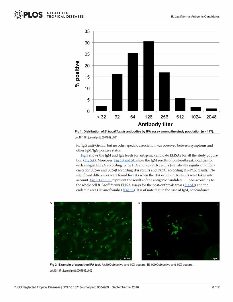
Serum samples were taken from 177 (89.4%) of the individuals. Of these 45 (25.4%) had anIFA titer� 256 being considered as positive by the IFA test (Figs 1 and 2, Table 4). The IFAresults were almost significantly lower in the endemic area than in the 4 post-outbreak villagestaken together (P = 0.0575).
For the whole cell B. bacilliformis IgM ELISA, the 3 standard deviation cut off methodresulted in 95.5% of volunteers presenting levels of IgM equivalent to evidence of infection,while with the use of FMM, the cut off was established at 0.35 (IgM) and 0.53 (IgG), withapproximately 34 and 27% of volunteers showing evidence of infection by IgM and IgG values,respectively. Thus, in order to differentiate the most positive samples we applied the FMM cutoff.
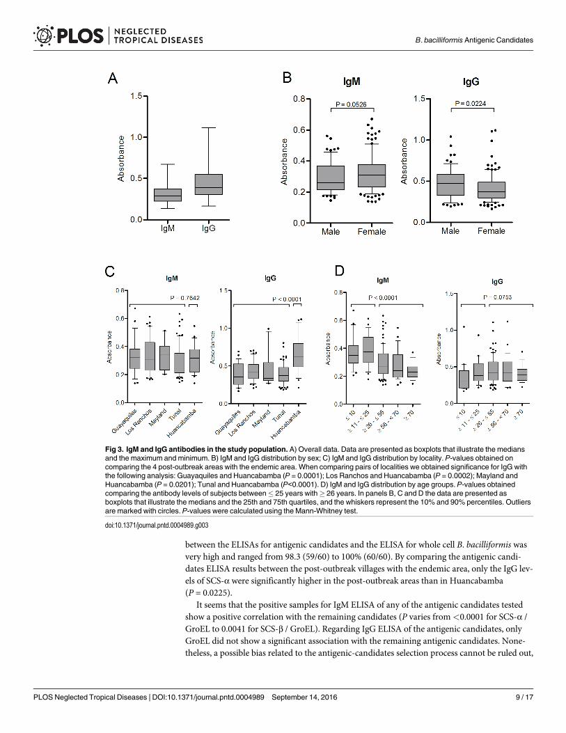
Differences in the antibody levels between genders were not found. However, it wasobserved that higher IgM levels tended to be more frequent in females (P = 0.0526) while IgGvalues were significantly higher in men (P = 0.0224). On analyzing the antibody levels by locali-ties, we saw that IgG levels were significantly higher (P<0.0001) in Huancabamba compared topost-outbreak areas where the disease had never been described before this outbreak. Thisshows the presence of a high percentage of healthy people with high levels of antibodies againstB. bacilliformis. Additionally, for IgG levels statistically significant differences (P values rangingfrom<0.0001 to 0.0229) were obtained on comparing each of the post-outbreak areas with theendemic area (Huancabamba) (Table 4). When the IgM levels were analyzed taking into con-sideration the different age groups, we observed that young people (�25 years) had signifi-cantly higher IgM levels (P<0.0001) than adults�26 years, while IgG levels only showed atendency to be higher in people�26 years (P = 0.0753). On comparing the IgG levels of theyounger population (�10 years) with each of the other age groups, we found statistically
Table 3. General data about the study population (n = 198).
Tunal Guayaquiles Los Ranchos Mayland Huancabamba Total
(n = 94) (n = 25) (n = 44) (n = 10) (n = 25)
n (%) 94 (47.5) 25 (12.6) 44 (22.2) 10 (5.1) 25 (12.6) 198 (100)
Mean age (min–max) 39.3 (1–94) 34.2 (6–67) 38.6 (5–77) 30.2 (6–73) 30.2 (9–68) 36.9 (1–94)
Male 31 (33) 8 (32) 17 (38.6) 4 (40) 16 (64) 76 (38.4)
Female 63 (67) 17 (68) 27 (61.4) 6 (60) 9 (36) 122 (61.6)
CD symptoms* 23 (24.5) 17 (68) 14 (31.8) 3 (30) 9 (36) 66 (33.3)
*The presence of at least one symptom compatible with Carrion’s disease, including fever, joint pain, headache, malaise, pallor, myalgia and warts.
doi:10.1371/journal.pntd.0004989.t003
B. bacilliformis Antigenic Candidates
PLOS Neglected Tropical Diseases | DOI:10.1371/journal.pntd.0004989 September 14, 2016 6 / 17

significant differences with all groups except people�70 years (between�11 -�25 P = 0.0281;�26 -�55 P = 0.0074;�56 -�70 P = 0.0246) (Fig 3).
No concordance was observed between clinical data and the positivity obtained with thediagnostic tools used. It is interesting to note that the case of Peruvian wart was only detectedby ELISA IgG (Table 5).
Table 6 shows the concordance between each pair of techniques used. Six samples had apositive result for at least 3 out of the 4 techniques used. This comparative analysis showed acertain degree of dispersion and a lack of concordance (Table 6).
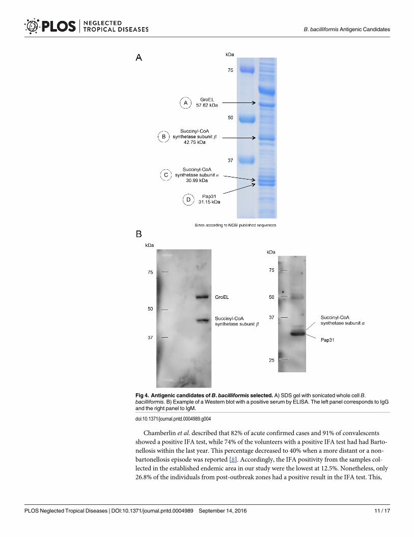
By doing the Western blots with the sera in the study and against whole sonicated B. bacilli-formis we chose four proteins as possible antigenic candidates, two identified with anti-humanIgM and two with anti-human IgG (Fig 4). Amino acid sequencing revealed that the IgM can-didates identified were Pap31 and succinyl-CoA synthetase subunit α (SCS-α) whereas for IgGwe identified GroEL and succinyl-CoA synthetase subunit β (SCS-β) (Table 7).
After cloning, expression and purification of the four antigenic candidates, we performedELISA for each IgM and IgG testing.
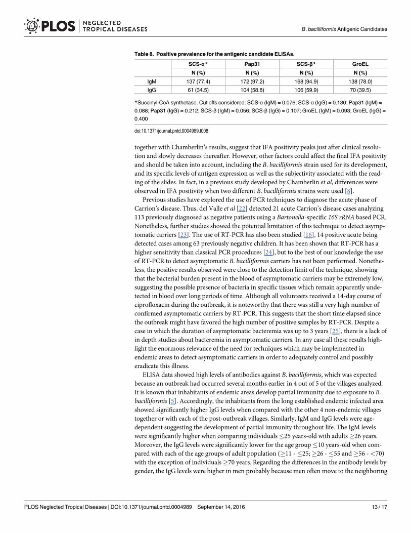
The IgM results showed a high prevalence of reactivity, from 77.4% for SCS-α to 97.2% forPap31. Meanwhile, the IgG reactivity in the ELISAs done for the antigens ranged from 34.5 toSCS-α and 59.9% to SCS-β IgG (Table 8). The person with the Verruga Peruana was positive
Table 4. Positive results for each technique in the different study sites.
Tunal Guayaquiles Los Ranchos Mayland Post-outbreakareas
Endemic area(Huancabamba)
P1 Total
n/N (%) n/N (%) n/N (%) n/N (%) n/N (%) n/N (%) n/N (%)
RT-PCR 30/94(31.9)a,c
15/25 (60)a,b 10/44(22.7)b,d,e
9/10(90)c,d
64/173 (37) 13/25 (52)e 0.1883 77/198(38.9)
IFA 21/75 (28) 8/25 (32) 8/43 (18.6) 4/10 (40) 41/153 (26.8) 3/24 (12.5) 0.0575‡ 45/177(25.4)
ELISA IgM 19/75 (25.3) 9/25 (36) 17/43 (39.5) 5/10 (50) 50/153 (32.7) 10/24 (41.7) 0.4869 60/177(33.9)
IgG 14/75 (18.7)f 6/25 (24)g 10/43 (23.3)h 2/10 (20)i 32/153 (20.9) 16/24 (66.7)f,g,h,i 0.0001** 48/177(27.1)
Positive forat least onetechnique†
55/75 (73.3) 23/25(92) 31/43 (72.1) 10/10(100)
119/153 (77.8) 22/24 (91.7) 0.1715 141/177(79.7)
†Only individuals with both blood and serum samples were considered (n = 177).1Statistical significance between the positivity in the 4 post-outbreak villages taken together and the positivity in the endemic area (Huancabamba).‡IFA results were almost significantly lower in the endemic area than in the 4 post-outbreak villages taken together (P = 0.0575).
**ELISA IgG values were significantly higher in Huancabamba than in the 4 post-outbreak villages taken together (P = 0.0001).
The superscript letters represent the statistically significant differences between the positivity in 2 specific areasa P = 0.0189b P = 0.0370c P = 0.0006d P = 0.0001e P = 0.0178f P<0.0001g P = 0.0041h P = 0.0007i P = 0.0229.
The RT-PCR values between Mayland and Huancabamba were almost statistically significant (P = 0.0548). In all cases the Fisher exact test was used.
doi:10.1371/journal.pntd.0004989.t004
B. bacilliformis Antigenic Candidates
PLOS Neglected Tropical Diseases | DOI:10.1371/journal.pntd.0004989 September 14, 2016 7 / 17
for IgG anti-GroEL, but no other specific association was observed between symptoms andother IgM/IgG positive status.
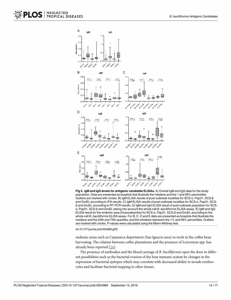
Fig 5 shows the IgM and IgG levels for antigenic candidate ELISAS for all the study popula-tion (Fig 5A). Moreover, Fig 5B and 5C show the IgM results of post-outbreak localities foreach antigen ELISA according to the IFA and RT-PCR results (statistically significant differ-ences for SCS-α and SCS-β according IFA results and Pap31 according RT-PCR results). Nosignificant differences were found for IgG when the IFA or RT-PCR results were taken intoaccount. Fig 5D and 5E represent the results of the antigenic candidate ELISAs according tothe whole cell B. bacilliformis ELISA assays for the post-outbreak areas (Fig 5D) and theendemic area (Huancabamba) (Fig 5E). It is of note that in the case of IgM, concordance
Fig 1. Distribution ofB. bacilliformis antibodies by IFA assay among the study population (n = 177).
doi:10.1371/journal.pntd.0004989.g001
Fig 2. Example of a positive IFA test. A) 20X objective and 10X oculars. B) 100X objective and 10X oculars.
doi:10.1371/journal.pntd.0004989.g002
B. bacilliformis Antigenic Candidates
PLOS Neglected Tropical Diseases | DOI:10.1371/journal.pntd.0004989 September 14, 2016 8 / 17
between the ELISAs for antigenic candidates and the ELISA for whole cell B. bacilliformis wasvery high and ranged from 98.3 (59/60) to 100% (60/60). By comparing the antigenic candi-dates ELISA results between the post-outbreak villages with the endemic area, only the IgG lev-els of SCS-α were significantly higher in the post-outbreak areas than in Huancabamba(P = 0.0225).
It seems that the positive samples for IgM ELISA of any of the antigenic candidates testedshow a positive correlation with the remaining candidates (P varies from<0.0001 for SCS-α /GroEL to 0.0041 for SCS-β / GroEL). Regarding IgG ELISA of the antigenic candidates, onlyGroEL did not show a significant association with the remaining antigenic candidates. None-theless, a possible bias related to the antigenic-candidates selection process cannot be ruled out,
Fig 3. IgM and IgG antibodies in the study population. A) Overall data. Data are presented as boxplots that illustrate the mediansand the maximum and minimum. B) IgM and IgG distribution by sex; C) IgM and IgG distribution by locality. P-values obtained oncomparing the 4 post-outbreak areas with the endemic area. When comparing pairs of localities we obtained significance for IgG withthe following analysis: Guayaquiles and Huancabamba (P = 0.0001); Los Ranchos and Huancabamba (P = 0.0002); Mayland andHuancabamba (P = 0.0201); Tunal and Huancabamba (P<0.0001). D) IgM and IgG distribution by age groups. P-values obtainedcomparing the antibody levels of subjects between� 25 years with� 26 years. In panels B, C and D the data are presented asboxplots that illustrate the medians and the 25th and 75th quartiles, and the whiskers represent the 10% and 90% percentiles. Outliersare marked with circles. P-values were calculated using the Mann-Whitney test.
doi:10.1371/journal.pntd.0004989.g003
B. bacilliformis Antigenic Candidates
PLOS Neglected Tropical Diseases | DOI:10.1371/journal.pntd.0004989 September 14, 2016 9 / 17
and thus a posterior analysis using samples from different endemic areas are needed to performan in depth analysis.
DiscussionCurrently, early diagnosis of Carrion’s disease remains an unsolved problem due to the socio-geographical context of the endemic areas; the illness is present in poor, rural and isolatedareas with precarious access, with neither the necessary equipment nor qualified personnel toperform molecular/immunological techniques. In endemic areas, the diagnosis is usually basedon clinical symptoms or thin blood smear, an expertise-dependent methodology with 96% ofspecificity but very low sensitivity (24–36%) [19]. This problem is further enhanced by theunspecific initial symptoms of Carrion’s disease, common to a several pathologies existing inthese areas, such as dengue and other arboviral diseases, malaria or tuberculosis [7,20]. All ofthis leads to misdiagnosis of patients [20,21] and the non treatment of asymptomatic carriers,thereby perpetuating disease transmission. The present data highlight the non-concordancebetween symptomatology and antibody levels. This is a relevant fact emphasizing again thechallenges that exist in diagnosing the illness clinically. Bacterial culture is not clinically usefuldue to the culture requirements and the slow bacterial growth rate [6,20]. Molecular and sero-logic tools, like PCR or IFA are able to detect acute cases more efficiently but are very difficultto be implemented in routine practice in remote endemic rural areas [1,8,22]. The use of sensi-tive and specific rapid diagnostic tools is a way to overcome these limitations.
Table 5. Distribution of the positive individuals for each diagnostic technique used according to the symptoms described.
N (%)* RT ELISA_IgM ELISA_IgG IFA
n (%)* n (%)* n (%)* n (%)*
Headache 48 (27.1)** 23 (47.9) 10 (20.8) 13 (27.1) 15 (31.3)
Joint pain 10 (5.6) 3 (30.0) 2 (20.0) 2 (20.0) 4 (40.0)
Fever 8 (4.5) 4 (50.0) 2 (25.0) 3 (37.5) 4 (50.0)
Malaise 8 (4.5) 4 (50.0) 2 (25.0) 3 (37.5) 4 (50.0)
Myalgia 8 (4.5) 4 (50.0) 2 (25.0) 4 (50.0) 3 (37.5)
Pallor 5 (2.8) 4 (80.0) 2 (40.0) 1 (20.0) 4 (80.0)
Peruvian wart 1 (0.6) 0 0 1 (100.0) 0
Any symptom 60 (33.9) 27 (45.0) 13 (21.7) 17 (28.3) 18 (30.0)
* percentage in the population (considering n = 177).
** 6 more individuals presented headache but were not considered in this table because only the blood sample were collected.
doi:10.1371/journal.pntd.0004989.t005
Table 6. Concordance among the techniques used.
RT-PCR IFA ELISA IgM
N (%) N (%) N (%)
+ - + - + -
IFA + 20 (11.3) 24 (13.6)
- 53 (29.9) 80 (45.2)
ELISA IgM + 24 (13.6) 36 (20.3) 14 (7.90) 46 (26.0)
- 49 (27.7) 68 (38.4) 30 (16.9) 87 (49.2)
ELISA IgG + 19 (10.7) 29 (16.4) 9 (5.1) 39 (22.0) 19 (10.7) 29 (16.4)
- 54 (30.5) 75 (42.4) 35 (19.8) 94 (53.1) 41 (23.2) 88 (49.7)
Only individuals with both blood and serum samples were considered (n = 177).
doi:10.1371/journal.pntd.0004989.t006
B. bacilliformis Antigenic Candidates
PLOS Neglected Tropical Diseases | DOI:10.1371/journal.pntd.0004989 September 14, 2016 10 / 17
Chamberlin et al. described that 82% of acute confirmed cases and 91% of convalescentsshowed a positive IFA test, while 74% of the volunteers with a positive IFA test had had Barto-nellosis within the last year. This percentage decreased to 40% when a more distant or a non-bartonellosis episode was reported [8]. Accordingly, the IFA positivity from the samples col-lected in the established endemic area in our study were the lowest at 12.5%. Nonetheless, only26.8% of the individuals from post-outbreak zones had a positive result in the IFA test. This,
Fig 4. Antigenic candidates of B. bacilliformis selected. A) SDS gel with sonicated whole cell B.bacilliformis. B) Example of a Western blot with a positive serum by ELISA. The left panel corresponds to IgGand the right panel to IgM.
doi:10.1371/journal.pntd.0004989.g004
B. bacilliformis Antigenic Candidates
PLOS Neglected Tropical Diseases | DOI:10.1371/journal.pntd.0004989 September 14, 2016 11 / 17
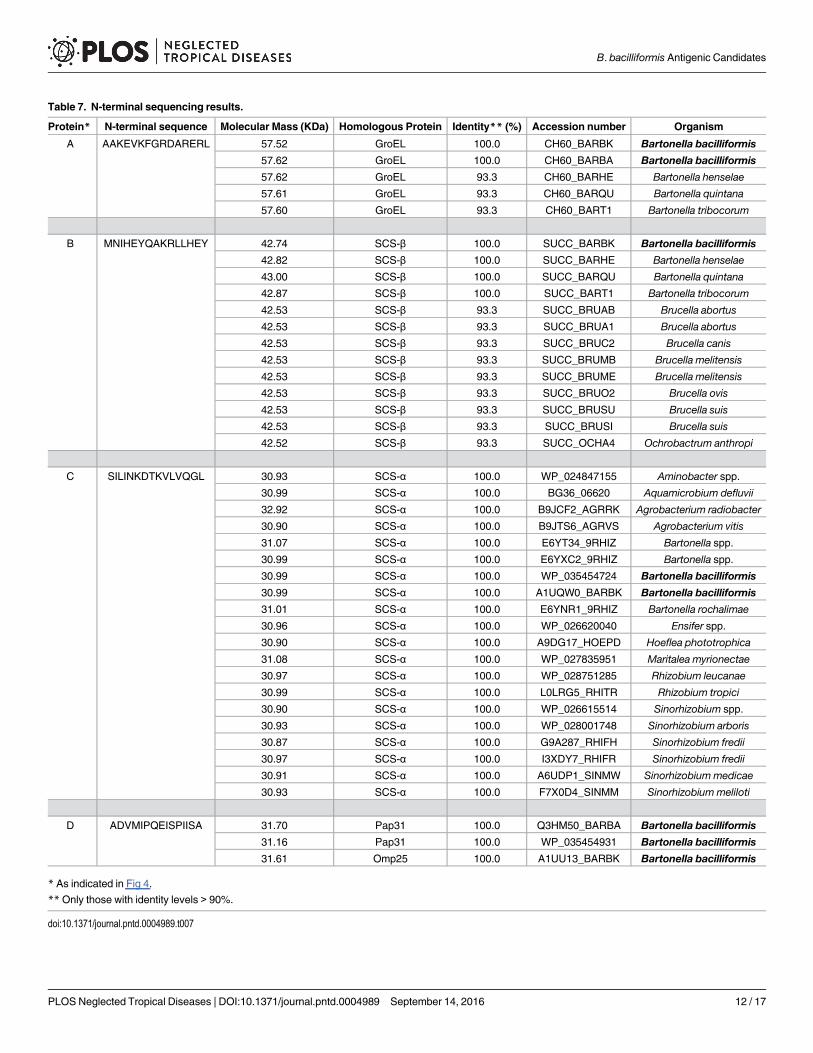
Table 7. N-terminal sequencing results.
Protein* N-terminal sequence Molecular Mass (KDa) Homologous Protein Identity** (%) Accession number Organism
A AAKEVKFGRDARERL 57.52 GroEL 100.0 CH60_BARBK Bartonella bacilliformis57.62 GroEL 100.0 CH60_BARBA Bartonella bacilliformis
57.62 GroEL 93.3 CH60_BARHE Bartonella henselae
57.61 GroEL 93.3 CH60_BARQU Bartonella quintana
57.60 GroEL 93.3 CH60_BART1 Bartonella tribocorum
B MNIHEYQAKRLLHEY 42.74 SCS-β 100.0 SUCC_BARBK Bartonella bacilliformis42.82 SCS-β 100.0 SUCC_BARHE Bartonella henselae
43.00 SCS-β 100.0 SUCC_BARQU Bartonella quintana
42.87 SCS-β 100.0 SUCC_BART1 Bartonella tribocorum
42.53 SCS-β 93.3 SUCC_BRUAB Brucella abortus
42.53 SCS-β 93.3 SUCC_BRUA1 Brucella abortus
42.53 SCS-β 93.3 SUCC_BRUC2 Brucella canis
42.53 SCS-β 93.3 SUCC_BRUMB Brucella melitensis
42.53 SCS-β 93.3 SUCC_BRUME Brucella melitensis
42.53 SCS-β 93.3 SUCC_BRUO2 Brucella ovis
42.53 SCS-β 93.3 SUCC_BRUSU Brucella suis
42.53 SCS-β 93.3 SUCC_BRUSI Brucella suis
42.52 SCS-β 93.3 SUCC_OCHA4 Ochrobactrum anthropi
C SILINKDTKVLVQGL 30.93 SCS-α 100.0 WP_024847155 Aminobacter spp.
30.99 SCS-α 100.0 BG36_06620 Aquamicrobium defluvii
32.92 SCS-α 100.0 B9JCF2_AGRRK Agrobacterium radiobacter
30.90 SCS-α 100.0 B9JTS6_AGRVS Agrobacterium vitis
31.07 SCS-α 100.0 E6YT34_9RHIZ Bartonella spp.
30.99 SCS-α 100.0 E6YXC2_9RHIZ Bartonella spp.
30.99 SCS-α 100.0 WP_035454724 Bartonella bacilliformis
30.99 SCS-α 100.0 A1UQW0_BARBK Bartonella bacilliformis31.01 SCS-α 100.0 E6YNR1_9RHIZ Bartonella rochalimae
30.96 SCS-α 100.0 WP_026620040 Ensifer spp.
30.90 SCS-α 100.0 A9DG17_HOEPD Hoeflea phototrophica
31.08 SCS-α 100.0 WP_027835951 Maritalea myrionectae
30.97 SCS-α 100.0 WP_028751285 Rhizobium leucanae
30.99 SCS-α 100.0 L0LRG5_RHITR Rhizobium tropici
30.90 SCS-α 100.0 WP_026615514 Sinorhizobium spp.
30.93 SCS-α 100.0 WP_028001748 Sinorhizobium arboris
30.87 SCS-α 100.0 G9A287_RHIFH Sinorhizobium fredii
30.97 SCS-α 100.0 I3XDY7_RHIFR Sinorhizobium fredii
30.91 SCS-α 100.0 A6UDP1_SINMW Sinorhizobium medicae
30.93 SCS-α 100.0 F7X0D4_SINMM Sinorhizobium meliloti
D ADVMIPQEISPIISA 31.70 Pap31 100.0 Q3HM50_BARBA Bartonella bacilliformis31.16 Pap31 100.0 WP_035454931 Bartonella bacilliformis
31.61 Omp25 100.0 A1UU13_BARBK Bartonella bacilliformis
* As indicated in Fig 4.
**Only those with identity levels > 90%.
doi:10.1371/journal.pntd.0004989.t007
B. bacilliformis Antigenic Candidates
PLOS Neglected Tropical Diseases | DOI:10.1371/journal.pntd.0004989 September 14, 2016 12 / 17
together with Chamberlin’s results, suggest that IFA positivity peaks just after clinical resolu-tion and slowly decreases thereafter. However, other factors could affect the final IFA positivityand should be taken into account, including the B. bacilliformis strain used for its development,and its specific levels of antigen expression as well as the subjectivity associated with the read-ing of the slides. In fact, in a previous study developed by Chamberlin et al, differences wereobserved in IFA positivity when two different B. bacilliformis strains were used [8].
Previous studies have explored the use of PCR techniques to diagnose the acute phase ofCarrion’s disease. Thus, del Valle et al [22] detected 21 acute Carrion’s disease cases analyzing113 previously diagnosed as negative patients using a Bartonella-specific 16S rRNA based PCR.Nonetheless, further studies showed the potential limitation of this technique to detect asymp-tomatic carriers [23]. The use of RT-PCR has also been studied [16], 14 positive acute beingdetected cases among 63 previously negative children. It has been shown that RT-PCR has ahigher sensitivity than classical PCR procedures [24], but to the best of our knowledge the useof RT-PCR to detect asymptomatic B. bacilliformis carriers has not been performed. Nonethe-less, the positive results observed were close to the detection limit of the technique, showingthat the bacterial burden present in the blood of asymptomatic carriers may be extremely low,suggesting the possible presence of bacteria in specific tissues which remain apparently unde-tected in blood over long periods of time. Although all volunteers received a 14-day course ofciprofloxacin during the outbreak, it is noteworthy that there was still a very high number ofconfirmed asymptomatic carriers by RT-PCR. This suggests that the short time elapsed sincethe outbreak might have favored the high number of positive samples by RT-PCR. Despite acase in which the duration of asymptomatic bacteremia was up to 3 years [25], there is a lack ofin depth studies about bacteremia in asymptomatic carriers. In any case all these results high-light the enormous relevance of the need for techniques which may be implemented inendemic areas to detect asymptomatic carriers in order to adequately control and possiblyeradicate this illness.
ELISA data showed high levels of antibodies against B. bacilliformis, which was expectedbecause an outbreak had occurred several months earlier in 4 out of 5 of the villages analyzed.It is known that inhabitants of endemic areas develop partial immunity due to exposure to B.bacilliformis [5]. Accordingly, the inhabitants from the long established endemic infected areashowed significantly higher IgG levels when compared with the other 4 non-endemic villagestogether or with each of the post-outbreak villages. Similarly, IgM and IgG levels were age-dependent suggesting the development of partial immunity throughout life. The IgM levelswere significantly higher when comparing individuals�25 years-old with adults�26 years.Moreover, the IgG levels were significantly lower for the age group�10 years-old when com-pared with each of the age groups of adult population (�11 -�25;�26 -�55 and�56 -<70)with the exception of individuals�70 years. Regarding the differences in the antibody levels bygender, the IgG levels were higher in men probably because men often move to the neighboring
Table 8. Positive prevalence for the antigenic candidate ELISAs.
SCS-α* Pap31 SCS-β* GroEL
N (%) N (%) N (%) N (%)
IgM 137 (77.4) 172 (97.2) 168 (94.9) 138 (78.0)
IgG 61 (34.5) 104 (58.8) 106 (59.9) 70 (39.5)
*Succinyl-CoA synthetase. Cut offs considered: SCS-α (IgM) = 0.076; SCS-α (IgG) = 0.130; Pap31 (IgM) =
0.088; Pap31 (IgG) = 0.212; SCS-β (IgM) = 0.056; SCS-β (IgG) = 0.107; GroEL (IgM) = 0.093; GroEL (IgG) =
0.400
doi:10.1371/journal.pntd.0004989.t008
B. bacilliformis Antigenic Candidates
PLOS Neglected Tropical Diseases | DOI:10.1371/journal.pntd.0004989 September 14, 2016 13 / 17
endemic areas such as Cajamarca department (San Ignacio area) to work in the coffee beanharvesting. The relation between coffee plantations and the presence of Lutzomyia spp. hasalready been reported [26].
The presence of antibodies and the blood carriage of B. bacilliformis open the door to differ-ent possibilities such as the bacterial evasion of the host immune system by changes in theexpression of bacterial epitopes which may correlate with decreased ability to invade erythro-cytes and facilitate bacterial trapping in other tissues.
Fig 5. IgM and IgG levels for antigenic candidate ELISAs. A) Overall IgM and IgG data for the studypopulation. Data are presented as boxplots that illustrate the medians and the 1 and 99% percentiles.Outliers are marked with circles. B) IgM ELISA results of post-outbreak localities for SCS-α, Pap31, SCS-βand GroEL according to IFA results. C) IgM ELISA results of post-outbreak localities for SCS-α, Pap31, SCS-β and GroEL according to RT-PCR results. D) IgM and IgG ELISA result of post-outbreak population for SCS-α, Pap31, SCS-β and GroEL taking into account the whole cell B. bacilliformis ELISA assay. E) IgM and IgGELISA result for the endemic area (Huancabamba) for SCS-α, Pap31, SCS-β and GroEL according to thewhole cell B. bacilliformis ELISA assay. For B, C, D and E data are presented as boxplots that illustrate themedians and the 25th and 75th quartiles, and the whiskers represent the 1% and 99% percentiles. Outliersare marked with circles. P-values were calculated using the Mann-Whitney test.
doi:10.1371/journal.pntd.0004989.g005
B. bacilliformis Antigenic Candidates
PLOS Neglected Tropical Diseases | DOI:10.1371/journal.pntd.0004989 September 14, 2016 14 / 17
In the present study we have identified four potential antigenic candidates, two of whichhave previously been reported in the literature, GroEL and Pap31 [9,10,13], while the remain-ing two are described for the first time. These new antigenic candidates, SCS-α and SCS-β areinvolved in the tricarboxylic acid cycle, an important cytosolic metabolic pathway. Indeed, sub-unit α has been described as an immunogenic protein of Brucella melitensis [27], and the β sub-unit was recently reported in Bartonella henselae pathogenesis [28].
Pap31 or Hemin-binding protein A has shown to be a good candidate in the ELISA tech-nique and is highly expressed in B. bacillifomis cultures [13]. Furthermore, Pap31 is an outermembrane protein that seems to be good candidate for the development of serodiagnostic toolsfor Bartonella infections, as proposed for Bartonella quintana infections [17].
Knobloch et al. [10] described that GroEL, a heat shock protein, never bound to BartonellaIgM antibodies, suggesting that GroEL may be a good antigenic candidate for the chronicphase. Similarly, in the present study GroEL was identified as reactive with IgG. However, wewere also able to detect IgM anti-GroEL. Differences in the time elapsed from the infection andthe sample collection may explain this finding. GroEL is present in the outer and inner mem-branes of B. bacilliformis, having also been detected in B. bacilliformis supernatants. Its pres-ence is correlated with mitogenic activity against human vascular endothelial cells which leadsto the development of verrucous lesions [29]. This mitogenic activity is inhibited in vitro by thepresence of specific anti-GroEL antibodies, suggesting the protective role of specific IgG inasymptomatic carriers. Indeed, the presence of specific IgG anti-GroEL was observed in thevolunteer presenting a Verruga Peruana. GroEL has also been described as a good candidatefor vaccine production and the development of diagnostic kits for Brucella melitensis [30].
The strong correlation between the positive results for IgM ELISAs anti-B. bacilliformiswhole cell and positive results for antigenic candidates, suggests that the antigens can be usedindistinctly in a rapid diagnostic test. These are preliminary results and more studies should bedone to characterize these B. bacilliformis antigens.
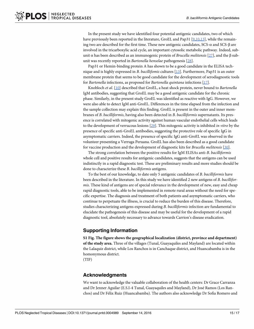
To the best of our knowledge, to date only 5 antigenic candidates of B. bacilliformis havebeen described in the literature. In this study we have identified 2 new antigens of B. bacillifor-mis. These kind of antigens are of special relevance in the development of new, easy and cheaprapid diagnostic tools, able to be implemented in remote rural areas without the need for spe-cific expertise. The diagnosis and treatment of both patients and asymptomatic carriers, whocontinue to perpetuate the illness, is crucial to reduce the burden of this disease. Therefore,studies characterizing antigens expressed during B. bacilliformis infection are fundamental toelucidate the pathogenesis of this disease and may be useful for the development of a rapiddiagnostic tool, absolutely necessary to advance towards Carrion’s disease eradication.
Supporting InformationS1 Fig. The figure shows the geographical localization (district, province and department)of the study area. Three of the villages (Tunal, Guayaquiles and Mayland) are located withinthe Lalaquiz district, while Los Ranchos is in Canchaque district, and Huancabamba is in thehomonymous district.(TIF)
AcknowledgmentsWe want to acknowledge the valuable collaboration of the health centers: Dr Grace Carranzaand Dr Jenner Aguilar (E.S.I-4 Tunal, Guayaquiles and Mayland), Dr José Ramos (Los Ran-chos) and Dr Félix Ruiz (Huancabamba). The authors also acknowledge Dr Sofia Romero and
B. bacilliformis Antigenic Candidates
PLOS Neglected Tropical Diseases | DOI:10.1371/journal.pntd.0004989 September 14, 2016 15 / 17
Dr Judith Chamberlin for their kind suggestions and help with the IFA protocol. CG acknowl-edges Sandra Martínez-Puchol for her support.
Author Contributions
Conceived and designed the experiments: CGo JR MM.
Performed the experiments: CGo NP CTVMJP IS.
Analyzed the data: CGo AMT JR MM.
Contributed reagents/materials/analysis tools: JdVM CGu.
Wrote the paper: CGo JR.
References1. Sanchez Clemente N, Ugarte-Gil CA, Solorzano N, Maguiña C, Pachas P, Blazes D, et al. Bartonella
bacilliformis: a systematic review of the literature to guide the research agenda for elimination. PloSNegl Trop Dis. 2012; 6: e1819. doi: 10.1371/journal.pntd.0001819 PMID: 23145188
2. Maguiña C, Gotuzzo E. Bartonellosis–new and old. Infect Dis Clin North Am. 2000; 14: 1–22. PMID:10738670
3. Ticona E, Huaroto L, Garcia Y, Vargas L, Madariaga MG. The pathophysiology of the acute phase ofhuman bartonellosis resembles AIDS. Med Hypotheses. 2010; 74: 45–9. doi: 10.1016/j.mehy.2009.06.054 PMID: 19665314
4. Ihler GM. Bartonella bacilliformis: dangerous pathogen slowly emerging from deep background. FEMSMicrobiol Lett. 1996; 144: 1–11. PMID: 8870245
5. Minnick MF, Anderson BE, Lima A, Battisti JM, Lawyer PG, Birtles RJ. Oroya fever and verruga per-uana: bartonelloses unique to South America. PloS Negl Trop Dis. 2014; 8: e2919. doi: 10.1371/journal.pntd.0002919 PMID: 25032975
6. Pachas P. Epidemiologia de Bartonelosis en el Peru. Lima, Peru: Ministerio de Salud, 2000.
7. Pachas P. Enfermedad de Carrión (Bartonellosis) en el Perú. Lima, Peru: Ministerio de Salud. Lima,2001.
8. Chamberlin J, Laughlin L, Gordon S, Romero S, Solórzano N, Regnery RL. Serodiagnosis of Bartonellabacilliformis infection by indirect fluorescence antibody assay: test development and application to apopulation in an area of Bartonellosis endemicity. J Clin Microbiol. 2000; 8: 4269–71.
9. Knobloch J. Analysis and preparation of Bartonella bacilliformis antigens. Am J Trop Med Hyg. 1988;39: 173–8. PMID: 3044156
10. Knobloch J, Schreiber M. Bb65, a major immunoreactive protein of Bartonella bacilliformis. Am J TropMed Hyg. 1990; 43: 373–9. PMID: 1700634
11. Anderson BE, NeumanMA. Bartonella spp. as emerging human pathogens. Clin Microbiol Rev. 1997;10: 203–19. PMID: 9105751
12. Padmalayam I, Kelly T, Baumstark B, Massung R. Molecular cloning, sequencing, expression, andcharacterization of an immunogenic 43-kilodalton lipoprotein of Bartonella bacilliformis that has homol-ogy to NlpD/LppB. Infect Immun. 2000; 68: 4972–9. PMID: 10948113
13. Taye A, Chen H, Duncan K, Zhang Z, Hendrix L, Gonzalez J, et al. Production of recombinant proteinPap31 and its application for the diagnosis of Bartonella bacilliformis infection. Ann N Y Acad Sci. 2005;1063: 280–5. PMID: 16481528
14. Guzmán Cuzcano J. Situación epidemiológica de la enfermedad de Carrión en el Perú, SE 35–2014.Boletín Epidemiológico, Lima. 2014; 23: 695–9.
15. Ministerio de Salud. Atención de la Bartonelosis o enfermedad de Carrión en el Perú. Norma técnica N° 048-MINSA/DGSP-V.01, Lima, Peru. Ministerio de Salud, 2007.
16. Smit PW, Peeling RW, Garcia PJ, Torres LL, Pérez-Lu JE, Moore D, et al. Dried blood spots for qPCRdiagnosis of acute Bartonella bacilliformis infection. Am J Trop Med Hyg. 2013; 89: 988–90. doi: 10.4269/ajtmh.13-0246 PMID: 24043691
17. Matsuoka M, Sasaki T, Seki N, Kobayashi M, Sawabe K, Sasaki Y, et al. Hemin-binding proteins aspotent markers for serological diagnosis of infections with Bartonella quintana. Clin Vaccine Immunol.2013; 20: 620–6. doi: 10.1128/CVI.00717-12 PMID: 23408526
B. bacilliformis Antigenic Candidates
PLOS Neglected Tropical Diseases | DOI:10.1371/journal.pntd.0004989 September 14, 2016 16 / 17
18. Bretscher MT, Supargiyono S, Wijayanti MA, Nugraheni D, Widyastuti AN, Lobo NF, et al. Measure-ment of Plasmodium falciparum transmission intensity using serological cohort data from Indonesianschoolchildren. Malar J. 2013; 12: 21. doi: 10.1186/1475-2875-12-21 PMID: 23327665
19. Ellis BA, Rotz LD, Leake JA, Samalvides F, Bernable J, Ventura G, et al. An outbreak of acute Bartonel-losis (Oroya fever) in the Urubamba region of Peru, 1998. Am J Trop Med Hyg. 1999; 61: 344–9.PMID: 10463692
20. Maguiña C, Garcia PJ, Gotuzzo E, Cordero L, Spach DH. Bartonellosis (Carrión's disease) in the mod-ern era. Clin Infect Dis. 2001; 33: 772–9. PMID: 11512081
21. Cornejo A, Gomes C, Suarez L, Martínez-Puchol S, Bustamante P, Pons MJ, et al. An unidentified clus-ter of infection in the Peruvian Amazon region. J Infect Dev Ctries. 2015; 9: 524–9. doi: 10.3855/jidc.6235 PMID: 25989173
22. del Valle Mendoza J, Silva CasoW, Tinco Valdez C, Pons MJ, del Valle LJ, Casabona Oré V, et al.Diagnosis of Carrion's disease by direct blood PCR in thin blood smear negative samples. PLoS One.2014; 9: e92283. doi: 10.1371/journal.pone.0092283 PMID: 24651298
23. Gomes C, Martinez-Puchol S, Pons MJ, Bazán J, Tinco C, del Valle J, et al. Evaluation of PCRApproaches for Detection of Bartonella bacilliformis in Blood Samples. PLoS Negl Trop Dis. 2016; 10(3): e0004529. doi: 10.1371/journal.pntd.0004529 PMID: 26959642
24. Diaz MH, Bai Y, Malania L, Winchell JM, Kosoy MY. Development of a novel genus-specific real-timePCR assay for detection and differentiation of Bartonella species and genotypes. J Clin Microbiol.2012; 50: 1645–9. doi: 10.1128/JCM.06621-11 PMID: 22378904
25. Lydy SL, Eremeeva ME, Asnis D, Paddock CD, NicholsonWL, Silverman DJ, et al. Isolation and char-acterization of Bartonella bacilliformis from an expatriate Ecuadorian. J Clin Microbiol. 2008; 46: 627–37. PMID: 18094131
26. Contreras-Gutiérrez MA, Velez I, Porter C, Uribe SI. An updated checklist of Phlebotomine sand flies(Diptera: Psychodidae: Phlebotominae) from the Colombian Andean coffee-growing region. Biomédica.2014; 34:483–98. doi: 10.1590/S0120-41572014000300017 PMID: 25504134
27. Teixeira-Gomes AP, Cloeckaert A, Bézard G, Dubray G, Zygmunt MS. Mapping and identification ofBrucella melitensis proteins by two-dimensional electrophoresis and microsequencing. Electrophore-sis. 1997; 18: 156–62. PMID: 9059838
28. Chang CC, Chen YJ, Tseng CS, Lai WL, Hsu KY, Chang CL, et al. A comparative study of the interac-tion of Bartonella henselae strains with human endothelial cells. Vet Microbiol. 2011; 149: 147–56. doi:10.1016/j.vetmic.2010.09.033 PMID: 21035278
29. Minnick MF, Smitherman LS, Samuels DS. Mitogenic effect of Bartonella bacilliformis on human vascu-lar endothelial cells and involvement of GroEL. Infect Immun. 2003; 71: 6933–42. PMID: 14638782
30. Sekhavati MH, Heravi RM, Tahmoorespur M, Yousefi S, Abbassi-Daloii T, Akbari R. Cloning, molecularanalysis and epitopics prediction of a new chaperone GroEL Brucella melitensis antigen. Iran J BasicMed Sci. 2015; 18: 499–505. PMID: 26124937
B. bacilliformis Antigenic Candidates
PLOS Neglected Tropical Diseases | DOI:10.1371/journal.pntd.0004989 September 14, 2016 17 / 17


![RESEARCHARTICLE Seabirdbycatchmitigationtrialsinartisanal ...diposit.ub.edu/dspace/bitstream/2445/130325/1/684037.pdfshearwaters appeartobethemostaffected seabirds bythisfishery[2,3].Combined](https://img.pdfslide.us/doc/110x75/60c15d9b0c7fb41dda2cd567/researcharticle-seabirdbycatchmitigationtrialsinartisanal-shearwaters-appeartobethemostaffected.jpg)