Embed Size (px)
Citation preview

Ge et al. BMC Neurosci (2017) 18:54 DOI 10.1186/s12868-017-0375-y
RESEARCH ARTICLE
Difference in the functional connectivity of the dorsolateral prefrontal cortex between smokers with nicotine dependence and individuals with internet gaming disorderXin Ge1†, Yawen Sun1†, Xu Han1, Yao Wang1, Weina Ding1, Mengqiu Cao1, Yasong Du2, Jianrong Xu1* and Yan Zhou1*
Abstract
Background: It has been reported that internet gaming disorder (IGD) and smokers with nicotine dependence (SND) share clinical characteristics, such as over-engagement despite negative consequences and cravings. This study is to investigate the alterations in the resting-state functional connectivity (rsFC) of the dorsolateral prefrontal cortex (DLPFC) observed in SND and IGD. In this study, 27 IGD, 29 SND, and 33 healthy controls (HC) underwent a resting-state functional magnetic resonance imaging (rs-fMRI) scan. DLPFC connectivity was determined in all participates by inves-tigating the synchronized low-frequency fMRI signal fluctuations using a temporal seed-based correlation method.
Results: Compared with the HC group, the IGD and SND groups showed decreased rsFC with DLPFC in the right insula and left inferior frontal gyrus with DLPFC. Compared with SND group, the IGD subjects exhibited increased rsFC in the left inferior temporal gyrus and right inferior orbital frontal gyrus and decreased rsFC in the right middle occipi-tal gyrus, supramarginal gyrus, and cuneus with DLPFC.
Conclusion: Our results confirmed that SND and IGD share similar neural mechanisms related to craving and impul-sive inhibitions. The significant difference in rsFC with DLPFC between the IGD and SND subjects may be attributed to the visual and auditory stimulation generated by long-term internet gaming.
Keywords: Functional magnetic resonance imaging, Internet gaming disorder, Nicotine dependence, Resting-state functional connectivity, Dorsolateral prefrontal cortex
© The Author(s) 2017. This article is distributed under the terms of the Creative Commons Attribution 4.0 International License (http://creativecommons.org/licenses/by/4.0/), which permits unrestricted use, distribution, and reproduction in any medium, provided you give appropriate credit to the original author(s) and the source, provide a link to the Creative Commons license, and indicate if changes were made. The Creative Commons Public Domain Dedication waiver (http://creativecommons.org/publicdomain/zero/1.0/) applies to the data made available in this article, unless otherwise stated.
BackgroundInternet gaming disorder (IGD), also known as problem-atic internet use, is the excessive and recurrent use of online internet games [1]. IGD is different from substance abuse or drug addiction such that no substance or chemi-cal intake is involved; however, excessive internet use may lead to physical dependence similar to that observed in other addictions [2]. Currently, IGD has become a serious
mental health issue around the world, thereby requiring additional investigation, as exemplified by its inclusion as a condition for further study in Section 3 of the Diag-nostic and Statistical Manual of Mental Disorders (5th Edition, DSM-5) [3]. The following diagnostic criteria for IGD were suggested: time distortion, time spent longer than initially intended and planned time, use of inter-net activity to cope with or escape problems, compulsive behavior, deception about the extent of use, failure to stop or control use, and preoccupation with internet use when offline [4–6]. Notably, many of these behavioral symp-toms resemble substance-related disorders [7–9].
Currently the precise pathogenesis of IGD remains unclear. A few studies suggested that the risk factor of
Open Access
BMC Neuroscience
*Correspondence: [email protected]; [email protected]; [email protected] †Xin Ge and Yawen Sun have contributed equally to this work1 Department of Radiology, Ren Ji Hospital, School of Medicine, Shanghai Jiao Tong University, Shanghai 200127, People’s Republic of ChinaFull list of author information is available at the end of the article

Page 2 of 10Ge et al. BMC Neurosci (2017) 18:54
IGD is related to the increased prevalence of substance dependence [10–12]. Numerous studies found that IGD and substance dependence shared similar neural mechanisms, such as nicotine dependence [9, 13, 14]. On the basis of behavioral addiction, researchers have been attempting to associate IGD with other behavioral problems that can lead to addiction, such as drug abuse, alcohol abuse, and nicotine dependence [7, 15]. Our pre-vious study revealed that smokers with IGD exhibited decreased resting-state functional connectivity (rsFC) in the right rectus gyrus and increased rsFC in the left middle frontal gyrus with post cingulate cortex (PCC), compared with nonsmokers with IGD. Furthermore, negatively correlation was found in the PCC connec-tivity with the right rectus gyrus with Chen’s internet addiction score (CIAS) of smokers with IGD before cor-rection. The results suggested that, compared with the nonsmokers with IGD, smokers with IGD had altera-tions of function in brain regions related to executive motivation and function [9]. However, Vergara et al. [16] delineated a general pattern of hypoconnectivity in the precuneus, insula, postcentral gyrus, and visual cortex of substance consumers. In addition, connectivity reduc-tion between postcentral and one resting state networks covering right fusiform and lingual gyri showed their sig-nificant association with severity of hazardous drinking. In smokers, hypoconnectivity between the thalamus and putamen was observed. By contrast, the angular gyrus showed hyper-connectivity with the precuneus linked to smoking and significantly correlated with the sever-ity of nicotine dependence. These results suggested that particular effects of alcohol and nicotine can be sepa-rated and identified. Han et al. [8] found IGD subjects and alcohol dependence (AD) have positive rsFC values in the dorsolateral prefrontal cortex (DLPFC) and cingu-late, cerebellum, as well as negative rsFC values between the DLPFC and orbitofrontal cortex. The AD group was found to have positive rsFC values between the DLPFC, striatal areas, and temporal lobe, whereas the IGD group shows negative rsFC values among these areas. They con-cluded that the both groups may have deficits in execu-tive function.
In this study, we attempted to detect the difference between the rsFC of individuals with IGD and those of smokers with nicotine dependence (SND), and explore the mechanism of this difference. According to Han et al. [8], cravings induced by particular substances such as alcohol are closely associated with DLPFC activity [17]. Furthermore, DLPFC is thought to play key roles in mediating clinical symptoms of executive dysfunction, alcohol dependence, including impulsivity, and aggrava-tion of abuse potential [18]. The present study aims to assess DLPFC-seeded rsFC in IGD and SND.
MethodsParticipantsThe current study was approved by the Research Ethics Committee of Ren Ji Hospital and School of Medicine, Shanghai Jiao Tong University, China No.[2016]079k(2) with written informed consent from all subjects. All par-ticipants were informed of the aims of our study before MRI examination. Of the 86 participants included in the study and were evaluated by brain MRI from Jan 2016 to Dec 2016, 27 had IGD, 29 SND, and 30 healthy con-trols (HC). As described in our previously study [9], the IGD subjects who met the diagnostic questionnaire for internet addiction (i.e., YDQ) test modified by Beard and Wolf [19] were recruited from the psychological outpatient clinic at the Shanghai Mental Health Center. While, the SND and HC groups were recruited through advertisements. The IGD group played internet game approximately 42–70 h (mean ± SD: 44.31 ± 10.27) per week. The appropriate questions from the Structured Clinical Interview for DSM-IV [20] was used to assess nicotine dependence. The participant from the IGD and HC groups had never smoked, and no participant self-reported daily alcohol consumption or other substance use disorder (SUD). All the SND subjects began smok-ing 2–10 years before the current study onset. They are all daily smokers, and they smoke approximately 10–45 cigarettes (mean ± SD: 21 ± 1.76) per day. CIAS [21], self-rating anxiety scale (SAS) [22], self-rating depression scale (SDS) [23], Barratt impulsiveness scale-11 (BIS-11) [24], and Fagerstrom test of nicotine dependence (FTND) [25] were performed to assess the clinical characteristics of the participants. CIAS is a self-reported measure with good reliability and validity and has been used to meas-ure the severity of internet addiction [26]. The FTND is a six-item self-report questionnaire used to assess the severity of nicotine dependence [25]. All questionnaires were initially written in English and then translated into Chinese.
All the participants were right handed, and none of participants had (1) previous hospitalization for a history of major psychiatric disorders or psychiatric disorders; (2) a substance use disorders other than nicotine addic-tion; (3) mental retardation; (4) neurological illness or injury; (5) intolerance to MRI.
MRI acquisitionImages were obtained using a 3.0T MRI scanner (GE Signa HDxt 3T, USA) with a standard head coil. Restrain-ing foam pads was used to reduce head motion and ear-plugs were used to reduce scanner noise. The SND group was required to abstain from smoking 1 h before scan-ning. Resting-state functional MRI data were acquired using a gradient-echo echo-planar sequence as described

Page 3 of 10Ge et al. BMC Neurosci (2017) 18:54
in our previously study [9]. Afterward, 34 transverse slices (repetition time [TR] = 2000 ms, echo time [TE] = 30 ms; field of view [FOV] = 230 × 230 mm2; 3.6 × 3.6 × 4 mm3 voxel size) were obtained aligned along the anterior commissure-posterior commissure line. Each fMRI scan lasted 440 s. During the scanning, the participants were instructed to stay awake with their eyes closed and do not think any specific subjects. After scanning, the subjects were asked to confirm they remain awake during the scan. In addition, high-reso-lution T1-weighted anatomical images (TR = 6.1 ms, TE = 2.8 ms, TI = 450 ms, slice thickness = 1 mm, gap = 0, flip angle = 15°, FOV = 256 × 256 mm2, num-ber of slices = 166, 1 × 1 × 1 mm3 voxel size) using a 3D fast spoiled gradient recalled sequence images.
Statistical analysisThe demographic and clinical measures of the groups were compared. One-way ANOVA tests were carried out using Statistical Package for the Social Sciences software (version 18) to assess the differences among the 3 groups. Bonferroni post hoc tests were then performed to assess the differences between each pair of groups. A 2-tailed p value of 0.05 was considered statistically significant for all analyses.
Functional MRI preprocessing was performed using a toolbox for data processing and analysis for brain imag-ing (http://rfmri.org/dpabi) [27]. After discarding the first 10 volumes of each functional time series, the remain-ing 210 images were preprocessed. Slice-timing correc-tion, realignment, and spatial normalization, as well as smoothing (6 mm full width at half maximum), were con-ducted. Nuisance covariates, including time-series pre-dictors for global, cerebrospinal fluid, white matter and six movement parameters were regressed out to improve the signal-to-noise ratio and minimize the motion arti-fact. No participant in this study exhibited movement greater than 1.5 mm with maximum translation in x, y, or z, axes or maximum rotation of 1.5° in the 3 axes. Moreover, the mean framewise displacement (FD) was computed by averaging the FDi of each subject from each time point [28]. No difference among the mean FD val-ues of the groups (p = 0.71). Then, we applied temporal filtering (0.01–0.08 Hz) to the time series of each voxel to reduce the influence of high-frequency noise and low-frequency drift [29–32]. DLPFC was used as the region of interest (ROI) seed in the current study, and the DLPFC template was made as described in previous research [8].
Then, the blood-oxygen-level-dependent signal time series of the in each voxel within the seed region were averaged to generate the reference time series. A correlation map for each subject was produced by
computing the correlation coefficients between the reference time series and time series from the other brain voxels. Z values were converted from the corre-lation coefficients by Fisher’s z-transform to improve the normality of the distribution [31]. Afterwards, the individual z-scores were entered into SPM8 for the one-sample t test in a voxel-wise manner, which was performed to determine the brain regions with signifi-cant positive or negative correlation with the DLPFC within each group. Individual scores were entered into SPM8 for random effect analysis, and then one-way ANOVA were performed.
Differences with regard to age, sex, education, SAS scores, SDS scores, and BIS-11 scores were regressed for each rsFC along the subject dimension. Multiple com-parison corrections were performed using the AlphaSim program in the Analysis of Functional Neuroimages (AFNI) software package (NIMH, Bethesda, MD USA; available at http://afni.nimh.nih.gov/afni) [33], as deter-mined by Monte Carlo simulations. Significant differ-ences were defined as those which survived a threshold of p < 0.05, AlphaSim corrected (a combined threshold of p < 0.001 for each voxel and a cluster size >11 voxels, yielding a corrected threshold of p < 0.05). Group inter-action analyses were then carried out with two-sample t-tests. The differences were obtained according to the results of ANOVA by applying the mask to limit the t-tests to the significant brain areas. AlphaSim corrected threshold p < 0.05 (a combined threshold of p < 0.001 and a cluster size >11 voxels) was performed as multi-ple comparison correction. Brain regions exhibiting sig-nificant differences were then masked on the MNI brain templates.
ResultsDemographic and clinical characteristicsTable 1 listed the demographic and clinical measures for each group. No significant difference was observed between the IGD and HC groups in terms of age and years of education. However, significant differences were found between the IGD and SND groups and between the HC and SND groups. Difference with respect to sex was obtained because no female smoker participated in the study. The IGD subjects had higher CIAS, SAS, SDS, and BIS-11 compared with other 2 groups.
DLPFC connectivity analysisOne‑way ANOVA analysis in three groupsSignificant differences were observed among the rsFC with the DLPFC in the left side of inferior temporal gyrus, insula, inferior frontal gyrus, right side of the middle temporal gyrus, supramarginal gyrus, cuneus,

Page 4 of 10Ge et al. BMC Neurosci (2017) 18:54
superior orbital frontal gyrus, insula, inferior orbital frontal gyrus, and superior frontal gyrus (Table 2; Fig. 1).
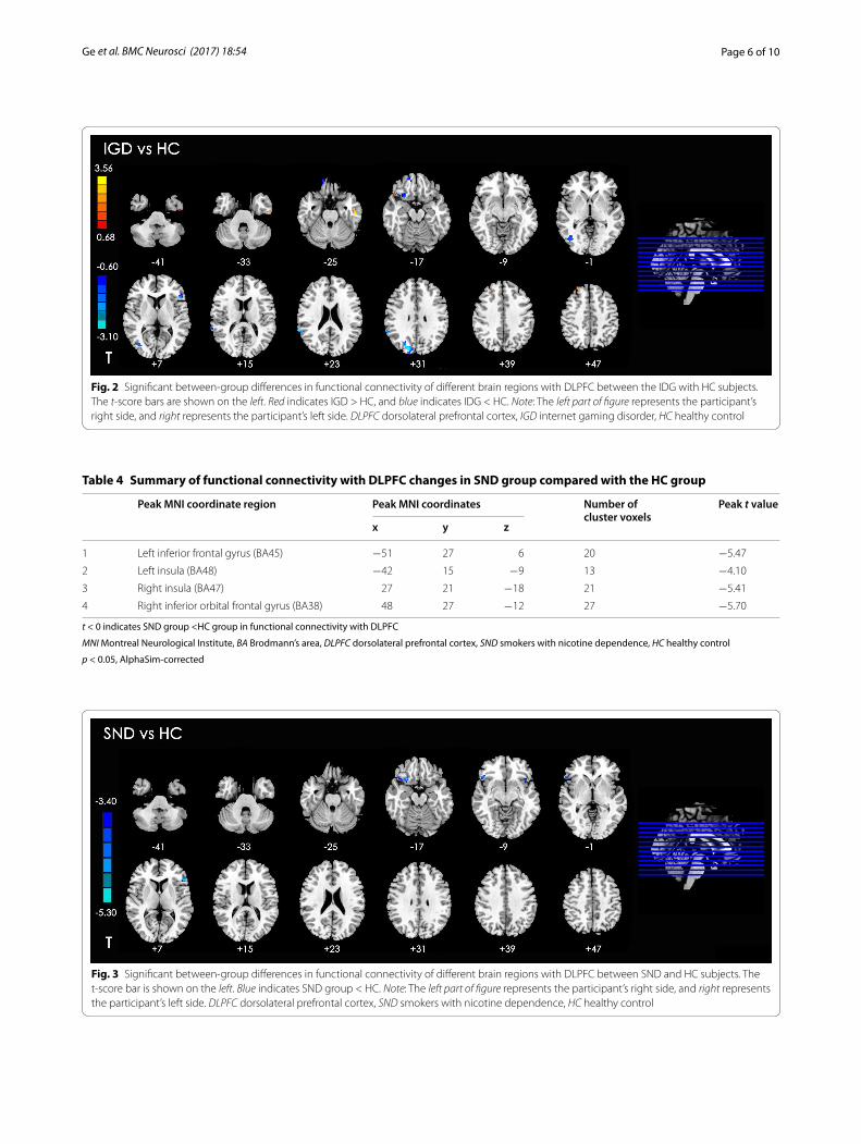
Between‑group analysis of DLPFC connectivity: IGD versus HCThe IGD group exhibited significantly increased rsFC in left inferior temporal gyrus, right superior temporal gyrus, and right middle frontal gyrus with the DLPFC, compared with the HC group. In addition, decreased rsFC was found in the left inferior frontal lobe, right side of the medial frontal orbital gyrus, insula, middle occipi-tal gyrus, superior temporal gyrus, and cuneus with the DLPFC (Table 3; Fig. 2).
Between‑group analysis of DLPFC connectivity: SND versus HCThe SND group showed significantly decreased rsFC in bilateral insula, left inferior frontal gyrus, and right infe-rior orbital frontal gyrus with the DLPFC (Table 4; Fig. 3).
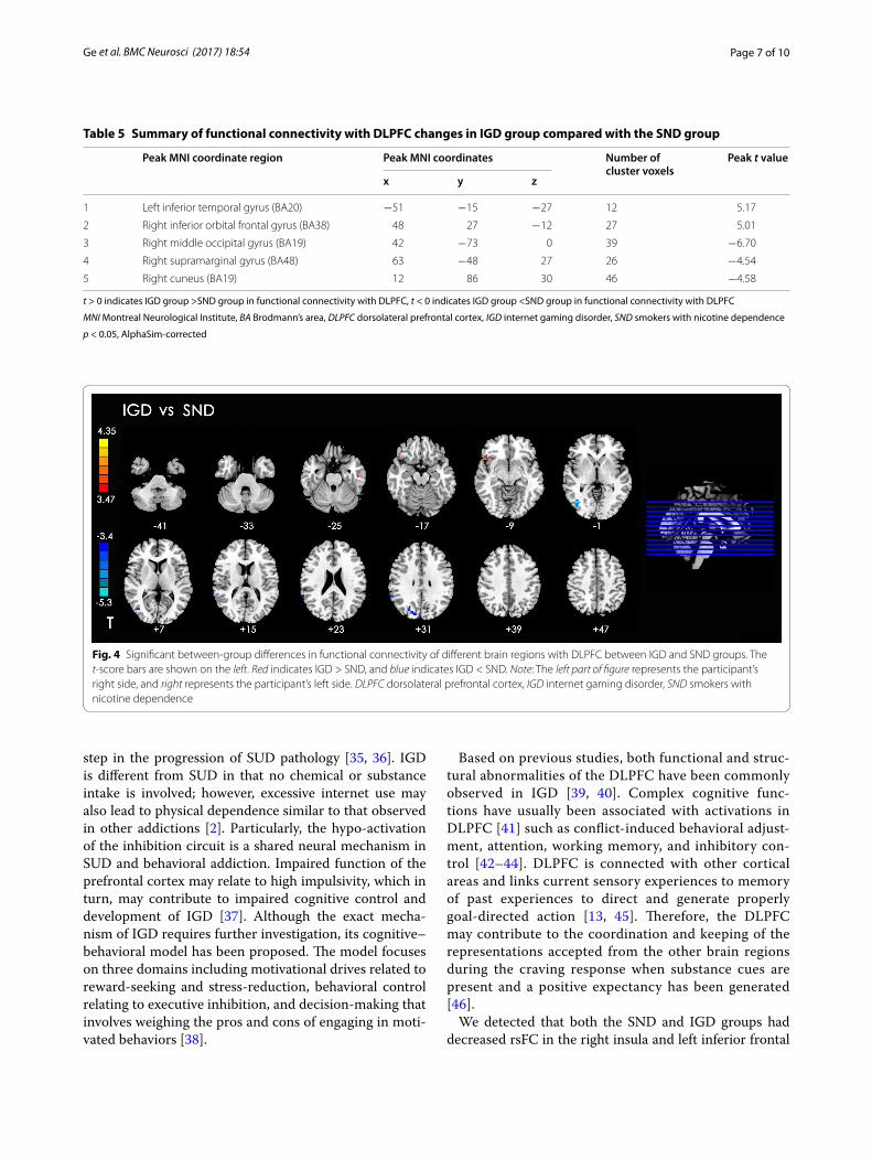
Between‑group analysis of DLPFC connectivity: IGD versus SNDCompared with the SND group, the IGD subjects had increased rsFC in the left inferior temporal gyrus and right inferior orbital frontal gyrus and decreased rsFC in the right side of the middle occipital gyrus, supramar-ginal gyrus, and cuneus with the DLPFC (Table 5; Fig. 4).
Table 1 Demographic and clinical characteristics of the three groups
p 1-2 for IGD group versus HC group, p 1-3 for IGD group versus SND group, p 2-3 for HC group versus SND group
SD standard deviation, HC healthy control, IGD internet gaming disorder, SND smokers with nicotine dependence, FTND Fagerstrom test of nicotine dependence
IGD (n = 27) HC (n = 33) SND (n = 29) F value (p value)
p1-2 p1-3 p2-3(Mean ± SD) (Mean ± SD) (Mean ± SD)
Age (years) 20.78 ± 2.20 20.78 ± 2.51 22.58 ± 2.41 5.56(0.005)
1 0.02 0.01
Sex (F/M) 8/19 6/27 0/33 12.091(0.001)
0.30 0.001 0.02
Education (years) 11.26 ± 1.67 12.67 ± 2.58 13.03 ± 2.06 5.19(0.007)
0.43 0.01 1
Chen Internet Addiction Scale (CIAS) 75.15 ± 9.95 44.27 ± 9.81 46.76 ± 8.34 98.29(<0.001)
<0.001 <0.001 0.45
Self-Rating Anxiety Scale (SAS) 51.33 ± 10.16 39.61 ± 6.39 44.48 ± 8.96 14.09(<0.001)
<0.001 0.01 0.08
Self-rating depression scale (SDS) 54.56 ± 10.80 43.12 ± 8.85 47.44 ± 9.13 10.66(<0.001)
<0.001 0.02 0.24
Barratt Impulsiveness Scale-11 (BIS-11) 61.22 ± 8.44 52.82 ± 6.64 52.41 ± 7.50 12.36(<0.001)
<0.001 <0.001 1
FTND 6.52 ± 2.11
Table 2 Significant differences in functional connectivity of different brain regions with DLPFC changes among the three groups
MNI Montreal Neurological Institute, BA Brodmann’s area, DLPFC dorsolateral prefrontal cortex
p < 0.05, AlphaSim-corrected
Peak MNI coordinate region Peak MNI coordinates Number of cluster voxels
Peak F value
x y z
1 Left inferior temporal gyrus (BA20) −51 −15 −27 29 15.69
2 Left insula (BA48) −36 16 −11 20 8.46
3 Left inferior frontal gyrus (BA45) −51 27 6 22 14.70
4 Right middle temporal gyrus (BA19) 42 −72 0 47 21.28
5 Right supramarginal gyrus (BA40) 63 −48 27 30 11.32
6 Right cuneus (BA19) 15 −84 30 75 13.43
7 Right superior orbital frontal gyrus (BA11) 15 54 −21 39 13.96
8 Right insula (BA47) 27 21 −18 22 13.61
9 Right inferior orbital frontal gyrus (BA38) 48 27 −12 61 10.56
10 Right superior frontal gyrus (BA9) 24 42 42 15 9.68

Page 5 of 10Ge et al. BMC Neurosci (2017) 18:54
Correlation between DLPFC connectivity and CIAS of IGD, DLPFC connectivity, and FTND of SNDCompared with the HC group, the IGD and SND both had decreased rsFC in the left inferior frontal gyrus and right insula with DLPFC. The rsFC strength values (mean zFC values) were extracted and averaged within a spherical ROI (radius of 10 mm) centered on the difference peak of the rsFC group (Tables 2, 3) in the IGD and SND groups. Pear-son correlations were performed between the rsFC values with CIAS of the IGD group and the FTND score in SND group. However, no significant correlation was found.
DiscussionIn this study, we observe both similar and different brain connectivities in IGD group related to SND group. We detected that both the SND and IGD groups had
decreased rsFC with DLPFC in the right insula and left inferior frontal gyrus. Furthermore, the IGD subjects exhibited different rsFC with DLPFC in the orbital fron-tal cortex and temporal, occipital, and parietal lobes.
Evidence revealed that many of the behavioral symp-toms, even the neural mechanisms underlying IGD, resemble SUD [14, 34]. SUD involves a chronic, recur-rent pattern of drug, nicotine, or alcohol use, and nico-tine dependence is one of its most common forms. SUD could result in neurological alterations, particularly in frontal lobe structures implicated in cognitive-behavio-ral control. The network of cortical regions dysfunction, including the DLPFC, anterior cingulate cortex and lat-eral parietal cortex, relates to deficits in behavioral inhi-bition. This dysfunction has been linked to the loss of control over substance intake, which could be a critical
Fig. 1 Significant differences in functional connectivity of different brain regions with DLPFC changes among the three groups. Note: The left part of figure represents the participant’s right side, and right represents the participant’s left side. DLPFC dorsolateral prefrontal cortex
Table 3 Summary of functional connectivity with DLPFC changes in IGD compared with the HC group
t > 0 indicates IGD group >HC group in functional connectivity, t < 0 indicates IGD group <HC group in functional connectivity with DLPFC
MNI Montreal Neurological Institute, BA Brodmann’s area, DLPFC dorsolateral prefrontal cortex, IGD internet gaming disorder, HC healthy control
p < 0.05, AlphaSim-corrected
Peak MNI coordinate region Peak MNI coordinates Number of cluster voxels
Peak t value
x y z
1 Left inferior temporal gyrus (BA20) −54 −21 −27 29 4.14
2 Right superior temporal gyrus (BA38) 48 24 −21 14 2.51
3 Right middle frontal gyrus (BA9) 27 42 42 15 3.15
4 Left inferior frontal gyrus (BA45) −45 24 0 22 −2.81
5 Right medial frontal orbital lobe (BA11) 18 57 −18 16 −1.88
6 Right insula (BA48) 27 15 −18 21 −2.22
7 Right middle occipital gyrus (BA19) 39 −75 3 47 −2.26
8 Right superior temporal gyrus (BA22) 65 −45 24 30 −3.62
9 Right cuneus (BA19) 18 −84 27 67 −3.67
Page 6 of 10Ge et al. BMC Neurosci (2017) 18:54
Fig. 2 Significant between-group differences in functional connectivity of different brain regions with DLPFC between the IDG with HC subjects. The t-score bars are shown on the left. Red indicates IGD > HC, and blue indicates IDG < HC. Note: The left part of figure represents the participant’s right side, and right represents the participant’s left side. DLPFC dorsolateral prefrontal cortex, IGD internet gaming disorder, HC healthy control
Table 4 Summary of functional connectivity with DLPFC changes in SND group compared with the HC group
t < 0 indicates SND group <HC group in functional connectivity with DLPFC
MNI Montreal Neurological Institute, BA Brodmann’s area, DLPFC dorsolateral prefrontal cortex, SND smokers with nicotine dependence, HC healthy control
p < 0.05, AlphaSim-corrected
Peak MNI coordinate region Peak MNI coordinates Number of cluster voxels
Peak t value
x y z
1 Left inferior frontal gyrus (BA45) −51 27 6 20 −5.47
2 Left insula (BA48) −42 15 −9 13 −4.10
3 Right insula (BA47) 27 21 −18 21 −5.41
4 Right inferior orbital frontal gyrus (BA38) 48 27 −12 27 −5.70
Fig. 3 Significant between-group differences in functional connectivity of different brain regions with DLPFC between SND and HC subjects. The t-score bar is shown on the left. Blue indicates SND group < HC. Note: The left part of figure represents the participant’s right side, and right represents the participant’s left side. DLPFC dorsolateral prefrontal cortex, SND smokers with nicotine dependence, HC healthy control
Page 7 of 10Ge et al. BMC Neurosci (2017) 18:54
step in the progression of SUD pathology [35, 36]. IGD is different from SUD in that no chemical or substance intake is involved; however, excessive internet use may also lead to physical dependence similar to that observed in other addictions [2]. Particularly, the hypo-activation of the inhibition circuit is a shared neural mechanism in SUD and behavioral addiction. Impaired function of the prefrontal cortex may relate to high impulsivity, which in turn, may contribute to impaired cognitive control and development of IGD [37]. Although the exact mecha-nism of IGD requires further investigation, its cognitive–behavioral model has been proposed. The model focuses on three domains including motivational drives related to reward-seeking and stress-reduction, behavioral control relating to executive inhibition, and decision-making that involves weighing the pros and cons of engaging in moti-vated behaviors [38].
Based on previous studies, both functional and struc-tural abnormalities of the DLPFC have been commonly observed in IGD [39, 40]. Complex cognitive func-tions have usually been associated with activations in DLPFC [41] such as conflict-induced behavioral adjust-ment, attention, working memory, and inhibitory con-trol [42–44]. DLPFC is connected with other cortical areas and links current sensory experiences to memory of past experiences to direct and generate properly goal-directed action [13, 45]. Therefore, the DLPFC may contribute to the coordination and keeping of the representations accepted from the other brain regions during the craving response when substance cues are present and a positive expectancy has been generated [46].
We detected that both the SND and IGD groups had decreased rsFC in the right insula and left inferior frontal
Table 5 Summary of functional connectivity with DLPFC changes in IGD group compared with the SND group
t > 0 indicates IGD group >SND group in functional connectivity with DLPFC, t < 0 indicates IGD group <SND group in functional connectivity with DLPFC
MNI Montreal Neurological Institute, BA Brodmann’s area, DLPFC dorsolateral prefrontal cortex, IGD internet gaming disorder, SND smokers with nicotine dependence
p < 0.05, AlphaSim-corrected
Peak MNI coordinate region Peak MNI coordinates Number of cluster voxels
Peak t value
x y z
1 Left inferior temporal gyrus (BA20) −51 −15 −27 12 5.17
2 Right inferior orbital frontal gyrus (BA38) 48 27 −12 27 5.01
3 Right middle occipital gyrus (BA19) 42 −73 0 39 −6.70
4 Right supramarginal gyrus (BA48) 63 −48 27 26 −4.54
5 Right cuneus (BA19) 12 86 30 46 −4.58
Fig. 4 Significant between-group differences in functional connectivity of different brain regions with DLPFC between IGD and SND groups. The t-score bars are shown on the left. Red indicates IGD > SND, and blue indicates IGD < SND. Note: The left part of figure represents the participant’s right side, and right represents the participant’s left side. DLPFC dorsolateral prefrontal cortex, IGD internet gaming disorder, SND smokers with nicotine dependence

Page 8 of 10Ge et al. BMC Neurosci (2017) 18:54
gyrus with DLPFC. The insula has been implicated in cue-induced craving and relapse in nicotine-dependent tobacco cigarette smokers [47]. And the orbitofrontal cor-tex is involved in the evaluation of the reward of stimuli and explicit representation of reward expectancy for the substance [7]. Our results were consistent with the pre-vious studies, which emphasized the brain regions, such as ventromedial prefrontal cortex, insula, thalamus, and cerebellum, which was critically linked with cigarette smoking. Structural MRI studies revealed that the integri-ties of the gray matters in the prefrontal cortex, anterior cingulate cortex, insula, thalamus, and cerebellum were reduced in smokers [48–50]. Liu et al. [51] investigated the brain function of IGD individuals using task-state fMRI. The IGD group showed increased activation in the right side of superior parietal lobule, insular lobe, precuneus, cingulated gyrus, superior temporal gyrus, and left side of brainstem. Internet video games activate the space, atten-tion, vision, and execution centers located in the tempo-ral, parietal, occipital, and frontal gyri. Abnormal brain function was noted in IGD subjects with hypofunction of the frontal cortex. Liu et al. detected IGD subjects that showed laterality activation of the right cerebral hemi-sphere, and they found that most areas were located in the right hemisphere. Neuroimaging studies in healthy sub-jects reported that the right hemisphere, especially in the right inferior frontal gyrus, is activated following success-ful response inhibition [52, 53]. During failed response inhibitions (i.e., trials that erroneously generated motor responses), the midline frontal structures, particularly the dorsomedial prefrontal cortex (dmPFC) encompassing pre-supplementary motor area and dorsal anterior cingu-late cortex, are usually activated [54]. Consequently, the right inferior frontal gyrus is critical for response inhibi-tion, whereas dmPFC is associate with response monitor-ing, particularly conflict and error monitoring [14].
The IGD subjects exhibited different rsFC with DLPFC in the orbital frontal cortex and temporal, occipital, and parietal lobes. Our result was partly similar with the result of a previous research compared rsFC with DLPFC in alcohol dependence with those in IGD [8]. They sug-gested that the connectivity observed in alcohol depend-ence is different from that in IGD because of the different comorbid diseases, early prevalence age, and visual and auditory stimulations in the former. Visual and auditory attentions are the results of the main sensory system inputs in response to internet game play [55]. Visual acu-ity loss or hearing problems may cause by extreme inter-net gaming [56]. Increased cortical volume within the parietal cortex was related to long-term gaming in pro-gamers, and thus may be related to increased visuospatial attention [57, 58].
Naturally, this study also comes with limitations. First, the cross-sectional design prevented us from determin-ing whether the group differences in the rsFC are vul-nerability factors for IGD and nicotine dependence. Second, the group sizes were unbalanced in our study, and the parameters such as sex, age, and education were not matched in the three groups. The unbalance group sizes might have influenced the results even though the variety was controlled during the statistical analysis. Third, the mean FTND in the SND group was 6.5, and thus the severity of nicotine dependence was not suffi-ciently high. Thus, increasing the number of participants is necessary.
ConclusionThe rsFC is a very powerful tool for exploring multifac-eted neuropsychiatric diseases, such as substance and non-substance addiction at system level. Our results confirmed that nicotine dependence and IGD may share similar mechanisms related to craving and impulsive inhibition. The observed difference between the rsFC of subjects with IGD and those of SND may be attributed to the impairments in audiovisual information processing by long-term internet gaming.
AbbreviationsIGD: internet gaming disorder; SND: smokers with nicotine dependence; rsFC: resting-state functional connectivity; DLPFC: dorsolateral prefrontal cortex; HC: healthy controls; rs-fMRI: resting-state functional magnetic resonance imaging; PCC: post cingulate cortex; CIAS: Chen’s internet addiction score; AD: alcohol dependence; SUD: substance-related disorders; SAS: self-rating anxiety scale; SDS: self-rating depression scale; BIS-11: Barratt impulsiveness scale-11; FTND: Fagerstrom test of nicotine dependence; TR: repetition time; TE: echo time; FOV: field of view; FD: framewise displacement; ROI: region of interest; AFNI: Analysis of Functional Neuroimages; dmPFC: dorsomedial prefrontal cortex.
Authors’ contributionsConceptualization: YZ and JX; Formal analysis: YS, MC, YW, and YZ; Investiga-tion: XG, YS, WD, MC, YD, and XH; Methodology: YW and YZ; Visualization: YS; Writing—original draft: XG, YS, and YZ; Writing—review and editing: YZ. All authors have read and approved the final version.
Author details1 Department of Radiology, Ren Ji Hospital, School of Medicine, Shanghai Jiao Tong University, Shanghai 200127, People’s Republic of China. 2 Department of Child & Adolescent Psychiatry, Shanghai Mental Health Center, Shanghai Jiao Tong University, Shanghai 200030, People’s Republic of China.
AcknowledgementsNot applicable
Competing interestsThe authors declare that the research was conducted in the absence of com-mercial and financial relationships that can be construed as potential conflicts of interest.
Availability of data and materialThe datasets used and analyzed during the current study are available from the corresponding author on reasonable request.
Page 9 of 10Ge et al. BMC Neurosci (2017) 18:54
Ethics approval and consent to participateThe current study was approved by the Research Ethics Committee of Ren Ji Hospital and School of Medicine, Shanghai Jiao Tong University, China No.[2016]079k(2). All participants were informed of the aims of our study before MRI examination. Each participant submitted a written informed consent.
FundingThis research was supported by the National Natural Science Foundation of China (No. 81571650), and Shanghai Science and Technology Committee Medical Guide Project (western medicine) (No. 17411964300). The funders had no role in study design, data collection and analysis, decision to publish, or preparation of the manuscript.
Publisher’s NoteSpringer Nature remains neutral with regard to jurisdictional claims in pub-lished maps and institutional affiliations.
Received: 15 March 2017 Accepted: 21 July 2017
References 1. Meng Y, Deng W, Wang H, Guo W, Li T. The prefrontal dysfunction in
individuals with internet gaming disorder: a meta-analysis of functional magnetic resonance imaging studies. Addict Biol. 2015;20(4):799–808.
2. Dong G, Hu Y, Lin X. Reward/punishment sensitivities among internet addicts: implications for their addictive behaviors. Prog Neuropsychop-harmacol Biol Psychiatry. 2013;46:139–45.
3. Potenza M. Perspective: behavioural addictions matter. Nature. 2015;522(7557):S62.
4. Young KS. Psychology of computer use: XL. Addictive use of the internet: a case that breaks the stereotype. Psychol Rep. 1996;79(3 Pt 1):899–902.
5. Atmaca M. A case of problematic internet use successfully treated with an SSRI-antipsychotic combination. Prog Neuropsychopharmacol Biol Psychiatry. 2007;31(4):961–2.
6. Shapira NA, Lessig MC, Goldsmith TD, Szabo ST, Lazoritz M, Gold MS, Stein DJ. Problematic internet use: proposed classification and diagnostic criteria. Depress Anxiety. 2003;17(4):207–16.
7. Ko CH, Liu GC, Yen JY, Yen CF, Chen CS, Lin WC. The brain activations for both cue-induced gaming urge and smoking craving among subjects comorbid with internet gaming addiction and nicotine dependence. J Psychiatr Res. 2013;47(4):486–93.
8. Han JW, Han DH, Bolo N, Kim B, Kim BN, Renshaw PF. Differences in func-tional connectivity between alcohol dependence and internet gaming disorder. Addict Behav. 2015;41:12–9.
9. Chen X, Wang Y, Zhou Y, Sun Y, Ding W, Zhuang Z, Xu J, Du Y. Dif-ferent resting-state functional connectivity alterations in smokers and nonsmokers with internet gaming addiction. Biomed Res Int. 2014;2014:825787.
10. Lee YS, Han DH, Kim SM, Renshaw PF. Substance abuse precedes internet addiction. Addict Behav. 2013;38(4):2022–5.
11. Padilla-Walker LM, Nelson LJ, Carroll JS, Jensen AC. More than a just a game: video game and internet use during emerging adulthood. J Youth Adolesc. 2010;39(2):103–13.
12. Aj VANR, Kuss DJ, Griffiths MD, Shorter GW, Schoenmakers MT. D VDM: the (co-)occurrence of problematic video gaming, substance use, and psychosocial problems in adolescents. J Behav Addict. 2014;3(3):157–65.
13. Ko CH, Liu GC, Hsiao S, Yen JY, Yang MJ, Lin WC, Yen CF, Chen CS. Brain activities associated with gaming urge of online gaming addiction. J Psychiatr Res. 2009;43(7):739–47.
14. de Ruiter MB, Oosterlaan J, Veltman DJ, van den Brink W, Goudriaan AE. Similar hyporesponsiveness of the dorsomedial prefrontal cortex in problem gamblers and heavy smokers during an inhibitory control task. Drug Alcohol Depend. 2012;121(1–2):81–9.
15. Sung J, Lee J, Noh HM, Park YS, Ahn EJ. Associations between the risk of internet addiction and problem behaviors among Korean Adolescents. Korean J Fam Med. 2013;34(2):115–22.
16. Vergara VM, Liu J, Claus ED, Hutchison K, Calhoun V. Alterations of resting state functional network connectivity in the brain of nicotine and alcohol users. Neuroimage. 2017;151:45–54.
17. George MS, Anton RF, Bloomer C, Teneback C, Drobes DJ, Lorberbaum JP, Nahas Z, Vincent DJ. Activation of prefrontal cortex and anterior thalamus in alcoholic subjects on exposure to alcohol-specific cues. Arch Gen Psychiatry. 2001;58(4):345–52.
18. Jasinska AJ, Stein EA, Kaiser J, Naumer MJ, Yalachkov Y. Factors modu-lating neural reactivity to drug cues in addiction: a survey of human neuroimaging studies. Neurosci Biobehav Rev. 2014;38:1–16.
19. Beard KW, Wolf EM. Modification in the proposed diagnostic criteria for internet addiction. Cyberpsychol Behav. 2001;4(3):377–83.
20. First MBSR, Gibbon M, Williams JBW. Structured clinical interview for DDS-IV axis I disorders, clinician version (SID-CV). Washington, DC: American Psychiatric Press; 1996.
21. Chen SHWL, Su YJ, Wu HM, Yang PF. Development of Chinese Inter-net Addiction Scale and its psychometric study. Chin J Psychol. 2003;45(3):279–94.
22. Zung WW. A rating instrument for anxiety disorders. Psychosomatics. 1971;12(6):371–9.
23. Zung WW. A self-rating depression scale. Arch Gen Psychiatry. 1965;12:63–70.
24. Patton JH, Stanford MS, Barratt ES. Factor structure of the Barratt impul-siveness scale. J Clin Psychol. 1995;51(6):768–74.
25. Heatherton TF, Kozlowski LT, Frecker RC, Fagerstrom KO. The Fagerstrom test for nicotine dependence: a revision of the Fagerstrom Tolerance Questionnaire. Br J Addict. 1991;86(9):1119–27.
26. Ko CH, Yen JY, Yen CF, Chen CC, Yen CN, Chen SH. Screening for internet addiction: an empirical study on cut-off points for the Chen Internet Addiction Scale. Kaohsiung J Med Sci. 2005;21(12):545–51.
27. Yan CG, Wang XD, Zuo XN, Zang YF. DPABI: data processing & analysis for (resting-state) brain imaging. Neuroinformatics. 2016;14(3):339–51.
28. Power JD, Barnes KA, Snyder AZ, Schlaggar BL, Petersen SE. Spurious but systematic correlations in functional connectivity MRI networks arise from subject motion. NeuroImage. 2012;59(3):2142–54.
29. Greicius MD, Krasnow B, Reiss AL, Menon V. Functional connectivity in the resting brain: a network analysis of the default mode hypothesis. Proc Natl Acad Sci USA. 2003;100(1):253–8.
30. Biswal B, Yetkin FZ, Haughton VM, Hyde JS. Functional connectivity in the motor cortex of resting human brain using echo-planar MRI. Magn Reson Med. 1995;34(4):537–41.
31. Lowe MJ, Mock BJ, Sorenson JA. Functional connectivity in single and multislice echoplanar imaging using resting-state fluctuations. Neuroim-age. 1998;7(2):119–32.
32. Rogers P. The cognitive psychology of lottery gambling: a theoretical review. J Gambl Stud. 1998;14(2):111–34.
33. Cox RW. AFNI: software for analysis and visualization of functional magnetic resonance neuroimages. Comput Biomed Res Int J. 1996;29(3):162–73.
34. Baggio S, Dupuis M, Studer J, Spilka S, Daeppen JB, Simon O, Berchtold A, Gmel G. Reframing video gaming and internet use addiction: empirical cross-national comparison of heavy use over time and addiction scales among young users. Addiction. 2016;111(3):513–22.
35. Motzkin JC, Baskin-Sommers A, Newman JP, Kiehl KA, Koenigs M. Neural correlates of substance abuse: reduced functional connectivity between areas underlying reward and cognitive control. Hum Brain Mapp. 2014;35(9):4282–92.
36. George O, Koob GF. Individual differences in prefrontal cortex function and the transition from drug use to drug dependence. Neurosci Biobe-hav Rev. 2010;35(2):232–47.
37. Weinstein A, Livny A, Weizman A. New developments in brain research of internet and gaming disorder. Neurosci Biobehav Rev. 2017;75:314–30.
38. Dong G, Potenza MN. A cognitive-behavioral model of internet gaming disorder: theoretical underpinnings and clinical implications. J Psychiatr Res. 2014;58:7–11.
39. Du X, Yang Y, Gao P, Qi X, Du G, Zhang Y, Li X, Zhang Q. Compensa-tory increase of functional connectivity density in adolescents with internet gaming disorder. Brain Imaging Behav. 2016. doi:10.1007/s11682-016-9655-x.
Page 10 of 10Ge et al. BMC Neurosci (2017) 18:54
• We accept pre-submission inquiries
• Our selector tool helps you to find the most relevant journal
• We provide round the clock customer support
• Convenient online submission
• Thorough peer review
• Inclusion in PubMed and all major indexing services
• Maximum visibility for your research
Submit your manuscript atwww.biomedcentral.com/submit
Submit your next manuscript to BioMed Central and we will help you at every step:
40. Yuan K, Qin W, Wang G, Zeng F, Zhao L, Yang X, Liu P, Liu J, Sun J, von Deneen KM, et al. Microstructure abnormalities in adolescents with internet addiction disorder. PLoS ONE. 2011;6(6):e20708.
41. Naghavi HR, Nyberg L. Common fronto-parietal activity in attention, memory, and consciousness: shared demands on integration? Conscious Cogn. 2005;14(2):390–425.
42. Scherf KS, Sweeney JA, Luna B. Brain basis of developmental change in visuospatial working memory. J Cogn Neurosci. 2006;18(7):1045–58.
43. Oldrati V, Patricelli J, Colombo B, Antonietti A. The role of dorsolateral prefrontal cortex in inhibition mechanism: a study on cognitive reflection test and similar tasks through neuromodulation. Neuropsychologia. 2016;91:499–508.
44. Mansouri FA, Buckley MJ, Tanaka K. Mnemonic function of the dorsolat-eral prefrontal cortex in conflict-induced behavioral adjustment. Science. 2007;318(5852):987–90.
45. Vanderschuren LJ, Everitt BJ. Behavioral and neural mechanisms of com-pulsive drug seeking. Eur J Pharmacol. 2005;526(1–3):77–88.
46. Bonson KR, Grant SJ, Contoreggi CS, Links JM, Metcalfe J, Weyl HL, Kurian V, Ernst M, London ED. Neural systems and cue-induced cocaine craving. Neuropsychopharmacology. 2002;26(3):376–86.
47. Moran-Santa Maria MM, Hartwell KJ, Hanlon CA, Canterberry M, Lematty T, Owens M, Brady KT, George MS. Right anterior insula connectivity is important for cue-induced craving in nicotine-dependent smokers. Addict Biol. 2015;20(2):407–14.
48. Fritz HC, Wittfeld K, Schmidt CO, Domin M, Grabe HJ, Hegenscheid K, Hosten N, Lotze M. Current smoking and reduced gray matter volume—a voxel-based morphometry study. Neuropsychopharmacology. 2014;39(11):2594–600.
49. Kuhn S, Romanowski A, Schilling C, Mobascher A, Warbrick T, Winterer G, Gallinat J. Brain grey matter deficits in smokers: focus on the cerebellum. Brain Struct Funct. 2012;217(2):517–22.
50. Franklin TR, Wetherill RR, Jagannathan K, Johnson B, Mumma J, Hager N, Rao H, Childress AR. The effects of chronic cigarette smoking on gray matter volume: influence of sex. PLoS ONE. 2014;9(8):e104102.
51. Liu J, Li W, Zhou S, Zhang L, Wang Z, Zhang Y, Jiang Y, Li L. Functional characteristics of the brain in college students with internet gaming disorder. Brain Imaging Behav. 2016;10(1):60–7.
52. Forman SD, Dougherty GG, Casey BJ, Siegle GJ, Braver TS, Barch DM, Stenger VA, Wick-Hull C, Pisarov LA, Lorensen E. Opiate addicts lack error-dependent activation of rostral anterior cingulate. Biol Psychiatry. 2004;55(5):531–7.
53. Hampshire A, Chamberlain SR, Monti MM, Duncan J, Owen AM. The role of the right inferior frontal gyrus: inhibition and attentional control. Neuroimage. 2010;50(3):1313–9.
54. Modirrousta M, Fellows LK. Dorsal medial prefrontal cortex plays a necessary role in rapid error prediction in humans. J Neurosci. 2008;28(51):14000–5.
55. Dong G, Huang J, Du X. Alterations in regional homogeneity of resting-state brain activity in internet gaming addicts. Behav Brain Funct. 2012;8:41.
56. Bovo R, Ciorba A, Martini A. Environmental and genetic factors in age-related hearing impairment. Aging Clin Exp Res. 2011;23(1):3–10.
57. Hyun GJ, Shin YW, Kim BN, Cheong JH, Jin SN, Han DH. Increased cortical thickness in professional on-line gamers. Psychiatry Investig. 2013;10(4):388–92.
58. Song WH, Han DH, Shim HJ. Comparison of brain activation in response to two dimensional and three dimensional on-line games. Psychiatry Investig. 2013;10(2):115–20.