Embed Size (px)
Citation preview

RESEARCH ARTICLE
Flavonoid Glycosides of Polygonumcapitatum Protect against InflammationAssociated with Helicobacter pylori InfectionShu Zhang1, Fei Mo1*, Zhaoxun Luo1*, Jian Huang2, Chaoqin Sun2, Ran Zhang3
1 Guiyang medical college, Guiyang, 550004, China, 2 Affiliated hospital of Guiyang Medical College,Guiyang, 550004, China, 3 Liaocheng people’s hospital, Shandong, 252000, China
* [email protected] (FM); [email protected] (ZXL)
AbstractThe antibacterial and anti-inflammatory activities, and protective effects of extracts (flavo-
noid glycosides) of Polygonum capitatum were investigated to detect the evidence for the
utilization of the herb in the clinical therapy of gastritis caused by H. pylori. A mouse gastritis
model was established using H. pylori. According to treating methods, model mice were
random assigned into a model group (MG group), a triple antibiotics group (TG group, clari-
thromycin, omeprazole and amoxicillin), low/middle/high concentrations of flavonoid glyco-
sides groups (LF, MF and HF groups) and low/middle/high concentrations of flavonoid
glycosides and amoxicillin groups (LFA, MFA and HFA groups). A group with pathogen-free
mice was regarded as a control group (CG group). The eradicate rates of H. pylori were100%, 93%, 89% in TG, MFA and HF groups. The serum levels of IFN-gamma and gastrin
were higher in a MG group than those from all other groups (P < 0.05). The serum levels of
IFN-gamma and gastrin were reduced significantly in LF, MF and HF groups (P < 0.05)
while little changes were observed in LFA, MFA and HFA groups. In contrast, the serum lev-
els of IL-4 were lower and higher in MG and CG groups compared with other groups
(P<0.05). The serum levels of IL-4 were increased significantly in LF, MF and HF groups (P< 0.05) while little changes were found in LFA, MFA and HFA groups. According to patho-
logical scores, flavonoid glycosides therapy showed better protection for gastric injuries
than the combination of flavonoid glycoside and amoxicillin (P < 0.05). The results sug-
gested that flavonoid glycoside has repairing functions for gastric injuries. The results sug-
gest that the plant can treat gastritis and protect against gastric injuries. The flavonoid
glycosides from Polygonum capitatum should be developed as a potential drug for the ther-
apy of gastritis caused by H. pylori.
IntroductionHelicobacter pylori (H. pylori) infection is the main cause of chronic inflammation (gastritis),which is the second leading cause of gastric cancer in the world [1].H. pylori infection also
PLOSONE | DOI:10.1371/journal.pone.0126584 May 18, 2015 1 / 23
OPEN ACCESS
Citation: Zhang S, Mo F, Luo Z, Huang J, Sun C,Zhang R (2015) Flavonoid Glycosides of Polygonumcapitatum Protect against Inflammation Associatedwith Helicobacter pylori Infection. PLoS ONE 10(5):e0126584. doi:10.1371/journal.pone.0126584
Academic Editor: Dipshikha Chakravortty, IndianInstitute of Science, INDIA
Received: December 19, 2014
Accepted: April 6, 2015
Published: May 18, 2015
Copyright: © 2015 Zhang et al. This is an openaccess article distributed under the terms of theCreative Commons Attribution License, which permitsunrestricted use, distribution, and reproduction in anymedium, provided the original author and source arecredited.
Data Availability Statement: Data are availableupon request due to ethical and legal restrictions.Data related to Polygonum capitatum and preparationof its extracts are available upon request to: Prof.Liyan Zhang, Guizhou Weimen Pharmaceutical Co.Ltd., Address No23, Gaoxin Road, Wudang District,Guiyang 550018, Guizhou Province, China;[email protected]. All other data are available uponrequest to author Fei Mo due to ethical restrictionsimposed by Ethic Committee of Guiyang MedicalCollege (Guiyang, China).
Funding: The project was supported by the joint fundof Science and Technology Department of Guizhou

causes gastrointestinal lymphoma, which develop in stomach[2]. Mucosa-associated lymphoidtissue (MALT) and diffusing large B-cell lymphoma are the common histologic characters ofgastric lymphoma[3]. Additionally,H. pylori infection is closely associated with the develop-ment of MALT lymphoma, which also results in gastric cancer[4]. The occurrence of selectedgenes such as gastrin and somatostatin[5], determine the pathogenicity of H. pylori. These pro-teins are pathogens contributing to peptic inflammation, ulceration, and cancer [6–8].H. pylorieradication can completely control the development of MALT lymphomas[9] and has becomethe main focus for the prevention of gastric disease.
Presently, metronidazole, clarithromycine and amoxicillin are mostly used medicine for thetherapy ofH. pylori [10, 11]. However, a high prevalence of medicine resistance has been wide-ly reported inH. pylori and the mechanisms for causing medicine resistance are complex [12–14]. The rate for the eradication ofH. pylori is even less than 50% in most places[15]. Amoxicil-lin is the most powerful medicine for the therapy of the bacteria, but the high prevalence ofmedicine resistance in H. pylori also limits its utilization [12, 16]. There is increasing evidencethat amoxicillin causes severe adverse effects in most patients [17].
Traditional Chinese medicine (TCM) has been implied in the Chinese health care for morethan two thousand years[18]. The side effects and adverse events of TCM are often regarded asgenerally mild and infrequent[19]. Polygonum capitatum (P. capitatum), a traditional ChineseMiao-nationality herb, has been widely used in the treatment of urologic diseases [20]. Recentpharmacological studies have demonstrated that the antibacterial and anti-inflammatory activ-ities of P. capitatum can be used for treating urinary tract infections at a clinical stage [21]. Fur-ther work extends the application of P. capitatum in treating diseases caused byH. pylori(Chinese patent No. CN102824417A), but the molecular mechanism remains unknown.
In immune system, interferon (IFN)-gamma can stimulate macrophage release and plays acritical role in the immune response against infection and controlling intracellular pathogens[22, 23]. Aberrant IFN-gamma expression is linked to many autoinflammatory and autoim-mune diseases. The importance of IFN-gamma in the immune system stems is mostly due toits immunostimulatory and immunomodulatory effects [24]. IFN-gamma is mainly producedby natural killer and natural killer T cells in immune response, and by cluster of differentiationCD4 Th1 and cytotoxic CD8 T cells when antigen-mediated immunity develops[25].H. pyloriinfection is one of the major causes for gastroduodenal pathologies. The long-term persistenceof bacterial infection and immune and inflammatory response will affect the levels of IFN-gamma. IFN-gamma promotes the severity of the induced gastric lesions during the host re-sponse toH. pylori [26].
The interleukin 4 (IL4) is a kind of cytokine inducing the differentiation of naive helper Tcells to Th2 cells[27]. Upon activation by IL-4, Th2 cell produces additional IL-4 in a positivefeedback way. The function of IL-4 is similar to that of Interleukin 13[28]. It has many biologicalroles, such as activating the proliferation of B cells and T cells. It plays an important role in hu-moral and adaptive immunity. IL-4 can induce B-cell class switching to the antibody IgE, andup-regulate the levels of MHC class II. IL-4 can decrease the levels of Th1 cells, macrophages,IFN-gamma, and dendritic cell IL-12. Elevated IL-4 is associated with inflammation and woundrepair[29]. IL-4 can modulate macrophage activation and polarization into M2 and inhibit acti-vation of macrophages into M1 cells[30]. An increase in M2macrophages is accompanied bythe production of IL-10 and TGF-β that result in a decrease of pathological inflammation[31].
Gastrin, a kind of peptide hormone, stimulates the production of gastric acid by the parietalcells from the stomach and promotes gastric motility. Gastrin is secreted by G cells from thepyloric antrum of the stomach[32]. Decreased levels of gastrin were found in healthy adults be-cause of the change ofH. pylori infection. The control of gastrin can prevent H. pylori-inducedgastritis. Gastrin plays a critical role in the inflammatory reaction of the gastric mucosa caused
Flavonoid Glycosides and Bacteria Infection
PLOS ONE | DOI:10.1371/journal.pone.0126584 May 18, 2015 2 / 23
province and Medical College of Guiyang (No. LG(2012)012 and LG(2012)054), Specialized ProgramConstruction Project in University (No. 21 (No.[2010]15), the Science and Technology Program of Guizhou(No. [2014]2027), The Ph.D. Programs Foundation ofAffiliated hospital of Guiyang Medical College (No.2014) and College students' innovativeentrepreneurial training program of Guizhou(No.201410660014). The funders had no role in studydesign, data collection and analysis, decision topublish, or preparation of the manuscript.
Competing Interests: The authors have declaredthat no competing interests exist.

byH. pylori infection [33]. Somatostatin, a kind of growth hormone-inhibiting hormone[34],is a peptide hormone regulating the endocrine system and affecting cell proliferation by inter-acting with G protein-coupled somatostatin receptors and inhibiting the release of secondaryhormones. Somatostatin cell is an important regulator of gastric acid secretion and alterationin its numbers plays a key role in gastroduodenal disease. The alterations may correlate withthe severity of inflammation and certain peptide-immune interactions in the gastric mucosacaused byH. pylori infection [35].
Flavonoid glycoside is one of main components of P. capitatum and shows anti-bacterial activ-ities [21]. Furthermore, amoxicillin is often combined with other medicine to treatH. pylori relat-ed disease. Therefore, we want to know the effects of flavonoid glycoside, which were comparedwith effects of combination therapy of flavonoid glycoside and amoxicillin onH. pylori infecteddisease. Meanwhile, the levels of IFN-gamma, IL-4, Gastrin and Somatostatin were measured.
Results
Sensitivity test of H. pylori to flavonoid glycosideIn vitro antibacterial test, 40 ug/mL or higher concentrations of flavonoid glycoside inhibited thegrowth ofH. pylori (Fig 1). Therefore, the MIC of flavonoid glycoside was 40 ug/mL, which wasmuch higher than MIC of amoxicillin (1 ug/mL). The resistance of MIC of flavonoid glycosidewas regarded as>40.0 μg/mL. The resistance of MIC of amoxicillin was regarded as>1.0 μg/ml.
The toxicity of flavonoid glycoside on mice lymphocyteHigh levels of flavonoid glycoside may inhibit the bioactivity of lymphocytes although flavonoidglycoside shows antibacterial activities [36]. There were not significant differences for meanOD values of lymphocytes when the concentrations were less than 64 ug/ml (P< 0.05). TheOD values of lymphocytes were reduced significantly when the concentrations were more than128 ug/ml (P< 0.05). The OD of lymphocytes reached the lowest level when the concentrationof flavonoid glycoside was 512 ug/mL (Table 1).The results suggest that certain concentrationsof flavonoid glycoside will not affect the proliferation of lymphocytes while the high concentra-tions of flavonoid glycoside will inhibit the proliferation of lymphocytes by its toxicity.
The establishment of a mouse model infected with H. pyloriThe establishment of H. pylori-infected mice model was evaluated from two aspects: the identi-fication of H. pylori isolated from gastric tissues of mice models and analysis of histochemistryand histopathology. H. pylori were identified via Gram staining, rapid urease test, oxidase testand catalase test. Gram staining differentiates bacteria by detecting peptidoglycan[37], whichlocates in the cell walls of gram-positive bacteria. After a Gram stain test, gram-positive bacte-ria showed the crystal violet dye, while a counterstain gave gram-negative bacteria a pink color(Fig 2A).Rapid urease test, is a rapid diagnostic test for H. pylori[38].H. pylori secreted the
Fig 1. Sensitivity test of H. pylori to flavonoid glycoside. A: blank control. B: 40 ug/mL flavonoidglycoside. C: 80 ug/mL flavonoid glycoside.
doi:10.1371/journal.pone.0126584.g001
Flavonoid Glycosides and Bacteria Infection
PLOS ONE | DOI:10.1371/journal.pone.0126584 May 18, 2015 3 / 23
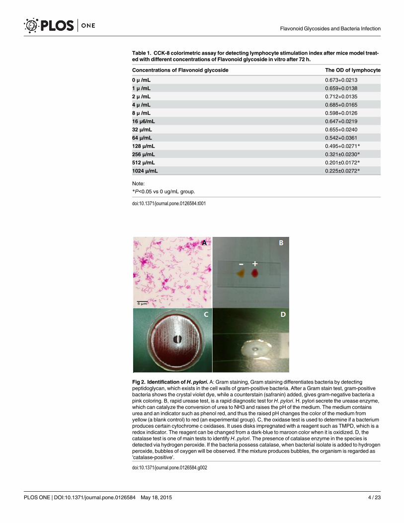
Table 1. CCK-8 colorimetric assay for detecting lymphocyte stimulation index after mice model treat-ed with different concentrations of Flavonoid glycoside in vitro after 72 h.
Concentrations of Flavonoid glycoside The OD of lymphocyte
0 μ /mL 0.673+0.0213
1 μ /mL 0.659+0.0138
2 μ /mL 0.712+0.0135
4 μ /mL 0.685+0.0165
8 μ /mL 0.598+0.0126
16 μ6/mL 0.647+0.0219
32 μ/mL 0.655+0.0240
64 μ/mL 0.542+0.0361
128 μ/mL 0.495+0.0271*
256 μ/mL 0.321±0.0230*
512 μ/mL 0.201±0.0172*
1024 μ/mL 0.225±0.0272*
Note:
*P<0.05 vs 0 ug/mL group.
doi:10.1371/journal.pone.0126584.t001
Fig 2. Identification of H. pylori. A: Gram staining, Gram staining differentiates bacteria by detectingpeptidoglycan, which exists in the cell walls of gram-positive bacteria. After a Gram stain test, gram-positivebacteria shows the crystal violet dye, while a counterstain (safranin) added, gives gram-negative bacteria apink coloring. B, rapid urease test, is a rapid diagnostic test for H. pylori. H. pylori secrete the urease enzyme,which can catalyze the conversion of urea to NH3 and raises the pH of the medium. The medium containsurea and an indicator such as phenol red, and thus the raised pH changes the color of the medium fromyellow (a blank control) to red (an experimental group). C, the oxidase test is used to determine if a bacteriumproduces certain cytochrome c oxidases. It uses disks impregnated with a reagent such as TMPD, which is aredox indicator. The reagent can be changed from a dark-blue to maroon color when it is oxidized. D, thecatalase test is one of main tests to identify H. pylori. The presence of catalase enzyme in the species isdetected via hydrogen peroxide. If the bacteria possess catalase, when bacterial isolate is added to hydrogenperoxide, bubbles of oxygen will be observed. If the mixture produces bubbles, the organism is regarded as'catalase-positive'.
doi:10.1371/journal.pone.0126584.g002
Flavonoid Glycosides and Bacteria Infection
PLOS ONE | DOI:10.1371/journal.pone.0126584 May 18, 2015 4 / 23

urease enzyme, which catalyzed the conversion of urea to NH3 and raised the pH of the medi-um. The medium contained urea and an indicator phenol red, and thus the raised pH changedthe color of the medium from yellow (a blank control) to red (an experimental group) (Fig 2B).The oxidase test was used to determine if a bacterium produced certain cytochrome c oxidases[39]. The disks were impregnated with a reagent such TMPD, which was a redox indicator.The reagent changed from dark-blue to maroon when it was oxidized (Fig 2C). The catalasetest is one of main tests to identify H. pylori[40]. The presence of catalase enzyme in the specieswas detected via hydrogen peroxide. If the bacteria possessed catalase, bubbles of oxygenwould be observed when bacterial isolate was added to hydrogen peroxide (Fig 2D).
Analysis of histochemistry and histopathology was performed via Giemsa staining, HE stain-ing of mice gastric mucosa, microaerophilic culture and Immunohistochemical staining of Gand D cells in Gastric mucosa-associated lymphoid tissue (MALT) lymphoma between controland model groups. Giemsa stain can be used to study the adherence of pathogenic bacteria tomammalians cells. In the control groups, noH. pylori were identified in the gastric mucosa. Incontrast,H. pylori were observed in the gastric mucosa from mice models (Fig 3). Microaero-philic culture conditions are necessary for the growth ofH. pylori[41]. Gastric biopsies weresubjected to microaerophilic culture and then identified by the growth ofH. pylori in mice mod-els. In contrast, no colonies were observed in control groups (Fig 3). H&E staining was con-ducted to observe morphological alterations in gastric mucosa afterH. pylori infection. Mucosaldestruction in the gastric mucosa were observed in mice models. Representative inflammatoryinfiltrates were found at the base of the mucosa with lymphocytes and polymorphonuclears inthe mice infected byH. pylori. In contrast, the mice showed normal gastric morphology withoutmucosal destruction and inflammatory infiltrates (Fig 3). Immunohistochemical staining of Gcells in MALT lymphoma showed that the number of G cells in model groups was more that ina control group. Similarly, immunohistochemical staining of D cells in MALT lymphomashowed that the number of D cells in model groups was more that in a control group (Fig 3).All above information indicated that aH. pylori-infected model was successfully established.
The effects of flavonoid glycoside on the eradication rate of H. pyloriDifferent groups showed different eradication rate of H. pylori (Table 2). Three kinds of antibi-otics eradicated H. pylori completely in a TG group. The combination of flavonoid glycosideand amoxicillin eradicated H. pylori by more than 93% in a MFA group while only flavonoidglycoside eradicatedH. pylori by 89% at most. According to the relative amounts ofH. pylori-specific CagA gene determined by qRT-PCR[42], the log cells of per g of tissues were 0 in a TGgroup while the number is 6.6 ± 0.5 in a MG group. Comparatively, the numbers were 5.4 ± 0.3in a MFA group and 5.8 ± 0.3 in a HF group. Flavonoid glycoside showed the similar antibacte-rial activities compared with the combination therapy of flavonoid glycoside and amoxicillin.
Flavonoid glycoside improves the pathology of H. pylori-infected micemodelWe established a mouse gastritis model using H. pylori and the degree of infection was evaluat-ed by H&E staining (Fig 4). In a CG group, gastric mucosa was found with a common mor-phology, and no inflammation was observed. These histopathological varies showed that amouse model was established successfully. In a MG group, the structures of normal gastric mu-cosa were destroyed, and mucosal destruction was observed. The degree of infection was signif-icantly alleviated and inflammatory cells (lymphocytes, monocytes, neutrophils) were reducedin TG and HF groups compared with those in MF, HF, LFA, MFA and HFA groups (Fig 4). InTG and HF groups, the gastric mucosa was in normal morphology and the pathological scores
Flavonoid Glycosides and Bacteria Infection
PLOS ONE | DOI:10.1371/journal.pone.0126584 May 18, 2015 5 / 23

were also lower than other groups. In contrast, the gastric mucosa were destroyed with differ-ent degrees in MG, LF, MF, LFA, MFA and HFA groups, especially the pathological scoreswere the highest in a MG group. The results showed that flavonoid glycoside could improvethe pathology of H. pylori infection significantly. More importantly, according to pathologicalscores, flavonoid glycoside showed better protect gastric tissues than the combination of flavo-noid glycoside and amoxicillin (P< 0.05) (Fig 4). The results suggested that flavonoid glyco-side has repairing functions for gastric injuries.
Fig 3. The comparision for histochemistry and histopathology between a control and a model group.Giemsa staining (×400), Giemsa stain was used to observe the adherence of pathogenic bacteria to gastriccells. Microaerophilic culturing of H. pylori, the extracellular H. pylori are microaerophilic. Hematoxylin andEosin staining of gastric tissues was used for the detection of H. pylori (HE×200). The staining of G cells ofGastric mucosa-associated lymphoid tissue (MALT) lymphoma (PV×200). The staining of D cells of GastricMALT lymphoma (PV×200).
doi:10.1371/journal.pone.0126584.g003
Flavonoid Glycosides and Bacteria Infection
PLOS ONE | DOI:10.1371/journal.pone.0126584 May 18, 2015 6 / 23
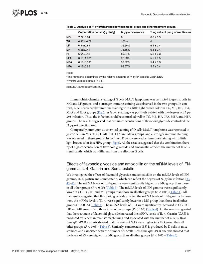
Immunohistochemical staining of G cells MALT lymphoma was restricted to gastric cells inMG and LF groups, and a stronger immune staining was observed in the two groups. In con-trast, G cells were weaker immune staining with a little light brown color in TG, MF, HF, LFA,MFA and HFA groups (Fig 5). A G cell staining was positively related with the degrees ofH. py-lori infection. Thus, the infection could be controlled well in TG, MF, HF, LFA, MFA and HFAgroups. The results suggested that certain concentrations of flavonoid glycoside controlled theH. pylori infection well.
Comparably, immunohistochemical staining of D cells MALT lymphoma was restricted togastric cells in MG, TG, LF, MF, HF, LFA and MFA groups, and a stronger immune stainingwas observed in these groups. In contrast, D cells were weaker immune staining with a littlelight brown color in a HFA group (Fig 6). All the results suggested that the combination thera-py of high concentration of flavonoid glycoside and amoxicillin affected the number of D cellssignificantly, which was different from the effect on G cells.
Effects of flavonoid glycoside and amoxicillin on the mRNA levels of IFN-gamma, IL-4, Gastrin and SomatostatinWe investigated the effects of flavonoid glycoside and amoxicillin on the mRNA levels of IFN-gamma, IL-4, gastrin and somatostatin, which can reflect the degrees of H. pylori infection [33,43–45]. The mRNA levels of IFN-gamma were significantly higher in a MG group than thosein all other groups (P< 0.05) (Table 3). The mRNA levels of IFN-gamma were significantlylower in CG, TG, HF and MF groups than those in all other groups (P< 0.05) (Table 4). Allthe results suggested that flavonoid glycoside affected the mRNA levels of IFN-gamma. In con-trast, the mRNA levels of IL-4 were significantly lower in a MG group than those in all othergroups (P< 0.05) (Table 4). The mRNA levels of IL-4 were significantly increased in CG, TG,HF and MF groups than those in all other groups (P< 0.05) (Table 4). All the results suggestedthat the treatment of flavonoid glycoside increased the mRNA levels of IL-4. Gastrin (GAS) isproduced by G cells in mice stomach lining and associated with the number of G cells. Real-time qRT-PCR analysis showed that the levels of GAS were higher in a MG group than allother groups (P< 0.05) (Table 5). Similarly, somatostain (SS) is produced by D cells in micestomach and associated with the number of D cells. Real-time qRT-PCR analysis showed thatthe levels of SS were higher in a MG group than all other groups (P< 0.05) (Table 6).
Table 2. Analysis ofH. pyloriclearance betweenmodel group and other treatment groups.
Colonization density(Ig cfu/g) H. pylori clearance aLog cells of per g of wet tissues
MG 7.27±0.94 0 6.6 ± 0.5
TG 6.35 ± 0.78 100% 0
LF 6.31±0.89 76.66% 6.1 ± 0.4
MF 6.56±0.41 76.15% 6.1 ± 0.4
HF 6.64±0.42 89.07% 5.8 ± 0.3
LFA 6.15±1.03* 92.09% 5.5 ± 0.5
MFA 6.10±0.59* 93.32% 5.4 ± 0.3
HFA 6.17±0.85 92.57% 5.5 ± 0.4
Note:aThe number is determined by the relative amounts of H. pylori-specific CagA DNA.
*P<0.05 vs model group (n = 8).
doi:10.1371/journal.pone.0126584.t002
Flavonoid Glycosides and Bacteria Infection
PLOS ONE | DOI:10.1371/journal.pone.0126584 May 18, 2015 7 / 23

Effects of flavonoid glycoside and amoxicillin on the protein levels ofinflammatory biomarkersBefore the study, the effects of flavonoid glycoside on lymphocytes were measured. The resultsshowed that the OD values of lymphocytes were affected by the concentration of flavonoid gly-coside. The OD values were the highest when no flavonoid glycoside was used and the valuesreached the lowest level when 512 ug/ml of flavonoid glycoside was used (P< 0.05) (Fig 7A).The levels of IFN-gamma from lymphocytes were also affected by flavonoid glycoside and thelevels reached the highest point when 32 ug/ml of flavonoid glycoside was used (P< 0.05) (Fig7B). Comparatively, the levels of IL-4 reached the highest point when 8 ug/ml of flavonoid glyco-side was used (P< 0.05) (Fig 7C). To explore the effects of flavonoid glycoside and amoxicillinon IFN-gamma and IL-4, the serum levels of IFN-gamma and IL-4 were measured in differentgroups. The results indicated that the serum levels of IFN-gamma were higher in a MG group
Fig 4. Hematoxylin and Eosin staining of gastric tissues (HE×200).Normal mice (without H. pylori infection) were assigned as a control group (CGgroup) and mice models infected with H. pylori were divided into model group (MG group, only treated with saline solution), triple combination therapy group(TG group, the daily medicine intake is 0.5 ug clarithromycin, 0.02 ug omeprazole, and 1 ug amoxicillin), low/middle/high concentrations of flavonoidglycoside group (LF/MF/HF group, treated with one daily dose of Flavonoid glycoside at 32/64/128 ug), low/middle/high concentrations of flavonoid glycosideand common concentration of amoxicillin group (LFA/MFA/HFA group, treated with one daily dose of flavonoid glycoside at 32/64/128 ug and amoxicillin at 1ug). Pathological score of mice gastric antrum in each group (±SD, n = 10). *P < 0.05 vs model group.
doi:10.1371/journal.pone.0126584.g004
Flavonoid Glycosides and Bacteria Infection
PLOS ONE | DOI:10.1371/journal.pone.0126584 May 18, 2015 8 / 23

than those in all other groups (P< 0.05) (Fig 7D). The groups only with flavonoid glycosidecould reduce the serum levels of IFN-gamma significantly (P< 0.05) while the combination offlavonoid glycoside and amoxicillin caused no obvious changes. In contrast, the serum levels ofIL-4 were lower in a MG group than those from all other groups while the levels in a CG groupwere higher than those from all other groups (P< 0.05) (Fig 7E). The groups only with flavonoidglycoside could increase the serum levels of IL-4 significantly (P< 0.05) while the combinationof flavonoid glycoside and amoxicillin caused no obvious changes. For gastrin, the serum levelswere higher in a MG group than those in all other groups except a LF group (P< 0.05) (Fig 7F).The concentrations of serum gastrin reached the lowest level in HF, TG and CG groups. Com-paratively, the serum levels of somatostatin were higher in all groups only with flavonoid glyco-side, a MFA group and a MG group than those from other groups (P< 0.05) (Fig 7G). Theconcentrations of serum somatostatin reached the lowest level in LFA and CG groups.
The effects of flavonoid glycosides on Gastric cells infected by H. pyloriBefore the study, the effects of flavonoid glycoside on gastric cells were measured. The resultsshowed that the OD values of gastric cells were affected by the concentrations of flavonoid
Fig 5. The staining of G cells of Gastric mucosa-associated lymphoid tissue (MALT) lymphoma (PV×200). Normal mice (without H. pylori infection)were assigned as a control group (CG group) and mice models infected with H. pylori were divided into model group (MG group, only treated with salinesolution), triple combination therapy group (TG group, the daily medicine intake is 0.5 ug clarithromycin, 0.02 ug omeprazole, and 1 ug amoxicillin), low/middle/high concentrations of flavonoid glycoside group (LF/MF/HF group, treated with one daily dose of Flavonoid glycoside at 32/64/128 ug), low/middle/high concentrations of flavonoid glycoside and common concentration of amoxicillin group (LFA/MFA/HFA group, treated with one daily dose of Flavonoidglycoside at 32/64/128 ug and amoxicillin at 1 ug). Comparison of G cells and gastrin gray value in each group (±s, n = 10). *P < 0.05 vs model group.
doi:10.1371/journal.pone.0126584.g005
Flavonoid Glycosides and Bacteria Infection
PLOS ONE | DOI:10.1371/journal.pone.0126584 May 18, 2015 9 / 23

Fig 6. The staining of D cells of Gastric mucosa-associated lymphoid tissue (MALT) lymphoma (PV×200).Normal mice (withoutH. pylori infection)were assigned as a control group (CG group) and mice models infected with H. pylori were divided into model group (MG group, only treated with salinesolution), triple combination therapy group (TG group, the daily medicine intake is 0.5 ug clarithromycin, 0.02 ug omeprazole, and 1 ug amoxicillin), low/middle/high concentrations of flavonoid glycoside group (LF/MF/HF group, treated with one daily dose of flavonoid glycoside at 32/64/128 ug), low/middle/high concentrations of flavonoid glycoside and common concentration of amoxicillin group (LFA/MFA/HFA group, treated with one daily dose of Flavonoidglycoside at 32/64/128 ug and amoxicillin at 1 ug). Analysis of G cells and somatostatin gray value in each group (n = 10). *P < 0.05 vs model group.
doi:10.1371/journal.pone.0126584.g006
Table 3. The mRNA levels of IFN-gamma in the gastric mucosa of H. pylori infected mice in differentgroups (n = 6).
Groups β-actin IFN-gamma CT value 2-44CT
MG 32.831±0.428 19.523±2.410 1▲
CG 35.000±1.004 18.513±3.261 0.110*
TG 33.796±2.416 17.768±1.945 0.150*
HF 33.093±1.024 16.995±3.584 0.166*
MF 33.964±3.739 18.669±3.105 0.269*
LF 35.000±0.692 21.377±5.042 0.451
HFA 31.179±1.296 18.513±2.178 0.874
MFA 30.198±1.397 17.029±3.791 0.912
▲P < 0.05 model group vs blank group;
*P < 0.05 vs model group.
doi:10.1371/journal.pone.0126584.t003
Flavonoid Glycosides and Bacteria Infection
PLOS ONE | DOI:10.1371/journal.pone.0126584 May 18, 2015 10 / 23

glycoside. The OD values were the highest when 16 flavonoid glycoside was used and the valuesreached the lowest level when the concentrations were more than 512 ug/ml (P< 0.05) (Fig8A). The levels of IFN-gamma from lymphocytes were also affected by flavonoid glycoside andthe levels reached the highest point when 16 ug/ml of flavonoid glycoside was used (P< 0.05)(Fig 8B). Comparatively, the levels of IL-4 and gastrin reached the highest point when 32 ug/mlof flavonoid glycoside was used (P< 0.05) (Fig 8C and 8D). The levels of somatostatin werenot affected by flavonoid glycoside if the concentrations were less than 32 ug/ml of flavonoidglycoside (P< 0.05) (Fig 8E). All these biomarkers would be reduced greatly if the concentra-tions were more than 64 ug/ml, suggesting that high concentrations of flavonoid glycosidehave toxicity toward gastric cells and inhibit the production of these molecules. All these resultsimplied that flavonoid glycoside may show its anti-inflammatory functions by increasing thelevels of IFN-gamma, IL-4 and gastrin.
DiscussionH. pylori infection is well known to be associated with the risk of many diseases. For example,H. pylori infection contributes to cardiovascular diseases and stroke, and Alzheimer's disease
Table 4. The mRNA levels of IL-4 in the gastric mucosa ofH. pylori infectedmice in different groups(n = 6).
Groups β-actin CT values IL-4 CT values 2-ΔΔCT
MG 32.501±0.138 17.283±1.383 1▲
CG 30.651±1.027 18.388±1.972 7.755*
TG 31.872±0.987 18.837±2.936 4.831*
HF 31.198±0.237 17.988±1.097 4.022*
MF 30.868±0.487 17.390±2.945 3.324*
LF 31.989±1.002 18.292±0.843 2.869*
HFA 33.946±0.948 18.387±1.075 1.257
MFA 32.202±0.739 17.639±2.382 1.354
LFA 35.662±1.024 21.377±2.261 1.669
Note:▲ P < 0.05 via a CG group,
*P < 0.05 via a MG group.
doi:10.1371/journal.pone.0126584.t004
Table 5. The mRNA levels of Gastrin in the gastric mucosa ofH. pylori infectedmice in differentgroups (n = 6).
Groups β-actin GAS(CT) 2-ΔΔCT LOG(2-ΔΔCT)
MG 20.68±0.97 20.56±0.79 1 0
CG 19.79±0.02 26.51±0.13 0.0093±0.0001* -2.03
TG 20.01±0.55 25.45±0.72 0.0225±0.0001* -1.65
HF 20.28±0.14 25.38±0.29 0.0286±0.0006* -1.54
MF 20.48±0.23 26.16±0.03 0.0192±0.0010* -1.72
LF 20.79±0.38 26.96±0.36 0.0135±0.0015* -1.87
HFA 20.52±0.33 25.29±0.07 0.0361±0.0053* -1.44
MFA 20.07±0.08 20.54±0.22 0.7095±0.0199 -0.15
*P < 0.05 vs model group
doi:10.1371/journal.pone.0126584.t005
Flavonoid Glycosides and Bacteria Infection
PLOS ONE | DOI:10.1371/journal.pone.0126584 May 18, 2015 11 / 23

(AD) [46]. Another example, H. pylori infection is most likely to cause chronic gastritis, pepticulcer disease and liver-related diseases[47].H. pylori infection significantly affects the life quali-ty of many people in the world and has become a global burden. H. pylori infection therapyoften includes pharmaceutical treatment, such as clarithromycin, omeprazole and amoxicillin[48–50]. However, all the medicine has side effects which greatly limit the usage: Omeprazoletreatment can cause hypergastrinemia and trophic effects in the stomach with an increase ofhistamine-producing enterochromaffin-like cells[51]; Combination therapy of clarithromycinand rabeprazole can increase the risk of neurotoxicity[52]; Amoxicillin therapy can also lead tosevere adverse effects and death[17]. Thus, it is necessary to explore the new medicine withfewer side effects and therapeutic efficacy. Flavonoid glycoside is a kind of Chinese herbs andhas been widely used for the therapy of urinary tract infection [21]. Recent work indicates thatflavonoid glycoside can be used for the treatment ofH. pylori infection with a few side effects(Chinese patent No. CN102824417A). Amoxicillin is often combined with other medicine forthe therapy of H. pylori infection [53], so the combination therapy of flavonoid glycoside andamoxicillin is an effective way for the therapy ofH. pylori infection.
To understand the effects of flavonoid glycoside and amoxicillin on aH. pylori infected dis-ease, a mouse gastritis model was established usingH. pylori. To explore the molecular mecha-nisms of effects of flavonoid glycoside and amoxicillin on a mouse gastritis model, IFN-gamma,IL-4, gastrin and somatostatin may be the best molecules for the purpose because of the follow-ing reasons:H. pylori infection can elevate IFN-gamma-mediated gastric inflammation [43];IL-4 is an anti-inflammatory and Th2-type cytokine.H. pylori infection in mammals inducesan immune response, which is characterized by an increase of IFN-gamma and absence of IL-4[54]; Serum levels of gastrin are higher inH. pylori-infected patients than in uninfected subjects,andH. pylori infection induces hypergastrinemia in mammals[55]; Somatostatin is a regulatorypeptide, which is mainly existed in the stomach. Somatostatin is needed for IL-4-mediated reso-lution ofH. pylori gastritis [56]. Flavonoid glycoside intervention can reduce the serum levels ofINF-gamma but not IL-4(Figs 7D and 8E). However, the combination of flavonoid glycosideand amoxicillin doesn’t change the levels of INF-gamma, which cannot improve the inflamma-tion of the tissues infected withH. pylori. The results suggested that only flavonoid glycosidetherapy can control the inflammation caused byH. pylori infection.
For further mechanism, flavonoid glycoside has the potential antioxidant activity and theantioxidant functions of flavonoid glycoside have been reported[57]. Most species of Polygo-num have bioactive constituents, which contribute to many medicinal properties. P. cuspida-tum and P. capitatum exhibit great antioxidant properties and are a potent resource of naturalbioactive antioxidants. Here, we found only flavonoid glycoside treatment repaired the gastric
Table 6. The mRNA levels of Somatostatinin the gastric mucosa of H. pylori infectedmice in different groups (n = 6).
Groups β-actin (CT) SS (CT) 2-ΔΔCT LOG(2-ΔΔCT)
MG 20.68±0.97 19.07±1.04 1 0
CG 19.79±0.02 20.92±0.02 0.150±0.013* -0.82
TG 20.01±0.55 20.61±0.37 0.217±0.015* -0.66
HF 20.28±0.14 20.40±0.05 0.303±0.004* -0.52
MF 20.48±0.23 19.86±0.25 0.506±0.018* -0.30
LF 20.79±0.38 21.27±0.38 0.236±0.012* -0.63
HFA 20.52±0.33 20.38±0.43 0.362±0.005* -0.44
MFA 20.07±0.08 19.05±0.20 0.556±0.064* -0.25
*P < 0.05 vs model group
doi:10.1371/journal.pone.0126584.t006
Flavonoid Glycosides and Bacteria Infection
PLOS ONE | DOI:10.1371/journal.pone.0126584 May 18, 2015 12 / 23

Fig 7. The effects of flavonoid glycoside on the number of lymphocytes, protein levels of inflammatory biomarkers. A, the effects of differentconcentrations of flavonoid glycoside on the number of lymphocytes. B, the effects of different concentrations of flavonoid glycoside on protein levels of INF-gamma. C, the effects of different concentrations of flavonoid glycoside on protein levels of IL-4. D, the serum levels of INF-gamma in different groups. E, theserum levels of IL-4 in different groups. F, the serum levels of gastrin in different groups. G, the serum levels of somatostatin in different groups. Normal mice(without H. pylori infection) were assigned as a control group (CG group) and mice models infected withH. pylori were divided into model group (MG group,only treated with saline solution), triple combination therapy group (TG group, the daily medicine intake is 0.5 ug clarithromycin, 0.02 ug omeprazole, and 1ug amoxicillin), low/middle/high concentrations of flavonoid glycoside group (LF/MF/HF group, treated with one daily dose of flavonoid glycoside at 32/64/128ug), low/middle/high concentrations of flavonoid glycoside and common concentration of amoxicillin group (LFA/MFA/HFA group, treated with one daily doseof flavonoid glycoside at 32/64/128 ug and amoxicillin at 1 ug). *P < 0.05 vs a model group (n = 10).
doi:10.1371/journal.pone.0126584.g007
Flavonoid Glycosides and Bacteria Infection
PLOS ONE | DOI:10.1371/journal.pone.0126584 May 18, 2015 13 / 23

injury compared with those treated with the combination of flavonoid glycoside and amoxicil-lin. The difference may be caused by the antioxidant bioactivities of flavonoid glycoside andside effects of amoxicillin for gastric mucosa. Therefore, we explored the protect function of fla-vonoid glycoside for gastric mucosa. A high concentration of amoxicillin may be harmful togastric mucosa although it can enhance the eradicate rate of H. pylori. Certainly, there aresome limits for present study and some important experiments are not performed. It would bebetter if inflammatory responses can be measured in the gastric tissues (for the local inflamma-tory responses) and within the spleen of infected mice. Furthermore, the functions of manyconstituents of P. capitatum were not explored. All the work will be performed in the future.Flavonoid glycosides show effects on many cytokines, which plays a critical role for the therapyofH. pylori infection. Flavonoid glycoside can affect the levels of IFN-gamma, IL-4 and gastrinbut not somatostatin (Fig 8), which is also the basis for further studying the mechanisms forthe functions of flavonoid glycoside.
Fig 8. The effects of flavonoid glycoside on gastric cells infected byH. pylori. A, the effects of different concentrations of flavonoid glycoside on thenumber of gastric cells MGC803. B, the effects of different concentrations of flavonoid glycoside on protein levels of INF-gamma. C, the effects of differentconcentrations of flavonoid glycoside on protein levels of IL-4. D, the effects of different concentrations of flavonoid glycoside on protein levels of gastrin. E,the effects of different concentrations of flavonoid glycoside on protein levels of somatostatin. *P < 0.05 vs a control group without flavonoid glycoside.
doi:10.1371/journal.pone.0126584.g008
Flavonoid Glycosides and Bacteria Infection
PLOS ONE | DOI:10.1371/journal.pone.0126584 May 18, 2015 14 / 23

We demonstrated that the protective and anti-bacterial functions of flavonoid glycosidefrom P. capitatum for the therapy of H. pylori-infected diseases. The function may be associat-ed with its protective functions of gastric mucosa, antioxidant bioactivities and regulation forthe levels of IFN-gamma, IL-4, gastrin and somatostatin. All these functions can reduce the in-jury of gastric tissues infected byH. pylori and improve the symptoms. We therefore proposethat flavonoid glycoside from P. capitatum is potential for theH. pylori infection and should bedeveloped a new drug for H. pylori infected diseases.
Materials and Methods
Materials and ReagentH. pylori SS1 were purchased from the Chinese Center for Disease Control (Beijing, China). Thestrains were cultured on selective agar (Wilkins—Chalgren agar supplemented with 5 percent ofhorse blood, 10.5 ug/mL vancomycin, 0.5 ug/mL cefsulodin, 1 ug/mL trimethoprim lactate, and1 ug/mL fungizone (Biogerm, Maia, Portugal)) and incubated at 37°C under microaerobic con-ditions for one day. The extracts of Polygonum capitatum, flavonoid glycosides were preparedaccording to a previous report[21, 58] and identified by Professor Ma Lin of the Institute of Ma-terial Medicine, Chinese Academy of Chinese Medical Sciences (Beijing, China). Amoxicillin,omeprazole and clarithromycin was purchased from Xinya Co., (Shanghai, China).
Minimum inhibitory concentration (MIC) testThe determination of MICs of flavonoid glycosides for theH. pylori was examined by use ofthe serial dilution method as described previously [59]. Briefly, the bacteria were sub-culturedon Mueller-Hinton agar supplemented with 5 percent sheep blood for two days. A bacterialsuspension with 107 CFU/ml was placed onto each flavonoid glycoside dilution agar plate.After incubation for three days, the MIC of each sample was determined. Quality control wasperformed withH. pylori SS1.
Lymphocyte proliferation assayLymphocytes were isolated from mice spleens using Amaxa Mouse T Cell Nucleofector Kit(Amaxa, Gaithersburg, USA). Lymphocyte proliferations upon the stimulation of flavonoidglycoside and controls were determined with the colorimetric Cell Counting Kit-8 (CCK-8,Beyotime, Shanghai, China). Isolated lymphocytes were plated in flat-bottom 96-well microti-tre plate at a density of 5×10⁵ cells/well, 10 μl of CCK-8 was added to each well and incubatedfor further 4 h, and absorbing value at 450 nm was measured to count cell proliferation. Thestimulation index (SI) was calculated as the ratio of mean OD value of the wells containing fla-vonoid glycoside-stimulated cells to mean OD value of the wells containing cells without flavo-noid glycoside stimulation. All assays were conducted in triplicates.
Model establishmentA total 100 mice C57BL/6 (6~8 weeks, male/female = 1:1, weight(20±5)g) were purchasedfrom Experimental Animals Center of Chongqing Medical University (license No. SYXK2007–0001). All animals were housed in cages with a 12 h light/dark cycle. The cages were keptat 23 ± 1°C with 50% relative humidity. Food and water could be available ad libitum. Animalcare and handling procedures were conducted according to the International Association forStudy of Pain guidelines for animals in pain research. All efforts were performed to minimizethe number of animals and their suffering in the experiment. Animals were provided with
Flavonoid Glycosides and Bacteria Infection
PLOS ONE | DOI:10.1371/journal.pone.0126584 May 18, 2015 15 / 23

sawdust bedding material and were housed under these conditions for at least 1 week prior tothe experiments. Mice were fasted for 12 h before all experimental studies.
Before the infection of mice, H. pylori from plate cultures were inoculated into in Brucellabroth culture medium (Becton Dickinson, Cockeysville, USA) containing 10% fetal bovineserum and were cultured for 18 h under microaerobic conditions. A total of 90 pathogen-freeC57BL/6 8-week-old mice were used in compliance with guidelines and a protocol approvedby the Animal Care and Use Committee of Guiyang medical college. Using a 20-gauge ball-point metal feeding tube (Harvard Apparatus, Inc., Holliston, MA, USA), 90 mice were inocu-lated intragastrically with 0.1 mL ofH. pylori SS1 cell suspension containing 108 colony-forming units /mL on three alternate days. Ten healthy mice were inoculated with saline solu-tion and used as a control.
After nine days, ten mice from a model group and ten from a control group were sacrificedusing cervical dislocation without anesthesia prior to the end of the experiment. Subsequently,the stomachs were isolated from the mice by cutting the tissues from the esophagus to the duo-denum. The non-glandular portion of fore-stomach was removed from the glandular stomach.The glandular stomach was dissected and rinsed with PBS, and divided into three longitudinalstrips, which were used for bacterial culture, RNA analysis, and histology.
Evaluation of a mouse model infected with H. pyloriGastric tissues were homogenized via Tissue Tearor (BioSpecProducts, Bartlesville, USA). Thehomogenate were placed on trypticase soy agar (TSA) with different dilutions, complementingwith 5 percent horse blood, 10 μg/mL nalidixic acid, 100 μg/mL vancomycin, 2 μg/mL ampho-tericin, and 200 μg/mL bacitracin (all antibiotics were from Sigma-Aldrich Shanghai TradingCo Ltd, Shanghai, China). After 5–7 d of culture under microaerobic conditions,H. pylori colo-nies were counted and the number of colony forming units per gram of tissue calculated (CFU/g). Colonies were used to test the bioactivity of urease, catalase, and oxidase. H. pylori colonieswere identified using the following methods: Gram-staining, H. pylori colonies were identifiedusing a Gram-staining kit (BD Biosciences, San Jose, USA) according to the manuscripture’sinstructions; Rapid urease test, rapid urease test was performed according to a previous report[60]. A biopsy was inoculated into 1mL of 10% urea dissolved in distilled water (pH 6.8), towhich two drops of one percent phenol red solution were added and incubated at 37°C for oneday. A color change from yellow to pink within 1 h from the start of the test was considered acriterion for the presence ofH. pylori infection; Catalase test, catalase test was conducted ac-cording to a previous report[61]. The colony grown in selective medium was placed on a slide,and bubbling following dropping 3% H2O2 was determined as positive reaction; Oxidase test,H. pylori uses disks impregnated with a reagent such as N,N,N',N'-tetramethyl-p-phenylene-diamine (TMPD), which is a redox indicator[62]. The reagent can be changed from a dark-blue to maroon color when it is oxidized.
GroupsAs Fig 9 showed, 90 mice were used for the establishment of mice models infected withH. pylo-ri and 10 normal mice were used as control group (CG). From the mice models, 10 mice wereused for the assessment of model and remaining mice were randomly divided into 8 groups. AsFig 9 showed, normal mice (pathogen free) were assigned as a control group (CG group) andmice models infected withH. pylori were divided into model group (MG group), triple combi-nation therapy group (TG group. According to a previous report, for an adult/50 kg, daily med-icine intake is 1000 mg clarithromycin, 40 mg omeprazole, and 2000 mg amoxicillin for theeradication ofH. pylori[63]. For a mouse/25 g, the daily medicine intake was 0.5 ug
Flavonoid Glycosides and Bacteria Infection
PLOS ONE | DOI:10.1371/journal.pone.0126584 May 18, 2015 16 / 23

clarithromycin, 0.02 ug omeprazole, and 1 ug amoxicillin), low concentration of flavonoid gly-coside group (LF group, treated with one daily dose of flavonoid glycoside at 32 ug), middleconcentration of flavonoid glycoside group (MF group, treated with one daily dose of flavonoidglycoside at 64 ug), high concentration of flavonoid glycoside group (HF group, treated withone daily dose of flavonoid glycoside at 128 ug), low concentration of flavonoid glycoside andamoxicillin group (LFA group, treated with one daily dose of flavonoid glycoside at 32 ug andamoxicillin at 1 ug), middle concentration of flavonoid glycoside and amoxicillin group (MFAgroup, treated with one daily dose of flavonoid glycoside at 64 ug and amoxicillin at 1 ug) andhigh concentration of flavonoid glycoside and amoxicillin group (LFA group, treated with onedaily dose of flavonoid glycoside at 128 ug and amoxicillin at 1 ug). Mice in CG and MG groupswere fed with saline solution. After two weeks, all mice were sacrificed using cervical disloca-tion without anesthesia prior to the end of the experiment. Each sample was fixed in 10 percentneutral formalin. The remains of tissues were stored at -80°C All the protocols for mice studieswere approved by the Animal Care and Use Committees of Guiyang Medical College (Guiyang,China). The therapeutic efficiency of these groups was assessed from two aspects: 1) eradica-tion rate of H. pylori; 2) the analysis of histochemistry and histopathology.
Fig 9. The flowchart of study.Normal mice (withoutH. pylori infection) were assigned as a control group (CG group) and mice models infected with H. pyloriwere divided into model group (MG group, only treated with saline solution), triple combination therapy group (TG group, the daily medicine intake is 0.5 ugclarithromycin, 0.02 ug omeprazole, and 1 ug amoxicillin), low/middle/high concentrations of flavonoid glycoside group (LF/MF/HF group, treated with onedaily dose of flavonoid glycoside at 32/64/128 ug), low/middle/high concentrations of flavonoid glycoside and common concentration of amoxicillin group(LFA/MFA/HFA group, treated with one daily dose of Flavonoid glycoside at 32/64/128 ug and amoxicillin at 1 ug). Gastrin, GAS; Somatostatin, SS.
doi:10.1371/journal.pone.0126584.g009
Flavonoid Glycosides and Bacteria Infection
PLOS ONE | DOI:10.1371/journal.pone.0126584 May 18, 2015 17 / 23

Eradication rate of H. pyloriThe eradication rate of H. pylori in different groups was performed according to a previous re-port[64].
Analysis of histochemistry and histopathologyHematoxylin and eosin (H&E) staining. A longitudinal strip from the greater curvature of thestomach was excised and placed in 10% normal buffered formalin for 24 h, embedded in paraf-fin and stained with for H&E staining according to a previous report[65]. Pathological scoresof gastric tissues of the mice were graded according to the criteria described in a previous re-port [66].
Immunohistochemical staining for G and D cells. For each mouse, half of the stomach wastotally processed for immunohistochemistry study to observe the mucosa from the distalesophagus to the duodenum. Two-micrometer-thick sections were cut from the buffered for-malin-fixed paraffin-embedded tissue blocks and placed onto Super frost plus slides (Menzel-Gläser, Braunschweig, Germany). After baking in an oven, the sections were dewaxed and re-hydrated. Endogenous peroxidase was blocked with 2% H2O2 in absolute methanol for 10 min.Monoclonal Mouse anti-Gastrin antibody (#G2020-08, Beijing Huamei Scientific, Beijing,China) and Monoclonal Mouse anti-somatostatin antibody (#MA5-17182, Thermo Fisher Sci-entific, Inc., Rockford, USA) were used and immunohistochemical staining for G and D cellswas conducted with the strept-avidin-biotin-peroxidase complex (Byotime, Shanghai, China).Five images were randomly viewed under microscope (200×) from each anti-gastrin immuno-histochemical staining section. The number and grey values of G cells were counted by a com-puter. The number and grey values of D cells were measured in the same way as the G cells.
Real-time qRT-PCRRNA was isolated from the stomach using t using a RNA isolation kit (Bioteke, Beijing, China).The cDNAs were synthesized from purified RNA with Reverse Transcription Kit (Takara, Da-lian, China). The mRNA levels of IFN-gamma, IL-4, Gastrin, Somatostatin and H. pylori spe-cific gene CagA (GenBank No. AB090103.1)[42], were measured using the primers weresynthesized as Table 7. For real time qRT-PCR, β-actin was used as the normalizer, and tissuefrom uninfected mouse stomachs served as a blank control. All cDNA samples were analyzedin triplicate, along with β-actin controls. Levels of mRNA are compared between the tissuefromH. pylori-infected mice and the tissue from uninfected mice. Relative units were calculat-ed as 2-ΔΔCt (Ct, cycle threshold) where ΔΔCt is equal to the difference between the ΔCt of thegene of interest of the experimental sample and the calibrator tissue. The ΔCt of target geneswere calculated as the difference between the cycle threshold of target genes and the cyclethreshold of β-actin.
PCR amplification was performed with an initial denaturation cycle at 95°C for 5 min, fol-lowed by 50 amplification cycles consisting of 95°C for 5 sec, annealing at 60°C for 10 sec, andextension at 72°C for 20 sec. After amplification, a melting step was performed, consisting of95°C for 5 sec, cooling to 45°C for 30 sec (with a temperature transition rate of 20°C per sec-ond), and finally a slow rise in the temperature to 85°C at a rate of 0.1°C per second with afluorescence decline.
ELISASerum sample was placed in each well with the same volume in Microtiter plates and ELISAwas performed by using ELISA Kit for according to an instruction manual (Mouse IL-4 ELISA
Flavonoid Glycosides and Bacteria Infection
PLOS ONE | DOI:10.1371/journal.pone.0126584 May 18, 2015 18 / 23

Kit, #RAB0300, Sigma-Aldrich Shanghai Trading Co Ltd., Shanghai, China; Mouse IFN-gamma ELISA Kit, #EM1001, Pierce Biotechnology, Inc., Chicago, USA; Mouse Gastrin ELISAKit, # CSB-E12924m, Wuhan Hi-tech Medical Devices Park, Wuhan, China; Mouse Somato-statin Elisa Kit, #CSB-E08205m, Wuhan Hi-tech Medical Devices Park, Wuhan, China). Theabsorption value for Nitrophenolate 158 was measured at 405 nm using Automated ELISA an-alyzer (Yantai Addcare Bio-Tech Co., Ltd., Yantai, China). The series of different concentra-tions of IFN-gamma, IL-4, Gastrin and Somatostatin were used to plot a standard curve.
The effects of flavonoid glycosides on Gastric cells infected by H. pyloriGastric cell line MGC803 from Shanghai Institutes for Biological Sciences, CAS (Shanghai,China). MGC803 cells were cultured in DMEMmedium containing 10% fetal bovine serum(FBS) at 37°C in 5% CO2.H. pylori were routinely grown on sheep blood agar plates in 10%CO2 at 37°C. Before infections, H. pylori was cultured for 24 h on BAP, harvested with centri-fuge, and resuspended in 1 ml of brucella broth. For biochemical assays, 5 × 10⁶ MGC803 cellswere plated in 6-cm-diameter dishes with DMEMmedium. The next day, the cells were washedwith PBS, pH 7.0, and serum starved in serum-free DMEM for 4 h. The media were then ex-changed for fresh DMEM with 2% FBS. The eukaryotic cells were then stimulated withH. pylo-ri at a multiplicity of infection of approximately 100:1 for 1 h and cultured for one day.Subsequently, the cells were treated with a series of diluted flavonoid glycoside. After three-dayculture, all the cells were washed with DMEM and lysed on ice with lysis buffer (50 mM Tris,pH 7.0, 100 mMNaCl, 1% Triton X-100, 1 mM EDTA, 1 mM EGTA). Supernatants were col-lected via centrifugation, and evaluated for IL-4, INF-gamma, gastrin and Somatostatin byELISA, as described before.
Statistical analysisAll data were analyzed via SPSS 20 software (Chicago, IL, USA). Histograms and the Kolmogo-rov—Smirnov methods were conducted to determine a normal distribution of the variables.With a normal distribution, quantitative data were presented as mean ± SD. T-test for indepen-dent means is used to test whether there is a difference between groups. P< 0.05 was regardedas statistically significant.
Table 7. Primers used in real-time qRT-PCR.
Genes Primers (5' to 3') Size(bp)
β-actin A0009 GAGACCTTCAACACCCCAGC 263
β-actin A0010 ATGTCACGCACGATTTCCC
CagA F1 ttcagtaaggtagagcaagc 180
CagA R1 caattctttcctgatatccg
GAS F1 TGCTGGCTCTAGCTACCTTCTC 230
GAS R1 TCCGTAGGCCTCTTCTTCTTC
SS F1 GAGCCCAACCAGACAGAGAAT 151
SS R1 AGAAGTTCTTGCAGCCAGCTT
IFN-gamma F1 TGGCTGTTTCTGGCTGTTACT 218
IFN-gamma R1 GATGGCCTGATTGTCTTTCAA
IL-4 F1 GTCCTCACAGCAACGAAGAAC 241
IL-4 R1 TGATGCTCTTTAGGCTTTCCA
doi:10.1371/journal.pone.0126584.t007
Flavonoid Glycosides and Bacteria Infection
PLOS ONE | DOI:10.1371/journal.pone.0126584 May 18, 2015 19 / 23

AcknowledgmentsWe are grateful to the anonymous reviewer for constructive criticism and strategic advicewhich greatly improved the article. The project was supported by the joint fund of Science andTechnology Department of Guizhou province and Medical College of Guiyang (No. LG(2012)012 and LG(2012)054), Specialized Program Construction Project in University (No.(No.[2010]15), the Science and Technology Program of Guizhou (No. [2014]2027), The Ph.D. Pro-grams Foundation of Affiliated hospital of Guiyang Medical College (No. 2014) and Collegestudents' innovative entrepreneurial training program of Guizhou (No.201410660014).
Author ContributionsConceived and designed the experiments: SZ FM. Performed the experiments: ZXL JH. Ana-lyzed the data: CQS. Contributed reagents/materials/analysis tools: RZ. Wrote the paper: RZ.
References1. Kodaman N, Pazos A, Schneider BG, Piazuelo MB, Mera R, Sobota RS, et al. Human and Helicobacter
pylori coevolution shapes the risk of gastric disease. Proceedings of the National Academy of Sciencesof the United States of America. 2014; 111(4):1455–60. doi: 10.1073/pnas.1318093111 PMID:24474772; PubMed Central PMCID: PMC3910595.
2. Roberts D, Hopkins M, Miller S, Schafer W. Gastric MALT lymphoma in the absence of Helicobacter py-lori infection presenting as an upper gastrointestinal hemorrhage. Southern medical journal. 2006; 99(10):1134–6. doi: 10.1097/01.smj.0000215746.17667.b1 PMID: 17100037.
3. Sena Teixeira Mendes L, DA A, CW A. Helicobacter pylori infection in gastric extranodal marginal zonelymphoma of mucosa-associated lymphoid tissue (MALT) lymphoma: a re-evaluation. Gut. 2014; 63(9):1526–7. doi: 10.1136/gutjnl-2014-307389 PMID: 24951256.
4. Ono S, Kato M, Takagi K, Kodaira J, Kubota K, Matsuno Y, et al. Metachronous gastric cancer followingcomplete remission of gastric MALT lymphoma. Annals of oncology: official journal of the European So-ciety for Medical Oncology / ESMO. 2009; 20(10):1748–9. doi: 10.1093/annonc/mdp389 PMID:19690056.
5. Mihaljevic S, Katicic M, Karner I, Vuksic-Mihaljevic Z, Dmitrovic B, Ivandic A. The influence of Helico-bacter pylori infection on gastrin and somatostatin values present in serum. Hepato-gastroenterology.2000; 47(35):1482–4 PMID: 11100382.
6. Shiota S, Murakami K, Okimoto T, KodamaM, Yamaoka Y. Serum Helicobacter pylori CagA antibodytiter as a useful marker for advanced inflammation in the stomach in Japan. Journal of gastroenterologyand hepatology. 2014; 29(1):67–73. doi: 10.1111/jgh.12359 PMID: 24033876; PubMed CentralPMCID: PMC3870047.
7. Chen MY, He CY, Meng X, Yuan Y. Association of Helicobacter pylori babA2 with peptic ulcer diseaseand gastric cancer. World journal of gastroenterology: WJG. 2013; 19(26):4242–51. doi: 10.3748/wjg.v19.i26.4242 PMID: 23864790; PubMed Central PMCID: PMC3710429.
8. Lin HJ, Perng CL, LoWC, Wu CW, Tseng GY, Li AF, et al. Helicobacter pylori cagA, iceA and vacA ge-notypes in patients with gastric cancer in Taiwan. World journal of gastroenterology: WJG. 2004; 10(17):2493–7 PMID: 15300891.
9. Guo Q, Guo S, Zhang Y. Treatment of gastric MALT lymphoma with a focus on Helicobacter pylori erad-ication. International journal of hematology. 2013; 97(6):735–42. doi: 10.1007/s12185-013-1348-2PMID: 23616223.
10. Nishizawa T, Maekawa T, Watanabe N, Harada N, Hosoda Y, Yoshinaga M, et al. Clarithromycin Ver-sus Metronidazole as First-line Helicobacter pylori Eradication: A Multicenter, Prospective, Random-ized Controlled Study in Japan. Journal of clinical gastroenterology. 2014. doi: 10.1097/MCG.0000000000000165 PMID: 24921211.
11. Gong EJ, Yun SC, Jung HY, Lim H, Choi KS, Ahn JY, et al. Meta-analysis of first-line triple therapy forhelicobacter pylori eradication in Korea: is it time to change? Journal of Korean medical science. 2014;29(5):704–13. doi: 10.3346/jkms.2014.29.5.704 PMID: 24851029; PubMed Central PMCID:PMC4024949.
12. Qureshi NN, Gallaher B, Schiller NL. Evolution of Amoxicillin Resistance of Helicobacter pylori In Vitro:Characterization of Resistance Mechanisms. Microbial drug resistance. 2014. doi: 10.1089/mdr.2014.0019 PMID: 24901497.
Flavonoid Glycosides and Bacteria Infection
PLOS ONE | DOI:10.1371/journal.pone.0126584 May 18, 2015 20 / 23

13. Tu IF, Liao JH, Yang FL, Lin NT, Chan HL, Wu SH. Lon protease affects the RdxA nitroreductase activi-ty and metronidazole susceptibility in Helicobacter pylori. Helicobacter. 2014; 19(5):356–66. doi: 10.1111/hel.12140 PMID: 24834789.
14. Yue JY, Yue J, Wang MY, SongWC, Gao XZ. CagA status & genetic characterization of metronidazoleresistant strains of H. pylori from: A region at high risk of gastric cancer. Pakistan journal of medical sci-ences. 2014; 30(4):804–8 PMID: 25097521; PubMed Central PMCID: PMC4121702.
15. Yilmaz O, Demiray E. Clinical role and importance of fluorescence in situ hybridization method in diag-nosis of H pylori infection and determination of clarithromycin resistance in H pylori eradication therapy.World journal of gastroenterology: WJG. 2007; 13(5):671–5 PMID: 17278188; PubMed CentralPMCID: PMC4065998.
16. Picoli SU, Mazzoleni LE, Fernandez H, De Bona LR, Neuhauss E, Longo L, et al. Resistance to amoxi-cillin, clarithromycin and ciprofloxacin of Helicobacter pylori isolated from Southern Brazil patients.Revista do Instituto de Medicina Tropical de Sao Paulo. 2014; 56(3):197–200 PMID: 24878996;PubMed Central PMCID: PMC4085860.
17. Gresser U. Amoxicillin-clavulanic acid therapy may be associated with severe side effects—review ofthe literature. European journal of medical research. 2001; 6(4):139–49 PMID: 11309226.
18. Liu P, Kong M, Yuan S, Liu J, Wang P. History and experience: a survey of traditional chinese medicinetreatment for Alzheimer's disease. Evidence-based complementary and alternative medicine: eCAM.2014; 2014:642128. doi: 10.1155/2014/642128 PMID: 24624220; PubMed Central PMCID:PMC3927560.
19. Xu LW, Jia M, Salchow R, Kentsch M, Cui XJ, Deng HY, et al. Efficacy and side effects of chinese herb-al medicine for menopausal symptoms: a critical review. Evidence-based complementary and alterna-tive medicine: eCAM. 2012; 2012:568106. doi: 10.1155/2012/568106 PMID: 23365599; PubMedCentral PMCID: PMC3551256.
20. He CY, Fu J, Ma JY, Feng R, Tan XS, Huang M, et al. Biotransformation and in vitro metabolic profile ofbioactive extracts from a traditional Miao-nationality herbal medicine, Polygonum capitatum. Molecules.2014; 19(7):10291–308. doi: 10.3390/molecules190710291 PMID: 25033057.
21. Liao SG, Zhang LJ, Sun F, Zhang JJ, Chen AY, Lan YY, et al. Antibacterial and anti-inflammatory ef-fects of extracts and fractions from Polygonum capitatum. Journal of ethnopharmacology. 2011; 134(3):1006–9. doi: 10.1016/j.jep.2011.01.050 PMID: 21296143.
22. Ni B, RajaramMV, LafuseWP, Landes MB, Schlesinger LS. Mycobacterium tuberculosis decreaseshuman macrophage IFN-gamma responsiveness through miR-132 and miR-26a. Journal of immunolo-gy. 2014; 193(9):4537–47. doi: 10.4049/jimmunol.1400124 PMID: 25252958.
23. Bi Y, Zhou J, Yang H, Wang X, Zhang X, Wang Q, et al. IL-17A produced by neutrophils protectsagainst pneumonic plague through orchestrating IFN-gamma-activated macrophage programming.Journal of immunology. 2014; 192(2):704–13. doi: 10.4049/jimmunol.1301687 PMID: 24337746.
24. Soo HooW, Jensen ER, Saadat A, Nieto D, Moss RB, Carlo DJ, et al. Vaccination with cell immuno-globulin mucin-1 antibodies and inactivated influenza enhances vaccine-specific lymphocyte prolifera-tion, interferon-gamma production and cross-strain reactivity. Clinical and experimental immunology.2006; 145(1):123–9. doi: 10.1111/j.1365-2249.2006.03107.x PMID: 16792682; PubMed CentralPMCID: PMC1941996.
25. Schoenborn JR, Wilson CB. Regulation of interferon-gamma during innate and adaptive immune re-sponses. Advances in immunology. 2007; 96:41–101. doi: 10.1016/S0065-2776(07)96002-2 PMID:17981204.
26. Vivas JR, Regnault B, Michel V, Bussiere FI, Ave P, Huerre M, et al. Interferon gamma-signature tran-script profiling and IL-23 upregulation in response to Helicobacter pylori infection. International journalof immunopathology and pharmacology. 2008; 21(3):515–26 PMID: 18831919.
27. Wagner B, Burton A, Ainsworth D. Interferon-gamma, interleukin-4 and interleukin-10 production by Thelper cells reveals intact Th1 and regulatory TR1 cell activation and a delay of the Th2 cell response inequine neonates and foals. Veterinary research. 2010; 41(4):47. doi: 10.1051/vetres/2010019 PMID:20374696; PubMed Central PMCID: PMC2865874.
28. Kelly-Welch AE, Hanson EM, Boothby MR, Keegan AD. Interleukin-4 and interleukin-13 signaling con-nections maps. Science. 2003; 300(5625):1527–8. doi: 10.1126/science.1085458 PMID: 12791978.
29. Salmon-Ehr V, Ramont L, Godeau G, Birembaut P, Guenounou M, Bernard P, et al. Implication of inter-leukin-4 in wound healing. Laboratory investigation; a journal of technical methods and pathology.2000; 80(8):1337–43 PMID: 10950124.
30. Holden JA, Attard TJ, Laughton KM, Mansell A, O'Brien-Simpson NM, Reynolds EC. Porphyromonasgingivalis LPS weakly activates M1 and M2 polarised mouse macrophages but induces inflammatorycytokines. Infection and immunity. 2014: IAI. 02325–14. doi: 10.1128/IAI.02325-14 PMID: 25047849
Flavonoid Glycosides and Bacteria Infection
PLOS ONE | DOI:10.1371/journal.pone.0126584 May 18, 2015 21 / 23

31. Cao Q, Wang Y, Zheng D, Sun Y, Wang Y, Lee VW, et al. IL-10/TGF-beta-modified macrophages in-duce regulatory T cells and protect against adriamycin nephrosis. Journal of the American Society ofNephrology: JASN. 2010; 21(6):933–42. doi: 10.1681/ASN.2009060592 PMID: 20299353; PubMedCentral PMCID: PMC2900959.
32. Ratto GB. Pyloric antrum gastrin cell hyperplasia after jejunum or colon transposition: effects of jejunalor colonic mucosa on G cells. European surgical research Europaische chirurgische ForschungRecherches chirurgicales europeennes. 1991; 23(5–6):327–32 PMID: 1802736.
33. Sordal O, WaldumH, Nordrum IS, Boyce M, Bergh K, Munkvold B, et al. The gastrin receptor antago-nist netazepide (YF476) prevents oxyntic mucosal inflammation induced by Helicobacter pylori infec-tion in Mongolian gerbils. Helicobacter. 2013; 18(6):397–405. doi: 10.1111/hel.12066 PMID:23865485.
34. Lucke C, Hoffken B, Muhlen A. Studies on the postponed growth hormone secretion following the infu-sion of somatostatin. Acta endocrinologica. 1976; 82(3):460–6 PMID: 947121.
35. Yamamoto S, Kaneko H, Konagaya T, Mori S, Kotera H, Hayakawa T, et al. Interactions among gastricsomatostatin, interleukin-8 and mucosal inflammation in Helicobacter pylori-positive peptic ulcer pa-tients. Helicobacter. 2001; 6(2):136–45 PMID: 11422469.
36. Datta BK, Datta SK, Chowdhury MM, Khan TH, Kundu JK, Rashid MA, et al. Analgesic, antiinflammato-ry and CNS depressant activities of sesquiterpenes and a flavonoid glycoside from Polygonum visco-sum. Die Pharmazie. 2004; 59(3):222–5 PMID: 15074598.
37. Kline EK. Bergey's Manual of Determinative Bacteriology, 6th edition. Am J Public Health N. 1948; 38(12):1700-. PMID: WOS:000206441200013.
38. Nisha KJ, Nandakumar K, Shenoy KT, Janam P. Periodontal disease and Helicobacter pylori infection:a community-based study using serology and rapid urease test. Journal of investigative and clinicaldentistry. 2014. doi: 10.1111/jicd.12122 PMID: 25175565.
39. Slesak G, Douangdala P, Inthalad S, Silisouk J, Vongsouvath M, Sengduangphachanh A, et al. FatalChromobacterium violaceum septicaemia in northern Laos, a modified oxidase test and post-mortemforensic family G6PD analysis. Annals of clinical microbiology and antimicrobials. 2009; 8:24. doi: 10.1186/1476-0711-8-24 PMID: 19640274; PubMed Central PMCID: PMC2725030.
40. Mohammadi M, Kashani SS, Garoosi YT, Tazehkand SJ. In vivo measurement of Helicobacter pylori in-fection. Methods in molecular biology. 2012; 921:239–56. doi: 10.1007/978-1-62703-005-2_26 PMID:23015509.
41. Cover TL. Perspectives on methodology for in vitro culture of Helicobacter pylori. Methods in molecularbiology. 2012; 921:11–5. doi: 10.1007/978-1-62703-005-2_3 PMID: 23015486; PubMed CentralPMCID: PMC3921885.
42. Siavoshi F, Salmanian AH, Akbari F, Malekzadeh R, Massarrat S. Detection of Helicobacter pylori-spe-cific genes in the oral yeast. Helicobacter. 2005; 10(4):318–22. doi: 10.1111/j.1523-5378.2005.00319.xPMID: 16104948.
43. Wang YC, Chen CL, Sheu BS, Yang YJ, Tseng PC, Hsieh CY, et al. Helicobacter pylori Infection Acti-vates Src Homology-2 Domain-Containing Phosphatase 2 To Suppress IFN-gamma Signaling. Journalof immunology. 2014; 193(8):4149–58. doi: 10.4049/jimmunol.1400594 PMID: 25225672.
44. Orsini B, Ottanelli B, Amedei A, Surrenti E, Capanni M, Del Prete G, et al. Helicobacter pylori cag patho-genicity island is associated with reduced expression of interleukin-4 (IL-4) mRNA and modulation ofthe IL-4delta2 mRNA isoform in human gastric mucosa. Infection and immunity. 2003; 71(11):6664–7PMID: 14573693; PubMed Central PMCID: PMC219570.
45. Zavros Y, Paterson A, Lambert J, Shulkes A. Expression of progastrin-derived peptides and somato-statin in fundus and antrum of nonulcer dyspepsia subjects with and without Helicobacter pylori infec-tion. Digestive diseases and sciences. 2000; 45(10):2058–64 PMID: 11117584.
46. Kountouras J, Gavalas E, Polyzos SA, Deretzi G, Kouklakis G, Grigoriadis S, et al. Association be-tween Helicobacter pylori burden and Alzheimer's disease. European journal of neurology: the officialjournal of the European Federation of Neurological Societies. 2014; 21(12):e100. doi: 10.1111/ene.12563 PMID: 25393387.
47. Siddique I, Al-Qabandi A, Al-Ali J, Alazmi W, Memon A, Mustafa AS, et al. Association between Helico-bacter pylori genotypes and severity of chronic gastritis, peptic ulcer disease and gastric mucosal inter-leukin-8 levels: Evidence from a study in the Middle East. Gut pathogens. 2014; 6(1):41. doi: 10.1186/s13099-014-0041-1 PMID: 25279005; PubMed Central PMCID: PMC4181383.
48. Yoon KH, Park SW, Lee SW, Kim BJ, Kim JG. Clarithromycin-based standard triple therapy can still beeffective for Helicobacter pylori eradication in some parts of the Korea. Journal of Korean medical sci-ence. 2014; 29(9):1240–6. doi: 10.3346/jkms.2014.29.9.1240 PMID: 25246742; PubMed CentralPMCID: PMC4168177.
Flavonoid Glycosides and Bacteria Infection
PLOS ONE | DOI:10.1371/journal.pone.0126584 May 18, 2015 22 / 23

49. Peng YC, Huang LR, Shyu CL, Cheng CC, Ho SP. Interaction of omeprazole and Helicobacter pylori-in-duced nuclear factor-kappaB activation and mediators in gastric epithelial cells. Journal of the ChineseMedical Association: JCMA. 2014. doi: 10.1016/j.jcma.2014.07.006 PMID: 25205289.
50. Lim HC, Lee YJ, An B, Lee SW, Lee YC, Moon BS. Rifabutin-based High-dose Proton-pump Inhibitorand Amoxicillin Triple Regimen as the Rescue treatment for Helicobacter pylori. Helicobacter. 2014.doi: 10.1111/hel.12147 PMID: 25231089.
51. Hakanson R, Axelson J, Tielemans Y, Johansson AG, Willems G, Sundler F. Unilateral vagal denerva-tion suppresses omeprazole-induced trophic effects on the denervated side of the rat stomach. Scandi-navian journal of gastroenterology. 1992; 27(1):65–70 PMID: 1736345.
52. Feng Z, Huang J, Xu Y, Zhang M, Hu S. Dissociative disorder induced by clarithromycin combined withrabeprazole in a patient with gastritis. The Journal of international medical research. 2013; 41(1):239–43. doi: 10.1177/0300060513475384 PMID: 23569151.
53. ChenMC, Lei WY, Lin JS, Yi CH,Wu DC, Hu CT. Levofloxacin-amoxicillin/clavulanate-rabeprazole ver-sus a standard seven-day triple therapy for eradication of Helicobacter pylori infection. BioMed re-search international. 2014; 2014:158520. doi: 10.1155/2014/158520 PMID: 24995271; PubMedCentral PMCID: PMC4066685.
54. Karttunen R, Karttunen T, Ekre HP, MacDonald TT. Interferon gamma and interleukin 4 secreting cellsin the gastric antrum in Helicobacter pylori positive and negative gastritis. Gut. 1995; 36(3):341–5PMID: 7698689; PubMed Central PMCID: PMC1382441.
55. Rieder G, Merchant JL, Haas R. Helicobacter pylori cag-type IV secretion system facilitates corpus col-onization to induce precancerous conditions in Mongolian gerbils. Gastroenterology. 2005; 128(5):1229–42 PMID: 15887107.
56. Kao JY, Pierzchala A, Rathinavelu S, Zavros Y, Tessier A, Merchant JL. Somatostatin inhibits dendriticcell responsiveness to Helicobacter pylori. Regulatory peptides. 2006; 134(1):23–9. doi: 10.1016/j.regpep.2005.11.002 PMID: 16375983.
57. Taiwo BJ, Igbeneghu OA. Antioxidant and antibacterial activities of flavonoid glycosides from ficusexasperata vahl-holl (moraceae) leaves. African journal of traditional, complementary, and alternativemedicines: AJTCAM / African Networks on Ethnomedicines. 2014; 11(3):97–101 PMID: 25371569;PubMed Central PMCID: PMC4202425.
58. Liao SG, Zhang LJ, Sun F, Wang Z, He X, Wang AM, et al. Identification and characterisation of pheno-lics in Polygonum capitatum by ultrahigh-performance liquid chromatography with photodiode array de-tection and tandemmass spectrometry. Phytochemical analysis: PCA. 2013; 24(6):556–68. doi: 10.1002/pca.2432 PMID: 24154994.
59. Kim JM, Kim JS, Jung HC, Kim N, Kim YJ, Song IS. Distribution of antibiotic MICs for Helicobacter pyloristrains over a 16-year period in patients from Seoul, South Korea. Antimicrobial agents and chemother-apy. 2004; 48(12):4843–7. doi: 10.1128/AAC.48.12.4843–4847.2004 PMID: 15561865; PubMed Cen-tral PMCID: PMC529181.
60. Patel SK, Mishra GN, Pratap CB, Jain AK, Nath G. Helicobacter pylori is not eradicated after triple ther-apy: a nested PCR based study. BioMed research international. 2014; 2014:483136. doi: 10.1155/2014/483136 PMID: 25054141; PubMed Central PMCID: PMC4094868.
61. Yang YH, Park D, Yang G, Lee SH, Bae DK, Kyung J, et al. Anti-Helicobacter pylori effects of IgY fromegg york of immunized hens. Laboratory animal research. 2012; 28(1):55–60. doi: 10.5625/lar.2012.28.1.55 PMID: 22474475; PubMed Central PMCID: PMC3315199.
62. Tsukita S, Koyanagi S, Nagata K, Koizuka H, Akashi H, Shimoyama T, et al. Characterization of a cb-type cytochrome c oxidase from Helicobacter pylori. Journal of biochemistry. 1999; 125(1):194–201PMID: 9880817.
63. Chen S, Chen Z, Bei L. Omeprazole, clarithromycin and amoxicillin therapy for Helicobacter pylori infec-tion. Zhonghua nei ke za zhi. 1996; 35(12):799–802 PMID: 9592303.
64. Park CS, Lee SM, Park CH, Koh HR, Jun CH, Park SY, et al. Pretreatment Antimicrobial Susceptibility-Guided Vs. Clarithromycin-Based Triple Therapy for Helicobacter pylori Eradication in a RegionWithHigh Rates of Multiple Drug Resistance. The American journal of gastroenterology. 2014; 109(10):1595–602. doi: 10.1038/ajg.2014.222 PMID: 25091062.
65. Irani S, Monsef Esfahani A, Bidari Zerehpoush F. Detection of Helicobacter pylori in Oral Lesions. Jour-nal of dental research, dental clinics, dental prospects. 2013; 7(4):230–7. doi: 10.5681/joddd.2013.037PMID: 24578822; PubMed Central PMCID: PMC3935555.
66. Tu S, Bhagat G, Cui G, Takaishi S, Kurt-Jones EA, Rickman B, et al. Overexpression of interleukin-1beta induces gastric inflammation and cancer and mobilizes myeloid-derived suppressor cells inmice. Cancer cell. 2008; 14(5):408–19. doi: 10.1016/j.ccr.2008.10.011 PMID: 18977329; PubMed Cen-tral PMCID: PMC2586894.
Flavonoid Glycosides and Bacteria Infection
PLOS ONE | DOI:10.1371/journal.pone.0126584 May 18, 2015 23 / 23