Embed Size (px)
DESCRIPTION
Research Techniques I (Biology 513). Staining. Staining. Why stain tissue? Most tissues are colorless and unless one uses diffraction interference contrast microscopy, one cannot view the tissue easily. Staining. Types of biological stains. 1) Natural dyes - PowerPoint PPT Presentation
Citation preview
+
Research Techniques I (Biology 513)
Staining
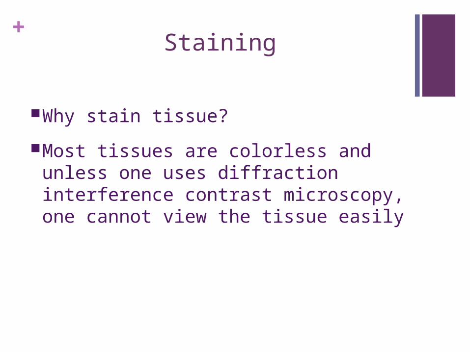
+Staining
Why stain tissue?
Most tissues are colorless and unless one uses diffraction interference contrast microscopy, one cannot view the tissue easily
+Staining
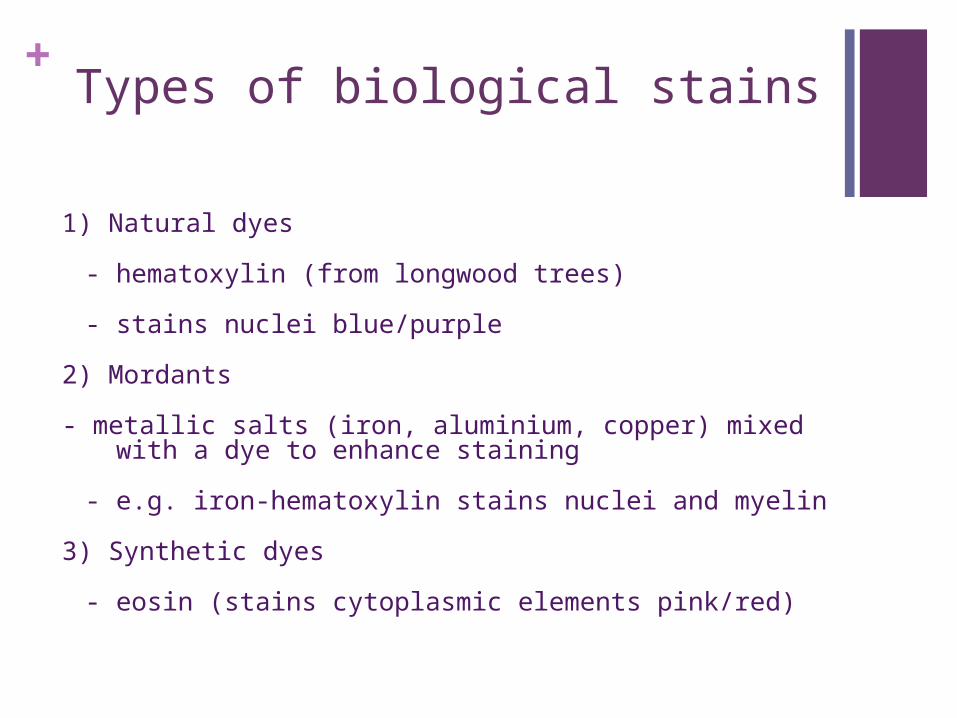
+Types of biological stains
1) Natural dyes
- hematoxylin (from longwood trees)
- stains nuclei blue/purple
2) Mordants
- metallic salts (iron, aluminium, copper) mixed with a dye to enhance staining
- e.g. iron-hematoxylin stains nuclei and myelin
3) Synthetic dyes
- eosin (stains cytoplasmic elements pink/red)
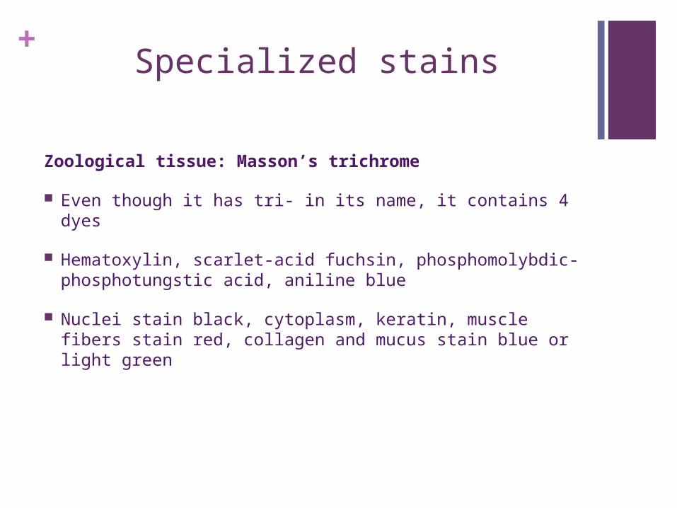
+Specialized stains
Zoological tissue: Masson’s trichrome
Even though it has tri- in its name, it contains 4 dyes
Hematoxylin, scarlet-acid fuchsin, phosphomolybdic-phosphotungstic acid, aniline blue
Nuclei stain black, cytoplasm, keratin, muscle fibers stain red, collagen and mucus stain blue or light green
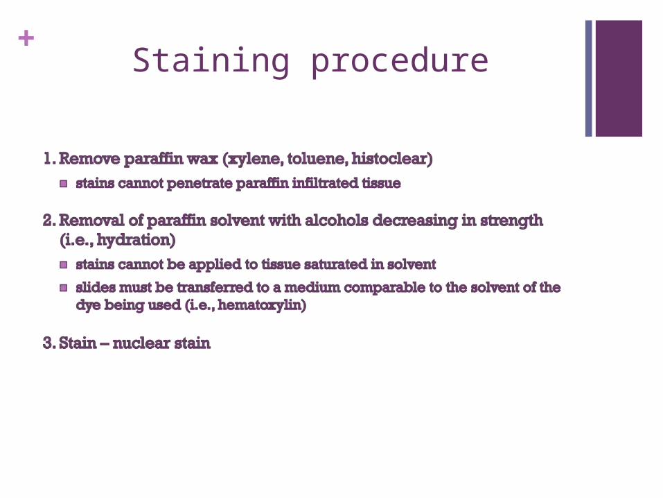
+Staining procedure
+Staining procedure
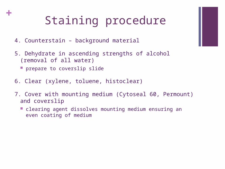
4. Counterstain – background material
5. Dehydrate in ascending strengths of alcohol (removal of all water) prepare to coverslip slide
6. Clear (xylene, toluene, histoclear)
7. Cover with mounting medium (Cytoseal 60, Permount) and coverslip clearing agent dissolves mounting medium ensuring an even
coating of medium
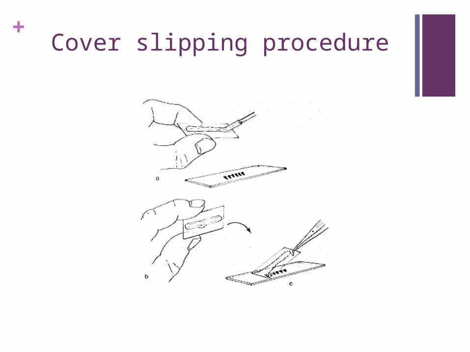
+Cover slipping procedure