Embed Size (px)
Citation preview

Brief Research StatementNicholas Cooley
University of Missouri, Department of [email protected] — 1 (614) 551 0167
Abstract
Current methods for monitoring blood glucoselevels in patients suffering from diabetes, thoughaccurate still require degrees of invasiveness thatimpinge upon monitoring fidelity (Figure 1). Thiscreates the potential for patients to fail to ac-curately monitor and control blood glucose lev-els. These failures lead to a significant portion ofhealthcare costs, decreases in quality of life, andmorbidities associated with diabetes. To addressthis, minimally invasive methods of monitoringblood glucose have been pursued. Building a sys-tem which can perform this type of glucose sens-ing is complex, and has largely been approachedthrough the use of fluorescent sensors which selec-tively bind to glucose, coupled with optical sens-ing equipment. This type of system has significantchallenges associated with it: the fluorescent sen-sor in use must selectively bind glucose at phys-iologically relevant concentrations, excitation andemission wavelengths must be farther red on theelectromagnetic spectrum than the absorbance ofhemoglobin - placing them firmly in the near in-frared(NIR), the sensor must provide stable and ro-bust signal output in vivo, and the sensor must be
persistent enough in vivo to present significant de-creases in overall invasiveness of monitoring.
Figure 1: Current methods of monitoring blood glucoseinvolve the ’finger prick’ method [1]
A system for monitoring blood glucose levelsin vivo, involving hiding a NIR sensor inside ofred blood cells, to prevent clearance, and allowfor long term monitoring was proposed, and someof the work involved with this project is describedherein.

IntroductionDiabetes causes decreases in quality of life, mor-bidity rates increase in patients who suffer from it,and it represents a large cost burden on health caresystems [2].
Significant literature exists on fluorescent sens-ing of glucose [3, 4]. The chemical interactions andsupramolecular architectures necessary to success-fully selectively bind glucose over other biologi-cally relevant diols are relatively well understood,however utilizing this body of knowledge to designa fluorescent sensor capable of working in vivo hasproven difficult. Though some glucose selectivefluorescent sensors exist, transitioning the motifsused previously to NIR scaffolds, or to completelywater soluble fluorophores is a synthetic chemistrychallenge. To successfully perform blood glucosedetection in vivo a fluorescent sensor must: be fullywater soluble, provide concentration independentsignal output, be stable to conditions in vivo, andhave excitation and emission wavelengths beyondthe absorbance of hemoglobin.
Additionally, the goal of providing minimallyinvasive detection means that the fluorescent sen-sor must resist clearance in blood serum, and per-sist for long periods of time. To achieve this goal,it was proposed that a fluorescent sensor couldbe hidden inside erythrocytes to maintain pres-ence in circulating blood [5, 6, 7, 8]. Erythro-cytes can be loaded with exogenous chemicals andproteins through a process involving manipulatingosmotic pressure. The process is colloquially re-ferred to as ’ghosting’ due to the loss of somehemoglobin, paling the bright red appearance ofhealthy erythrocytes[9]. Loaded erythrocytes canbe reintroduced into the individual from which theywere originally obtained, and will continue circu-lating for the lifespan of healthy erythrocyte. Uti-lizing erythrocyte loading, in conjunction with anappropriate NIR fluorescent sensor, and an opticalsystem capable of exciting the sensor and receiv-ing a signal output in vivo, it is hypothesized thatsuccessful long term monitoring of blood glucosecan be achieved (Figure 2).
measure glucose levels through an enzymatic reaction between glucose oxidase on the electrode and glucose in the interstitial fluid [14,17,18]. However, these systems require frequent calibrations with the standard finger stick test to account for variations in the reaction and diffusion kinetics [19]. Also, changes in the glucose levels of the interstitial fluid temporally lag those of the blood [14,17,18,20]. Generally, implanted sensors are viewed by the body as foreign objects, eliciting an immune response that ultimately leads to fibrous encapsulation and precludes proper, long-term functioning [21,22]. Although efforts to reduce in vivo biofouling of the continuous glucose monitoring sensors through application of specialized membranes have led to observed stability over the lifetime of the system [16,18], the implanted electrode still has an operational lifetime of only 3-7 days [14,16–18].
As an alternative, we propose that the sensing chemistry may be entrapped within red blood cells (RBCs) through the hypotonic dilution method. This method takes advantage of the RBC membrane’s ability to reversibly swell in response to the osmolarity of the extracellular environment [23–26]. In this procedure, the RBCs are placed in a low osmolarity solution (i.e., lysis solution) at 0°C, which causes the membranes to swell and develop pores that are estimated to range from 10 to 500 nm [23,24,27,28]. While the pores are open, hemoglobin (Hb) and other molecules within the cell and fluorescent probes as well as salts in the lysis solution equilibrate. Once physiological osmolarity and temperature are restored, the pores reseal, entrapping the fluorescent probe. These carriers exhibit biodegradability and, as autologous cells, biocompatibility, thereby avoiding the deleterious effects of the immune system response [23,25,27,29–31]. Furthermore, resealed ghosts that have been returned to the body are able to circulate with a lifetime similar to normal RBCs [23,25,26,32,33]. Thus the sensors would have a lifetime of up to 120 days, depending upon the age of the RBCs at removal.
Lyseand Load
Reseal
Replace
Remove
HemoglobinFITC
Lyseand Load
Reseal
Replace
Remove
Lyseand Load
Reseal
Replace
Remove
HemoglobinFITCHemoglobinFITCHemoglobinFITC
Replace and Measure
90
mg/dl
Fig. 1. Cartoon illustrating the RBC sensing platform concept. A small portion of blood is removed from the patient; the blood is washed, loaded with the analyte-sensitive fluorescent dye, resealed, and then transfused back into the patient. An excitation light source is then directed at the wrist; the emission from the resealed red cell ghosts is collected and converted into an analyte concentration. Clip art used with permission from Microsoft.
Efforts to entrap chemicals within RBCs began in the early 1950s with the successful encapsulation of ATP (as reviewed in [23,34]) and then, in 1959, various molecular weight dextrans [35]. In 1973, RBCs were first used as delivery vehicles for therapeutic agents by two independent groups [36,37]. Since these early studies, the advantages of RBCs (e.g., biocompatibility, biodegradability, long life time) have continued to drive the development of RBC carriers for delivery of biopharmaceuticals. Several excellent reviews on drug delivery using RBCs as biocompatible carriers are available [29,30,38]. In general, the means of
#146861 - $15.00 USD Received 2 May 2011; revised 17 Jun 2011; accepted 17 Jun 2011; published 22 Jun 2011(C) 2011 OSA 1 July 2011 / Vol. 2, No. 7 / BIOMEDICAL OPTICS EXPRESS 2015
Figure 2: Loading Erythrocytes with exogenous fluorophores [10]
1

ResultsBecause working with NIR fluorophores holds in-trinsic challenges in relation to chemical reactiv-ity and stability, this work began with the goal ofbuilding a simple sugar sensor which could showbinding in aqueous conditions in cuvette experi-ments. A relatively unexplored cyanine platformsimilar IR-820 was chosen as the basis for the pro-posed sensor. This decision was made based onthe necessity of working in fully aqueous systems,and the presence of four sulfonate groups on thecyanine starting material. The charged sulfonateshelp to provide water solubility to an otherwiserigid and hydrophobic compound, and provide theadded benefit of hindering cell permeability. If acompound containing multiple charged groups issuccessfully sealed inside a carrier erythrocyte, itis unlikely to passively transition back across themembrane into serum.
N+N
NaO3S SO3Na
Cl
N+N
NaO3S SO3Na
N
DMF70oCPd(PPh3)45 Hr51%
NH
HN
HN
SO3-NaO3SSO3
-NaO3S
N+N
NaO3S SO3Na
N
N
SO3-NaO3S
BOH
OH
H2O/MeOH/ACN1:3:385oC22 Hr55%
Br
BHO OH
12
3
Figure 3: Synthesis of NIR sugar sensor
The commercially available cyanine 1, provedto be resistant to modification through normaladdition-elimination chemistry common in manycyanine modifications, however it was amenable
to palladium coupling, providing 2 in reasonableyield. Alkylation of the secondary amine, thoughrequiring conditions generally unfavorable to SN2chemistry, still proceeded in reasonable yield toprovide 3 as a simple sugar sensor (Figure 3).
Sensing is achieved through a PET mecha-nism. Simple carbohydrates and diols bind toboronic acids through a hydroxyl exchange mech-anism to form a boronate ester. The formation ofthe boronate ester then effectively lowers the pKa
of the nearby amine, increasing the protonation insolution upon binding. With the amine unproto-nated, the lone pair electrons act as PET quenchersputting the sensor in an ”off” state, upon bindingand protonation of the amine, the lone pair now be-comes tied up in a bond, and can no longer quench.This puts the sensor in an ”on” state, increasing flu-orescence intensity (Figure 4).
N+N
NaO3S SO3Na
SO3-NaO3S
N
NH+
B-
OOHHO O
O OH
N+N
NaO3S SO3Na
SO3-NaO3S
N
N
BOH
OH
OOHHOHO
HO OH
HO
Figure 4: Scheme illustrating binding of Sensor 3 withFructose
Sensor 3 provides a starting point for thisproject. It presents excitation and emission prop-erties deep in the NIR and was fully water solubleup to Molar concentrations, however providing abinding constant of 271mM for fructose (Figure5) put a binding strength significantly weaker thanwhat has been reported for simple mono-boronicacid – carbohydrate binding systems[11].
2
0"
0.5"
1"
1.5"
2"
815" 865" 915" 965"
Intensity
((A.U)(
Wavelength((nm)(
npc(4(178(Fructose(Binding(Ex(780nm(
0"
0.5"
1"
1.5"
2"
2.5"
3"
3.5"
4"
4.5"
0" 0.2" 0.4" 0.6" 0.8" 1"
Emission,(Excita-on(@(780nm(10μm((
Figure 5: Fluorescence Emission and Binding Isothermof Sensor 3 upon addition of Fructose. Excitation at780nm, Emission Intensity for inset at 825nm.
Fructose was used as a benchmark because ofall the simple carbohydrates, phenylboronic acidsbind fructose with the strongest affinity. Also,when titrated with glucose to determine a bindingconstant, the concentrations of glucose used wereso high that viscosity effects on the fluorescenceintensity rendered the data unsuitable for determin-ing a binding constant.
ConclusionsThe end goal of this project is to build a fluores-cent sensor for glucose monitoring in vivo, and thispresents a starting point for that process. It meetsall of the important criteria necessary to achievethat goal except for having an appropriate bind-ing constant. However this compound was not ex-pected to provide an appropriate binding constant,so failure to meet that goal was expected. Movingforward this project must address that hurdle di-rectly. Over-engineered examples of synthetic re-ceptors for glucose exist[12]. Notably, the struc-ture and mechanisms of fluorescence enhancementof our designed sensor differ from those published
and interrogated previously, this is due to the ne-cessity of both working in fully aqueous systems,and working in the NIR[13]. Utilizing the lessonslearned from these receptors, it is the hope thatmoving forward with this project, glucose selectivebinding can be achieved.
Future Work
Some of this work has already been published[14],while some is in stages of preparation. However asignificant amount remains to be performed. Thechallenge of building a sensor with an appropriatebinding constant for glucose in vivo is not to be un-derestimated. Building the sensor described here-in though ultimately synthetically simple, was pre-ceded by attempts at other proposed sensor struc-tures that were ultimately unsuccessful. Unsurpris-ingly, the current attempts at constructing a glucoseselective sensor, or even a sensor with strongerbinding constants are challenging.
Undiscussed so far in this research statementis the work in this project on erythrocyte loading.Partially this is due to an attempt at keeping thisstatement brief, and partially this is due to the factthat the compounds designed herein have excita-tion and emission wavelengths beyond the oper-ational range of the fluorescence microscopes wehave had access to throughout this project, makingclear data collection for determination of loadingconcentrations and uniformity difficult. The struc-tural differences between the synthesized com-pound, and commercially available or syntheticallyaccessible analogs make comparisons with differ-ent fluorophores unclear in their relevance.
Optimizing erythrocyte loading is a significantchallenge, and due to the nature of the experimentsdesigned, this work has so far been performed withrat, sheep, and horse erythrocytes, none of which
3
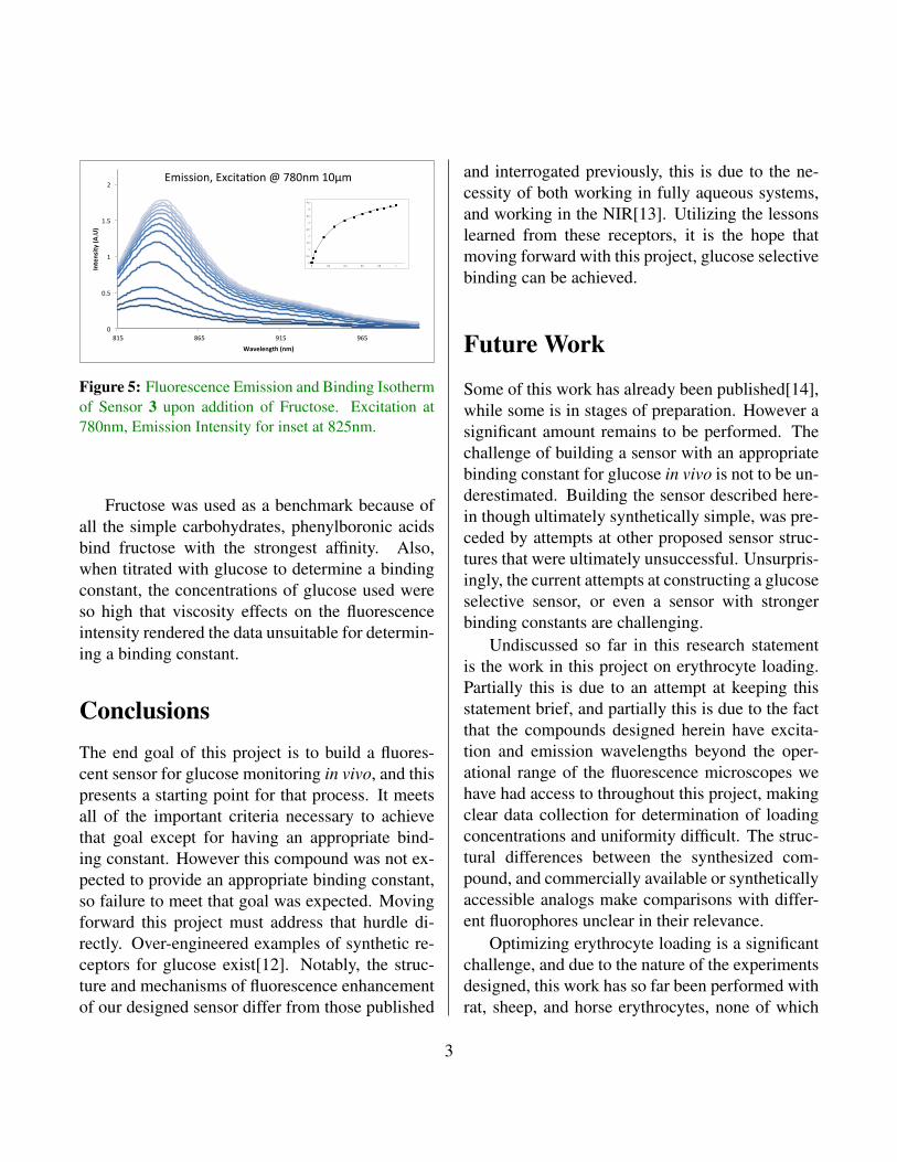
transport glucose across their membranes. Whilebeing unable to probe loading efficiency optically,we can observe populations of erythrocytes postloading via Scanning Electron Microscopy (SEM),and the results indicate that the procedure does notprovide a uniformly healthy population (Figure 6).
Figure 6: SEM images of rat erythrocytes followingloading procedure. Some erythrocytes are deformed,while others appear healthy, also present are unidenti-fied structures which may be aggregates of cellular de-bris from the loading process
Moving forward, part of this project will re-volve around optimization of these procedures toensure that erythrocytes loaded with dye will re-main healthy and robust. The diversity of erythro-cyte morphology between species also presents an-other challenge for this project[15, 16]. Proof ofconcept studies so far have involved rat models,where glucose sensing through red cells is not fea-sible, consequently erythrocyte loading has beenoptimized towards rat erythrocytes. Transitioningthe project further to a model system which canprovide glucose detection feasibility will require
re-optimization of erythrocyte loading to a differ-ent cell morphology.
This step will be required for every model nec-essary to prove efficacy and safety as the projectworks towards an end goal of clinical trials in hu-mans. These are steep challenges to address, andthe success of the project moving forward will re-quire clear and achievable solutions to them. Someof those solutions are already available, as erythro-cyte loading has already been performed in humansclinically, and the literature surrounding synthesisand modification of NIR fluorophores continues toexpand from where it stood at the outset of thisproject.
AcknowledgementsThe work discussed and presented herein was ini-tiated by Dr. Mark Milanick, and expanded to in-clude Dr. Kennith Meissner and his research groupat Texas A & M, and Dr. Timothy Glass and thegroup in which I have been working towards myPhD. Dr.’s Milanick, Meissner, and Glass have allbeen intimately involved in the planning, execu-tion, and funding of this project to it’s current state.Project Dr. Meissner’s group recently moved to theUniversity of Swansea. Dr. Sarah Ritter and San-dra C. Bustamante Lopez working under the su-pervision of Dr. Meissner have been responsiblefor designing optical detection equipment vital tothe success of this project, but undiscussed herein,and in optimizing erythrocyte loading proceduresbriefly discussed herein. Dr. Xiaole Shao beganthe chemical synthesis portion of this project underthe supervision of Dr. Glass, which I have contin-ued. Hamidreza Sepasazingbadi is currently work-ing with me, under the supervision of Dr. Glassto continue the project following my graduation inDecember.
4

Bibliography
[1] Blausen.com. Glucose meter and usage.
[2] Centers for Disease Control and Prevention.
[3] Jon Stefan Hansen, Jørn Bolstad Christensen,Johannes Fabritius Petersen, Thomas Hoeg-Jensen, and Jens Christian Norrild. Aryl-boronic acids: A diabetic eye on glucosesensing. Sensors and Actuators B: Chemical,161(1):45–79, 2012.
[4] Xiaolong Sun and Tony D. James. Glu-cose Sensing in Supramolecular Chem-istry. Chemical Reviews, 115(15):8001–8037, 2015.
[5] Carmen Gutierrez Millan, Clara IsabelColino Gandarillas, Marıa Luisa SayaleroMarinero, and Jose M Lanao. Cell-baseddrug-delivery platforms. Therapeutic Deliv-ery, 3(1):25–41, 2012.
[6] G. Schwoch and H. Passow. Prepara-tion and properties of human erythrocyteghosts. Molecular and Cellular Biochem-istry, 2(2):197–218, dec 1973.
[7] Carmen Gutierrez Millan, Aranzazu ZarzueloCastaneda, Ma. Luisa Sayalero Marinero, andJose M. Lanao. Factors associated withthe performance of carrier erythrocytes ob-tained by hypotonic dialysis. Blood Cells,
Molecules, and Diseases, 33(2):132–140,2004.
[8] Elsa Briones, Clara Isabel Colino, andJose M. Lanao. Delivery systems to in-crease the selectivity of antibiotics in phago-cytic cells. Journal of Controlled Release,125(3):210–227, 2008.
[9] Sara Biagiotti, Maria Filomena Paoletti,Alessandra Fraternale, Luigia Rossi, andMauro Magnani. Drug delivery by red bloodcells. IUBMB Life, 63(8):621–631, 2011.
[10] Sarah C Ritter, Mark A Milanick, andKenith E Meissner. Encapsulation of FITC tomonitor extracellular pH: a step towards thedevelopment of red blood cells as circulatingblood analyte biosensors. Biomedical opticsexpress, 2(7):2012–21, jul 2011.
[11] Jun Yan, Greg Springsteen, Susan Deeter,and Binghe Wang. The relationship amongpKa, pH, and binding constants in the in-teractions between boronic acids and diolsitis not as simple as it appears. Tetrahedron,60(49):11205–11209, 2004.
[12] Wei Yang, Huan He, and Dale G. Drueck-hammer. Computer-guided design in molec-ular recognition: Design and synthesis of aglucopyranose receptor. Angewandte Chemie- International Edition, 40(9):1714–1718,may 2001.
[13] Susumu Arimori, Michael L. Bell, , ChanS. Oh, and Tony D. *. A Modular Fluo-rescence Intramolecular Energy Transfer Sac-charide Sensor. Organic Letters, 4(24):4249–4251, 2002.
[14] Sarah C. Ritter, Xiaole Shao, Nicholas Coo-ley, Mark A. Milanick, Timothy E. Glass, and
5

Kenith E. Meissner. Blood analyte sensingusing fluorescent dye-loaded red blood cells.SPIE Proceedings, 8951:895109, feb 2014.
[15] K Ohta, F Gotoh, M Tomita, N Tanahashi,M Kobari, T Shinohara, Y Terayama, B Mi-hara, and H Takeda. Animal species differ-ences in erythrocyte aggregability. The Amer-ican journal of physiology, 262(4):H1009–
H1012, apr 1992.
[16] U. Windberger, A. Bartholovitsch, R. Plasen-zotti, K. J. Korak, and G. Heinze. Wholeblood viscosity, plasma viscosity and erythro-cyte aggregation in nine mammalian species:reference values and comparison of data. Ex-perimental physiology, 88(3):431–440, may2003.
6