Embed Size (px)
Citation preview
BRAINRESEARCHPROTOCOLS
ELSEVIER Brain Research Protocols ID (2002) 69-74www.elsevier.com/locate/brainresprot
Research report
N eonatal prefrontal cortex les ion using CO 2 laser techniqueLaura Sánchez-Huertaa, Griselda Ayalaa, Javier Marroquína, Rafael Calderóna,
Adriana B. Silva-Gómeza, Konstantin S. Khotiaintsevb, Larisa DegtiarevaC,Sergei N. Khotiaintsevd, Gonzalo Floresa,*
"Instituto de Fisiología, Universidad Autónoma de Puebla, 14 Sur 6301, Puebla CP 72570, MexicbFacultad de Ciencias de la Computación, Universidad Autónoma de Puebla, Puebla, Mexico
cResearch Institute for Human Ecology, Kiev, Ukraine"Facultad de Ingeniería, Universidad Nacional Autónoma de México, Mexico
Accepted 23 August 2002
Abstract
Prefrontal cortex (PFC) is a large afea of the brain and its neonatal lesion with ibotenic or kainic acid is used to study fue earlyabnorrnalities in neurodevelopment that lead to behavioral changes linked to schizophrenia. However, these exitotoxic drugs produce alarge and asymmetric damage in the PFC. We produced the bilaterallesions of fue dorsal part of the PFC of neonatal Sprague-Dawleyrats (postnatal day 7, P7) at fue anteroposterior +2.5 mm and mediolateral :tO.4 coordinates by fue new laser technique that employ fueconfined radiation of fue CO2 laser in fue pulsed mode. The laser was used because its coherent tadiation can be focused in a very smallspot and as small as of several tens of rnicrometers in diameters. The CO2 laser radiation is strongly absorbed by water that is present inany soft tissue. Thereafter, fue configuration of the heated zone and, consequently, that of fue lesion does not depend on fue morphologicalnon-homogeneity of particular structures. We obtained fue symmetric, conical in shape and small-size bilateral lesions of fue PFC. Thesize of the lesion depended on fue beam spot-size and could be as small as several dozens of micrometers in diameter. Our data suggeststhat the laser technique will be used for fue anatomical-functional studies of fue PFC in fue brain.@ 2002 EIsevier Science B.V. AII rights reserved.
Theme: Disorders of fue nervous system
Topic: Developmental disorders
Keywords: Prefrontal cortex; Schizophrenia; Animal model; Laser; Neurodevelopment
animal. This includes the preparation of fue surgical fieldby assuring fue afea is sufficiently sterile to rnitigate theprobability of sepsis, induction of unconsciousness withanesthesia by hypotherrnia, making a 0.5 cm incision,recording of initial stereotaxic coordinates, bilateral appli-cation of confined laser radiation during several seconds,closing fue skin with veterinary tissue adhesive at conclu-sion of procedure, and postoperative monitoring of animal sfor proper warmth.
1. Type of research
1. Neurodevelopmental studies in rats.2. Laser-induced lesions to frontal cortex.3. Pyrarnidal neuron death.
2. Time required
2.1. Surgical procedure
2.2. Tissue preparationThe surgical procedure of bilateral prefrontal cortex
(PFC) laser application takes approximately 30 min per At postnatal day 64 (P64), rats were anesthetized withsodium pentobarbital (75 mg/kg, i.p.) and brains wererapidly removed, frozen in isopentane maintained at-40 °C, and stored at -80 °C until use. Frozen brains
*Corresponding author. Tel.: +522-244-1657; fax: +522-233-4511
E-mail address: [email protected] (G. Flores).
1385-299X/02/$ - see front matter @ 2002 EIsevier Science B.V. AII rights reserved.
PII: S1385-299X(02)00183-6
70 L. Sánchez-Huerta et al. I Brain Research Protocols 10 (2002) 69-74
were 'sectioned at 50 IJ.m thickness on the coronal planusing a Leica cryostat. Sections at fue level oí frontalcortex were collected on cleaned, gelatin-coated micro-scope slides (4 sections/slide) and then stored at -80 °Cuntil the dar of stain. Sections were stained with 0.5%cresyl violet and examined under microscope where le-sions were visualized. Perfusion and brain removal took 20min per animal, cryosectioning took 30 min per animal andstaining took 3 h.
3.4. Policy issues
Thc proccdurcs described herein have been conducted incompliance with the policies of the Society for Neuro-science for conducting neuroscicnce research and with theguidelines qf the Laws and Codes of Mexico in TheSeventh Title of the Regulatio~s of th~ General Law ofHealth Regarding Health Research. :
4. Detailed procedures3. Materials
4.1. Laser applied3.1. Animals
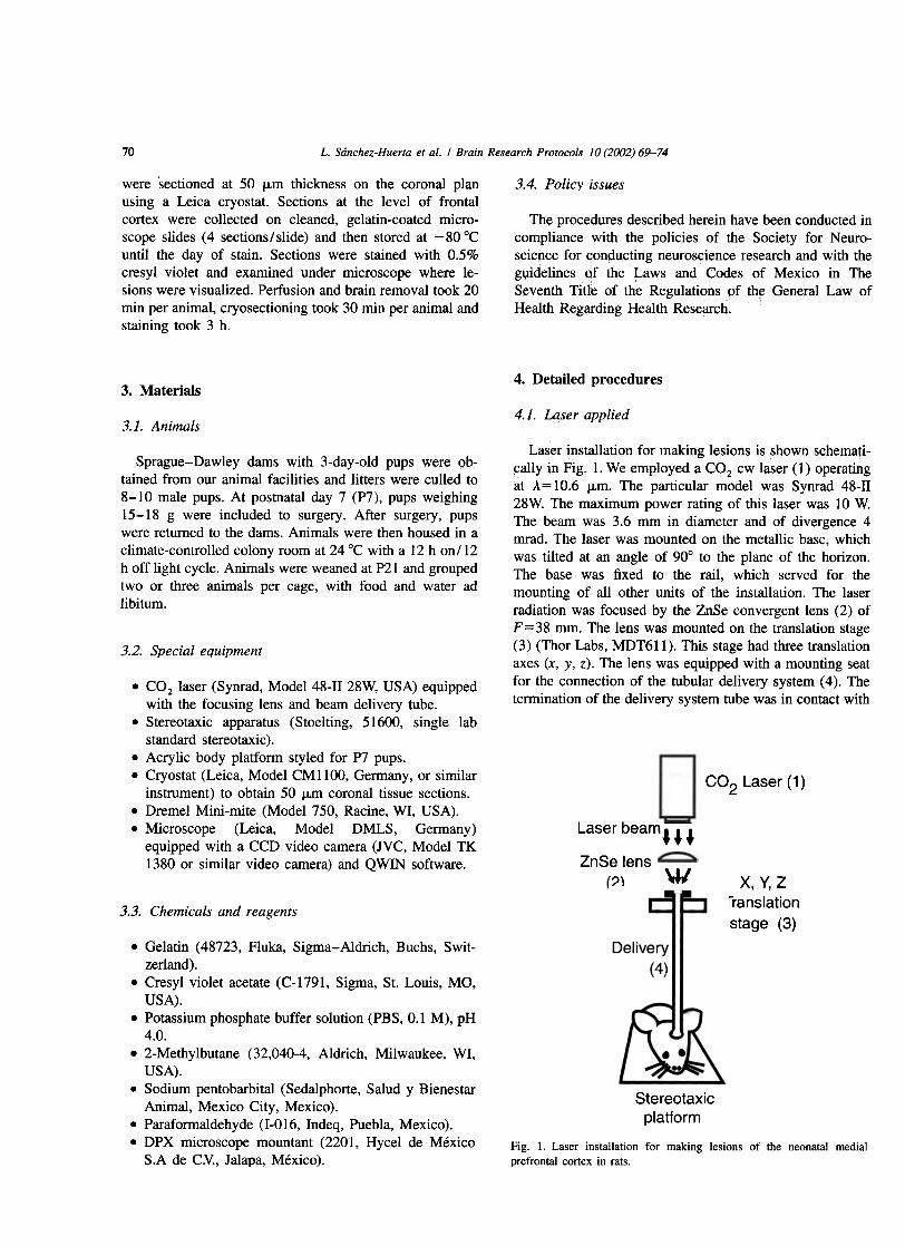
Laser installation for making lesions is shown schemati-~ally in Fig. 1. We employed a CO2 cw 1aser (1) operatingat Á= 10.6 ~m. The particular model was Synrad 48-1128W. The maximum power rating of this laser was 10 W.The beam was 3.6 mm in diameter and of divergence 4rnrad. The laser was mounted on the metallic base, whichwas tilted at an angle of 90° to the plane of fue horizon.The base was fixed to t4e rail, which served for fuemounting of all other units of fue installation. The laserradiation was focused by fue ZnSe convergent lens (2) OfF=38 mm. The lens was mounted on the translation stage(3) (Thor Labs, MDT61 1). This stage had three translationaxes (x, y, z). The lens was equipped with a mounting seatfor the connection of the tubular delivery system (4). Thetermination of fue delivery system tube was in contact with
Sprague-Dawley darns with 3-day-old pups were ob~tained from OuT animal facilities and litters were culled to8-10 male pups. At postnatal day 7 (P7), pups weighing15-18 g were included to surgery. After surgery, pupswere retumed to the dams. Animals were then housed in aclimate-controlled colony room at 24 °C with a 12 h on/12h off light cycle. Animals were weaned at P21 and groupedtwo or three animals per cage, with food and water adlibitum.
3.2. Special equipment
CO2 Laser (1)
Laser beam m
. COz laser (Synrad, Model 48-11 28W, USA) equippedwith fue focusing lens and beam delivery tube.
. Stereotaxic apparatus (Stoelting, 51600, single labstandard stereotaxic).
. Acrylic body platform styled for P7 pups.
. Cryostat (Leica, Model CMII00, Germany, or similarinstrument) to obtain 50 IJ.m corona! tissue sections.
. Dremel Mini-mite (Model 750, Racine, WI, USA).
. Microscope (Leica, Model DMLS, Germany)equipped with a CCD video camera (JVC, Model TK1380 or similar video camera) and QWIN software. ZnSe lens
(2) W X,Y,ZTranslation
stage (3)3.3. Chemicals and reagents
tube
Stereotaxic
platform
. Gelatin (48723, Fluka, Sigma-Aldrich, Buchs, Swit-
zerland).. Cresyl violet acetate (C-1791, Sigma, Sto Louis, MO,
USA).. Potassium phosphate buffer solution (PBS, 0.1 M), pH
4.0.. 2-Methylbutane (32,040-4, Aldrich, Milwaukee, WI,
USA).. Sodium pentobarbital (Sedalphorte, Salud y Bienestar
Animal, Mexico City, Mexico).. Paraformaldehyde (1-016, Indeq, Puebla, Mexico).. DPX microscope mountant (2201, Hycel de México
S.A de C:V:, Jalapa, México).Fig. l. Laser installation for making lesions of fue neonatal media!prefrontal cortex in rats.
L. Sánchez-Huerta et al. / Brain Research Protocols 10 (2002) 69-74 71
4 °C overnight, and then stored at -80 °C until fue dar oífue staining procedure.
(i) Slides were dried by cold air dry using a drier.(ii) Follow Nissl-type protocol for staining and dehydra-
tion as follows: load slides a slide tray, and then washthem for 10 min in PBS. After gently shaking tray ofslides to remove fue excess PBS, stain with a 15%diluted mixture of 0.5 mg cresyl violet acetate in 100ml distilled water (DDH20) by submersion for 1-2mino After desired depth of stain is achieved, dip inDDH20, followed by 5-min baths in each of 70, 90and 100% ethanol to dehydrate, followed by two5-min baths in xylene to defat. After xylene baths,remove slides individually from xylene with tweezersand wipe excess from bottom edge of slide, then coverslip with DPX mountant or equivalent. The dehydra-tion, defatting and coverslipping steps should beconducted under a fume hood while wearing latexgloves and eye protection, as chemicals involved aresomewhat toxic.
(iii)Using a Leica DMLS (or equivalent) microscope at40X equipped with a JVC CCD video camera (ModelTK 1380 or equivalent), fue slides of PFC regiDos offue sham and neonatal PFC laser-lesioned rats wereanalyzed with fue Leica Image software (ModelQWIN).
fue animal head at right angle to it and against the aperturemade in fue animal's skull.
The delivery system was a hollow circular metallic tube(a stainless steel needle of fue medical syringe). It was ofinner diameter 1.2 rom, outer diameter 1.8 rom, and 80 romlong. The tube mount was concentric with fue needle axisand fitted the mounting seat of the lens (3). The relativelylarge length of the tube facilitated fue rnixing of fue laserbeam. In addition, fue substantially rough inner surface offue tube contributed to fue diffuse reflection and, hence, tointense beam mixing. This perrnitted to deliver the con-fined laser radiation of quasi-uniform density distributionacross fue beam to the object. The tube was not subjectedto any special processing, e.g., polishing. However, eachtime before use, the tube's interior was cleaned mechani-cally and then treated with alcohol and phosphoric acid insequence. The stainless steel tube featured relatively highattenuation per unit length at 10.6 ~m wavelength ascompared to some specially designed infrared waveguides[12,9,13,5,15]. However, because of fue relatively shortlength of delivery system and excess of fue optical poweravailable from fue laser, fue tube attenuation was of almostno importance in fue present installation. These considera-tions and fue low syringe cost deterrnined our choice infavor of this delivery system.
The tilt of fue laser and fue delivery tube at an angle of-900 with respect to fue plane of fue horizon elirninatedfue possibility of the tube clogging by some biologicalsubstance. In our experience, small quantities of blood offue wound of the experimental animal mar penetrate intofue tube aperture during fue exposition of animal to laserradiation. The tilt of the delivery tube presented animportant practical improvement with respect to fue previ-ous design [11] that featured the horizontal delivery tube.
Finally, each PFC received fue laser radiation in fueforro of a series of pulses. Each series comprised threepulses of 1 s duration each; fue interval between fue pulseswas 5 s.
5. Results
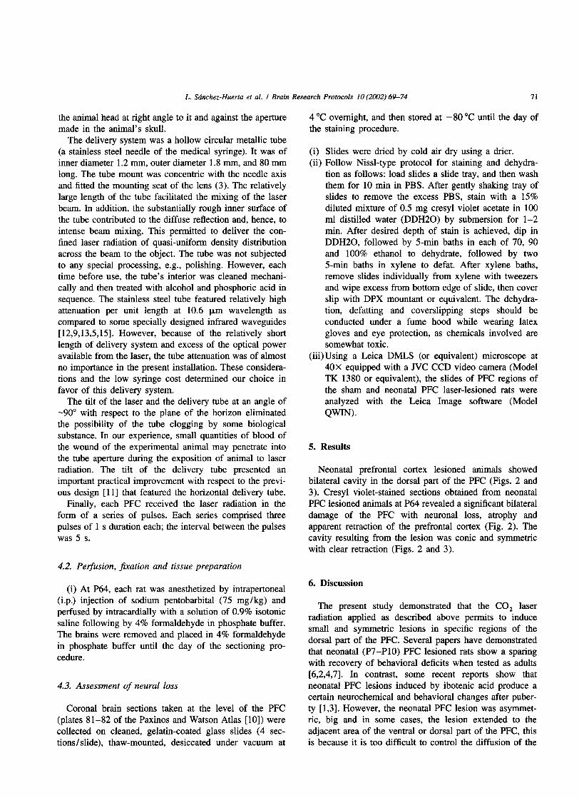
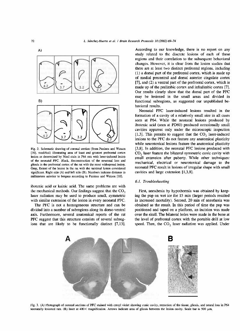
Neonatal prefrontal cortex lesioned animals showedbilateral cavity in fue dorsal part of fue PFC (Figs. 2 and3). Cresyl violet-stained sections obtained from neonatalPFC lesioned animal s at P64 revealed a significant bilateraldamage of fue PFC with neuronal loss, atrophy andapparent retraction of fue prefrontal cortex (Fig. 2). Thecavity resulting from fue lesion was conic and syrnmetricwith clear retraction (Figs. 2 and 3).
4.2. Peifusion, fixation and tissue preparation
6. Discussion(i) At P64, each rat was anesthetized by intrapertoneal
(i.p.) injection of sodium pentobarbital (75 mg/kg) andperfused by intracardially with a solution of 0.9% isotonicsaline following by 4% formaldehyde in phosphate buffer.The brains were removed and placed in 4% formaldehydein phosphate buffer until fue day of the sectioning pro-cedure.
The present study demonstrated that fue CO2 laserradiation applied as described above permits to inducesmall and symmetric lesions in specific regions of fuedorsal part of fue PFC. Several papers have demonstratedthat neonatal (P7-P1O) PFC lesioned rats show a sparingwith recovery of behavioral deficits when tested as adults[6,2,4,7]. In contrast, some recent reports show thatneonatal PFC lesions induced by ibotenic acid produce acertain neurochemical and behavioral changes after puber-ty [1,3]. However, fue neonatal PFC lesion was asymmet-ric, big and in some cases, fue lesion extended to fueadjacent afea of fue ventral or dorsal part of fue PFC, thisis because it is too difficult to control the diffusion of fue
4.3. Assessment of neural loss
Corona! brain sections taken at fue level of fue PFC(plates 81-82 of fue Paxinos and Watson Atlas [10]) werecollected on cleaned, gelatin-coated glass slides (4 sec-tions I slide), thaw-mounted, desiccated under vacuum at
L. Sánchez-Huerta et al. I Brain Research Protocols 10 (2002) 69-7472
A)
B)
According to our knowledge, there is no report on anystudy related to fue disc!ete lesions of each of fueseregions and their correlation to fue subsequent behavioralchanges. However, it is clear from the lesion studies thatthere are at least two distinct prefrontal regions, including(1) a dorsal part of the prefrontal cortex, which is made upof medial precentral and dorsal anterior cingulate cortex[7], and (2) a ventral part of the prefrontal cortex, which ismade up of the prelimbic cortex and infralimbic cortex [7].Our results clearly show that the dorsal part of fue PFCmar be lesioned in fue small afeas and divided infunctional subregions, as suggested our unpublished be-havioral results.
Neonatal PFC laser-induced lesions resulted in fueformation of a cavity of a relatively small size in all casesseen at P64. While the neonatal lesions produced byibotenic acid (seen at PD60) produced occasionally smallcavities apparent only under the microscopic inspection[1,3]. This permits to suggest that fue CO2 laser-inducedlesions to the PFC do not feature any anatomical plasticitywhile neurotoxicallesions feature fue anatomical plasticity[3,8]. In addition, fue neonatal PFC lesions produced withCO2 laser feature the bilateral syrnmetric conic cavity withsmall extension after puberty. While other techniques:mechanical, electrical or neurotoxical damage to fueneonatal PFC result in lesions of irregular shape with smallcavities and large extension [1,3,8].
~", -
3.7
Fig. 2. Schematic drawing of coronaJ section (from Paxinos and Watson[10], modified) illustrating area of least and greatest prefrontaJ cortexlesion as determined by Nissl stain in P64 rats with laser-i¡lduced lesio\1of fue neonataJ PFC. Black, Reconstruction of fue neuronaJ loss andgliosis in fue prefrontaJ cortex of fue rat with !h~ most widesP!"ead lesioll.Gray, Extent of fue lesion in fue rat with the mini~al lesion consideredsigmficant. Right side (A) and left side (B). Numbers indicate distance inmillimeters anterior to bregma according to Paxinos and Watson [10].
6.1. Troubleshootingibotenic acid or kainic acid. The same problems are withthe mechanical methods. Our findings suggest that the cazlaser radiation mar be used to produce small, symrnetricwith similar extension of fue lesion in every neonatal PFC.
The PFC is not a homogeneous structure and can bedivided into a number of subregions along its dorso-ventralaxis. Furthermore, several anatomical reports of fue ratPFC suggest that this structure consists of several subreg-ions that are likely to be functionally distinct [7,13].
First, anesthesia by hypothermia was obtained by keep-ing the pup on wet ice for 15 min (larger periods resu1tedin increased mort~lity). Second, 20 min of anesthesia wasobtained as the resu1t. In this period of time the pup waspositioned and taped on a p1atform, an incision was madeover the sku11. The bilateral ho1es were made in the boDe atthe 1eve1 of prefrontal cortex with the portab1e dri11 at 10wspeed. Then, the CO2 laser radiation was applied. Under
B
Fig. 3. (A) Photograph of coronal sections of PFC stained with cresyl violet showing conic cavity, retraction of fue tissue, gliosis, and neuralloss in P64neonatally lesioned rats. (B) Inset at 400X inagnification. Arrows indicate area of gliosis between the lesion cavity. Scale bar is 500 ¡Lm.
í2.7 I
L. Sánchez-Huerta et al. I Brain Research Protocols 10 (2002) 69-74
the appropriate preparation of instruments, etc., and aftersome practice, we could do all this procedurein les s than20 min. Following fue precautionary steps describedabove, and keeping fue pups warm, both during and aftersurgery, and monitoring their respiration after surgery, itwas possible to maximize fue pup survival rateo
(iv) Carefully make a l-mm incision in the pup's scalp toexpose fue skull. Locate bregma and note fue initialstereotactic coordinates.
(v) Use fue following coordinates: AP -2.5 mm and ML:!:O.4 [1,3].
(vi) Apply fue CO2 laser radiation.(vii) Mark pups for later identification, then place fue pups
on head pad. After recovery, retum pups to dam.(viii)Wean fue pups at P2l, and then separate into groups
of three per cage with free access to food and water.
6.2. Alternative and support protocols
6.2.1. Laser les ionThe exposition time of the PFC to fue COz laser
radiation is critical. As described in detail in Section 4.1,expositions longer than 10 s decreased fue animal survivalrate, this may be due to fue excess heating of fue brillo[11]. However, this problem was overcome by using aseries of pulses of laser radiation. Each series comprisedthree pulses of 1 s duration each; fue interval between fuepulses was 5 s. This technique increased the pup survivalrate closely to 100%.
7.2. Perfusion and stain
6.2.2. Excitotoxic lesionIbotenic and kainic acid are fue most common drugs
used to produce excitotoxic damage of fue cortex. Thevolume and concentration of fue excitotoxic drugs arecritical. More than 300 f1l per side into the neonatal brainrat, increase its mortality [3]. The lesion size depends onboth, concentration and diffusion of the drug. However,fue majar problem with this drugs is that fue lesion size inthe neonatal rats is not really small and symmetric with airregular shape. Other secondary problem is the mortalityof fue pups, in special with fue kainic acid.
(i) At P64, previously anesthesia induced by sodiumpentobarbital (100 mg/kg), transcardially perfusedwith a primary flush of 50 mI of phosphate-bufferedsaline (PBS, 0.1 M, pH 7.4) followed by 20 min of 4%paraformaldehyde-PBS via gravity drip.
(ii) Immediately brain was removed and stored at -80 °Cuntil fue sectioning day.
(iii) Frozen rat brains were sectioned at 50 ~m thicknesseson the coronal plane using a cryostat. Sections at fuelevel of fue PFC were collected on gelatin-coatedmicroscope slides and stored at - 80 °C until the day
of staining.(iv) Violet cresyl staining procedure (as described in detail
in Section 4.3) was used to assess fue neural loss.
8. Essentialliterature references
Original papers: [1,3,11].6.2.3. Classical aspirarían lesion
The aspiration pressure in fue neonatal cortical lesion iscritical. High pressure in fue aspiration cause big darnagein fue neonatal brain [2,14]. The blood volume in theneonatal rat is low, so small breeding is another problemwith this procedure. Furthermore, the lesion size is notconsistent, with lack of symmetry between sides withirregular shape in comparison with fue laser procedure[2,14]. However, the aspiration procedure is uncomplicatedand not expensive technique.
Acknowledgements
This study was supported in part by grants fromCONACyT (30675-M, 35001-A), UNAM (INl13799) andFundación Mexicana para la Salud AC. We are grateful toDr. Carlos Escamilla for bis help and suggestions related tokeeping of animals. We thank Juan J. RamÍrez and JesusSánchez for correcting fue manuscript. L.S.-H., R.C. andA.S.-G. acknowledge fue CONACYT for fue studentship.G.F. and S.N.K. acknowledge fue National ResearchSystem of Mexico for membership.7. Quick procedure
7.1. Laser lesionReferences
(i) Prepare surgical field so that fue area is sufficientlysterile so as to mitigate the probability of sepsis.
(ii) Securely attach the acrylic body platform for P7 pups.(iii) Anesthetize fue P7 pup by keeping it for 15 min on
wet ice, then position fue animal on fue body platformwith head presentation well-centered in a horizontalplane and snout positioned in a proper proximity ton()~ec()ne
[1] W.G. Brake, G. Flores, D. Francis, M.J. Meaney, L.K. Srivastava, A.Gratton, Enhanced nucleus accumbens dopamine and plasma cor-ticosterona stress responses in adult rats witb neonatal excitotoxiclesions to fue medial prefrontal cortex, Neuroscience 96 (2000)
687-695.[2] J.M. De Brabander, C.G. van Eden, J.P. de Bruin, Neuroanatomical
correlates of sparing of function after neonatal medial prefrontalcortex lesion in rats, Brain Res. 568 (1991) 24-34.
L. Sánchez-Huerta et al. / Brain Research Protocols 10 (2002) 69-7474
[10] G. Paxinos, C. Watson, The Rat Brain in Stereotactic Coordinates,2nd Edition, Academic Press, New York, 1986.
[11] L. Sanchez-Huerta, A. Hemandez, G. Ayala, J. Marroquin, A.B.Silva, K.S. Khotiaintsev, VA. Svirid, G. Flores, S.N. Khotiaintsev,Laser technique for anatomical-functional study of the medialprefrontal cortex of the brain, in: A.I. Melker (Ed.), Proceedings ofSPIE 3687, Nondestructive Testing and Computer Simulations inScience and Engineering, 1999, pp. 34-41.
[12] S. Shalem, A. German, A. Katzir, Optical properties of silver-halidecore/clad IR fibers, in: Proceedings of SPIE 2631, Medical Sensorsand Fiber Optic Sensors, 1995, pp. 216-225.
[13] C.G. van Eden, H.B.M. Uylings, Cytoarchitectonic development ofthe prefrontal cortex in the rat, J. Comp. Neurol. 241 (1985)
253-267.[14] C.G. van Eden, A. Rinkens, H.B.M. Uylings, Retrograde degenera-
tion of the thalamic neurons in the mediodorsal nucleus afterneonatal and adult aspiration lesions of the medial prefrontal cortexin the rato Implications for the mechanisms of functional recovery,Eur. J. Neurosci. 10 (1998) 1581-1589.
[15] H. Watanabe, H. Hiraga, Y. Abe, M. Miyagi, Fabrication ofdielectric-coated silver hollow nickel waveguide with multiple innerdielectric layers for Er: YAG laser transmission, in: N.I. Croitoru, R.Aviv, M. Frenz (Eds.), Proceedings of SPIE 2928, BiomedicalSystems and Technologies, 1996, pp. 19-27.
[3] G. Flores, G.K. Wood, J.J. Liang, R. Quirion, L.K. Srivastava,Enhanced amphetamine sensitivity and increased expression ofdopamine D2 receptors in postpubertal rats after neonatal excitotoxiclesions of fue medial prefrontal cortex, J. Neurosci. 16 (1996)7366-7375.
[4] H.J. Freeman, M.E. Stanton, Medial prefrontal cortex lesions andspatial delayed altemation in fue developing rat: recovery or sparing,Behav. Neurosci. 106 (1992) 924-932.
[5] S. Inberg, M. Oksman, N.I. Croitoru, Novel copper hollow wave-guides for IR laser radiation, in: N.I. Croitoru, R. Aviv, M. Frenz(Eds.), Proceedings of SPIE 2928, Biochemical Systems and Tech-nologies, 1996, pp. 28-38.
[6] B. Kolb, I.Q. Whishaw, Neonatal frontallesion in fue rat: sparing oflearning but not species-typical behavior in fue presence of reducedbrain weight and cortical thickness, J. Comp. Physiol. Psychol. 95
(1981) 863-879.[7] B. Kolb, G. Gibb, Possible anatomical basis of recovery of function
after neonatal frontallesions in rats, Behav. Neurosci. 107 (1993)799-811.
[8] B.K. Lipska, H.A. AI-Amin, D.R. Weinberger, Excitotoxic lesions offue rat medial prefrontal cortex. Effects on abnonnal behaviorsassociated with neonatal hippocampal damage, Neuropsychophar-macology 19 (1999) 451-464.
[9] Y. Matsuura, M. Miyagi, Silver-halide fiber tip as a beam homogen-izer for infrared hollow waveguides, Optics Lett. 22 (1997) 1308-
1310.


![The Prefrontal Cortex: A Basic Embryological, Histological ... · III. Orbital prefrontal cortex [orbitofrontal cortex]: Brodmann’s areas 11, 12, and 13 constitute the orbital PFC](https://img.pdfslide.us/doc/110x75/5fc315edd007e71901019aff/the-prefrontal-cortex-a-basic-embryological-histological-iii-orbital-prefrontal.jpg)