Embed Size (px)
Citation preview
www.elsevier.com/locate/brainres
Brain Research 1000 (2004) 92–101
Research report
Group I metabotropic glutamate receptor actions in oriens/alveus
interneurons of rat hippocampal CA1 region
Christine E. Gee1, Jean-Claude Lacaille*
Centre de Recherche en Sciences Neurologiques and Departement de Physiologie, Faculte de Medecine, Universite de Montreal, C.P. 6128,
Succursale Centre-ville, Montreal, Quebec, Canada H3C 3J7
Accepted 12 November 2003
Abstract
Group I metabotropic glutamate receptors (mGluRs) are important for hippocampal interneuron function. We used whole-cell recording
and confocal imaging to characterize group I mGluR actions in CA1 oriens/alveus interneurons in slices. In tetrodotoxin and ionotropic
glutamate receptor antagonists, the group I mGluR specific agonist DHPG increased intradendritic Ca2 + levels and depolarized interneurons,
whereas the group II mGluR specific agonist DCG-IV and the group III mGluR specific agonist L-AP4 did not. DHPG-induced depolarizing
and Ca2 + responses were antagonized by the group I mGluR antagonist 4CPG, but only Ca2 + responses were significantly inhibited by the
mGluR1 antagonist CPCCOEt. DHPG-induced depolarizing responses were not blocked by the inositol-1,4,5-trisphosphate (IP3) receptor
inhibitor heparin, the protein kinase C (PKC) antagonists GF-109203X, or the inhibitor of phospholipase C (PLC) U73122. Thus, these
responses to DHPG may not be transduced by the PLC! IP3/diacylglycerol (DAG) pathway classically linked to group I mGluRs. DHPG-
induced depolarizations were not blocked by intracellular GDPhS or bath-application of N-ethylmaleimide (NEM), suggesting the
involvement of a G protein-independent pathway. Our findings indicate that group I mGluRs induce a depolarization of oriens/alveus
interneurons via a G protein-independent mechanism different from their classic signalling pathway. Since depolarizations are associated with
intracellular Ca2 + rises, these actions may be important for their synaptic plasticity and vulnerability to excitotoxicity.
D 2003 Elsevier B.V. All rights reserved.
Theme: Neurotransmitters, modulators, transporters, and receptors
Topic: Excitatory amino acid receptors: physiology, pharmacology and modulation
Keywords: Confocal microscopy; Ca2+ imaging; Whole-cell recording; Second messenger; mGluR1a; Inhibitory interneuron
1. Introduction receptor potential-like non-selective cationic conductance
Metabotropic glutamate receptors (mGluRs) are mem-
bers of the heptahelical G protein-coupled family of recep-
tors (reviewed in Ref. [37]). To date, eight mGluRs have
been cloned (mGluR1–8) which have been divided into
three groups based on their amino acid sequences, as well as
their pharmacological and signalling characteristics (re-
viewed by Ref. [12]). Group I consists of mGluR1 and
mGluR5 which are coupled to phosphoinositide hydrolysis
and mobilize Ca2 + from intracellular stores sensitive to
inositol-1,4,5-trisphosphate (IP3). Neuronal excitability
may be enhanced by group I mGluR activation of a transient
0006-8993/$ - see front matter D 2003 Elsevier B.V. All rights reserved.
doi:10.1016/j.brainres.2003.11.046
* Corresponding author. Tel.: +1-514-343-6347; fax: +1-514-343-7072.
E-mail address: [email protected] (J.-C. Lacaille).1 Present address: Brain Research Institute, University of Zurich,
Winterthurerstr. 190, CH-8057 Zurich, Switzerland.
[7,11,13,19,21,23,38], a decrease in potassium conductance
[6,8,20], activation of a Na+/Ca2 + exchanger [24,26,40] or
amplification of Ca2 + signals [9,32]. Group I mGluRs are
localized perisynaptically on the soma and dendrites of
hippocampal neurons [27] and play a key role in modulating
synaptic connections [12]. In addition, blocking activation
of group I mGluRs can be neuroprotective following seiz-
ures or ischaemic insults [33].
Interneurons play an important role in the hippocampal
network by controlling the activity of principal cells [17].
Immunohistochemical studies show that the mGluR1a splice
variant is preferentially expressed in interneurons of the
oriens/alveus layers (OA) of the CA1 region of the hippo-
campus, whereas mGluR5 immunoreactivity is high in
principal cells and scattered interneurons of both stratum
oriens and stratum radiatum of CA1 [27]. Analysis by single
cell reverse transcriptase polymerase chain reaction showed
C.E. Gee, J.-C. Lacaille / Brain Research 1000 (2004) 92–101 93
that OA interneurons express mGluR1 and/or mGluR5 [42].
Previous investigations have shown that, in the CA1 region,
application of the group I/II mGluR agonist (1S,3R)-1-
aminocyclopentane-1,3-dicarboxylic acid (ACPD) increases
intracellular Ca2 + and depolarizes OA interneurons, but has
little effect on interneurons of stratum radiatum/lacunosum
moleculare [5,44]. Interestingly, OA interneurons in CA1
are preferentially lost in the kainate model of epilepsy
whereas interneurons of stratum radiatum/lacunosum
moleculare remain [3,30]. Antagonism of group I mGluRs
reduces interneuron loss following kainic acid injections
[39]. In addition, excitatory synapses of CA1 OA interneur-
ons undergo an mGluR1a-dependent form of long-term
potentiation, which is absent in CA1 stratum radiatum/
lacunosum moleculare interneurons [34,36].
In view of the importance of mGluRs in modulating the
excitability, the strength of synaptic connections, and the
vulnerability of OA interneurons, the aim of the present
study was to further characterize the actions of group I
mGluRs in hippocampal OA interneurons. More specifical-
ly, we sought to examine which subtypes of mGluRs are
involved in the intracellular Ca2 + rise and depolarization of
OA interneurons, and to determine the signalling pathway
that mediates these responses.
2. Materials and methods
2.1. Slice preparation
Transverse hippocampal slices were prepared from
young (13–19 days) male Sprague–Dawley rats [44]. All
animal experiments were carried out in accordance with the
Canadian Institutes of Health Research, as well as the US
National Institutes of Health, guidelines for the care and use
of laboratory animals. Briefly, rats were anaesthetized with
halothane, decapitated and the brains rapidly dissected in
cold (5 jC), oxygenated (95% O2/5% CO2) artificial cere-
brospinal fluid (aCSF) containing (in mM) 124 NaCl,
2.5 KCl, 2 CaCl2, 26 NaHCO3, 1.25 NaH2PO4, 2 MgSO4,
10 glucose, pH 7.35–7.4,f 305 mosM. Blocks of brain
containing the hippocampus were affixed with cyanoacry-
late to a vibratome stage and cut into 300 Am thick slices.
Slices were allowed to recover in aCSF at room temperature
(22–24 jC) for at least one h before use.
2.2. Electrophysiology
Slices were transferred to a recording chamber that was
perfused with oxygenated aCSF (18–21 jC, 1–3 ml/min).
Patch pipettes were pulled from borosilicate glass (1 mm
O.D., A-M Systems, Everett, WA USA) and filled with (in
mM) 130K-methylsulfate, 1MgCl2, 8 NaCl, 2 Tris–ATP, 0.4
Tris–GTP, 10 HEPES, 0.1 EGTA, 10 Na2-phosphocreatine,
0.15% biocytin and 10–20 AM oregon green BAPTA-I
(Molecular Probes, Eugene OR USA), titrated with KOH to
pH 7.2–7.25, and adjusted to 280–290 mosM (electrode
resistance 4–6 MV). Except when indicated, 0.5 AM tetro-
dotoxin (TTX), 100 AM (F )-2-amino-5-phosphopentanoic
acid (APV) and 10 AM 6-cyano-7-nitroquinoxaline-2,3,-
dione (CNQX) were present to block action potentials and
ionotropic glutamate receptor activation. Whole-cell patch
clamp recordings were obtained under visual control using a
40� long-range water-immersion objective from CA1 inter-
neurons with somata located near the oriens/alveus border
(OA), as previously described [44,18]. Changes in membrane
voltage were monitored using an Axoclamp-2B amplifier
(Axon Instruments, Foster City, CA, USA) in bridge mode.
Signals were digitized at 22 kHz and recorded on videotape.
In addition, signals were filtered at 1 kHz, digitized at 2 kHz
(TL-1, Axon Instruments), stored on a PC and analysed using
pClamp software (Axon Instruments). The bridge balance
was monitored and adjusted using the bridge circuit. Record-
ings were discontinued if series resistance was more than 20
MV at the beginning of the experiment or if it increased to
more than 30 MV during experiments. Cells with stable
resting membrane potential, overshooting action potentials
and an input resistance greater than 160 MV (before appli-
cation of TTX, APV and CNQX) were accepted.
2.3. Calcium imaging
Ca2 + imaging was performed using a laser scanning
confocal microscope, as described previously [44,18]. After
obtaining the whole-cell configuration, at least 20 min were
allowed for intracellular diffusion of the fluorophore. The
fluorophore was excited using a 488-nm argon laser. Emis-
sion was detected through a high-pass filter (cutoff 515 nm)
and recorded to a PC using the MPL software (BioRad) with
the confocal aperture opened fully. Time-lapse images were
collected at 0.133 Hz. The images were analyzed off-line
using Cfocal and Bfocal software (provided by M. Charlton,
University of Toronto, Toronto, ON, Canada). The fluores-
cence intensity (F) was averaged for a delimited region of
interest of a dendrite for each frame. Changes in fluores-
cence were calculated for each frame relative to the aver-
aged baseline fluorescence (Frest) and expressed as:
%DF=F ¼ ½ðF � FrestÞ=Frest� � 100
2.4. Materials
All metabotropic glutamate receptor agonists (S)-3,
5-dihydroxyphenylglycine (DHPG), (2S,2VR,3VR)-2-(2V,3-dicarboxycyclopropyl)glycine (DCG-IV), L(+)-2-amino-
4-phosphonobutyric acid (L-AP4) and antagonists
(S)-4-carboxyphenylglycine (4CPG), (RS)-1-aminoindan-
1,5-dicarboxylic acid (AIDA), 7-(hydroxyimino)cyclopro-
pa[b]chromen-1a-carboxylate ethyl ester (CPCCOEt) were
purchased from Tocris Cookson (Ballwin, MO, USA). DL-2-
amino-5-phosphonopentanoic acid (APV) and 6-cyano-7-
nitroquinoxaline-2,3-dione (CNQX) were purchased
C.E. Gee, J.-C. Lacaille / Brain Research 1000 (2004) 92–10194
from RBI (Natick, MA, USA) or Tocris Cookson. 1-[6-
[[(17h)-3-methoxyestra-1,3,5(10)-trien-17-yl]amino]hexyl]-
1H-pyrrole-2,5-dione (U73122), 1-[6-[[(17h)-3-methoxyes-
tra-1,3,5(10)-trien-17-yl]amino]hexyl]-2,5-pyrrolidinedione
(U73343) and N-ethylmaleimide (NEM) were purchased
from Calbiochem (La Jolla, CA, USA). 2-[1-(3-
dimethylaminopropyl)indo-3-yl]-3-(indo-3-yl)maleimide
(GF-109203X) was obtained from Tocris Cookson. K-meth-
ylsulphate was purchased from ICN (Costa Mesa, CA, USA).
Oregon green BAPTA-1 was from Molecular Probes
(Eugene, OR, USA). Other chemicals were purchased from
Sigma (Oakville, ON, Canada).
2.5. Histology
After recording, the slices containing biocytin-filled cells
were transferred to a freshly prepared solution of 4%
paraformaldehyde in 0.1 M phosphate buffer and fixed
overnight at 4 jC. Slices were washed, stored in 0.1 M
phosphate buffer for up to two weeks and processed using
the Vectastain ABC kit (Vector Laboratories, Burlingame,
CA, USA) followed by 3,3V-diaminobenzidine with nickel-
intensification as previously described [40]. Sections were
mounted in DPX mounting medium and examined under a
light microscope.
2.6. Statistics
Unless otherwise indicated, data are expressed as mean -
F S.E.M. Statistical analysis was performed using appro-
priate tests, as indicated in results and figure legends, and
the statistical software SigmaPlot or SPSS. Significance
level was set at p < 0.05.
3. Results
3.1. Actions of mGluR agonists in OA interneurons
Whole-cell current-clamp recordings were obtained from
OA interneurons in the hippocampal CA1 region. All slices
were processed after electrophysiological recordings to
reveal biocytin labelling. Biocytin labelling indicated un-
ambiguously that the vast majority of OA cells (65 of 73
cells) were interneurons [17,44]. One OA cell had a trian-
gular-shaped and horizontally oriented soma, dendrites
entering stratum radiatum, but the axon was not sufficiently
labelled to make a clear identification. For the remaining
seven cells, the non-pyramidal nature could not be verified.
Bath application of the specific agonist of group I/II
mGluRs ACPD increases somatic as well as dendritic Ca2 +
levels, and depolarizes OA interneurons [44,18]. To deter-
mine which subtype of mGluRs mediates these effects,
selective agonists for group I, II and III mGluRs were
bath-applied. Peak depolarizing and Ca2 + responses were
quantified, as these were found to be the most stable
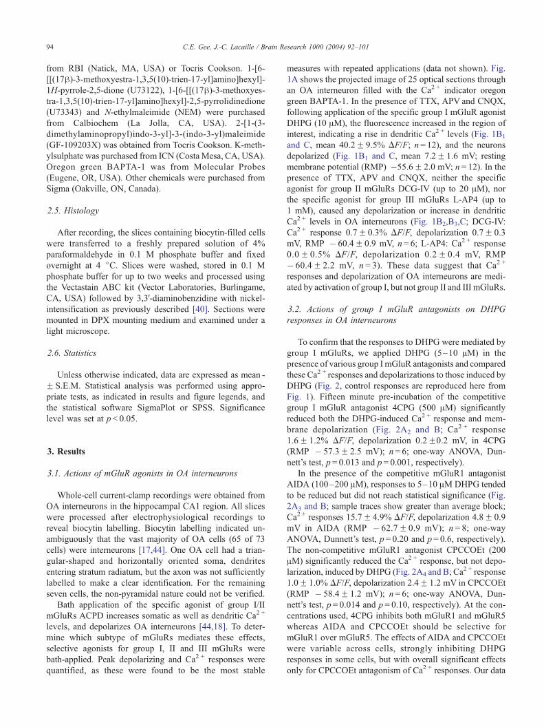
measures with repeated applications (data not shown). Fig.
1A shows the projected image of 25 optical sections through
an OA interneuron filled with the Ca2 + indicator oregon
green BAPTA-1. In the presence of TTX, APV and CNQX,
following application of the specific group I mGluR agonist
DHPG (10 AM), the fluorescence increased in the region of
interest, indicating a rise in dendritic Ca2 + levels (Fig. 1B1
and C, mean 40.2F 9.5% DF/F; n= 12), and the neurons
depolarized (Fig. 1B1 and C, mean 7.2F 1.6 mV; resting
membrane potential (RMP) �55.6F 2.0 mV; n = 12). In the
presence of TTX, APV and CNQX, neither the specific
agonist for group II mGluRs DCG-IV (up to 20 AM), nor
the specific agonist for group III mGluRs L-AP4 (up to
1 mM), caused any depolarization or increase in dendritic
Ca2 + levels in OA interneurons (Fig. 1B2,B3,C; DCG-IV:
Ca2 + response 0.7F 0.3% DF/F, depolarization 0.7F 0.3
mV, RMP � 60.4F 0.9 mV, n = 6; L-AP4: Ca2 + response
0.0F 0.5% DF/F, depolarization 0.2F 0.4 mV, RMP
� 60.4F 2.2 mV, n = 3). These data suggest that Ca2 +
responses and depolarization of OA interneurons are medi-
ated by activation of group I, but not group II and III mGluRs.
3.2. Actions of group I mGluR antagonists on DHPG
responses in OA interneurons
To confirm that the responses to DHPG were mediated by
group I mGluRs, we applied DHPG (5–10 AM) in the
presence of various group I mGluR antagonists and compared
these Ca2 + responses and depolarizations to those induced by
DHPG (Fig. 2, control responses are reproduced here from
Fig. 1). Fifteen minute pre-incubation of the competitive
group I mGluR antagonist 4CPG (500 AM) significantly
reduced both the DHPG-induced Ca2 + response and mem-
brane depolarization (Fig. 2A2 and B; Ca2 + response
1.6F 1.2% DF/F, depolarization 0.2F0.2 mV, in 4CPG
(RMP � 57.3F 2.5 mV); n = 6; one-way ANOVA, Dun-
nett’s test, p = 0.013 and p= 0.001, respectively).
In the presence of the competitive mGluR1 antagonist
AIDA (100–200 AM), responses to 5–10 AMDHPG tended
to be reduced but did not reach statistical significance (Fig.
2A3 and B; sample traces show greater than average block;
Ca2 + responses 15.7F 4.9% DF/F, depolarization 4.8F 0.9
mV in AIDA (RMP � 62.7F 0.9 mV); n = 8; one-way
ANOVA, Dunnett’s test, p = 0.20 and p= 0.6, respectively).
The non-competitive mGluR1 antagonist CPCCOEt (200
AM) significantly reduced the Ca2 + response, but not depo-
larization, induced by DHPG (Fig. 2A4 and B; Ca2 + response
1.0F 1.0% DF/F, depolarization 2.4F 1.2 mV in CPCCOEt
(RMP � 58.4F 1.2 mV); n = 6; one-way ANOVA, Dun-
nett’s test, p = 0.014 and p = 0.10, respectively). At the con-
centrations used, 4CPG inhibits both mGluR1 and mGluR5
whereas AIDA and CPCCOEt should be selective for
mGluR1 over mGluR5. The effects of AIDA and CPCCOEt
were variable across cells, strongly inhibiting DHPG
responses in some cells, but with overall significant effects
only for CPCCOEt antagonism of Ca2 + responses. Our data
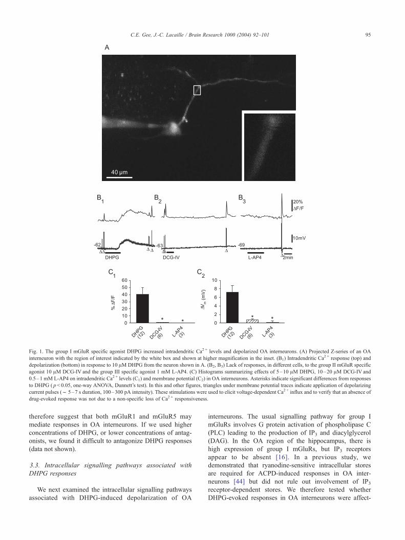
Fig. 1. The group I mGluR specific agonist DHPG increased intradendritic Ca2 + levels and depolarized OA interneurons. (A) Projected Z-series of an OA
interneuron with the region of interest indicated by the white box and shown at higher magnification in the inset. (B1) Intradendritic Ca2 + response (top) and
depolarization (bottom) in response to 10 AM DHPG from the neuron shown in A. (B2, B3) Lack of responses, in different cells, to the group II mGluR specific
agonist 10 AM DCG-IV and the group III specific agonist 1 mM L-AP4. (C) Histograms summarizing effects of 5–10 AM DHPG, 10–20 AM DCG-IV and
0.5–1 mM L-AP4 on intradendritic Ca2 + levels (C1) and membrane potential (C2) in OA interneurons. Asterisks indicate significant differences from responses
to DHPG ( p< 0.05, one-way ANOVA, Dunnett’s test). In this and other figures, triangles under membrane potential traces indicate application of depolarizing
current pulses (f 5–7 s duration, 100–300 pA intensity). These stimulations were used to elicit voltage-dependent Ca2 + influx and to verify that an absence of
drug-evoked response was not due to a non-specific loss of Ca2 + responsiveness.
C.E. Gee, J.-C. Lacaille / Brain Research 1000 (2004) 92–101 95
therefore suggest that both mGluR1 and mGluR5 may
mediate responses in OA interneurons. If we used higher
concentrations of DHPG, or lower concentrations of antag-
onists, we found it difficult to antagonize DHPG responses
(data not shown).
3.3. Intracellular signalling pathways associated with
DHPG responses
We next examined the intracellular signalling pathways
associated with DHPG-induced depolarization of OA
interneurons. The usual signalling pathway for group I
mGluRs involves G protein activation of phospholipase C
(PLC) leading to the production of IP3 and diacylglycerol
(DAG). In the OA region of the hippocampus, there is
high expression of group I mGluRs, but IP3 receptors
appear to be absent [16]. In a previous study, we
demonstrated that ryanodine-sensitive intracellular stores
are required for ACPD-induced responses in OA inter-
neurons [44] but did not rule out involvement of IP3receptor-dependent stores. We therefore tested whether
DHPG-evoked responses in OA interneurons were affect-
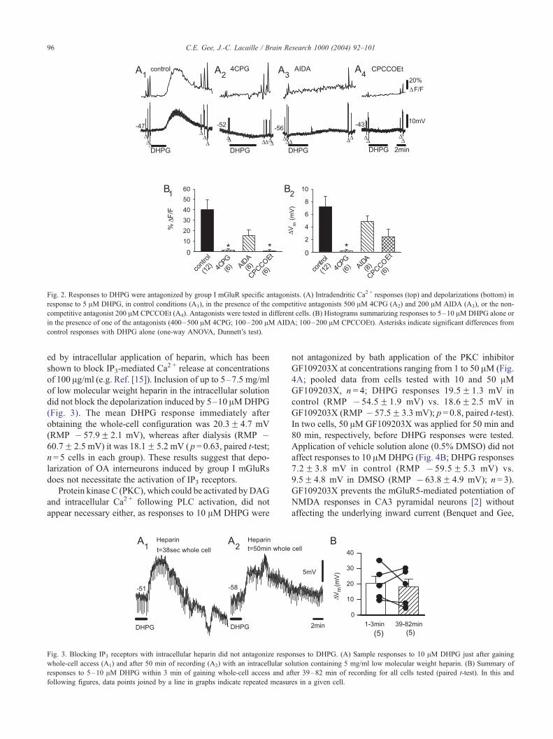
Fig. 2. Responses to DHPG were antagonized by group I mGluR specific antagonists. (A) Intradendritic Ca2 + responses (top) and depolarizations (bottom) in
response to 5 AM DHPG, in control conditions (A1), in the presence of the competitive antagonists 500 AM 4CPG (A2) and 200 AM AIDA (A3), or the non-
competitive antagonist 200 AM CPCCOEt (A4). Antagonists were tested in different cells. (B) Histograms summarizing responses to 5–10 AM DHPG alone or
in the presence of one of the antagonists (400–500 AM 4CPG; 100–200 AM AIDA; 100–200 AM CPCCOEt). Asterisks indicate significant differences from
control responses with DHPG alone (one-way ANOVA, Dunnett’s test).
C.E. Gee, J.-C. Lacaille / Brain Research 1000 (2004) 92–10196
ed by intracellular application of heparin, which has been
shown to block IP3-mediated Ca2 + release at concentrations
of 100 Ag/ml (e.g. Ref. [15]). Inclusion of up to 5–7.5 mg/ml
of low molecular weight heparin in the intracellular solution
did not block the depolarization induced by 5–10 AMDHPG
(Fig. 3). The mean DHPG response immediately after
obtaining the whole-cell configuration was 20.3F 4.7 mV
(RMP � 57.9F 2.1 mV), whereas after dialysis (RMP �60.7F 2.5 mV) it was 18.1F 5.2 mV ( p = 0.63, paired t-test;
n = 5 cells in each group). These results suggest that depo-
larization of OA interneurons induced by group I mGluRs
does not necessitate the activation of IP3 receptors.
Protein kinase C (PKC), which could be activated byDAG
and intracellular Ca2 + following PLC activation, did not
appear necessary either, as responses to 10 AM DHPG were
Fig. 3. Blocking IP3 receptors with intracellular heparin did not antagonize respo
whole-cell access (A1) and after 50 min of recording (A2) with an intracellular so
responses to 5–10 AM DHPG within 3 min of gaining whole-cell access and a
following figures, data points joined by a line in graphs indicate repeated measur
not antagonized by bath application of the PKC inhibitor
GF109203X at concentrations ranging from 1 to 50 AM (Fig.
4A; pooled data from cells tested with 10 and 50 AMGF109203X, n = 4; DHPG responses 19.5F 1.3 mV in
control (RMP � 54.5F 1.9 mV) vs. 18.6F 2.5 mV in
GF109203X (RMP � 57.5F 3.3 mV); p = 0.8, paired t-test).
In two cells, 50 AMGF109203X was applied for 50 min and
80 min, respectively, before DHPG responses were tested.
Application of vehicle solution alone (0.5% DMSO) did not
affect responses to 10 AMDHPG (Fig. 4B; DHPG responses
7.2F 3.8 mV in control (RMP � 59.5F 5.3 mV) vs.
9.5F 4.8 mV in DMSO (RMP � 63.8F 4.9 mV); n = 3).
GF109203X prevents the mGluR5-mediated potentiation of
NMDA responses in CA3 pyramidal neurons [2] without
affecting the underlying inward current (Benquet and Gee,
nses to DHPG. (A) Sample responses to 10 AM DHPG just after gaining
lution containing 5 mg/ml low molecular weight heparin. (B) Summary of
fter 39–82 min of recording for all cells tested (paired t-test). In this and
es in a given cell.
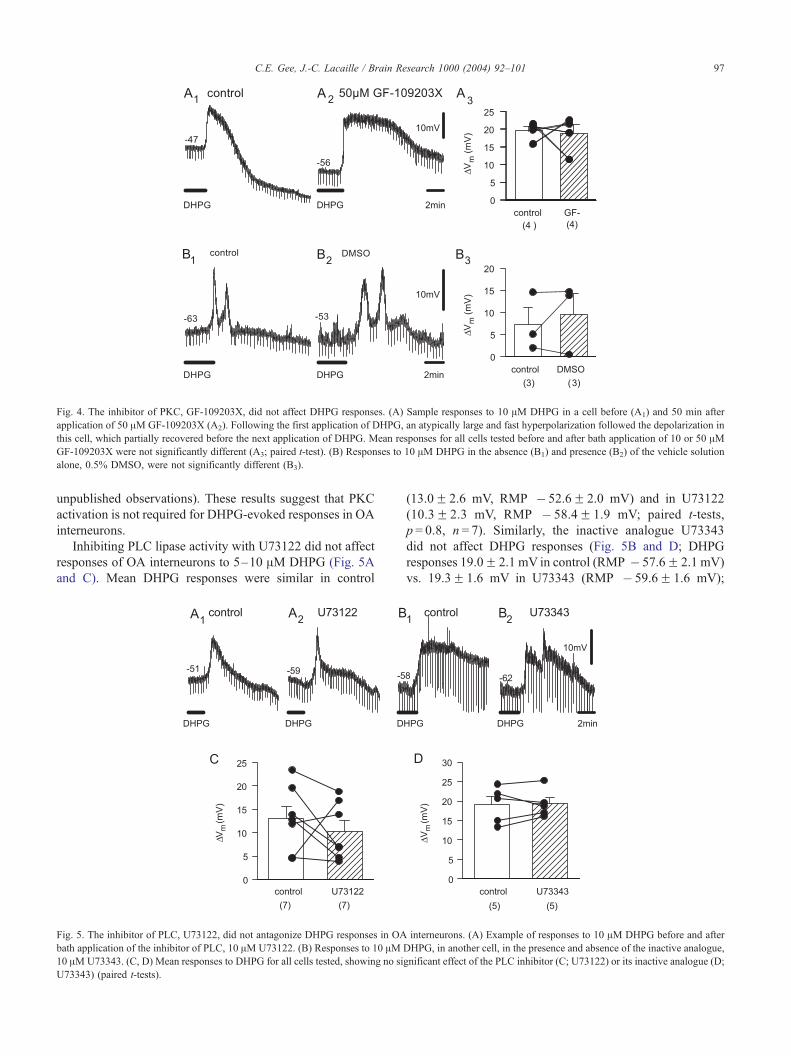
Fig. 4. The inhibitor of PKC, GF-109203X, did not affect DHPG responses. (A) Sample responses to 10 AM DHPG in a cell before (A1) and 50 min after
application of 50 AM GF-109203X (A2). Following the first application of DHPG, an atypically large and fast hyperpolarization followed the depolarization in
this cell, which partially recovered before the next application of DHPG. Mean responses for all cells tested before and after bath application of 10 or 50 AMGF-109203X were not significantly different (A3; paired t-test). (B) Responses to 10 AM DHPG in the absence (B1) and presence (B2) of the vehicle solution
alone, 0.5% DMSO, were not significantly different (B3).
C.E. Gee, J.-C. Lacaille / Brain Research 1000 (2004) 92–101 97
unpublished observations). These results suggest that PKC
activation is not required for DHPG-evoked responses in OA
interneurons.
Inhibiting PLC lipase activity with U73122 did not affect
responses of OA interneurons to 5–10 AM DHPG (Fig. 5A
and C). Mean DHPG responses were similar in control
Fig. 5. The inhibitor of PLC, U73122, did not antagonize DHPG responses in OA
bath application of the inhibitor of PLC, 10 AM U73122. (B) Responses to 10 AM10 AMU73343. (C, D) Mean responses to DHPG for all cells tested, showing no si
U73343) (paired t-tests).
(13.0F 2.6 mV, RMP � 52.6F 2.0 mV) and in U73122
(10.3F 2.3 mV, RMP � 58.4F 1.9 mV; paired t-tests,
p = 0.8, n = 7). Similarly, the inactive analogue U73343
did not affect DHPG responses (Fig. 5B and D; DHPG
responses 19.0F 2.1 mV in control (RMP � 57.6F 2.1 mV)
vs. 19.3F 1.6 mV in U73343 (RMP � 59.6F 1.6 mV);
interneurons. (A) Example of responses to 10 AM DHPG before and after
DHPG, in another cell, in the presence and absence of the inactive analogue,
gnificant effect of the PLC inhibitor (C; U73122) or its inactive analogue (D;
C.E. Gee, J.-C. Lacaille / Brain Research 1000 (2004) 92–10198
paired t-tests, p = 0.9, n = 5). In spinal cord slices, U73122 is
effective at blocking the long-term depression of synaptic
transmission, without blocking the acute synaptic inhibition,
induced by group I mGluRs, thus blocking some but not all
actions of group I mGluRs [10]. These data suggest that
lipase activity of PLC may not be required for DHPG-
induced responses in OA interneurons.
3.4. G protein-independent signalling pathway associated
with DHPG responses
We next evaluated whether DHPG responses in OA
interneurons involve a G protein-dependent mechanism.
GDPhS (up to 1 mM) was included in the patch pipette
solution to block G protein activation. Depolarizations were
still evoked by DHPG up to one h after obtaining whole-cell
access in cells recorded with GDPhS containing solution
(Fig. 6B3 and C2; 8.3F 2.1 mV in five cells recorded with
GDPhS (RMP � 53.2F 2.5 mV) vs. 9.8F 2.9 mV in six
control cells recorded with GTP (RMP � 57.0F 2.7 mV),
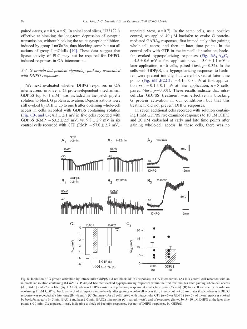
Fig. 6. Inhibition of G protein activation by intracellular GDPhS did not block D
intracellular solution containing 0.4 mM GTP, 40 AM baclofen evoked hyperpolar
(A1, BAC1) and 22 min later (A2, BAC2), whereas DHPG evoked a depolarizing
containing 1 mM GDPhS, baclofen evoked a response immediately after gaining
response was recorded at a later time (B3; 48 min). (C) Summary, for all cells tested
by baclofen at early ( < 5 min; BAC1) and later (>5 min; BAC2) time points (C1; p
points (>30 min; C2; unpaired t-test), indicating a block of baclofen responses, b
unpaired t-test, p = 0.7). In the same cells, as a positive
control, we applied 40 AM baclofen to evoke G protein-
mediated GABAB responses, first immediately after gaining
whole-cell access and then at later time points. In the
control cells with GTP in the intracellular solution, baclo-
fen evoked hyperpolarizing responses (Fig. 6A1,A2,C1;
� 4.5F 0.6 mV at first application vs. � 3.0F 1.1 mV at
later application, n = 6 cells, paired t-test, p= 0.32). In the
cells with GDPhS, the hyperpolarizing responses to baclo-
fen were present initially, but were blocked at later time
points (Fig. 6B1,B2,C1; � 4.1F 0.8 mV at first applica-
tion vs. � 0.1F 0.1 mV at later application, n = 5 cells,
paired t-test, p = 0.001). These results indicate that intra-
cellular GDPhS treatment was effective in blocking
G protein activation in our conditions, but that this
treatment did not prevent DHPG responses.
In seven additional cells recorded with solution contain-
ing 1 mM GDPhS, we examined responses to 10 AMDHPG
and 20 AM carbachol at early and late time points after
gaining whole-cell access. In these cells, there was no
HPG responses in OA interneurons. (A) In a control cell recorded with an
izing responses within the first few minutes after gaining whole-cell access
response at a later time point (35 min). (B) In a cell recorded with solution
whole-cell access (B1; 2 min) but not 30 min later (B2,), whereas a DHPG
with intracellular GTP (n= 6) or GDPhS (n= 5), of mean responses evoked
aired t-tests), and of responses elicited by 5–10 AM DHPG at the later time
ut not of DHPG responses, by GDPhS.
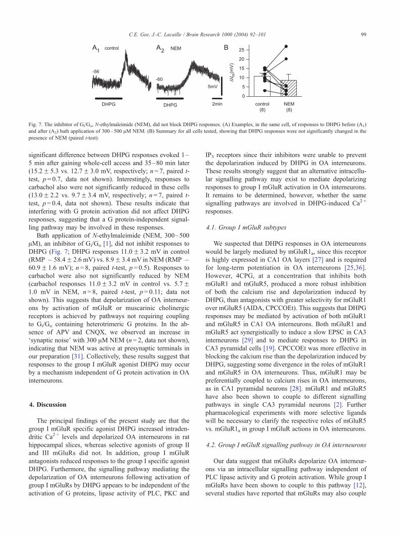
Fig. 7. The inhibitor of Gi/Go, N-ethylmaleimide (NEM), did not block DHPG responses. (A) Examples, in the same cell, of responses to DHPG before (A1)
and after (A2) bath application of 300–500 AM NEM. (B) Summary for all cells tested, showing that DHPG responses were not significantly changed in the
presence of NEM (paired t-test).
C.E. Gee, J.-C. Lacaille / Brain Research 1000 (2004) 92–101 99
significant difference between DHPG responses evoked 1–
5 min after gaining whole-cell access and 35–80 min later
(15.2F 5.3 vs. 12.7F 3.0 mV, respectively; n = 7, paired t-
test, p = 0.7, data not shown). Interestingly, responses to
carbachol also were not significantly reduced in these cells
(13.0F 2.2 vs. 9.7F 3.4 mV, respectively; n= 7, paired t-
test, p = 0.4, data not shown). These results indicate that
interfering with G protein activation did not affect DHPG
responses, suggesting that a G protein-independent signal-
ling pathway may be involved in these responses.
Bath application of N-ethylmaleimide (NEM, 300–500
AM), an inhibitor of Gi/Go [1], did not inhibit responses to
DHPG (Fig. 7; DHPG responses 11.0F 3.2 mV in control
(RMP � 58.4F 2.6 mV) vs. 8.9F 3.4 mV in NEM (RMP �60.9F 1.6 mV); n = 8, paired t-test, p = 0.5). Responses to
carbachol were also not significantly reduced by NEM
(carbachol responses 11.0F 3.2 mV in control vs. 5.7F1.0 mV in NEM, n = 8, paired t-test, p = 0.11; data not
shown). This suggests that depolarization of OA interneur-
ons by activation of mGluR or muscarinic cholinergic
receptors is achieved by pathways not requiring coupling
to Gi/Go containing heterotrimeric G proteins. In the ab-
sence of APV and CNQX, we observed an increase in
‘synaptic noise’ with 300 AM NEM (n = 2, data not shown),
indicating that NEM was active at presynaptic terminals in
our preparation [31]. Collectively, these results suggest that
responses to the group I mGluR agonist DHPG may occur
by a mechanism independent of G protein activation in OA
interneurons.
4. Discussion
The principal findings of the present study are that the
group I mGluR specific agonist DHPG increased intraden-
dritic Ca2 + levels and depolarized OA interneurons in rat
hippocampal slices, whereas selective agonists of group II
and III mGluRs did not. In addition, group I mGluR
antagonists reduced responses to the group I specific agonist
DHPG. Furthermore, the signalling pathway mediating the
depolarization of OA interneurons following activation of
group I mGluRs by DHPG appears to be independent of the
activation of G proteins, lipase activity of PLC, PKC and
IP3 receptors since their inhibitors were unable to prevent
the depolarization induced by DHPG in OA interneurons.
These results strongly suggest that an alternative intracellu-
lar signalling pathway may exist to mediate depolarizing
responses to group I mGluR activation in OA interneurons.
It remains to be determined, however, whether the same
signalling pathways are involved in DHPG-induced Ca2 +
responses.
4.1. Group I mGluR subtypes
We suspected that DHPG responses in OA interneurons
would be largely mediated by mGluR1a, since this receptor
is highly expressed in CA1 OA layers [27] and is required
for long-term potentiation in OA interneurons [25,36].
However, 4CPG, at a concentration that inhibits both
mGluR1 and mGluR5, produced a more robust inhibition
of both the calcium rise and depolarization induced by
DHPG, than antagonists with greater selectivity for mGluR1
over mGluR5 (AIDA, CPCCOEt). This suggests that DHPG
responses may be mediated by activation of both mGluR1
and mGluR5 in CA1 OA interneurons. Both mGluR1 and
mGluR5 act synergistically to induce a slow EPSC in CA3
interneurons [29] and to mediate responses to DHPG in
CA3 pyramidal cells [19]. CPCCOEt was more effective in
blocking the calcium rise than the depolarization induced by
DHPG, suggesting some divergence in the roles of mGluR1
and mGluR5 in OA interneurons. Thus, mGluR1 may be
preferentially coupled to calcium rises in OA interneurons,
as in CA1 pyramidal neurons [28]. mGluR1 and mGluR5
have also been shown to couple to different signalling
pathways in single CA3 pyramidal neurons [2]. Further
pharmacological experiments with more selective ligands
will be necessary to clarify the respective roles of mGluR5
vs. mGluR1a in group I mGluR actions in OA interneurons.
4.2. Group I mGluR signalling pathway in OA interneurons
Our data suggest that mGluRs depolarize OA interneur-
ons via an intracellular signalling pathway independent of
PLC lipase activity and G protein activation. While group I
mGluRs have been shown to couple to this pathway [12],
several studies have reported that mGluRs may also couple
C.E. Gee, J.-C. Lacaille / Brain Research 1000 (2004) 92–101100
to alternative G protein-independent pathways in neurons
[2,19,21,23]. GDPhS blocked the hyperpolarizing response
to the GABAB agonist baclofen, suggesting that the inhibi-
tion of G proteins was effective. The signalling pathway
mediating the group I mGluR responses in OA interneurons
remains to be determined, but in recent years several of the
heptahelical receptors have been shown to signal through G
protein-independent mechanisms (reviewed by Ref. [22]).
mGluRs are associated with a variety of other receptors and
proteins in the postsynaptic density, some of which could be
involved in the G protein-independent signalling (e.g. Ref.
[43]). Transient receptor potential (TRP) channels, which
are postulated to underlie mGluR- and muscarinic cholin-
ergic receptor-mediated inward currents in CA3 pyramidal
cells [19,41], can be opened in response to agonists by
lipase-independent activity of PLC [35]. As U73122 only
interferes with lipase activity of PLC, PLC may well be
involved in coupling mGluRs to neuronal depolarization.
Another candidate for mediating group I mGluR signalling
is the Homer family of proteins, which bind the carboxyl
termini of mGluR1a and mGluR5 [4], as well as the
ryanodine receptor [14]. Thus, Homer proteins could be in
a position to mediate the Ca2 + release from ryanodine-
sensitive stores in OA interneurons [44]. Further experi-
ments aimed at identifying the G protein-independent sig-
nalling pathway involved in group I mGluR actions will be
important to clarify the actions linked to these receptors and
their role in OA interneuron function.
4.3. Functional implications
Group I mGluRs have been shown to be important in OA
interneuron function. First, OA interneurons are selectively
vulnerable to excitotoxicity in the kainate (KA) model of
epilepsy [30]. Although the exact mechanism of this selec-
tive vulnerability remains to be determined, recent evidence
suggests that treatment with a group I mGluR antagonist
reduced the OA interneuron loss in KA-treated rats [39].
Thus, activation of group I mGluRs may play a role in the
vulnerability of these interneurons to glutamate excitotox-
icity during seizures. In addition, the mGluR1a subtype of
group I mGluRs is necessary for the induction of long-term
potentiation of glutamatergic synapses onto OA interneur-
ons [25,36]. The selective vulnerability of OA cells to
excitotoxicity in the KA model [30] and the presence of
long-term potentiation selectively in OA interneurons [36] is
consistent with the finding that group I mGluR activation
results in a coupling between Ca2 + influx via voltage-
dependent Ca2 + channels and release from ryanodine-sen-
sitive intracellular stores, selectively in OA interneurons,
and not in interneurons of stratum radiatum and lacunosum-
moleculare [44]. The present findings further suggest that a
signalling pathway independent from G proteins, PLC,
DAG, PKC and IP3, may be involved in linking group I
mGluR activation to intracellular depolarizations. Since
these depolarizations are associated with intracellular Ca2 +
rises, such cell-specific actions and their signalling mecha-
nisms may be important for synaptic plasticity and the
selective vulnerability of these interneurons.
Acknowledgements
This work was supported by grants to J.C.L. from the
Canadian Institutes of Health Research, Fonds de la
recherche en sante du Quebec, Fonds pour la Formation
des Chercheurs et l’Aide a la Recherche. J.C.L. is supported
by a Canada Research Chair in Cellular and Molecular
Neurophysiology. C.G. was supported by a postdoctoral
fellowship from the Natural Sciences and Engineering
Research Council of Canada. The authors thank F. Cartier
for typing the manuscript.
References
[1] T. Asano, N. Ogasawara, Uncoupling of gamma-aminobutyric acid B
receptors from GTP-binding proteins by N-ethylmaleimide: effect of
N-ethylmaleimide on purified GTP-binding proteins, Mol. Pharmacol.
29 (1986) 244–249.
[2] P. Benquet, C.E. Gee, U. Gerber, Two distinct signaling pathways
upregulate NMDA receptor responses via two distinct metabotropic
glutamate receptor subtypes, J. Neurosci. 22 (2002) 9679–9686.
[3] N. Best, J. Mitchell, H.V. Wheal, Changes in parvalbumin-immuno-
reactive neurons in CA1 area of hippocampus following a kainic acid
injection, Acta Neuropathol. 87 (1993) 187–195.
[4] P.R. Brakeman, A.A. Lanahan, R. O’Brien, K. Roche, C.A. Barnes,
R.L. Huganir, P.F. Worley, Homer: a protein that selectively binds
metabotropic glutamate receptors, Nature 386 (1997) 284–288.
[5] L. Carmant, G. Woodhall, M. Ouardouz, R. Robitaille, J.-C. Lacaille,
Interneuron-specific Ca2 + responses linked to metabotropic and ion-
otropic glutamate receptors in rat hippocampal slices, Eur. J. Neuro-
sci. 9 (1997) 1625–1635.
[6] S. Charpak, B.H. Gahwiler, K.Q. Do, T. Knopfel, Potassium conduc-
tances in hippocampal neurons blocked by excitatory amino-acid
transmitters, Nature 347 (1990) 765–767.
[7] S.C. Chuang, R. Bianchi, R.K.S. Wong, Group I mGluR activation
turns on a voltage-gated inward current in hippocampal pyramidal
cells, J. Neurophysiol. 83 (2000) 2844–2853.
[8] S.C. Chuang, R. Bianchi, D. Kim, H.S. Shin, R.K.S. Wong, Group I
metabotropic receptors elicit epileptiform discharges in the hippocam-
pus through PLCh1 signaling, J. Neurosci. 21 (2001) 6387–6394.
[9] P. Chavis, L. Fagni, J.B. Lansman, J. Bockaert, Functional coupling
between ryanodine receptors and L-type calcium channels in neurons,
Nature 382 (1996) 719–722.
[10] J. Chen, B. Heinke, J. Sandkuhler, Activation of group I metabotropic
glutamate receptors induces long-term depression at sensory synapses
in superficial spinal dorsal horn, Neuropharmacology 39 (2000)
2231–2243.
[11] P. Congar, X. Leinekugel, Y. Ben-Ari, V. Crepel, A long-lasting cal-
cium-activated nonselective cationic current is generated by synaptic
stimulation or exogenous activation of group I metabotropic gluta-
mate receptors in CA1 pyramidal neurons, J. Neurosci. 17 (1997)
5366–5379.
[12] P.J. Conn, J.P. Pin, Pharmacology and functions of metabotropic glu-
tamate receptors, Annu. Rev. Pharmacol. Toxicol. 37 (1997) 205–237.
[13] V. Crepel, L. Aniksztejn, Y. Ben-Ari, C. Hammond, Glutamate me-
tabotropic receptors increase a Ca(2+)-activated nonspecific cationic
current in CA1 hippocampal neurons, J. Neurophysiol. 72 (1994)
1561–1569.
C.E. Gee, J.-C. Lacaille / Brain Research 1000 (2004) 92–101 101
[14] W. Feng, J. Tu, T. Yang, P.S. Vernon, P.D. Allen, P.F. Worley, I.N.
Pessah, Homer regulates gain of ryanodine receptor type I channel
complex, J. Biol. Chem. 277 (2002) 44722–44730.
[15] E.A. Finch, G.J. Augustine, Local calcium signalling by inositol-
1,4,5-trisphosphate in Purkinje cell dendrites, Nature 396 (1998)
753–756.
[16] M. Fotuhi, A.H. Sharp, C.E. Glatt, P.M. Hwang, M. von Krosigk,
S.H. Snyder, T.M. Dawson, Differential localization of phosphoinosi-
tide-linked metabotropic glutamate receptor (mGluR1) and the inosi-
tol 1,4,5-trisphosphate receptor in rat brain, J. Neurosci. 13 (1993)
2001–2012.
[17] T.F. Freund, G. Buzsaki, Interneurons of the hippocampus, Hippo-
campus 6 (1996) 347–470.
[18] C.E. Gee, G. Woodhall, J.-C. Lacaille, Synaptically-activated calcium
responses in dendrites of hippocampal oriens–alveus interneurons,
J. Neurophysiol. 85 (2001) 1603–1613.
[19] C.E. Gee, P. Benquet, U. Gerber, Group I metabotropic glutamate
receptors activate a calcium-sensitive transient receptor potential-
like conductance in rat hippocampus, J. Physiol. 546 (2003)
655–664.
[20] N.C. Guerineau, B.H. Gahwiler, U. Gerber, Reduction of resting K+
current by metabotropic glutamate and muscarinic receptors in rat
CA3 cells: mediation by G-proteins, J. Physiol. 474 (1994) 27–33.
[21] N.C. Guerineau, J.L. Bossu, B.H. Gahwiler, U. Gerber, Activation of
a nonselective cationic conductance by metabotropic glutamatergic
and muscarinic agonists in CA3 pyramidal neurons of the rat hippo-
campus, J. Neurosci. 15 (1995) 4395–4407.
[22] R.A. Hall, R.T. Premont, R.J. Lefkowitz, Heptahelical receptor signal-
ing: beyond the G protein paradigm, J. Cell Biol. 145 (1999) 927–932.
[23] C. Heuss, M. Scanziani, B.H. Gahwiler, U. Gerber, G-protein-inde-
pendent signaling mediated by metabotropic glutamate receptors, Nat.
Neurosci. 2 (1999) 1070–1077.
[24] N.B. Keele, V.L. Arvanov, P. Shinnick-Gallagher, Quisqualate-prefer-
ring metabotropic receptor activates Na+–Ca2 + exchange in rat baso-
lateral amygdala neurones, J. Physiol. 499 (1997) 87–104.
[25] V. Lapointe, F. Morin, S. Ratte, A. Croce, F. Conquet, J.-C. La-
caille, Synapse-specific mGluR1-dependent long-term potentiation
in interneurons regulates mouse hippocampal inhibition, J. Physiol.
(in press).
[26] K. Lee, P.R. Boden, Characterization of the inward current induced by
metabotropic glutamate receptor stimulation in rat ventromedial hy-
pothalamic neurons, J. Physiol. 504 (1997) 649–663.
[27] R. Lujan, Z. Nusser, J.D. Roberts, R. Shigemoto, P. Somogyi, Peri-
synaptic location of metabotropic glutamate receptors mGluR1 and
mGluR5 on dendrites and dendritic spines in the rat hippocampus,
Eur. J. Neurosci. 8 (1996) 1488–1500.
[28] G. Mannaioni, M.J. Marino, O. Valenti, S.F. Traynellis, P.J. Conn,
Metabotropic glutamate receptors 1 and 5 differentially regulate CA1
pyramidal cell function, J. Neurosci. 21 (2001) 5925–5934.
[29] M. Mori, U. Gerber, Slow feedback inhibition in the CA3 area of the
rat hippocampus by synergistic activation of mGluR1 and mGluR5,
J. Physiol. 544 (2002) 793–799.
[30] F. Morin, C. Beaulieu, J.-C. Lacaille, Selective loss of GABA neurons
in area CA1 of the rat hippocampus after intraventricular kainate,
Epilepsy Res. 32 (1998) 363–369.
[31] W. Morishita, S.A. Kirov, T.A. Pitler, L.A. Martin, R.A. Lenz, B.E.
Alger, N-ethylmaleimide blocks depolarization-induced suppression
of inhibition and enhances GABA release in the rat hippocampal slice
in vitro, J. Neurosci. 17 (1997) 941–950.
[32] T. Nakamura, J.G. Barbara, K. Nakamura, W.N. Ross, Synergistic
release of Ca2 + from IP3-sensitive stores evoked by synaptic activa-
tion of mGluRs paired with backpropagating action potentials, Neu-
ron 24 (1999) 727–737.
[33] F. Nicoletti, V. Bruno, M.V. Catania, G. Battaglia, A. Copani, G.
Barbagallo, V. Cena, J. Sanchez-Prieto, P.F. Spano, M. Pizzi,
Group-I metabotropic glutamate receptors: hypotheses to explain their
dual role in neurotoxicity, neuroprotection, Neuropharmacology 38
(1999) 1477–1484.
[34] M. Ouardouz, J.-C. Lacaille, Mechanisms of selective long-term po-
tentiation of excitatory synapses in stratum oriens/alveus interneurons
of rat hippocampal slices, J. Neurophysiol. 73 (1995) 810–819.
[35] R.L. Patterson, D.B. van Rossum, D.L. Ford, K.J. Hurt, S.S. Bae,
P.-G. Suh, T. Kurosaki, S.H. Snyder, D.L. Gill, Phospholipase C-g is
required for agonist-induced Ca2 + entry, Cell 111 (2002) 529–541.
[36] Y. Perez, F. Morin, J.-C. Lacaille, A hebbian form of long-term po-
tentiation dependent on mGluR1a in hippocampal inhibitory inter-
neurons, Proc. Natl. Acad. Sci. U. S. A. 98 (2001) 9401–9406.
[37] J.P. Pin, R. Duvoisin, The metabotropic glutamate receptors: structure
and functions, Neuropharmacology 34 (1995) 1–26.
[38] L.D. Pozzo Miller, J.J. Petrozino, J.A. Connor, G protein-coupled
receptors mediate a fast excitatory postsynaptic current in CA3
pyramidal neurons in hippocampal slices, J. Neurosci. 15 (1995)
8320–8330.
[39] J. Renaud, M. Emond, S. Meilleur, C. Psarropoulou, L. Carmant,
AIDA, a class I metabotropic glutamate-receptor antagonist limits
kainate-induced hippocampal dysfunction, Epilepsia 43 (2002)
1306–1317.
[40] C. Staub, I. Vranesic, T. Knopfel, Responses to metabotropic gluta-
mate receptor activation in cerebellar purkinje cells: induction of an
inward current, Eur. J. Neurosci. 4 (1992) 832–839.
[41] C. Strubing, G. Krapivinsky, L. Krapivinsky, D.E. Clapham, TRPC1
and TRPC5 form a novel cation channel in mammalian brain, Neuron
29 (2001) 645–655.
[42] J.A. van Hooft, R. Giuffrida, M. Blatow, H. Monyer, Differential ex-
pression of group I metabotropic glutamate receptors in functionally
distinct hippocampal interneurons, J. Neurosci. 20 (2000) 3544–3551.
[43] R.S. Walikonis, O.N. Jensen, M. Mann, D.W.J. Provance, J.A. Mer-
cer, M.B. Kennedy, Identification of proteins in the postsynaptic
density fraction by mass spectrometry, J. Neurosci. 20 (2000)
4069–4080.
[44] G. Woodhall, C.E. Gee, R. Robitaille, J.-C. Lacaille, Membrane po-
tential and intracellular Ca2 + oscillations activated by mGluRs in
hippocampal stratum oriens/alveus interneurons, J. Neurophysiol. 81
(1999) 371–382.


![Glutamate carboxypeptidase II gene knockout attenuates ... · metabotropic glutamate receptor (mGluR3) [- 7–9]. Acti vating mGluR3 by NAAG reduces the synaptic glutamate ... (Leica](https://img.pdfslide.us/doc/110x75/5c4d740293f3c34aee567cc7/glutamate-carboxypeptidase-ii-gene-knockout-attenuates-metabotropic-glutamate.jpg)


![RESEARCH Open Access Metabotropic glutamate receptor 5 ... · taneous nocifensive behavior as well as thermal hyper-algesia and allodynia in rats [29,30]. I.t. pretreatment with the](https://img.pdfslide.us/doc/110x75/5fd8fafd78d69f50705b2706/research-open-access-metabotropic-glutamate-receptor-5-taneous-nocifensive-behavior.jpg)