Embed Size (px)
Citation preview
Research Report 2015
Schaller Research Groups
at the University of Heidelberg and the DKFZ
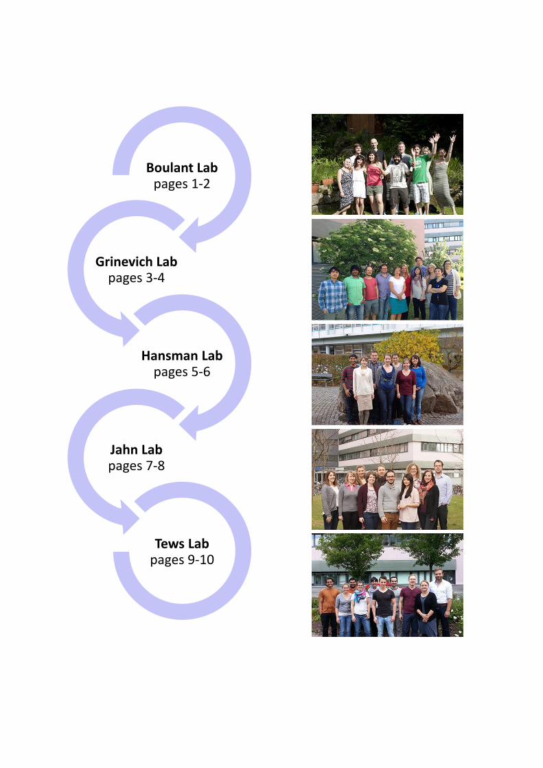
Boulant Lab pages 1-2
Grinevich Lab pages 3-4
Hansman Lab pages 5-6
Jahn Lab pages 7-8
Tews Lab pages 9-10
Steeve Boulant, PhD [email protected]
+49 (0) 6221-42-1560
2012 - present: Group leader Department Infectious Disease, Virology University Hospital, Heidelberg, Germany
Schaller Research Group at the University of Heidelberg and the DKFZ
2008 - 2012: Postdoctoral Associate Harvard Medical School, Boston MA, USA 2005 – 2008: Marie-Curie Postdoctoral Fellow MRC virology unit, Glasgow, UK 2001 – 2004: PhD, Molecular Biology and Biochemistry IBCP-CNRS, Lyon, France
Dynamics of viral infection
Research Aims: The laboratory is interested in how intestinal epithelial cells (IECs) that line the surface of the gut can tolerate the presence of the commensal microflora and at the same time recognize and fight enteric pathogens. Background: Intestinal epithelial cells (IECs) constitute the primary barrier that separates us from the outside environment. The mechanisms by which IECs can tolerate the presence of the commensal bacteria present in the gut have started to be revealed. Recently, the presence of a large amount of viruses in the gut and the potential role of these viruses in maintaining homeostasis has begun to be appreciated. Whether IECs have developed mechanisms to tolerate the presence of these enteric viruses and whether these viruses have developed strategies to subvert IECs innate immune response remain to be characterized. Importantly, whether the presence of these viruses is beneficial and whether they should be considered as “commensal viruses” remain to be fully addressed. Research Highlights: Viral strategies to infect intestinal epithelial cells: Reovirus is a widely used model virus to study antiviral innate immune response and pathogenesis. During the natural
course of infection, virion particles are converted in the lumen of the gut to intermediate subviral particles (ISVPs) by proteolytic digestion. The reasons for this conversion remained unclear. However, we recently found that ISVPs have developed specific strategies to not trigger innate immune response in IECs. On the contrary, infection of IECs by virions induces a strong immune response that leads to cellular death. Interestingly, we found that ISVP infected cells secrete a pro-survival factor that protects IECs against virion induced cellular death. We propose that conversion of reovirus to ISVPs in the lumen of the gut is a viral strategy to initiate primary infection by subverting IECs innate immune system and by counteracting cellular-death pathways. Polarized antiviral response IECs, strategies to tolerate the intestinal commensal flora: IECs are structurally polarized with an apical side facing the lumen of the gut and a basolateral side facing the lamina propia side. While the luminal side is constantly
exposed to the commensal flora, the basolateral side is sterile. From this observation we hypothesize that one way for IECs to tolerate the presence of the ever-present microbial flora would be to segregate their innate immune function to the basolateral side and be “blind” from their apical side. We found that the extent of immune response is a function of the site of infection. Infection of IECs from the basolateral side triggered a stronger response of IECs compared to infection of the apical side. We are currently identifying the molecular mechanisms that lead to this polarized antiviral response and are testing whether it could be a strategy to avoid excessive response against the commensal flora while maintaining full responsiveness against viruses that have passed the epithelium barrier. Functional differences in type I versus type III interferon mediated immunity in IECs: While type I interferon (IFN) mediated immunity is ubiquitous to all cells, type III IFN mediated immunity is confined to epithelial cells due to the restricted expression of its receptor. It is currently believe that both IFNs have redundant functions; however, the epithelium specificity of type III IFN strongly suggests that both IFNs must have functional differences at epithelial surfaces. In the lab we have defined, for the first time, functional differences between type I and type III IFN signaling in epithelial cells and we propose that type III IFN signaling is specifically tailored to efficiently combat infection without inducing an excessive pro-inflammatory response.
Group Composition: Dr. Megan Stanifer, Postdoctoral Fellow Anja Rippert, Technician Pranav Shah, PhD student Delia Bucher, PhD student Kalliopi Pervolaraki, PhD student Marta Fratini, PhD student Markus Mukenhirn, PhD student Christian Kischnick, PhD student Last Year Publications: Boulant S., Stanifer, M., Lozach P.Y.
(2015): Dynamics of virus-receptor interactions in virus binding, signaling, and endocytosis. Review in Viruses 7(6):2794-2815.
Odendall C., Dixit, E., Stavru, F., Bierne, H., Franz, K.M., Durbin, A.F., Boulant, S., Gehrke, L., Cossart, P., Kagan, J.C., (2014): Diverse intracellular pathogens activate type III interferon expression from peroxisomes. Nat Immunol. 15(8):717-26.
Grinevich, V., Knobloch, H. S., Roth, L.C., Althammer, F., Domansky, A., Vinnikov, I., Stanifer, M., Boulant, S. (2015): Somatic transgenesis (Viral vectors). Book chapter in Master Classes in Molecular Neuroendocrinology, Wiley (in production).
External Funding: DFG SFB1129: Integrative analysis of
pathogen replication and spread. European FP7, Marie Curie integration
grant Teaching Activities: Molecular Virology Master Student
Practical HBIGS course, practical Molecular Virology Post-transcriptional
regulation tutorial DKFZ “Progress in Cancer Research” DKFZ journal club: Infection and Cancer Frontiers in Biosciences Master Student
lecture International Graduate School:
Pathogen-Host Interactions at Cellular Barriers, Muenster, Germany
Valery Grinevich, MD, PhD
[email protected] +49 (0) 6221-42-1581
2012 – present: Group leader
Schaller Research Group on Neuropeptides at the University of Heidelberg and the DKFZ
2008 – 2012: Group Leader
Department of Molecular Neurobiology, Max Planck Institute for Medical Research, Heidelberg, Germany
2003 – 2007: Postdoctoral Fellow
Department of Molecular Neurobiology, Max Planck Institute for Medical Research, Heidelberg, Germany
Schaller Research Group on Neuropeptides
Research Aims: To study mechanisms of neuropeptide actions in the mammalian brain. Background: Neuropeptides represent a group of non-canonical neuromodulators which modify activity of neuronal ensembles, resulting in specific changes of behavior. Among approximately 100 neuropeptides, the most popular and the best-studied one is oxytocin. Despite very pronounced effects on various forms of behaviors, ranging from anxiolytic action to social attachment, the basic question as to how oxytocin (OT) exerts its effects is far from being answered. Our team focuses on dissecting neuropeptidergic circuits controlling distinct forms of behavior, regulating pain and fear processing as well as reproduction. Research Highlights: Identification of a novel oxytocin circuit controlling nociception: Previously, we showed that magnocellular oxytocin neurons release OT into the blood from the posterior pituitary and innervate numerous forebrain nuclei (Knobloch et al., Neuron, 2012). Recently, we detected a novel type of OT neuron which simultaneously terminates on magnocellular OT neurons and neurons of deep layers of the spinal cord. Activation
of these OT neurons induces profound analgesia (Knobloch-Bollmann et al., in revision). Study of functional heterogeneity within the central oxytocin system: The finding made in the previous project, as well as indications in the literature, suggested functional specialization within the central OT system. To explore this hypothesis, we apply new genetic techniques to tag OT neurons, activated by fear. We found that a small fraction of fear-activated OT neurons is able to especially control the fear response. This finding provides evidence for functional specialization within the OT system (Kernert et al., in preparation), which led us to initiate study on specific OT circuits modulating sexual and feeding behaviors. Central mechanisms of stress-induced inhibition of reproductive function: Since the early 1990s, based on pharmacological experiments, it was postulated that the major neuropeptide of stress, corticotropin releasing hormone (CRH) via its receptor type 1 (CRH-R1), suppresses hypothalamic-pituitary-gonadal (HPG) axis activity. However,

after ablation of the CRH-R1 gene, we were not able to rescue stress-induced inhibition of the HPG axis. On the contrary, our viral-based tracing studies revealed innervation of the central limb of the HPG axis (anterior preoptic area) by CRH axons. Based on this anatomical observation, we proposed that CRH is capable to inhibit HPG via its second type of receptor, CRH-R2, which has a lower affinity to CRH than CRH-R1. Currently, we are testing this hypothesis employing mice lacking the CRH-R2 gene. Group Composition: Dr. Marina Eliava, Postdoctoral Fellow Dr. Apar Jain, Postdoctoral Fellow Dr. Niki Raftogianni, Postdoctoral Fellow Miriam Kernert, PhD student Ferdinand Althammer, PhD student Flame Tang, Guest scientist Jonas Metz, Bachelor student Anastasia Binder, Lab rotation student Judith Müller, Technical Assistant Heike Böhli, Technical Assistant Last Year Publications: Grinevich, V., Knobloch-Bollmann, H.S.,
Eliava, M., Busnelli, M., Chini, B. (2015): Assembling the puzzle: Pathways of oxytocin signaling in the brain. Biol. Psychiatry (in press).
Grinevich, V., Knobloch, H. S., Roth, L.C., Althammer, F., Domansky, A., Vinnikov, I., Stanifer, M., Boulant, S. (2015): Somatic transgenesis (Viral vectors). Book chapter in Master Classes in Molecular Neuroendocrinology, Wiley (in production).
Grinevich, V., Desarménien, M., Chini, B., Tauber, M., Muscatelli, F. (2015): The central oxytocin pathways in mammalian ontogenesis: the late maturation and psychosocial diseases. Front. Neuroanat. 8:164.
Vinnikov, I., Hajdukiewicz, K., Reymann, J., Czajkowski, R., Roth, L.C., Novak, M., Roller, A., Dörner, N., Erfle, H., Theis F.J., Schütz, G., Grinevich, V., Konopka, W. (2014): Hypothalamic miR-103 protects
from hyperphagic obesity in mice. J. Neurosci. 34: 10659-10674.
Knobloch, H.S., Grinevich, V. (2014): Evolution of central oxytocin pathways in vertebrates. Front. Behav. Neurosci. 8:31.
Eckstein, M., Becker, B., Scheele, D., Scholz, C., Preckel, K., Schlaepfer, T.E., Grinevich, V., Kendrick, K.M., Maier, W., Hurlemann, R. (2014): Oxytocin facilitates the extinction of conditioned fear in humans. Biol. Psychiatry (in press).
External Funding: Human Frontier Science Program, 2015-
2018. SFB-1158 “From nociception to chronic
pain: Structure-function properties of neural pathways and their reorganization”, 2015-2019.
SFB-1134 “Neuronal ensembles: cellular components, patterned activity and plasticity of co-active neurons in local networks”, 2015-2018.
PROCOPE program (DAAD/German Academic Exchange Service and Government of France), 2015-2016.
DAAD, Partnership Program University Tsukuba-DAAD, 2013-2014.
DFG, German Research Foundation grant GR 3619/4-1, 2012-2015.
Royal Society Edinburgh Award, Award year 2013, 2013-2015.
Awards and Patents: Human Frontier Research Grant Royal Society Edinburgh Award Teaching Activities: Lecture for PhD students at the
Interdisciplinary Center of Neuroscience (IZN), University of Heidelberg, 2014.
Neuroscience summer lecture series for Master students, University of Heidelberg, June 2014 and 2015.
Lecture “Progress in Cancer Research” for Master and PhD students of the DKFZ, May 2014.
Practical courses for Bachelor students, University of Heidelberg, 2014, 2015.
Grant Hansman, PhD [email protected] +49 (0)6221-42-1520
2012 - present: Group Leader Schaller Research Group at the University
of Heidelberg and the DKFZ 2005 - 2010: Senior Scientist, National Institutes of Infectious Diseases, Japan 2001 – 2005: PhD at Tokyo University, Japan
Norovirus Study Group
Research Aims: The purpose of my research group is to better understand norovirus capsid flexibility with respect to receptor binding interactions and virus evolution with the ultimate aim of developing norovirus antivirals. Background: Human noroviruses are the dominant cause of outbreaks of gastroenteritis around the world and infect all age groups. There are no antivirals or vaccines against noroviruses, mainly because these viruses cannot be grown in cell culture. The prevention and treatment of human norovirus are of major public health concerns. Research Highlights: Nanobody Binding to a Conserved Epitope Promotes Norovirus Particle Disassembly: We investigated occluded epitopes and capsid flexibility using a panel of Nanobodies raised against GII.10 norovirus virus-like particles (VLPs). We showed that Nano-85 was broadly reactive to GII noroviruses and was also able to detect norovirus virions from clinical samples. Interestingly, the Nano-85 binding interaction with intact particles caused the particles to disassemble in vitro. Our data indicated that Nano-85 has the potential to function as a diagnostic
reagent for the detection of human norovirus in clinical specimens and also as a possible antiviral. Rabbit hemorrhagic disease virus binding to histo-blood group antigens (HBGAs): We analyzed a rabbit hemorrhagic disease virus (RHDV) using X-ray crystallography. Amino acids directly interacting with the ABH-fucose of the HBGA were highly conserved among different RHDV strains, suggesting that the HBGA binding pocket might have been preserved over time. Interestingly, several HBGA binding characteristics between RHDV and human GII noroviruses were similar, which indicates a possible convergent evolution of HBGA binding interactions and discloses the possibility to use RHDV as a surrogate model for human norovirus. Human norovirus’ fondness of histo-blood group antigens: We investigated P domains from epidemic GII.4 human noroviruses from 2004, 2006, and 2012 and co-crystallized them with a panel of HBGAs. We demonstrated that
the interaction between HBGAs and the norovirus P domain was dynamic and involved P domain loop movements and alternative HBGA conformations. The complexity of GII.4 norovirus-HBGA binding mechanisms could explain their worldwide dominance in the human population over the past decade. Structural analysis of a feline norovirus protruding domain: In this study, we determined the first structure of a feline norovirus P domain using X-ray crystallography. The unbound GIV.2 P domain structure was reminiscent of human norovirus P domains, except for a novel P2 subdomain α-helix and an extended P1 subdomain interface loop. Fucose binding to norovirus: We identified two new fucose-binding pockets (termed fucose-3/4 sites) on a norovirus P dimer using X-ray crystallography. Fucose-3/4 sites were located between two previously determined HBGA binding pockets (termed fucose-1/2 sites). We found that four fucose molecules were capable of binding altogether at fucose-1/2/3/4 sites. We showed that the P dimer was asymmetrical and this likely influenced a step-wise binding of fucose to the P dimer. Group Composition: Bishal Singh, PhD student (began Mar
2013), CHS funding Mila Leuthold, PhD student (began Apr
2013), DKFZ funding Anna Koromyslova, PhD student (began
June 2013), CHS funding Sylvie Doerflinger, PhD student (began
Oct 2013), DKFZ funding Karoline dos Anjos, PhD student (began
Apr 2014), University of Brazil funding Last Year Publications: Koromyslova A.D., Hansman G.S. (2015):
Nanobody binding to a conserved epitope promotes norovirus particle disassembly. J Virol. 89(5):2718-2730.
Leuthold M.M., Dalton K.P., Hansman G.S. (2015): Structural analysis of a rabbit hemorrhagic disease virus binding to histo-blood group antigens. J Virol. 89(4):2378-2387.
Singh B.K., Leuthold M.M., Hansman G.S. (2015): Human noroviruses' fondness for histo-blood group antigens. J Virol. 89(4):2024-2040.
Singh B.K., Glatt S., Ferrer J.L., Koromyslova A.D., Leuthold M.M., Dunder J., Hansman G.S. (2015): Structural analysis of a feline norovirus protruding domain. Virology 474:181-185.
Koromyslova A.D., Leuthold M.M., Bowler M.W., Hansman G.S. (2015): The sweet quartet: Binding of fucose to the norovirus capsid. Virology 483:203-208.
External Funding: Helmholtz-CAS Research Grant. 2nd year
(Three years of funding) Awards and Patents: Patent pending: Koromyslova AD and
Hansman GS. 2014. Nanobodies against human noroviruses, detection and therapy (BK12914EP, 14.10.2014).
Development of antivirals against human norovirus-NAD. Ministerium für Wissenschaft, Forschung und Kunst Baden-Württemberg (2014).
L’Oreal and UNESCO award “for women in science” and career-promoting projects (2014).
Teaching Activities: Frontiers in Biosciences, Molecular
Virology, Teaching, Pymol concepts, 02.02.2015
HBIGS Course; Practical, Purification of virus-spike and Nanobody complex, 31.03.2014-04.04.2014
Infectious Diseases; Discussion, Drug and vaccine pipeline, 21.05.2014
Progress in Cancer Research, DKFZ, 04.12.2014
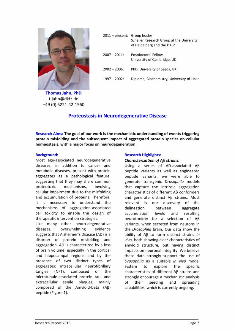
Thomas Jahn, PhD
[email protected] +49 (0) 6221-42-1560
2011 – present: Group leader
Schaller Research Group at the University of Heidelberg and the DKFZ
2007 – 2011: Postdoctoral Fellow University of Cambridge, UK 2002 – 2006: PhD, University of Leeds, UK 1997 – 2002: Diploma, Biochemistry, University of Halle
Proteostasis in Neurodegenerative Disease
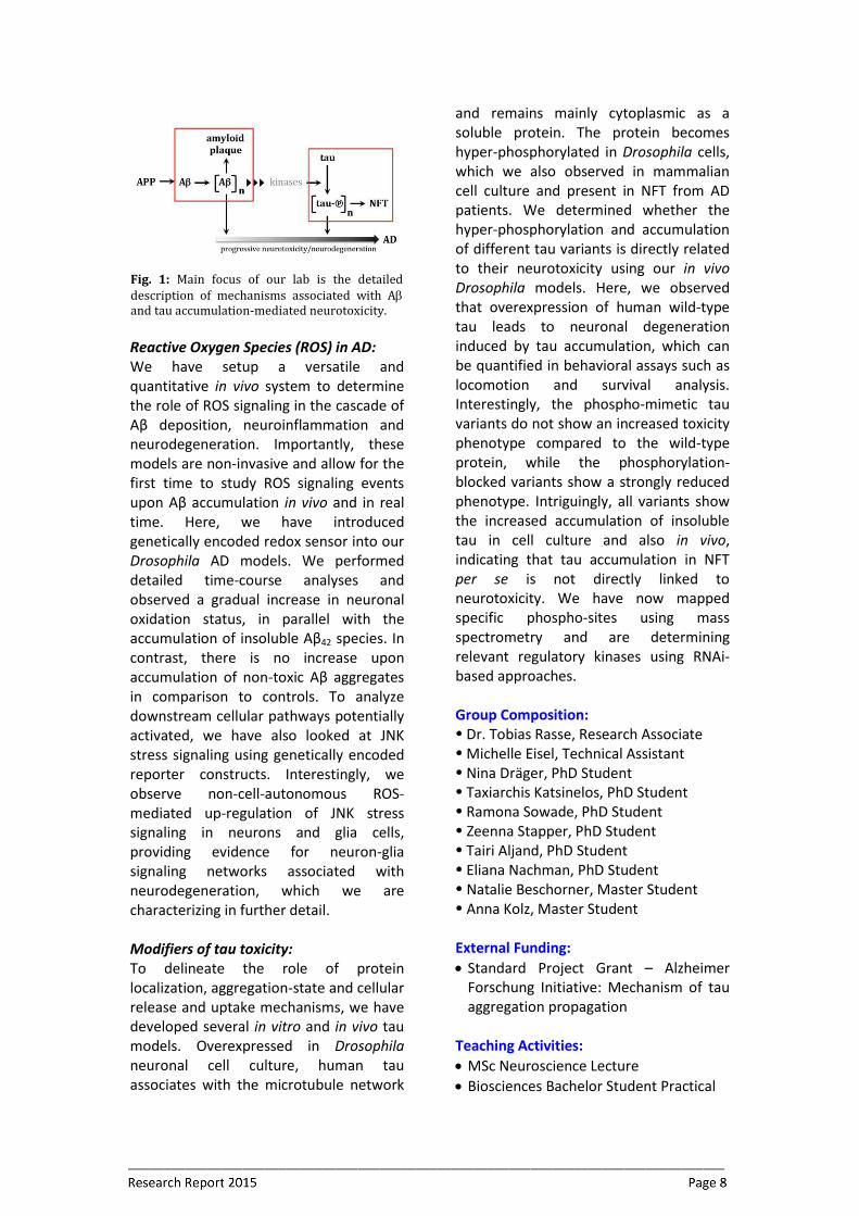
Research Aims: The goal of our work is the mechanistic understanding of events triggering protein misfolding and the subsequent impact of aggregated protein species on cellular homeostasis, with a major focus on neurodegeneration. Background: Most age-associated neurodegenerative diseases, in addition to cancer and metabolic diseases, present with protein aggregates as a pathological feature, suggesting that they may share common proteotoxic mechanisms, involving cellular impairment due to the misfolding and accumulation of proteins. Therefore, it is necessary to understand the mechanisms of aggregation-associated cell toxicity to enable the design of therapeutic intervention strategies. Like many other neuro-degenerative diseases, overwhelming evidence suggests that Alzheimer’s Disease (AD) is a disorder of protein misfolding and aggregation. AD is characterized by a loss of brain volume, especially in the cortical and hippocampal regions and by the presence of two distinct types of aggregates: intracellular neurofibrillary tangles (NFT), composed of the microtubule-associated protein tau, and extracellular senile plaques, mainly composed of the Amyloid-beta (Aβ) peptide (Figure 1).
Research Highlights: Characterization of A strains: Using a series of AD-associated Aβ peptide variants as well as engineered peptide variants, we were able to generate transgenic Drosophila models that capture the intrinsic aggregation characteristics of different Aβ conformers and generate distinct Aβ strains. Most relevant is our discovery of the delineation between aggregate accumulation levels and resulting neurotoxicity for a selection of Aβ variants, when secreted from neurons in the Drosophila brain. Our data show the ability of Aβ to form distinct strains in vivo, both showing clear characteristics of amyloid structure, but having distinct impacts on neuronal integrity. We believe these data strongly support the use of Drosophila as a suitable in vivo model system to explore the specific characteristics of different Aβ strains and strongly encourage a mechanistic analysis of their seeding and spreading capabilities, which is currently ongoing.
Reactive Oxygen Species (ROS) in AD: We have setup a versatile and quantitative in vivo system to determine the role of ROS signaling in the cascade of Aβ deposition, neuroinflammation and neurodegeneration. Importantly, these models are non-invasive and allow for the first time to study ROS signaling events upon Aβ accumulation in vivo and in real time. Here, we have introduced genetically encoded redox sensor into our Drosophila AD models. We performed detailed time-course analyses and observed a gradual increase in neuronal oxidation status, in parallel with the accumulation of insoluble Aβ42 species. In contrast, there is no increase upon accumulation of non-toxic Aβ aggregates in comparison to controls. To analyze downstream cellular pathways potentially activated, we have also looked at JNK stress signaling using genetically encoded reporter constructs. Interestingly, we observe non-cell-autonomous ROS- mediated up-regulation of JNK stress signaling in neurons and glia cells, providing evidence for neuron-glia signaling networks associated with neurodegeneration, which we are characterizing in further detail. Modifiers of tau toxicity: To delineate the role of protein localization, aggregation-state and cellular release and uptake mechanisms, we have developed several in vitro and in vivo tau models. Overexpressed in Drosophila neuronal cell culture, human tau associates with the microtubule network
and remains mainly cytoplasmic as a soluble protein. The protein becomes hyper-phosphorylated in Drosophila cells, which we also observed in mammalian cell culture and present in NFT from AD patients. We determined whether the hyper-phosphorylation and accumulation of different tau variants is directly related to their neurotoxicity using our in vivo Drosophila models. Here, we observed that overexpression of human wild-type tau leads to neuronal degeneration induced by tau accumulation, which can be quantified in behavioral assays such as locomotion and survival analysis. Interestingly, the phospho-mimetic tau variants do not show an increased toxicity phenotype compared to the wild-type protein, while the phosphorylation-blocked variants show a strongly reduced phenotype. Intriguingly, all variants show the increased accumulation of insoluble tau in cell culture and also in vivo, indicating that tau accumulation in NFT per se is not directly linked to neurotoxicity. We have now mapped specific phospho-sites using mass spectrometry and are determining relevant regulatory kinases using RNAi-based approaches. Group Composition: Dr. Tobias Rasse, Research Associate Michelle Eisel, Technical Assistant Nina Dräger, PhD Student Taxiarchis Katsinelos, PhD Student Ramona Sowade, PhD Student Zeenna Stapper, PhD Student Tairi Aljand, PhD Student Eliana Nachman, PhD Student Natalie Beschorner, Master Student Anna Kolz, Master Student External Funding: Standard Project Grant – Alzheimer
Forschung Initiative: Mechanism of tau aggregation propagation
Teaching Activities: MSc Neuroscience Lecture Biosciences Bachelor Student Practical
Fig. 1: Main focus of our lab is the detailed description of mechanisms associated with A and tau accumulation-mediated neurotoxicity.
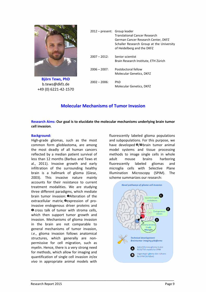
Björn Tews, PhD [email protected]
+49 (0) 6221-42-1570
2012 – present: Group leader Translational Cancer Research German Cancer Research Center, DKFZ
Schaller Research Group at the University of Heidelberg and the DKFZ
2007 – 2012: Senior scientist Brain Research Institute, ETH Zürich 2006 – 2007: Postdoctoral fellow Molecular Genetics, DKFZ 2002 – 2006: PhD Molecular Genetics, DKFZ
Molecular Mechanisms of Tumor Invasion
Research Aims: Our goal is to elucidate the molecular mechanisms underlying brain tumor cell invasion. Background: High-grade gliomas, such as the most common form glioblastoma, are among the most deadly of all human cancers reflected by a median patient survival of less than 12 months (Barbus and Tews et al., 2011). Invasive growth and early infiltration of the surrounding healthy brain is a hallmark of glioma (Giese, 2003). This invasive nature mainly accounts for their resistance to current treatment modalities. We are studying three different paradigms, which mediate brain tumor invasion: Alteration of the extracellular matrix; expression of pro-invasive endogenous driver proteins and
cross talk of tumor with stroma cells, which then support tumor growth and invasion. Mechanisms of glioma invasion in the brain are not comparable to general mechanisms of tumor invasion, i.e., glioma invasion follows anatomical structures, which generally are non-permissive for cell migration, such as myelin. Hence, there is a very strong need for methods, which allow for imaging and quantification of single cell invasion in/ex vivo in appropriate animal models with
fluorescently labeled glioma populations and subpopulations. For this purpose, we have developed / brain tumor animal model systems and tissue processing methods to image single cells in whole adult mouse brains harboring fluorescently labeled gliomas and microglia cells with Selective Plane Illumination Microscopy (SPIM). The scheme summarizes our research:
Research Highlights: Glioma invasion along white matter tracts: The Rho family of small GTPases comprises important regulators of cell migration and invasion (Kempf and Tews et al., 2014). Increased RhoA/ROCK activation is linked to impaired cell migration by induction of profound morphological actin cytoskeleton changes. Interestingly, glioma cells express S1PR2, which senses inhibitory myelin signals and subsequently activates RhoA leading to stress fiber formation. We could show that especially PTEN-negative glioma cells activate a pathway that abolishes RhoA activation, enabling them to invade along white matter structures very efficiently. Glioma microglia X-talk is mediated via sphingolipids: Increasing evidence accumulates that the glioma microenvironment converts the glioma-associated microglia into glioma-supportive, immunosuppressive cells. We investigate the role of S1P and its derivatives as a potential messenger enabling communication between microglia and glioma cells. We have established primary mouse microglia extraction protocols using Cx3cr1-GFP (Jung et al., 2000) and RCAS/TVA mice. We study co-cultured green microglia with mouse glioma cells expressing RFP as well as modulated levels of various S1P-metabolizing enzymes. SPIM is applied to study microglia - glioma interaction and its dependence on S1P in RCAS-driven mouse models of glioma in vivo. Light sheet microscopy (SPIM) to study single cell invasion: We have generated ex and in vivo glioma animal models to image glioma invasion as well as glioma - stroma interaction using light sheet microscopy. We have developed an imaging platform for small molecule screens to identify substances interfering with glioma white matter invasion.
Group Composition: Dr. Julia Bode, Postdoctoral Fellow Peter Wirthschaft, PhD student Anika Simon, PhD student Rakesh Sharma, PhD student Himanshu Soni, PhD student Fabio Dietrich, Technician Bettina Keller, Master student Tatjana Alexander, Bachelor student Stefan Kling, Technician (DKFZ trainee) Last Year Publications: Vajda F. et al. (2014): Cell type-specific
Nogo-A gene ablation promotes axonal regeneration in the injured adult optic nerve. Cell Death Differ. 22(2):323-35.
Petrasek T. et al. (2014). Nogo-A-deficient Transgenic Rats Show Deficits in Higher Cognitive Functions, Decreased Anxiety, and Altered Circadian Activity Patterns. Front Behav Neurosci. 8:90.
Enkel T. et al. (2014). Reduced expression of Nogo-a leads to motivational deficits in rats. Front Behav Neurosci. 8:10.
Kempf A., Tews B. et al. (2014). The Sphingolipid Receptor S1PR2 Is a Receptor for Nogo-A Repressing Synaptic Plasticity. PLoS Biol. 12(1).
Petrasek T. et al. (2014). Nogo-A downregulation impairs place avoidance in the Carousel maze but not spatial memory in the Morris water maze. Neurobiol Learn Mem. 107:42-9.
External Funding: BMBF e:Med: Systems biology of the
Unfolded Protein Response in Glioma (SUPR-G; 01ZX1401A; Coordinator Björn Tews); 1.500 000 €; subproject funding for Tews: 319 161 €.
Otto-Bayer Fellowship Teaching Activities: DKFZ Major Cancer Biology Lecture “Progress in Cancer Research”,
16.10.2014 RNA Summer School Regensburg