Embed Size (px)
Citation preview
1
Research Paper: Ultrasound-Assisted Liquid-Liquid Extraction for Analyzing Methadone in Urine Samples by Gas Chromatography-Mass Spectrometry
Ahmad Shekari1 , Rooholah Valipour2 , Mehrdad Setareh1,2 , Kambiz Soltaninejad2*
1. Laboratory of Forensic Toxicology, Legal Medicine Center, Zanjan, Iran.2. Department of Forensic Toxicology, Legal Medicine Research Center, Legal Medicine Organization, Tehran, Iran.
* Corresponding Author: Kambiz Soltaninejad, PhD.Address: Department of Forensic Toxicology, Legal Medicine Research Center, Legal Medicine Organization, Tehran, Iran.Tel: +98 (21) 55613731E-mail: [email protected]
Background: Methadone abuse and dependence are a growing concern in some countries. The present study developed and validated a rapid, simple, easy, and sensitive method for analyzing methadone in urine specimens. This approach was intended for use in clinical and forensic toxicology and drug screening laboratories.
Methods: We determined methadone in urine samples by Gas Chromatography / Mass Spectrometry (GC / MS). Besides, we used Ultrasound-Assisted Liquid-Liquid Extraction (UALLE) method for the extraction and preconcentration of methadone before analysis.
Results: The detection limit was 2.1 ng/mL and the limit of quantification equaled 7 ng/mL. The methadone calibration curve was linear and ranged from 7 to 10000 ng/mL, and the correlation coefficient was calculated as 0.9984. The tested method was accurate and precise. Recovery fell in the range of 81.3% to 97.4% and the enrichment factor was measured as 8.7. The method was successfully used for determining methadone in real clinical and postmortem urine samples.
Conclusion: The presented method was a rapid, easy, simple, and sensitive procedure; thus, it could be applied in clinical and forensic toxicology laboratories as a routine approach for analyzing methadone.
A B S T R A C T
Keywords:
Gas Chromatography-Mass Spectrometry (GC-MS), Methadone, Ultrasound-Assisted liquid-liquid microextraction, Urine
Citation: Shekari A, Valipour R, Setareh M, Soltaninejad K. Ultrasound-Assisted Liquid-Liquid Extraction for Analyzing Methadone in Urine Samples by Gas Chromatography-Mass Spectrometry. International Journal of Medical Toxicology and Forensic Medicine. 2020; 10(3):29457. https://doi.org/10.32598/ijmtfm.v10i3.29457
: https://doi.org/10.32598/ijmtfm.v10i3.29457
Use your device to scan and read the article online
Article info: Received: 13 March 2020First Revision: 25 March 2020Accepted: 01 Jul 2020
Published: 27 Oct 2020
1. Introduction
ethadone is a synthetic long-acting opioid analgesic, i.e., used in the maintenance treatment of Opioid Use Disorder (OUD) and the relief of moderate to severe pain [1, 2]. Metha-
done, as a mu-opioid receptor full agonist, generates
effects, such as analgesia, euphoria, sedation, miosis, constipation, nausea, and respiratory depression [2]. Methadone Maintenance Therapy (MMT) reduces the risk of illegal drug use, criminal acts, hospital admis-sions, and death in individuals with OUD; however, MMT is associated with some adverse consequences, including accidental and deliberate methadone poison-ing and mortality [3-5].
M
Summer 2020, Volume 10, Number 3
2
Therefore, methadone abuse and dependence are a growing concern in some countries, like Iran [6]. Ac-cordingly, the analysis of methadone in biological sam-ples critically affects the diagnosis, treatment, monitor-ing, and prevention of methadone abuse/dependence in clinical, forensic, and workplace settings.
In clinical, forensic, and workplace settings, deter-mining methadone in bio-samples, such as blood, plas-ma, serum, and saliva is correlated with limitations. Such restrictions include interferences, short detection window, invasive sample collection method, infection risk, and high costs. Thus, urine is a bio-sample choice for drug abuse screening tests in clinical and forensic laboratories. In comparison to blood and plasma, urine is a simple matrix with a longer duration of excretion (detection window). Furthermore, it provides higher concentrations of parent drugs and metabolites than blood; it is available in sufficient quantities as well as in point-of-care tests. Additionally, well-established and cost-effective methods for collecting the sample are accessible [7].
Preparing biological samples is the most significant pre-analytical step for the purification and concentra-tion of the desired analyte(s). It also controls the in-terfering compounds. Rapid and appropriate sample preparation procedures crucially impact achieving robust analytical methods. Multiple conventional ana-lytical techniques, such as Liquid-Liquid Extraction (LLE) and Solid-Phase Extraction (SPE) have been in-troduced for the extraction and analysis of drugs, like methadone in bio-samples [8, 9]. Implementing these techniques for sample preparation is associated with some disadvantages. Conventional LLE procedure is time-consuming and requires using plenty of volumes of pure and hazardous solvents. SPE technique has a relatively desirable efficacy; however, it is time-con-suming due to the need for washing, elution, and evap-oration of the solvents [10]. Therefore, it is necessary to design rapid, feasible, and environmentally compatible sample preparation methods.
Ultrasonic-Assisted Liquid-Liquid Extraction (UAL-LE) technique, compared with the conventional LLE method, presents several advantages. Such benefits in-clude increasing the efficiency and decreasing the time of extraction and volume of used solvents [11]. In this method, a low-frequency ultrasound wave induces a shearing action. It enhances the mass transfer and pro-duces cavitations (acoustic cavitations phenomenon). The sample with added solvent is incorporated in an
ultrasonic bath and exposed to ultrasonic waves for a few minutes [12-14].
Several analytical techniques have been established for analyzing methadone and its metabolites in bio-samples, such as gas chromatography and liquid chro-matography with SPE or LLE, as sample preparation procedures [8-18]; however, data on the urine sample preparation with UALLE for determining methadone by Gas Chromatography/Mass Spectrometry (GC/MS) are scarce.
In this study, we validated and optimized a rapid, easy, and sensitive UALLE-GC-MS for the extraction, preconcentration, and analysis of methadone in human urine samples.
2. Materials and Methods
Methadone standard solution (100 µg/mL) was ob-tained from Sigma Aldrich (St. Louis, USA). Metha-nol, chloroform, sodium hydroxide, and double-dis-tilled water were obtained from Merck (Darmstadt, Germany). All chemicals were of analytical grade.
Methadone standard solutions were prepared by dilu-tion with methanol to a final concentration of 10 µg/mL and were used for sample calibration. A calibration curve was drawn by analyzing drug-free urine samples spiked with methadone at concentrations of 7, 10, 500, 5000, and 10000 ng/mL.
Blank human urine samples (drug-free) were obtained from healthy volunteers in our laboratory and analyzed by the routine GC/MS method. The collected samples were stored in containers and were kept frozen at -20º C before analysis. The frozen urine specimens were thawed at room temperature. Additionally, we em-ployed the optimized extraction method for analyzing 50 real urine samples from patients undergoing MMT; postmortem urine samples were also collected from the cadavers with positive opioids abuse/poisoning history. These samples were transferred to the forensic toxicol-ogy laboratory of the Zanjan legal medicine center (Za-njan City, Iran).
In this study, we used and modified our previously-applied method [18]. In the screw cap test tube, a 1mL urine sample for an appropriate amount of sodium hy-droxide (4mol/L) was added and the pH of the samples was adjusted at 10 (780 pH Meter, Metrohm, Switzer-land). Then, the samples were immersed in an ultrason-ic bath (305W, 40KHz, Soltec, Italy) and ultrasonicated
Shekari A, et al. Ultrasound-Assisted Liquid-Liquid Extraction for Analyzing Methadone in Urine. IJMTFM. 2020; 10(3):29457.
Summer 2020, Volume 10, Number 3
3
for 3 min. After ultrasonication, 1mL chloroform was added to the sample. Next, the samples were vortexed (Velp ZX3, South Korea) for 1 min, followed by ultra-sonication for 5 min, again.
The samples were centrifuged at 1500 g for 5 min. After centrifugation, the liquid phase was discarded and the sediment phase was removed with a microsy-ringe (Hamilton, USA) and transferred to a vial. After the evaporation of the solvent under the nitrogen gas stream, the residue was dissolved in 100μl methanol and filtrated by a 0.22 μm filter (Macherey-Nagel GmbH, Germany) and injected into the GC-MS.
A gas chromatograph (7890, Agilent, USA) equipped with a split/splitless injector was used for GC-MS analysis. The GC column was HP-5MS (5% phenyl & 95% dimethyl polysiloxane, 30 m length x 0.25 mm ID x 0.25 μm film thickness) (Agilent, USA). The mass analyzer (Quadrupole, Agilent, USA) was connected to the column. Helium 99.999% was used as the car-rier gas. The injection port temperature and the transfer line temperatures were 250° C and 280° C, respective-ly. The initial column oven was set to 60° C and held constant for 1 min. The temperature program rate was 20° C/min and the final temperature was set to 280° C and eventual hold for 15 minutes. The mass spectrome-ter was operated by electron impact (70 eV) in positive full scan mode (50-550 m/z). Wiley and NIST libraries were used for the analysis of samples.
The optimization and validation of the UALLE-GC-MS procedure were as follows. The pH and ultrasoni-cation time were regarded as procedure parameters af-fecting the UALLE procedure.
The validation parameters, including precision, accu-racy, linearity, the limits of detection and quantifica-tion, recovery, enrichment factor, and selectivity were evaluated [19]. The method validation was performed on the human urine samples.
The calibration curve was drawn by analyzing 5 urine samples spiked with methadone in the concentration range of 7-10000 ng/mL. For each standard solution, we conducted triplicate measurements.
The Limit of Quantification (LOQ) was determined during the evaluation of the linear range of the calibra-tion curve. The Limit of Detection (LOD) and LOQ were calculated according to LOD =3.3𝜎/S and LOQ = 10𝜎/S; where 𝜎 is the standard deviation of the re-sponse, and S demonstrates the calibration curve slope.
Accuracy and precision were evaluated based on 3 replicates of 4 urine samples spiked with methadone in the concentrations of 7, 10, 400, and 8000 ng/mL. Ac-curacy and precision were expressed as the percentage of the true value (relative error percentage).
Reproducibility was determined by the same afore-mentioned range of concentrations. Each concentration was measured in 3 replicates over 5 consecutive days. For each value, we statistically determined its relative standard deviation by Microsoft Office Excel (Micro-soft, USA).
The extraction recovery was defined and determined as the ratio between the analyte amount in the sediment and the initial amount of the analyte within the sample [19]. The method recovery was measured at 3 concen-tration levels, including 1 µg/mL, 1.5 µg/mL, and 2 µg/mL. The same value was reported as a percentage of the experimentally derived concentration to the nomi-nal concentration. Furthermore, the Enrichment Factor (EF) was calculated according to a previous study [16].
Method selectivity was evaluated by the analysis of 6 blank urine samples to exclude potential interferences. Then, it was tested by analyzing urine samples spiked with a mixture of common licit and illicit drugs in con-centrations ranged from 100-500 ng/mL.
The extraction method was determined and validated with the greatest response. The validated method was used for analyzing methadone concentrations on real urine samples.
3. Results
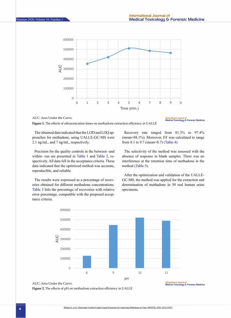
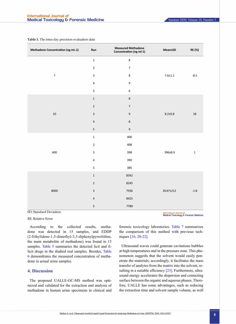
The variables of UALLE efficiency were selected based on preliminary experiments on the distinct re-sponses of the variables to achieve maximum AUC. Figure 1 shows the effect of sonication time on the ex-traction efficiency. The relevant results suggested that 5 min is an optimum time for ultrasonication. Accord-ing to the optimization results, pH=10 provided the optimum effect on the extraction of methadone from urine samples (Figure 2).
The standard calibration curve for methadone was linear in the concentration range of 7-10000 ng/mL; with a regression equation of Y=994.38X + 73553, and a correlation coefficient of 0.9984. These results were generally considered as evidence for indicating desir-able linearity in the concentration range.
Shekari A, et al. Ultrasound-Assisted Liquid-Liquid Extraction for Analyzing Methadone in Urine. IJMTFM. 2020; 10(3):29457.
Summer 2020, Volume 10, Number 3
4
The obtained data indicated that the LOD and LOQ ap-proaches for methadone, using UALLE-GC-MS were 2.1 ng/mL, and 7 ng/mL, respectively.
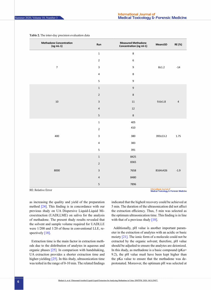
Precision for the quality controls in the between -and within- run are presented in Table 1 and Table 2, re-spectively. All data fell in the acceptance criteria. These data indicated that the optimized method was accurate, reproducible, and reliable.
The results were expressed as a percentage of recov-eries obtained for different methadone concentrations. Table 3 lists the percentage of recoveries with relative error percentage, compatible with the proposed accep-tance criteria.
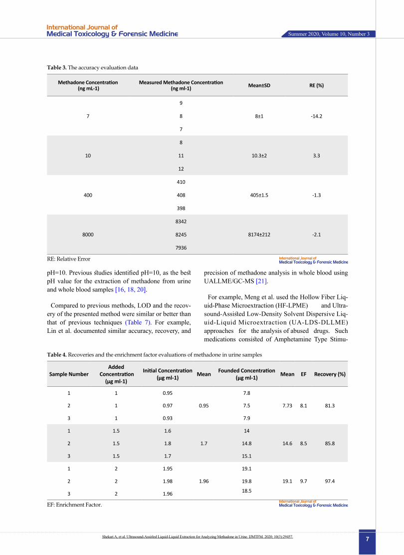
Recovery rate ranged from 81.3% to 97.4% (mean=88.1%). Moreover, EF was calculated to range from 8.1 to 9.7 (mean=8.7) (Table 4).
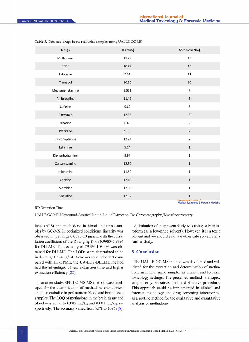
The selectivity of the method was assessed with the absence of response in blank samples. There was no interference at the retention time of methadone in the method (Table 5).
After the optimization and validation of the UALLE-GC-MS, the method was applied for the extraction and determination of methadone in 50 real human urine specimens.
AUC: Area Under the Curve.
0
100000
200000
300000
400000
500000
600000
0 1 2 3 4 5 6 7 8 9 10
AU
C
Time (min.)
Figure 1. The effects of ultrasonication times on methadone extraction efficiency in UALLE
0
100000
200000
300000
400000
500000
600000
8 9 10 11
AU
C
pH
Figure 2. The effects of pH on methadone extraction efficiency in UALLEAUC: Area Under the Curve.
Shekari A, et al. Ultrasound-Assisted Liquid-Liquid Extraction for Analyzing Methadone in Urine. IJMTFM. 2020; 10(3):29457.
Summer 2020, Volume 10, Number 3
5
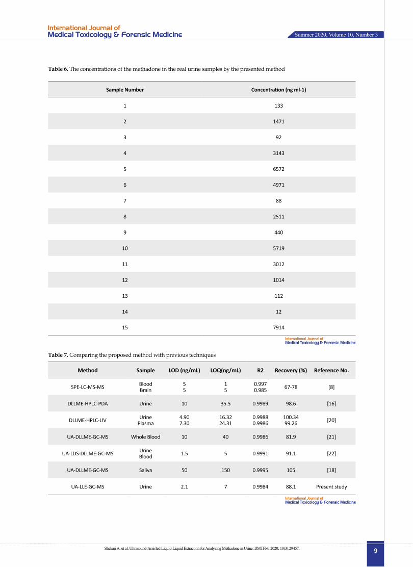
According to the collected results, metha-done was detected in 15 samples, and EDDP (2-Ethylidene-1,5-dimethyl-3,3-diphenylpyrrolidine, the main metabolite of methadone) was found in 13 samples. Table 5 summaries the detected licit and il-licit drugs in the studied real samples. Besides, Table 6 demonstrates the measured concentration of metha-done in actual urine samples.
4. Discussion
The proposed UALLE-GC-MS method was opti-mized and validated for the extraction and analysis of methadone in human urine specimens in clinical and
forensic toxicology laboratories. Table 7 summarizes the comparison of this method with previous tech-niques [16, 20-22].
Ultrasound waves could generate cavitations bubbles at high temperatures and in the pressure zone. This phe-nomenon suggests that the solvent would easily pen-etrate the materials; accordingly, it facilitates the mass transfer of analytes from the matrix into the solvent, re-sulting in a suitable efficiency [23]. Furthermore, ultra-sound energy accelerates the dispersion and contacting surface between the organic and aqueous phases. There-fore, UALLE has some advantages, such as reducing the extraction time and solvent sample volume, as well
Table 1. The intra-day precision evaluation data
RE (%)Mean±SDMeasured Methadone Concentration (ng ml-1)RunMethadone Concentration (ng mL-1)
-8.57.6±1.1
81
7
72
83
94
65
188.2±0.8
81
10
72
93
84
95
1396±8.9
4001
400
4082
3983
3904
3855
-1.88147±212
83421
8000
82452
79363
84254
77895
SD: Standard Deviation.
RE: Relative Error
Shekari A, et al. Ultrasound-Assisted Liquid-Liquid Extraction for Analyzing Methadone in Urine. IJMTFM. 2020; 10(3):29457.
Summer 2020, Volume 10, Number 3
6
as increasing the quality and yield of the preparation method [24]. This finding is in concordance with our previous study on UA Dispersive Liquid-Liquid Mi-croextraction (UADLLME) on saliva for the analysis of methadone. The present study results revealed that the solvent and sample volume required for UADLLE were 1/200 and 1/20 of those in conventional LLE, re-spectively [18].
Extraction time is the main factor in extraction meth-ods due to the distribution of analytes in aqueous and organic phases [25]. In comparison with handshaking, UA extraction provides a shorter extraction time and higher-yielding [25]. In this study, ultrasonication time was tested in the range of 0-10 min. The related findings
indicated that the highest recovery could be achieved at 5 min. The duration of the ultrasonication did not affect the extraction efficiency. Thus, 5 min was selected as the optimum ultrasonication time. This finding is in line with that of a previous study [18].
Additionally, pH value is another important param-eter in the extraction of analytes with an acidic or basic moiety [21]. The ionic form of a molecule could not be extracted by the organic solvent; therefore, pH value should be adjusted to ensure the analytes are deionized. In this study, as methadone is a basic compound (pKa= 9.2), the pH value must have been kept higher than the pKa value to ensure that the methadone was de-protonated. Moreover, the optimum pH was selected at
Table 2. The inter-day precision evaluation data
RE (%)Mean±SDMeasured Methadone Concentration (ng ml-1)RunMethadone Concentration
(ng mL-1)
-148±1.2
81
7
62
93
84
95
49.6±1.8
91
10
82
113
124
85
1.75393±13.2
4051
400
4102
3803
3834
3915
-1.98164±426
84251
8000
83652
76583
84804
78965
RE: Relative Error
Shekari A, et al. Ultrasound-Assisted Liquid-Liquid Extraction for Analyzing Methadone in Urine. IJMTFM. 2020; 10(3):29457.
Summer 2020, Volume 10, Number 3
7
pH=10. Previous studies identified pH=10, as the best pH value for the extraction of methadone from urine and whole blood samples [16, 18, 20].
Compared to previous methods, LOD and the recov-ery of the presented method were similar or better than that of previous techniques (Table 7). For example, Lin et al. documented similar accuracy, recovery, and
precision of methadone analysis in whole blood using UALLME/GC-MS [21].
For example, Meng et al. used the Hollow Fiber Liq-uid-Phase Microextraction (HF-LPME) and Ultra-sound-Assisted Low-Density Solvent Dispersive Liq-uid-Liquid Microextraction (UA-LDS-DLLME) approaches for the analysis of abused drugs. Such medications consisted of Amphetamine Type Stimu-
Table 4. Recoveries and the enrichment factor evaluations of methadone in urine samples
Recovery (%)EFMeanFounded Concentration (µg ml-1)MeanInitial Concentration
(µg ml-1)
Added Concentration
(µg ml-1)Sample Number
81.38.17.73
7.8
0.95
0.9511
7.50.9712
7.90.9313
85.88.514.6
14
1.7
1.61.51
14.81.81.52
15.11.71.53
97.49.719.1
19.1
1.96
1.9521
19.81.9822
18.51.9623
EF: Enrichment Factor.
Table 3. The accuracy evaluation data
RE (%)Mean±SDMeasured Methadone Concentration (ng ml-1)
Methadone Concentration (ng mL-1)
-14.28±1
9
7 8
7
3.310.3±2
8
10 11
12
-1.3405±1.5
410
400 408
398
-2.18174±212
8342
8000 8245
7936
RE: Relative Error
Shekari A, et al. Ultrasound-Assisted Liquid-Liquid Extraction for Analyzing Methadone in Urine. IJMTFM. 2020; 10(3):29457.
Summer 2020, Volume 10, Number 3
8
lants (ATS) and methadone in blood and urine sam-ples by GC-MS. In optimized conditions, linearity was observed in the range 0.0030-10 μg/mL with the corre-lation coefficient of the R ranging from 0.9985-0.9994 for DLLME. The recovery of 79.3%-103.4% was ob-tained for DLLME. The LODs were determined to be in the range 0.5-4 ng/mL. Scholars concluded that com-pared with HF-LPME, the UA-LDS-DLLME method had the advantages of less extraction time and higher extraction efficiency [22].
In another study, SPE-LC-MS-MS method was devel-oped for the quantification of methadone enantiomers and its metabolite in postmortem blood and brain tissue samples. The LOQ of methadone in the brain tissue and blood was equal to 0.005 mg/kg and 0.001 mg/kg, re-spectively. The accuracy varied from 95% to 109% [8].
A limitation of the present study was using only chlo-roform (as a low-price solvent). However, it is a toxic solvent and we should evaluate other safe solvents in a further study.
5. Conclusion
The UALLE-GC-MS method was developed and val-idated for the extraction and determination of metha-done in human urine samples in clinical and forensic toxicology settings. The presented method is a rapid, simple, easy, sensitive, and cost-effective procedure. This approach could be implemented in clinical and forensic toxicology and drug screening laboratories, as a routine method for the qualitative and quantitative analysis of methadone.
Table 5. Detected drugs in the real urine samples using UALLE-GC-MS
Drugs RT (min.) Samples (No.)
Methadone 11.22 15
EDDP 10.72 13
Lidocaine 9.91 11
Tramadol 10.26 10
Methamphetamine 5.551 7
Amitriptyline 11.49 5
Caffeine 9.82 3
Phenytoin 12.36 3
Nicotine 6.63 2
Pethidine 9.20 2
Cyproheptadine 12.24 2
ketamine 9.14 1
Diphenhydramine 9.97 1
Carbamazepine 12.30 1
Imipramine 11.62 1
Codeine 12.40 1
Morphine 12.60 1
Sertraline 12.32 1
RT: Retention Time.
UALLE-GC-MS: Ultrasound-Assisted Liquid-Liquid Extraction-Gas Chromatography/Mass Spectrometry.
Shekari A, et al. Ultrasound-Assisted Liquid-Liquid Extraction for Analyzing Methadone in Urine. IJMTFM. 2020; 10(3):29457.
Summer 2020, Volume 10, Number 3
9
Table 6. The concentrations of the methadone in the real urine samples by the presented method
Sample Number Concentration (ng ml-1)
1 133
2 1471
3 92
4 3143
5 6572
6 4971
7 88
8 2511
9 440
10 5719
11 3012
12 1014
13 112
14 12
15 7914
Table 7. Comparing the proposed method with previous techniques
Method Sample LOD (ng/mL) LOQ(ng/mL) R2 Recovery (%) Reference No.
SPE-LC-MS-MS BloodBrain
55
15
0.9970.985 67-78 [8]
DLLME-HPLC-PDA Urine 10 35.5 0.9989 98.6 [16]
DLLME-HPLC-UV UrinePlasma
4.907.30
16.3224.31
0.99880.9986
100.3499.26 [20]
UA-DLLME-GC-MS Whole Blood 10 40 0.9986 81.9 [21]
UA-LDS-DLLME-GC-MS UrineBlood 1.5 5 0.9991 91.1 [22]
UA-DLLME-GC-MS Saliva 50 150 0.9995 105 [18]
UA-LLE-GC-MS Urine 2.1 7 0.9984 88.1 Present study
Shekari A, et al. Ultrasound-Assisted Liquid-Liquid Extraction for Analyzing Methadone in Urine. IJMTFM. 2020; 10(3):29457.
Summer 2020, Volume 10, Number 3
10
Ethical Considerations
Compliance with ethical guidelines
All procedures performed in the study involving hu-man participants were following the ethical standards of the Legal Medicine Research Center’s ethical com-mittee.
Funding
This research was supported by the Legal Medicine Research Center, Legal Medicine Organization, Teh-ran- Iran (Grant No. 20742).
Author's contributions
All authors contributed in preparing this article.
Conflict of interest
The authors declared no conflicts of interest.
Acknowledgements
We gratefully acknowledge all technical personnel of the Forensic Toxicology Laboratory of the Legal Medicine Center of Zanjan Province for their techni-cal assistance. Furthermore, we would like to show our gratitude to the Legal Medicine Research Center, Legal Medicine Organization, Tehran City, Iran for financial-ly supporting this study.
References
[1] Noble F, Marie N. Management of opioid addiction with opioid substitution treatments: Beyond methadone and bu-prenorphine. Front Psychiatry. 2019; 9:742. [DOI:10.3389/fpsyt.2018.00742] [PMID] [PMCID]
[2] Mercadante S, David F, Villari P, Spedale VM, Casuccio A. Methadone versus morphine for postoperative pain in patients undergoing surgery for gynecological cancer: A randomized controlled clinical trial. J Clin Anesth. 2020; 61:109627. [DOI:10.1016/j.jclinane.2019.109627] [PMID]
[3] Bell J, Zador D. A risk-benefit analysis of methadone maintenance treatment. Drug Saf. 2000; 22(3):179-90. [DOI:10.2165/00002018-200022030-00002] [PMID]
[4] Clausen T, Anchersen K, Waal H. Mortality prior to, during and after opioid maintenance treatment (OMT): A national prospective cross-registry study. Drug Alcohol Depend.
2008; 94(1-3):151-7. [DOI:10.1016/j.drugalcdep.2007.11.003] [PMID]
[5] Soltaninejad K, Hassanian-Moghaddam H, Shadnia S. Methadone related poisoning on the rise in Tehran, Iran. Asia Pac J Med Toxicol. 2014; 3:104-9. [DOI: 10.22038/AP-JMT.2014.3378]
[6] Soroosh D, Neamatshahi M, Zarmehri B, Nakhaee S, Mehr-pour O. Drug-induced prolonged corrected QT interval in patients with methadone and opium overdose. Subst Abuse Treat Prev Policy. 2019; 14(1):8. [DOI:10.1186/s13011-019-0196-3] [PMID] [PMCID]
[7] Hadland SE, Levy S. Objective testing: Urine and other drug tests. Child Adolesc Psychiatr Clin N Am. 2016; 25(3):549-65. [DOI:10.1016/j.chc.2016.02.005] [PMID] [PM-CID]
[8] Dollerup Holm KM, Linnet K. Chiral analysis of metha-done and its main metabolite, EDDP, in postmortem brain and blood by automated SPE and liquid chromatography-mass spectrometry. J Anal Toxicol. 2012; 36(7):487-96. [DOI:10.1093/jat/bks057] [PMID]
[9] Castro AL, Tarelho S, Silvestre A, Teixeira HM. Simultane-ous analysis of some club drugs in whole blood using solid phase extraction and gas chromatography-mass spectrom-etry. J Forensic Leg Med. 2012; 19(2):77-82. [DOI:10.1016/j.jflm.2011.12.006] [PMID]
[10] Shamsipur M, Fattahi N. Extraction and determination of opium alkaloids in urine samples using dispersive liq-uid-liquid microextraction followed by high performance liquid chromatography. J Chromatogr B Analyt Technol Biomed Life Sci. 2011; 879(28):2978-83. [DOI:10.1016/j.jchromb.2011.08.033] [PMID]
[11] Rezaeepour R, Heydari R, Ismaili A. Ultrasound and salt-assisted liquid-liquid extraction as an efficient method for natural product extraction. Anal Methods. 2015, 7:3253-9. [DOI:10.1039/C5AY00150A]
[12] Ridgway K, Lalljie SPD, Smith RM. Sample preparation techniques for the determination of trace residues and con-taminants in foods. J Chromatogr A. 2007; 1153(1-2):36-53. [DOI:10.1016/j.chroma.2007.01.134] [PMID]
[13] Pavlovic’ DM, Babic’ S, Horvat AJM, Marija-Kaštelan M. Sample preparation in analysis of pharmaceuticals. Trends Analyt Chem. 2007; 26(11):1062-5. [DOI:10.1016/j.trac.2007.09.010]
[14] Sushma DD, Jaimala JA, Priya BP. Ultrasonica-tion: A unique extraction method of crude drugs. J In-dian Sys Med. 2015; 3(4):203-5. http://oaji.net/arti-cles/2016/3020-1456050200.pdf
[15] Cheng PS, Lee CH, Liu C, Chien CS. Simultaneous de-termination of ketamine, tramadol, methadone, and their metabolites in urine by gas chromatography-mass spec-trometry. J Anal Toxicol. 2008; 32(3):253-9. [DOI:10.1093/jat/32.3.253] [PMID]
[16] Alahyari E, Setareh M, Shekari A, Roozbehani G, Solta-ninejad K. Analysis of opioids in postmortem urine samples by dispersive liquid-liquid microextraction and high per-formance liquid chromatography with photo diode array detection. Egypt J Forensic Sci. 2018; 8:13. [DOI:10.1186/s41935-018-0046-x]
Shekari A, et al. Ultrasound-Assisted Liquid-Liquid Extraction for Analyzing Methadone in Urine. IJMTFM. 2020; 10(3):29457.
Summer 2020, Volume 10, Number 3
11
[17] Bratinčević MV, Visković T, Sutlović D. Comparison of the solid phase and liquid-liquid extraction methods for methadone determinationin human serum and whole blood samples using gas chromatography/mass spectrometry. Arh Hig Rada Toksikol. 2017; 68(4):308-14. [DOI:10.1515/aiht-2017-68-2953] [PMID]
[18] Shekari A, Forouzesh M, Valipour R, Fallah F, Shojaei P. Validation and optimization of ultrasound-assisted disper-sive liquid- liquid microextraction as a preparation method for detection of methadone in saliva with gas chromatogra-phy-mass spectrometry technique. Adv Pharm Bull. 2020; 10(2):329-33. [DOI:10.34172/apb.2020.040] [PMID] [PMCID]
[19] Scientific Working Group for Forensic Toxicology. Sci-entific Working Group for Forensic Toxicology (SWGTOX) standard practices for method validation in forensic toxicol-ogy. J Anal Toxicol. 2013; 37(7):452-74. [DOI:10.1093/jat/bkt054] [PMID]
[20] Ranjbari E, Golbabanezhad-Azizi AA, Hadjmohammadi MR. Preconcentration of trace amounts of methadone in human urine, plasma, saliva and sweat samples using dis-persive liquid-liquid microextraction followed by high per-formance liquid chromatography. Talanta. 2012; 94:116-22. [DOI:10.1016/j.talanta.2012.03.004] [PMID]
[21] Lin Z, Li J, Zhang X, Qiu M, Huang Z, Rao Y. Ultra-sound-assisted dispersive liquid-liquid microextraction for the determination of seven recreational drugs in human whole blood using gas chromatography-mass spectrom-etry. J Chromatogr B Analyt Technol Biomed Life Sci. 2017; 1046:177-84. [DOI:10.1016/j.jchromb.2017.01.024] [PMID]
[22] Meng L, Zhang W, Meng P, Zhu B, Zheng K. Compari-son of hollow fiber liquid-phase microextraction and ultra-sound-assisted low-density solvent dispersive liquid-liquid microextraction for the determination of drugs of abuse in biological samples by gas chromatography-mass spectrom-etry. J Chromatogr B Analyt Technol Biomed Life Sci. 2015; 989:46-53. [DOI:10.1016/j.jchromb.2015.02.039] [PMID]
[23] Zhong Z, Li G, Zhong X, Luo Z, Zhu B. Ultrasound-assist-ed low-density solvent dispersive liquid-liquid extraction for the determination of alkanolamines and alkylamines in cosmetics with ion chromatography. Talanta. 2013; 115:518-25. [DOI:10.1016/j.talanta.2013.04.045] [PMID]
[24] Piao C, Chen L, Wang Y. A review of the extraction and chromatographic determination methods for the analysis of parabens. J Chromatogr B Analyt Technol Biomed Life Sci. 2014; 969:139-48. [DOI:10.1016/j.jchromb.2014.08.015] [PMID]
[25] Chen PS, Haung WY, Huang SD. Analysis of triazine her-bicides using an up-and-down-shaker-assisted dispersive liquid-liquid microextractioncoupled with gas chromatog-raphy-mass spectrometry. J Chromatogr B Analyt Tech-nol Biomed Life Sci. 2014; 955-956:116-23. [DOI:10.1016/j.jchromb.2014.02.032] [PMID]
Shekari A, et al. Ultrasound-Assisted Liquid-Liquid Extraction for Analyzing Methadone in Urine. IJMTFM. 2020; 10(3):29457.
Summer 2020, Volume 10, Number 3
This Page Intentionally Left Blank