Embed Size (px)
Citation preview
Research Paper
www.aging-us.com 3223 AGING (Albany NY)
www.aging-us.com AGING 2016, Vol. 8, Advance
INTRODUCTION
Cells continually experience stress and damage from
exogenous and endogenous sources, and their responses
range from complete recovery to senescence and cell
death [1]. Proliferating cells cannot divide indefinitely
due to the progressive shortness of their telomeres and after almost 60 population doublings (Hayflick limit)
they stop to grow while remaining metabolically active
[1]. Cells can also become senescent prematurely as a
result of stressful events such as oncogene over-
expression and exposure to DNA damage (for example
induced by UV radiation), or oxidative stress (ROS).
This phenomenon, referred as premature senescence,
occurs rapidly after the triggering event and is a
mechanism implicated in cancer and aging. Recent
studies have identified a novel type of cellular
senescence response that occurs rapidly after
inactivation of PTEN, the major regulator of the PI3K/AKT/mTOR pathway in both mouse and human
primary cells [2]. Senescence driven by loss of PTEN is
mediated by activation of mTOR that actively translate
Identification of Salvia haenkei as gerosuppressant agent by using an integrated senescence-screening assay
Ivana Matic1,3, Ajinkya Revandkar8, Jingjing Chen8, Angela Bisio2, Stefano Dall’Acqua5, Veronica Cocetta5, Paola Brun6, Giorgio Mancino3, Martina Milanese7, Maurizio Mattei4, Monica Montopoli5, Andrea Alimonti1,8 1Laboratory for Research and Development in Aging, Atrahasis S.r.l., 00189 Rome, Italy 2Department of Chemistry and Pharmaceutical Technologies, University of Genova, 16126 Genova, Italy 3Research Center, San Pietro “Fatebenefratelli”, 00189 Rome, Italy 4Animal Technology Facility of University Tor Vergata, 00173 Rome, Italy 5Department of Pharmaceutical and Pharmacological Sciences, University of Padova, 35121 Padova, Italy 6Department of Molecular Medicine, University of Padova, 35121 Padova, Italy 7Studio Associato Gaia Snc, 16121 Genova, Italy 8Institute of Oncology Research (IOR), Bellinzona CH 6500, Switzerland Correspondence to: Andrea Alimonti; Monica Montopoli; email: [email protected] ; [email protected] Keywords: senescence-screening assay, senescence, Salvia haenkei, PICS, gerosuppressant Received: August 1, 2016 Accepted: November 14, 2016 Published: December 1, 2016
ABSTRACT
Cellular senescence is a stable cell cycle arrest that is the causative process of aging. The PI3K/AKT/mTOR pathway is implicated in the control of cellular senescence and inhibitors of this pathway have been successfully used for life span prolongation experiments in mammals. PTEN is the major regulator of the PI3K/AKT/mTOR pathway and loss of PTEN promotes a senescence response termed PICS. Here we report a novel-screening assay, for the identification of compounds that block different types of senescence response. By testing a library of more than 3000 natural and chemical compounds in PTEN deficient cells we have found that an extract from Salvia haenkei (SH), a native plant of Bolivia is a potent inhibitor of PICS. SH also decreases replicative and UV-mediated senescence in human primary fibroblasts and in a model of in vitro reconstructed
human epidermis. Mechanistically, SH treatment affects senescence driven by UV by interfering with IL1- signalling. Pre-clinical and clinical testing of this extract by performing toxicity and irritability evaluation in vitro also demonstrate the safety of SH extract for clinical use as anti-aging skin treatment.
www.aging-us.com 3224 AGING (Albany NY)
p53, a potent inducer of senescence [2]. Activation of
the PI3K/AKT/mTOR pathway independent of PTEN
loss is also implicated in replicative senescence, and
inhibition of mTOR was shown to prevent ageing in
different experimental in vivo models [3-6].
Interestingly, rapamycin and metformin two potent
mTOR inhibitors, suppress geroconversion, prevent
cancer and have minor side effects when administered
long-term in anti-aging doses [7-22]. Activation of the
PI3K/AKT pathway is also implicated in UV induced
cellular senescence, a phenomenon known as photo
ageing. Recent findings show that UV irradiation can
activate AKT and mTOR, thus boosting senescence and
photo aging [23-26]. Considering the need for cost
effective active agents that prevent or arrest cellular
senescence, efforts have been made to develop an assay
for the identification of novel anti-senescence
compounds [27, 28]. Natural compounds represent an
extraordinary inventory of high diversity structural
scaffolds that can offer promising candidates in the
major healthcare challenge of delaying ageing [29].
Plant extracts provide a substantial source of potentially
active compounds, however so far only few natural
compounds have been reported to have anti-senescence
effects [30-34]. Based on our previous research results
[2], we developed an assay that uses Pten null cells as a
tool to rapidly identify compounds that decrease
senescence in primary cells. Positive hits are later on
tested in human primary cells to validate their anti-
senescence efficacy in replicative and UV-mediated
senescence assays.
Here, we report the results of the screening of more than
3000 substances of both natural (plants and marine
extracts) and chemical source. Our data demonstrate
that an extract derived from the Salvia haenkei (SH)
plant is a strong inhibitor of senescence driven by loss
of Pten, senescence associated to replicative stress and
photo aging, both in mouse and human primary cells.
Furthermore, we have evaluated in vitro the toxicity and
irritability of SH on a model of reconstructed human
epidermis (EpiSkin) demonstrating SH safety for the
human skin and anti-senescence activity.
RESULTS
A screening platform for the identification of anti-
senescence compounds
Loss of Pten drives a cellular senescence response in
primary cells termed Pten loss induced cellular
senescence (PICS) [2]. We have recently developed an
effective method for identification of pro-senescence compounds to be used for cancer therapy [35]. By
modifying this screening assay, we developed a
screening platform, for identification of compounds
with anti-senescence activity for the treatment of aging
and aging-related disorders (Fig. 1). As previously
reported [2], upon inactivation of Pten, 30-40% of the
cells undergo to senescence within 4 days. This
provides a screening window to identify hits that affect
senescence in a short time frame, something that would
be complicated by using a different senescence assay
(e.g. replicative senescence). Compounds that decreased
the percentage of senescent cells in the screening
platform were designated as anti-senescence
compounds based on two parameters: 1) cell
proliferation and 2) inhibition of SA-β-galactosidase
staining (SA-β-gal), a prototypical senescence marker
[36]. For the identification of new anti-senescence hits,
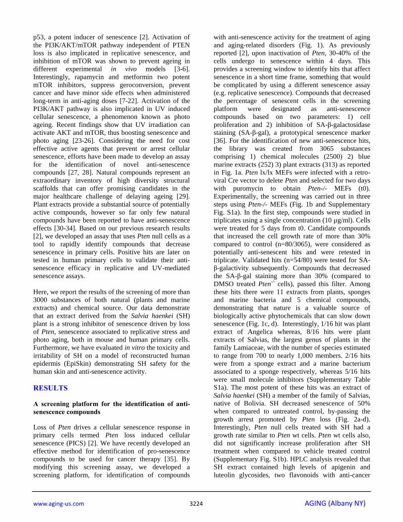
the library was created from 3065 substances
comprising 1) chemical molecules (2500) 2) blue
marine extracts (252) 3) plant extracts (313) as reported
in Fig. 1a. Pten lx/lx MEFs were infected with a retro-
viral Cre vector to delete Pten and selected for two days
with puromycin to obtain Pten-/- MEFs (t0).
Experimentally, the screening was carried out in three
steps using Pten-/- MEFs (Fig. 1b and Supplementary
Fig. S1a). In the first step, compounds were studied in
triplicates using a single concentration (10 µg/ml). Cells
were treated for 5 days from t0. Candidate compounds
that increased the cell growth rate of more than 30%
compared to control (n=80/3065), were considered as
potentially anti-senescent hits and were retested in
triplicate. Validated hits (n=54/80) were tested for SA-
β-galactivity subsequently. Compounds that decreased
the SA-β-gal staining more than 30% (compared to
DMSO treated Pten-/-
cells), passed this filter. Among
these hits there were 11 extracts from plants, sponges
and marine bacteria and 5 chemical compounds,
demonstrating that nature is a valuable source of
biologically active phytochemicals that can slow down
senescence (Fig. 1c, d). Interestingly, 1/16 hit was plant
extract of Angelica whereas, 8/16 hits were plant
extracts of Salvias, the largest genus of plants in the
family Lamiaceae, with the number of species estimated
to range from 700 to nearly 1,000 members. 2/16 hits
were from a sponge extract and a marine bacterium
associated to a sponge respectively, whereas 5/16 hits
were small molecule inhibitors (Supplementary Table
S1a). The most potent of these hits was an extract of
Salvia haenkei (SH) a member of the family of Salvias,
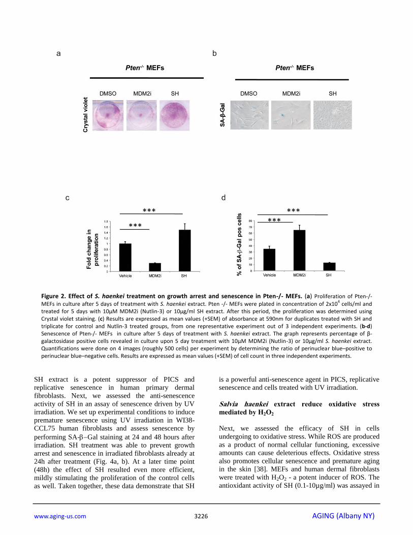
native of Bolivia. SH decreased senescence of 50%
when compared to untreated control, by-passing the
growth arrest promoted by Pten loss (Fig. 2a-d).
Interestingly, Pten null cells treated with SH had a
growth rate similar to Pten wt cells. Pten wt cells also,
did not significantly increase proliferation after SH
treatment when compared to vehicle treated control
(Supplementary Fig. S1b). HPLC analysis revealed that
SH extract contained high levels of apigenin and
luteolin glycosides, two flavonoids with anti-cancer
www.aging-us.com 3225 AGING (Albany NY)
properties [37] (Supplementary Fig. S1c and Supple-
mentary Table S1b).
Identification of compounds that prevent replicative
and radiation-driven senescence
To assess whether identified hits decrease replicative
senescence in vitro, we used human dermal fibroblasts.
To this extent we carried on a series of experiments
using the 3T3 protocol in the WI38-CCL75 cells for a
period of over 3 months. Cells were plated and
subsequently passed and re-plated in the same number
every 3 days, in the presence or absence of selected hits.
Only four out of 16 hits (2 plants and 2 marine extracts)
were able to decrease replicative senescence and were
further developed in our screening cascade. Among
these extracts, SH showed again the most relevant
activity (Supplementary Fig. S2a). As represented in
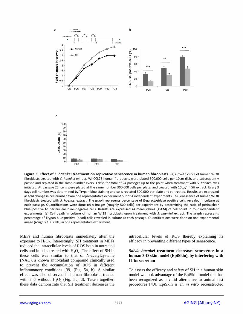
Fig. 3a, while untreated cells stopped growing at
passage 30, cells treated with SH continued to
proliferate. Moreover, senescence in treated cells was
significantly decreased when compared to control as
assessed by the SA--Gal staining (Fig. 3b). The
reduction in the percentage of SA--Gal staining in
these cells was comparable to the one observed in Pten
null MEFs showing a correspondence between these
two models. Importantly, treatment of cells with SH for a period of three months was not associated to increased
cell death, as demonstrated by the cell viability assay
(Fig. 3c). Taken together, these data demonstrate that
Figure 1. Schematic representation of the platform for the in vitro identification of anti-senescent compounds. (a)
Number of chemical and natural extracts from the plants and blue marine ecosystem used for the screening. (b) Schematic representation of the screening steps. (c-d) Cytostatic and cytotoxic compounds were excluded from the screening and only anti-senescence hits progressed. Compounds that induced a statistically significant increase (of 40% or more) in cell growth were considered potential anti-senescent candidates. Instead, compounds that induced a statistically significant decrease in cell number were considered pro-senescent (40% to 60% decrease), and cytotoxic (more than 60% decrease).
www.aging-us.com 3226 AGING (Albany NY)
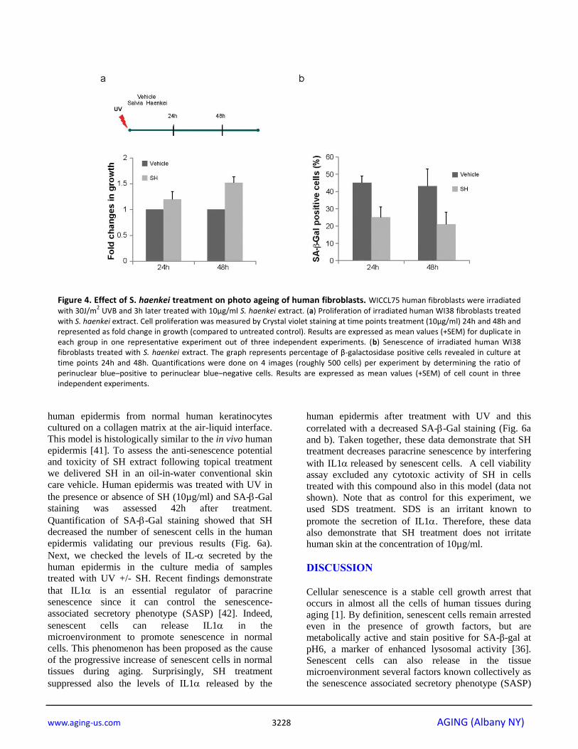
SH extract is a potent suppressor of PICS and
replicative senescence in human primary dermal
fibroblasts. Next, we assessed the anti-senescence
activity of SH in an assay of senescence driven by UV
irradiation. We set up experimental conditions to induce
premature senescence using UV irradiation in WI38-
CCL75 human fibroblasts and assess senescence by
performing SA-Gal staining at 24 and 48 hours after
irradiation. SH treatment was able to prevent growth
arrest and senescence in irradiated fibroblasts already at
24h after treatment (Fig. 4a, b). At a later time point
(48h) the effect of SH resulted even more efficient,
mildly stimulating the proliferation of the control cells
as well. Taken together, these data demonstrate that SH
is a powerful anti-senescence agent in PICS, replicative
senescence and cells treated with UV irradiation.
Salvia haenkei extract reduce oxidative stress
mediated by H2O2
Next, we assessed the efficacy of SH in cells
undergoing to oxidative stress. While ROS are produced
as a product of normal cellular functioning, excessive
amounts can cause deleterious effects. Oxidative stress
also promotes cellular senescence and premature aging in the skin [38]. MEFs and human dermal fibroblasts
were treated with H2O2 - a potent inducer of ROS. The
antioxidant activity of SH (0.1-10µg/ml) was assayed in
Figure 2. Effect of S. haenkei treatment on growth arrest and senescence in Pten-/- MEFs. (a) Proliferation of Pten-/-
MEFs in culture after 5 days of treatment with S. haenkei extract. Pten -/- MEFs were plated in concentration of 2x10⁴ cells/ml and
treated for 5 days with 10µM MDM2i (Nutlin-3) or 10µg/ml SH extract. After this period, the proliferation was determined using Crystal violet staining. (c) Results are expressed as mean values (+SEM) of absorbance at 590nm for duplicates treated with SH and triplicate for control and Nutlin-3 treated groups, from one representative experiment out of 3 independent experiments. (b-d) Senescence of Pten-/- MEFs in culture after 5 days of treatment with S. haenkei extract. The graph represents percentage of β-galactosidase positive cells revealed in culture upon 5 day treatment with 10µM MDM2i (Nutlin-3) or 10µg/ml S. haenkei extract. Quantifications were done on 4 images (roughly 500 cells) per experiment by determining the ratio of perinuclear blue–positive to perinuclear blue–negative cells. Results are expressed as mean values (+SEM) of cell count in three independent experiments.
www.aging-us.com 3227 AGING (Albany NY)
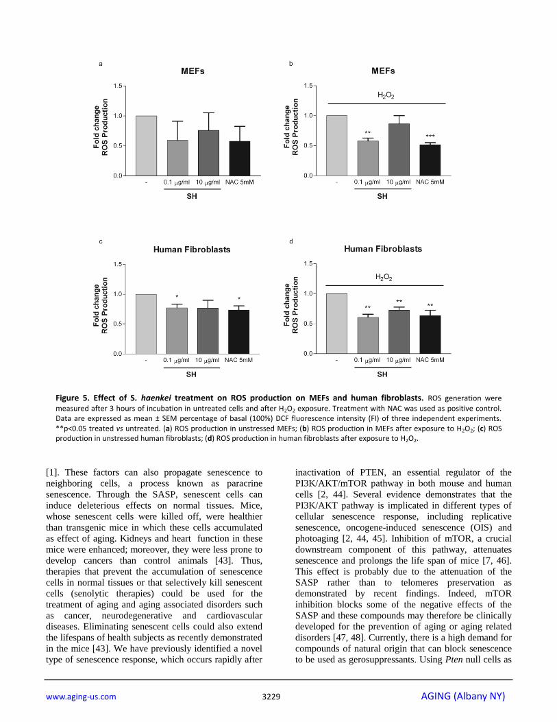
MEFs and human fibroblasts immediately after the
exposure to H2O2. Interestingly, SH treatment in MEFs
reduced the intracellular levels of ROS both in untreated
cells and in cells treated with H2O2. The effect of SH in
these cells was similar to that of N-acetylcysteine
(NAC), a known antioxidant compound clinically used
to prevent the accumulation of ROS in different
inflammatory conditions [39] (Fig. 5a, b). A similar effect was also observed in human fibroblasts treated
with and without H2O2 (Fig. 5c, d). Taken together,
these data demonstrate that SH treatment decreases the
intracellular levels of ROS thereby explaining its
efficacy in preventing different types of senescence.
Salvia haenkei treatment decreases senescence in a
human 3-D skin model (EpiSkin), by interfering with
IL1 secretion
To assess the efficacy and safety of SH in a human skin model we took advantage of the EpiSkin model that has
been recognized as a valid alternative to animal test
procedures [40]. EpiSkin is an in vitro reconstructed
Figure 3. Effect of S. haenkei treatment on replicative senescence in human fibroblasts. (a) Growth curve of human WI38
fibroblasts treated with S. haenkei extract. WI-CCL75 human fibroblasts were plated 300.000 cells per 10cm dish, and subsequently passed and replated in the same number every 3 days for total of 24 passages up to the point when treatment with S. haenkei was initiated. At passage 25, cells were plated at the same number 300.000 cells per plate, and treated with 10µg/ml SH extract. Every 3 days cell number was determined by Trypan blue staining and cells replated 300.000 per plate and re-treated. Results are expressed as fold change in cell number from one representative experiment out of 4 independent experiments. (b) Senescence of human WI38 fibroblasts treated with S. haenkei extract. The graph represents percentage of β-galactosidase positive cells revealed in culture at each passage. Quantifications were done on 4 images (roughly 500 cells) per experiment by determining the ratio of perinuclear blue–positive to perinuclear blue–negative cells. Results are expressed as mean values (+SEM) of cell count in four independent experiments. (c) Cell death in culture of human WI38 fibroblasts upon treatment with S. haenkei extract. The graph represents percentage of Trypan blue positive (dead) cells revealed in culture at each passage. Quantifications were done on one experimental image (roughly 100 cells) in one representative experiment.
www.aging-us.com 3228 AGING (Albany NY)
human epidermis from normal human keratinocytes
cultured on a collagen matrix at the air-liquid interface.
This model is histologically similar to the in vivo human
epidermis [41]. To assess the anti-senescence potential
and toxicity of SH extract following topical treatment
we delivered SH in an oil-in-water conventional skin
care vehicle. Human epidermis was treated with UV in
the presence or absence of SH (10µg/ml) and SA--Gal
staining was assessed 42h after treatment.
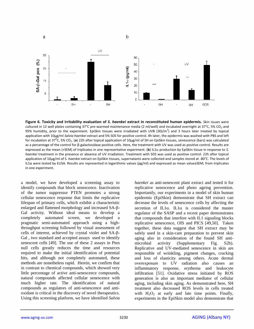
Quantification of SA--Gal staining showed that SH
decreased the number of senescent cells in the human
epidermis validating our previous results (Fig. 6a).
Next, we checked the levels of IL- secreted by the
human epidermis in the culture media of samples
treated with UV +/- SH. Recent findings demonstrate
that IL1 is an essential regulator of paracrine
senescence since it can control the senescence-
associated secretory phenotype (SASP) [42]. Indeed,
senescent cells can release IL1 in the
microenvironment to promote senescence in normal
cells. This phenomenon has been proposed as the cause
of the progressive increase of senescent cells in normal
tissues during aging. Surprisingly, SH treatment
suppressed also the levels of IL1 released by the
human epidermis after treatment with UV and this
correlated with a decreased SA--Gal staining (Fig. 6a
and b). Taken together, these data demonstrate that SH
treatment decreases paracrine senescence by interfering
with IL1released by senescent cells. A cell viability
assay excluded any cytotoxic activity of SH in cells
treated with this compound also in this model (data not
shown). Note that as control for this experiment, we
used SDS treatment. SDS is an irritant known to
promote the secretion of IL1. Therefore, these data
also demonstrate that SH treatment does not irritate
human skin at the concentration of 10µg/ml.
DISCUSSION
Cellular senescence is a stable cell growth arrest that
occurs in almost all the cells of human tissues during
aging [1]. By definition, senescent cells remain arrested
even in the presence of growth factors, but are
metabolically active and stain positive for SA-β-gal at
pH6, a marker of enhanced lysosomal activity [36].
Senescent cells can also release in the tissue
microenvironment several factors known collectively as
the senescence associated secretory phenotype (SASP)
Figure 4. Effect of S. haenkei treatment on photo ageing of human fibroblasts. WICCL75 human fibroblasts were irradiated with 30J/m
2 UVB and 3h later treated with 10μg/ml S. haenkei extract. (a) Proliferation of irradiated human WI38 fibroblasts treated
with S. haenkei extract. Cell proliferation was measured by Crystal violet staining at time points treatment (10μg/ml) 24h and 48h and represented as fold change in growth (compared to untreated control). Results are expressed as mean values (+SEM) for duplicate in each group in one representative experiment out of three independent experiments. (b) Senescence of irradiated human WI38 fibroblasts treated with S. haenkei extract. The graph represents percentage of β-galactosidase positive cells revealed in culture at time points 24h and 48h. Quantifications were done on 4 images (roughly 500 cells) per experiment by determining the ratio of perinuclear blue–positive to perinuclear blue–negative cells. Results are expressed as mean values (+SEM) of cell count in three independent experiments.
www.aging-us.com 3229 AGING (Albany NY)
[1]. These factors can also propagate senescence to
neighboring cells, a process known as paracrine
senescence. Through the SASP, senescent cells can
induce deleterious effects on normal tissues. Mice,
whose senescent cells were killed off, were healthier
than transgenic mice in which these cells accumulated
as effect of aging. Kidneys and heart function in these
mice were enhanced; moreover, they were less prone to
develop cancers than control animals [43]. Thus,
therapies that prevent the accumulation of senescence
cells in normal tissues or that selectively kill senescent
cells (senolytic therapies) could be used for the
treatment of aging and aging associated disorders such
as cancer, neurodegenerative and cardiovascular
diseases. Eliminating senescent cells could also extend
the lifespans of health subjects as recently demonstrated
in the mice [43]. We have previously identified a novel
type of senescence response, which occurs rapidly after
inactivation of PTEN, an essential regulator of the
PI3K/AKT/mTOR pathway in both mouse and human
cells [2, 44]. Several evidence demonstrates that the
PI3K/AKT pathway is implicated in different types of
cellular senescence response, including replicative
senescence, oncogene-induced senescence (OIS) and
photoaging [2, 44, 45]. Inhibition of mTOR, a crucial
downstream component of this pathway, attenuates
senescence and prolongs the life span of mice [7, 46].
This effect is probably due to the attenuation of the
SASP rather than to telomeres preservation as
demonstrated by recent findings. Indeed, mTOR
inhibition blocks some of the negative effects of the
SASP and these compounds may therefore be clinically
developed for the prevention of aging or aging related
disorders [47, 48]. Currently, there is a high demand for
compounds of natural origin that can block senescence
to be used as gerosuppressants. Using Pten null cells as
Figure 5. Effect of S. haenkei treatment on ROS production on MEFs and human fibroblasts. ROS generation were
measured after 3 hours of incubation in untreated cells and after H2O2 exposure. Treatment with NAC was used as positive control. Data are expressed as mean ± SEM percentage of basal (100%) DCF fluorescence intensity (FI) of three independent experiments. **p<0.05 treated vs untreated. (a) ROS production in unstressed MEFs; (b) ROS production in MEFs after exposure to H2O2; (c) ROS production in unstressed human fibroblasts; (d) ROS production in human fibroblasts after exposure to H2O2.
www.aging-us.com 3230 AGING (Albany NY)
a model, we have developed a screening assay to
identify compounds that block senescence. Inactivation
of the tumor suppressor PTEN promotes a strong
cellular senescence response that limits the replicative
lifespan of primary cells, which exhibit a characteristic
enlarged and flattened morphology and increased SA-β-
Gal activity. Without ideal means to develop a
completely automated screen, we developed a
pragmatic semi-automated approach using a high-
throughput screening followed by visual assessment of
cells of interest, achieved by crystal violet and SA-β-
Gal , two standard and accepted assays used to identify
senescent cells [49]. The use of these 2 assays in Pten
null cells greatly reduces the time and resources
required to make the initial identification of potential
hits, and although not completely automated, these
methods are nonetheless rapid. Herein, we confirm that
in contrast to chemical compounds, which showed very
little percentage of active anti-senescence compounds,
natural compounds affected cellular senescence with
much higher rate. The identification of natural
compounds as regulators of anti-senescence and anti-
oxidant is critical in the discovery of novel therapeutics.
Using this screening platform, we have identified Salvia
haenkei as anti-senescent plant extract and tested it for
replicative senescence and photo ageing prevention.
Importantly, our experiments in a model of skin human
epidermis (EpiSkin) demonstrate that SH extract can
decrease the levels of senescence cells by affecting the
secretion of IL1. IL1 is considered the master
regulator of the SASP and a recent paper demonstrates
that compounds that interfere with IL1 signaling blocks
replicative senescence, OIS and PICS [49,50]. Taken
together, these data suggest that SH extract may be
safely used in a skin-care preparation to prevent skin
aging also in consideration of the found SH anti-
microbial activity (Supplementary Fig. S2b).
Replicative and UV-mediated senescence in skin are
responsible of wrinkling, pigment changes, cracking
and loss of elasticity among others. Acute dermal
overexposure to UV radiation also causes an
inflammatory response, erythema and leukocyte
infiltration [51]. Oxidative stress initiated by ROS
generation is also an important mediator of cellular
aging, including skin aging. As demonstrated here, SH treatment also decreased ROS levels in cells treated
with H2O2 at early and late time points. Finally,
experiments in the EpiSkin model also demonstrate that
Figure 6. Toxicity and irritability evaluation of S. haenkei extract in reconstituted human epidermis. Skin issues were
cultured in 12 well plates containing 37°C pre-warmed maintenance media (2 ml/well) and incubated overnight at 37°C, 5% CO2 and 95% humidity, prior to the experiment. EpiSkin tissues were irradiated with UVB (30J/m
2) and 3 hours later treated by topical
application with 10µg/ml Salvia haenkei extract and 5% SDS for positive control. 4h later, the epidermis was washed with PBS and left for incubation at 37
0C, 5% CO2. (a) 22h after topical application of 10μg/ml of SH on EpiSkin tissues, senescence (bars) was calculated
as a percentage of the control for β-galactosidase positive cells. Here, the treatment with UV was used as positive control. Results are
expressed as the mean (+SEM) of triplicates in one representative experiment. (b) IL1 production by EpiSkin tissue in response to S. haenkei treatment in the presence or absence of UV irradiation. Treatment with SDS was used as positive control. 22h after topical application of 10μg/ml of S. haenkei extract on EpiSkin tissues, supernatants were collected and samples stored at -80°C. The levels of IL1α were tested by ELISA. Results are represented in logarithmic values (pg/ml) and expressed as mean value±SEM, from triplicates in one experiment.
www.aging-us.com 3231 AGING (Albany NY)
a skin care preparation containing SH extract is safe and
not irritant for the human skin. In sum, our findings
describe novel screening assays for the identification of
gerosuppressant agents. Previous screenings have
reported the identification of anti-aging compounds
using different biological systems (e.g. yeast) and
assays (e.g. in vitro assays, computer screening). Since
the pathways that control aging in mammals have
homologs in yeast, flies, and worms, several of these
screenings have been performed in invertebrates instead
that in mammalian cells [52-54]. These screenings have
contributed to the identification of several
gerosuppressants active compounds such as rapamycin,
metformin and resveratrol whose efficacy have been
later on validated in mammalian cells. Our screening
based on the use of PTEN deficient mouse embryonic
fibroblasts in a first step and in the consecutive
validation of positive HITs in human cells offers a
promising alternative to these models for the rapid
identification of effective gerosuppressants.
MATERIALS AND METHODS
Plant material and preparation of plant extract
The extract of Salvia haenkei was kindly provided by
Dr. Bisio from Dept. of Chemistry and Pharmaceutical
Technologies, University of Genoa, Italy. The plant
material was harvested and leaves were put in a
ventilated stove at 45°C for 24 hours, and then ground
as fine powder using a mixer IKA universal M20. A
quantity of 20.0g of powdered dried plant was weighed
in a 100ml conical flask to which 70ml of hexane
(purity 99%) was added for the pre-extraction. The flask
was placed in a bath sonicator (Branson 8210) and
sonicated at a temperature of 40°C for 30 minutes. The
mixture was filtered with filter paper, followed by
washing with 20ml of hexane and then with 50ml of
hexane. The filtrate was poured into a flask and the
solvent was concentrated under vacuum (about
11mmHg) up to 5-10 ml by rotavapor, using a water
bath at 40°C. This residue as poured into a glass
container followed by evaporation of the solvent. The
filtrate was left open overnight in a well-ventilated hood
until complete evaporation of the last traces of solvent.
The solids collected on the filter, were divided and air-
dried overnight in the hood. The dried material is
extracted in the same way with methanol-water (90:10).
The dried material from the filters was dissolved in
70ml of 90% methanol. The mixture was sonicated at
40°C for 30 minutes, after being filtered, then washed
with 20 ml of 90% methanol. The filtrate was poured
into a flask and the solvent completely evaporated under vacuum. The dry extract was dissolved in 90%
methanol in the least possible amount of absolute
methanol, using sonication and poured into a glass
container to evaporate overnight in the hood. The
extract was reconstituted with pure DMSO at a
concentration of 10mg/ml and kept at -20°C until
dilution for the treatment of cell cultures. The SH
extract was analysed by HPLC-DAD and HPLC-MS
obtaining a phytochemical fingerprint. The identified
constituents are summarized in supplementary data
(Supplementary Fig. S1c and Supplementary Table
S1b).
MEFs isolation
Ptenlx/lx
MEFs were prepared as previously described
[36]. Briefly, pregnant female mice at day 13
postcoitum (assuming as day one the first day the plug
was observed) were sacrificed by cervical dislocation.
The uterine horns were dissected out, briefly rinsed in
70% (v/v) ethanol and placed into a petri dish
containing PBS (Gibco 14190-169, without bivalent
cations). Each embryo was separated from its placenta
and surrounding membranes, the brain and dark red
organs were cut out. Embryos were washed with fresh
PBS, removing as much blood as possible. Using a
minimal amount of PBS and razor blades, the embryos
were finely minced into a suspension of cells to which
several ml of trypsin-EDTA (about 1-2ml per embryo,
Gibco 25300-096) was added. Following incubation
with gentle shaking at 37°C for 15min the resulting cell
suspension was pelleted and resuspended in fresh
DMEM (ReadyMix, PAA) containing 10% FCS, 2mM
L-glutamine, 2mM penicillin, 50µg/ml streptomycin.
Cells were plated out at 1 embryo equivalent per 10cm
dish ("passage No. 0"). The adherent fibroblasts reached
confluence at day 4 when they were collected and
stored at -80°C prior to use in the APICS assay (for
details of this assay see also Fig. 1 and Supplementary
Fig. S1a)
Cell cultures and infections
Ptenlx/lx
MEFs were isolated as described previously
[36]. To produce Pten-/-
MEFs, Ptenlx/lx
MEFs were
subsequently infected with a viral vector retro- Cre -
recombinase (Adgene Plasmid pMSCV PIG Cre (Cre
IRES Puro vector)). This retro-Cre was produced by
transfection of Phoenix cells (Eco and Ampho from Life
Technologies) at 70-80% confluence using
Lipofectamine 2000 (Invitrogen). At 70% confluence,
Ptenlx/lx
MEFs were infected with supernatant from
Phoenix cells, collected after 48h of transfection with
retro-Cre vector. To increase the efficiency of infection
5μg/ml Polybrene (Santa Cruz) was used. 12h after the
first infection, Ptenlx/lx
MEFs infection was repeated.
24h later, infected Ptenlx/lx
MEFs were selected with
3μg/ml puromycin. 48h later, Pten-/-
were plated and
treated with compounds within APICS molecular

www.aging-us.com 3232 AGING (Albany NY)
screening assay. As control (Ptenwt
) cells in APICS
assay we used Ptenlx/lx
MEFs infected with a viral vector
retro- PIG (Adgene plasmid pMSCVPIG (Pure
IRESGFPvector)), resistant to puromycin, by following
the same protocol as described for Pten-/-
MEFs. Human
WI38-CCL75 fibroblast cell line (ATCC) was used for
3T3 assay and UV irradiation experiments. All cell
cultures were maintained in fresh DMEM (ReadyMix,
PAA) containing 10% FCS, 2mM L-glutamine, 2mM
penicillin, 50µg/ml streptomycin.
Cell proliferation and viability
Cell proliferation was measured using staining with
Crystal violet colour (Sigma Aldrich). Cells were per-
fixed with 4% formaldehyde for 15min., washed with
PBS and stained with 0.1% Crystal violet for 20
minutes. After 3 wash cycles with PBS, cells were lysed
in 10% acetic acid and color intensity read at 590nm on
SUNRISE ELISA reader (Tecan, Switzerland). Growth
curve analysis was carried out as previously described
in literature [55]. Cell viability was assessed using
Trypan blue exclusion.
SA-β-galactosidase assay
Senescence staining was performed using the
commercial Senescence Detection Kit (Calbiochem,
#JA7633), designed to histochemically detect β-gal
activity in cultured cells at pH 6.0. β-gal at pH 6.0 is
present only in senescent cells and is not found in
presenescent, quiescent, or immortal cells. Standard
protocols were followed [56], and quantifications were
done on 4 images (roughly 500 cells) per experiment by
determining the ratio of perinuclear blue–positive to
perinuclear blue–negative cells. Fluorescent nuclear
staining was performed using 4',6-diamidino-2-
phenylindole (DAPI), purchased from Sigma Aldrich.
3T3 protocol
Human primary fibroblasts WI38-CCL75 were plated in
10cm2 dishes (3x10
5cells/dish), and subsequently
passed and re-plated in the same number every 3 days
for total of 24 passages up to the point when treatment
was initiated. At passage 25, cells were plated at the
same number and treated with the SH extract in single
concentration (10µg/ml). Every 3 days cell number was
determined by Trypan blue counting, cells re-plated and
re-treated. At passages 28, 29 and 30 senescence was
evaluated by measuring β-gal expression.
UV irradiation assay
We tested SH extract for the ability to prevent
senescence in a model of UVB irradiated human
fibroblast primary cells. To this purpose, WI38-CCL75
human fibroblasts were irradiated with the optimized
non cytotoxic dose (30J/m2) of UVB irradiation that
causes senescence. 3h after irradiation, positive hits
were added in single concentration (10µg/ml). Cell
proliferation was determined at different time points
using crystal violet assay. Senescence was measured by
β-gal expression.
ROS production
ROS were quantified using 2′,7′-dichlorofluorescin-
diacetate (H2-DCF-DA, Sigma-Aldrich), as previously
described [57]. Upon cleavage of the acetate groups by
intracellular esterase and oxidation, the H2-DCF-DA is
converted to the fluorescent 2',7'-dichlorofluorescein
(DCF). Briefly, the cells (5×10³) were seeded into 96-
well plates and allowed to adhere overnight. ROS level
was measured after the exposure to SH extract for 3
hours in the absence or presence of H2O2, and
subsequent addition of 50 μM H2-DCF-DA, further
incubation for 30 min at 37°C and washing with
phosphate-buffered saline (PBS). DCF fluorescence
intensity was measured at excitation 485 nm—emission
535 nm, using a Multilabel Plate Reader VICTOR X3
(PerkinElmer). Fold increase in ROS production was
calculated using the equation: (Ftreatment—
Fblank)/(Fcontrol—Fblank), where F is the fluorescence
reading.
EpiSkinLM
The EpiSkinLM model (LM: large model;
manufactured by EPISKIN S.N.C., Lyon, France) is a
reconstructed organotypic culture of human adult
keratinocytes that reproduce a multilayered and
differentiated human epidermis. Briefly, human adult
keratinocytes were seeded on a dermal substitute
consisting of a collagen I matrix coated with a layer of
collagen IV fixed to the bottom of a plastic chamber.
Epithelial differentiation was obtained by an air-
exposed step leading to a 3-dimensional epidermis
construct (1.07cm2 surface), with basal, spinous,
granular layers (with specific markers) and a stratum
corneum. EpiSkinLM units were delivered to the
laboratory within 24 hours after preparation. Upon
arrival, tissues were transferred to 12 well plates
containing 37°C pre-warmed maintenance media (2
ml/well) and incubated overnight at 37°C, 5% CO2 and
95% humidity. Skin units were treated with 30J/m2 of
UVB irradiation. SH extract (10µg/ml) was formulated
in a standard oil emulsion and applied topically to the
surface of the epidermis. 4h after UV irradiation, the
epidermis was washed with PBS and left for incubation
at 37°C, 5% CO2. 42h later, supernatants were collected
and stored at -80°C. To assess toxicity of the extract,
www.aging-us.com 3233 AGING (Albany NY)
cell viability test was performed using MTT assay (In
Vitro Toxicology Assay Kit, Sigma Aldrich) (data not
shown) and to assess the release of IL-1α, we analysed
collected supernatants by ELISA (Abcam) for presence
of IL-1α. For the quantification of senescence in the
EpiSkinLM modelfrozen sections of skin units (6μm
thick) were stained for SA-β-Gal as described above,
42h after irradiation +/- SH treatment at (10µg/ml).
Cytokine assay
Supernatants of EpiSkin epidermis, derived in different
conditions (negative control-PBS, positive control-SDS
and treatment with SH extract 10µg/ml) were collected
and stored at -800C. IL-1α (limit of sensitivity < 10
pg/ml) levels were determined by ELISA kit (Abcam)
according to the manufacturer’s specifications. Results
are expressed as pg/ml and reported as means from
three independent experiments.
Statistical analysis
All values obtained are means of at least three
independent experiments performed in duplicate or
triplicate. Results are presented as mean value ± SEM.
Control and treated groups were compared using the
analysis of variance (ANOVA) test. In all analyses, a p-
value of <0.05 was considered statistically significant.
Data were processed using Assistat (version 7.6b) and
Microsoft Excel software.
ACKNOWLEDGEMENTS
The authors wish to thank J. Cadau and Dr. A. Pagetta
for their technical and software assistance.
AUTHOR CONTRIBUTIONS
Conceived and designed the experiments: IM, MM, AA.
Performed the experiments: IM, AR, JC, AB, MaM,
VC, PB, SDA, SC, MI. Analyzed the data: IM, MM,
AA. Wrote the paper: IM, MM, AA.
CONFLICTS OF INTEREST
Andrea Alimonti, MD has stock options in Juvenor
LLC a skin care company that has developed Salvia haenkei extracts as skin care products.
FUNDING
This work was supported by ERC starting grant to AA
(nr. 261342). I.M is supported by Grant within the EU
project SPECIAL (FP7-KBBE-2010-4-266033). MM is
supported from the University of Padova
(CPDA124517/12).
REFERENCES
1. Campisi J, d’Adda di Fagagna F. Cellular senescence: when bad things happen to good cells. Nat Rev Mol Cell Biol. 2007; 8:729–40. doi: 10.1038/nrm2233
2. Alimonti A, Nardella C, Chen Z, Clohessy JG, Carracedo A, Trotman LC, Cheng K, Varmeh S, Kozma SC, Thomas G, Rosivatz E, Woscholski R, Cognetti F, et al. A novel type of cellular sense-cence that can be enhanced in mouse models and human tumor xenografts to suppress prostate tumorigenesis. J Clin Invest. 2010; 120:681–93. doi: 10.1172/JCI40535
3. Powers RW 3rd, Kaeberlein M, Caldwell SD, Kennedy BK, Fields S. Extension of chronological life span in yeast by decreased TOR pathway signaling. Genes Dev. 2006; 20:174–84. doi: 10.1101/gad.1381406
4. Jia K, Chen D, Riddle DL. The TOR pathway interacts with the insulin signaling pathway to regulate C. elegans larval development, meta-bolism and life span. Development. 2004; 131:3897–906. doi: 10.1242/dev.01255
5. Kapahi P, Zid BM, Harper T, Koslover D, Sapin V, Benzer S. Regulation of lifespan in Drosophila by modulation of genes in the TOR signaling pathway. Curr Biol. 2004; 14:885–90. doi: 10.1016/j.cub.2004.03.059
6. Vellai T, Takacs-Vellai K, Zhang Y, Kovacs AL, Orosz L, Müller F. Genetics: influence of TOR kinase on lifespan in C. elegans. Nature. 2003; 426:620. doi: 10.1038/426620a
7. Harrison DE, Strong R, Sharp ZD, Nelson JF, Astle CM, Flurkey K, Nadon NL, Wilkinson JE, Frenkel K, Carter CS, Pahor M, Javors MA, Fernandez E, Miller RA. Rapamycin fed late in life extends lifespan in genetically heterogeneous mice. Nature. 2009; 460:392–95. doi: 10.1038/nature08221.
8. Blagosklonny MV. Immunosuppressants in cancer prevention and therapy. OncoImmunology. 2013; 2:e26961. doi: 10.4161/onci.26961
9. Blagosklonny MV. Rejuvenating immunity: “anti-aging drug today” eight years later. Oncotarget. 2015; 6:19405–12. doi: 10.18632/oncotarget.3740
10. Bravo-San Pedro JM, Senovilla L. Immunostimulatory activity of lifespan-extending agents. Aging (Albany NY). 2013; 5:793–801. doi: 10.18632/aging.100619
11. Ross C, Salmon A, Strong R, Fernandez E, Javors M, Richardson A, Tardif S. Metabolic consequences of long-term rapamycin exposure on common marmoset monkeys (Callithrix jacchus). Aging (Albany NY). 2015; 7:964–73.
www.aging-us.com 3234 AGING (Albany NY)
doi: 10.18632/aging.100843
12. Liu Y, Diaz V, Fernandez E, Strong R, Ye L, Baur JA, Lamming DW, Richardson A, Salmon AB. Rapamycin-induced metabolic defects are reversible in both lean and obese mice. Aging (Albany NY). 2014; 6:742–54. doi: 10.18632/aging.100688
13. Kondratov RV, Kondratova AA. Rapamycin in preventive (very low) doses. Aging (Albany NY). 2014; 6:158–59. doi: 10.18632/aging.100645
14. Fang Y, Bartke A. Prolonged rapamycin treatment led to beneficial metabolic switch. Aging (Albany NY). 2013; 5:328–29. doi: 10.18632/aging.100554
15. Pospelova TV, Leontieva OV, Bykova TV, Zubova SG, Pospelov VA, Blagosklonny MV. Suppression of replicative senescence by rapamycin in rodent embryonic cells. Cell Cycle. 2012; 11:2402–07. doi: 10.4161/cc.20882
16. Blagosklonny MV. TOR-centric view on insulin resistance and diabetic complications: perspective for endocrinologists and gerontologists. Cell Death Dis. 2013; 4:e964. doi: 10.1038/cddis.2013.506
17. Dodds SG, Livi CB, Parihar M, Hsu HK, Benavides AD, Morris J, Javors M, Strong R, Christy B, Hasty P, Sharp ZD. Adaptations to chronic rapamycin in mice. Pathobiol Aging Age Relat Dis. 2016; 6:31688. doi: 10.3402/pba.v6.31688
18. Pellegrini C, Columbaro M, Capanni C, D’Apice MR, Cavallo C, Murdocca M, Lattanzi G, Squarzoni S. All-trans retinoic acid and rapamycin normalize Hutchinson Gilford progeria fibroblast phenotype. Oncotarget. 2015; 6:29914–28. doi: 10.18632/oncotarget.4939.
19. Menendez JA, Joven J. One-carbon metabolism: an aging-cancer crossroad for the gerosup-pressant metformin. Aging (Albany NY). 2012; 4:894–98. doi: 10.18632/aging.100523
20. Verlingue L, Dugourd A, Stoll G, Barillot E, Calzone L, Londono-Vallejo A. A comprehensive approach to the molecular determinants of lifespan using a Boolean model of geroconversion. Aging Cell. 2016; 15:1018–26; Epub ahead of print. doi: 10.1111/acel.12504
21. Kolesnichenko M, Hong L, Liao R, Vogt PK, Sun P. Attenuation of TORC1 signaling delays replicative and oncogenic RAS-induced sense-cence. Cell Cycle. 2012; 11:2391–401. doi: 10.4161/cc.20683
22. Sousa-Victor P, García-Prat L, Muñoz-Cánoves P. Dual mTORC1/C2 inhibitors: gerosuppressors with potential anti-aging effect. Oncotarget. 2015; 6:23052–54. doi: 10.18632/oncotarget.5563
23. Syed DN, Afaq F, Mukhtar H. Differential activation of signaling pathways by UVA and UVB radiation in normal human epidermal keratinocytes. Photochem Photobiol. 2012; 88:1184–90. doi: 10.1111/j.1751-1097.2012.01115.x
24. Zhang QS, Maddock DA, Chen JP, Heo S, Chiu C, Lai D, Souza K, Mehta S, Wan YS. Cytokine-induced p38 activation feedback regulates the prolonged activation of AKT cell survival pathway initiated by reactive oxygen species in response to UV irradiation in human keratinocytes. Int J Oncol. 2001; 19:1057–61.
25. Cao C, Wan Y. Parameters of protection against ultraviolet radiation-induced skin cell damage. J Cell Physiol. 2009; 220:277–84. doi: 10.1002/jcp.21780
26. Strozyk E, Kulms D. The role of AKT/mTOR pathway in stress response to UV-irradiation: implication in skin carcinogenesis by regulation of apoptosis, autophagy and senescence. Int J Mol Sci. 2013; 14:15260–85. doi: 10.3390/ijms140815260
27. Dabhade P, Kotwal S. Tackling the Aging Process With Bio-Molecules: A possible role for caloric restriction, food-derived nutrients, vitamins, amino acids, peptides, and minerals. J Nutr Gerontol Geriatr. 2013; 32:24–40. doi: 10.1080/21551197.2012.753777
28. Mik V, Szüčová L, Smehilová M, Zatloukal M, Doležal K, Nisler J, Grúz J, Galuszka P, Strnad M, Spíchal L. N9-substituted derivatives of kinetin: effective anti-senescence agents. Phytochemistry. 2011; 72:821–31. doi: 10.1016/j.phytochem.2011.02.002
29. Argyropoulou A, Aligiannis N, Trougakos IP, Skaltsounis AL. Natural compounds with anti-ageing activity. Nat Prod Rep. 2013; 30:1412–37. doi: 10.1039/c3np70031c
30. Xie H, Zhu H, Cheng C, Liang Y, Wang Z. Echinacoside retards cellular senescence of human fibroblastic cells MRC-5. Pharmazie. 2009; 64:752–54.
31. Gupta, Shyam K. Walker, Linda (CARDIFF, CA, US) Prevention of Cellular Senescence in Mammals by Natural Peptide Complexes United States Patent Application 20110190202
32. Gruber, James Vincent Composition For Delaying Cellular Senescence US Patent Application 20110052676
33. Jin J, Liang Y, Xie H, Zhang X, Yao X, Wang Z. Dendroflorin retards the senescence of MRC-5 cells. Pharmazie. 2008; 63:321–23.
34. Corominas-Faja B, Santangelo E, Cuyàs E, Micol V, Joven J, Ariza X, Segura-Carretero A, García J,
www.aging-us.com 3235 AGING (Albany NY)
Menendez JA. Computer-aided discovery of biological activity spectra for anti-aging and anti-cancer olive oil oleuropeins. Aging (Albany NY). 2014; 6:731–41. doi: 10.18632/aging.100691
35. Kalathur M, Toso A, Chen J, Revandkar A, Danzer-Baltzer C, Guccini I, Alajati A, Sarti M, Pinton S, Brambilla L, Di Mitri D, Carbone G, Garcia-Escudero R, et al. A chemogenomic screening identifies CK2 as a target for pro-senescence therapy in PTEN-deficient tumours. Nat Commun. 2015; 6:7227. doi: 10.1038/ncomms8227
36. Dimri GP, Lee X, Basile G, Acosta M, Scott G, Roskelley C, Medrano EE, Linskens M, Rubelj I, Pereira-Smith O. A biomarker that identifies senescent human cells in culture and in aging skin in vivo. Proc Natl Acad Sci USA. 1995; 92:9363–67. doi: 10.1073/pnas.92.20.9363
37. Lin Y, Shi R, Wang X, Shen HM. Luteolin, a flavonoid with potential for cancer prevention and therapy. Curr Cancer Drug Targets. 2008; 8:634–46. doi: 10.2174/156800908786241050
38. Velarde MC, Flynn JM, Day NU, Melov S, Campisi J. Mitochondrial oxidative stress caused by Sod2 deficiency promotes cellular senescence and aging phenotypes in the skin. Aging (Albany NY). 2012; 4:3–12. doi: 10.18632/aging.100423
39. Lan CC, Ho PY, Wu CS, Yang RC, Yu HS. LED 590 nm photomodulation reduces UVA-induced metalloproteinase-1 expression via upregula-tion of antioxidant enzyme catalase. J Dermatol Sci. 2015; 78:125–32. doi: 10.1016/j.jdermsci.2015.02.018
40. Spielmann H, Hoffmann S, Liebsch M, Botham P,
Fentem JH, Eskes C, Roguet R, Cotovio J, Cole T,
Worth A, Heylings J, Jones P, Robles C, et al. The
ECVAM international validation study on in vitro
tests for acute skin irritation: report on the validity
of the EPISKIN and EpiDerm assays and on the Skin
Integrity Function Test. Altern Lab Anim. 2007;
35:559–601.
41. EpiSkin skin irritation test, ECVAM ESAC statement in
2007 · OECD TG 439 published in 2010.
42. Laberge RM, Sun Y, Orjalo AV, Patil CK, Freund A,
Zhou L, Curran SC, Davalos AR, Wilson-Edell KA, Liu S,
Limbad C, Demaria M, Li P, et al. MTOR regulates the
pro-tumorigenic senes-cence-associated secretory
phenotype by promoting IL1A translation. Nat Cell
Biol. 2015; 17:1049–61. doi: 10.1038/ncb3195
43. Childs BG, Durik M, Baker DJ, van Deursen JM.
Cellular senescence in aging and age-related disease:
from mechanisms to therapy. Nat Med. 2015;
21:1424–35. doi: 10.1038/nm.4000
44. Astle MV, Hannan KM, Ng PY, Lee RS, George AJ, Hsu AK, Haupt Y, Hannan RD, Pearson RB. AKT induces senescence in human cells via mTORC1 and p53 in the absence of DNA damage: implications for targeting mTOR during malignancy. Oncogene. 2012; 31:1949–62. doi: 10.1038/onc.2011.394
45. Strozyk E, Kulms D. The role of AKT/mTOR pathway in stress response to UV-irradiation: implication in skin carcinogenesis by regulation of apoptosis, autophagy and senescence. Int J Mol Sci. 2013; 14:15260–85. doi: 10.3390/ijms140815260
46. Popovich IG, Anisimov VN, Zabezhinski MA, Semenchenko AV, Tyndyk ML, Yurova MN, Blagosklonny MV. Lifespan extension and cancer prevention in HER-2/neu transgenic mice treated with low intermittent doses of rapamycin. Cancer Biol Ther. 2014; 15:586–92. doi: 10.4161/cbt.28164
47. Herranz N, Gallage S, Mellone M, Wuestefeld T, Klotz S, Hanley CJ, Raguz S, Acosta JC, Innes AJ, Banito A, Georgilis A, Montoya A, Wolter K, et al. mTOR regulates MAPKAPK2 translation to control the senescence-associated secretory phenotype. Nat Cell Biol. 2015; 17:1205–17. doi: 10.1038/ncb3225
48. Laberge RM, Sun Y, Orjalo AV, Patil CK, Freund A, Zhou L, Curran SC, Davalos AR, Wilson-Edell KA, Liu S, Limbad C, Demaria M, Li P, et al. MTOR regulates the pro-tumorigenic sense-cence-associated secretory phenotype by promoting IL1A translation. Nat Cell Biol. 2015; 17:1049–61. doi: 10.1038/ncb3195
49. Kalathur M, Toso A, Chen J, Revandkar A, Danzer-Baltzer C, Guccini I, Alajati A, Sarti M, Pinton S, Brambilla L, Di Mitri D, Carbone G, Garcia-Escudero R, et al. A chemogenomic screening identifies CK2 as a target for pro-senescence therapy in PTEN-deficient tumours. Nat Commun. 2015; 6:7227. doi: 10.1038/ncomms8227
50. Acosta JC, Banito A, Wuestefeld T, Georgilis A, Janich P, Morton JP, Athineos D, Kang TW, Lasitschka F, Andrulis M, Pascual G, Morris KJ, Khan S, et al. A complex secretory program orchestrated by the inflammasome controls paracrine senescence. Nat Cell Biol. 2013; 15:978–90. doi: 10.1038/ncb2784
51. Alimonti A, Carracedo A, Clohessy JG, Trotman LC, Nardella C, Egia A, Salmena L, Sampieri K, Haveman WJ, Brogi E, Richardson AL, Zhang J, Pandolfi PP. Subtle variations in Pten dose determine cancer susceptibility. Nat Genet. 2010; 42:454–58. doi: 10.1038/ng.556
52. Petrascheck M, Ye X, Buck LB. An antidepres-sant that extends lifespan in adult Caenorhab-ditis elegans. Nature. 2007; 450:553–56. doi: 10.1038/nature05991

www.aging-us.com 3236 AGING (Albany NY)
53. Alavez S, Vantipalli MC, Zucker DJ, Klang IM, Lithgow GJ. Amyloid-binding compounds maintain protein homeostasis during ageing and extend lifespan. Nature. 2011; 472:226–29. doi: 10.1038/nature09873
54. Alavez S, Vantipalli MC, Zucker DJ, Klang IM, Lithgow GJ. Amyloid-binding compounds maintain protein homeostasis during ageing and extend lifespan. Nature. 2011; 472:226–29. doi: 10.1038/nature09873
55. Ishiyama M, Tominaga H, Shiga M, Sasamoto K, Ohkura Y, Ueno K. A combined assay of cell viability and in vitro cytotoxicity with a highly water-soluble tetrazolium salt, neutral red and crystal violet. Biol Pharm Bull. 1996; 19:1518–20. doi: 10.1248/bpb.19.1518
56. Debacq-Chainiaux F, Erusalimsky JD, Campisi J, Toussaint O. Protocols to detect senescence-associated beta-galactosidase (SA-betagal) activity, a biomarker of senescent cells in culture and in vivo. Nat Protoc. 2009; 4:1798–806. doi: 10.1038/nprot.2009.191
57. Catanzaro D, Rancan S, Orso G, Dall’Acqua S, Brun P, Giron MC, Carrara M, Castagliuolo I, Ragazzi E, Caparrotta L, Montopoli M. Boswellia serrata prevents intestinal epithelial barrier from oxidative and inflammatory damage. PLoS One. 2015; 10:e0125375. doi: 10.1371/journal.pone.0125375
www.aging-us.com 3237 AGING (Albany NY)
SUPPLEMENTARY MATERIALS
Supplementary Methods
HPLC-MS and HPLC-DAD-ELSD quali-quantitative analysis
Qualitative and quantitative constituents of Salvia haenkeii were performed by high-performance liquid
chromatography-tandem mass spectrometry (HPLC-MS) and high-performance liquid chromatography coupled with a
diode array detector and an evaporative light scattering detector (HPLC-DAD-ELSD). For HPLC-MS analysis a
Varian 212 binary chromatograph equipped with 500MS ion trap and Prostar 430 autosampler was used (Varian Inc.,
USA). For the HPLC-DAD-ELSD analysis an Agilent 1100 Series chromatograph with 1100 Diode Array detector
and Sedex LX 60 Evaporative Light Scattering Detector (ELSD) was used. As stationary phase an Agilent Eclipse
XDB-C8 2.1 x 150 mm, 3.5 μm (Agilent Tecnologies, USA) was used. Quantification of phenolic constituents was
obtained with the method of calibration curve: rutin (Sigma Aldrich, St. Louis, MO, USA) was used as external
standard for flavonoid quantification. Calibration curves were Y = 144232X + 112 (R2 = 0.9998) for rutin.
Microbiological assessments
Streptococcus pyogenes and Staphylococcus aureus were isolated from clinical specimens of human skin, cultured on
agar plates and identified according to the appearance of colonies, growth conditions and metabolic enzymatic
activities. Bacteria were grown in lysogeny broth (LB) at 37°C until mid-log phase determined by spectrophotometric
analysis, reading the optical density (O.D.) at 600 nm. Bacterial cultures were then diluted in LB to reach
concentration of 1x10⁶ colony-forming units (CFU)/mL and incubated at 37°C for 16 hours with SH at the indicated
concentrations. At the end of incubation, bacterial load was estimated by reading the O.D. using a spectrophotometer
(Sunrise, Tecan; Switzerland). Data were expressed as mean ± standard error of the percentage of O.D. calculated
versus the respective untreated samples.
www.aging-us.com 3238 AGING (Albany NY)
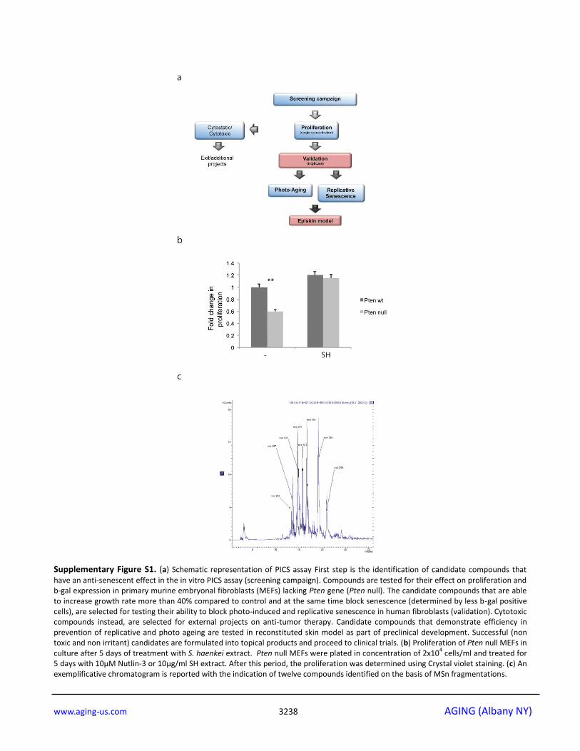
Supplementary Figure S1. (a) Schematic representation of PICS assay First step is the identification of candidate compounds that
have an anti-senescent effect in the in vitro PICS assay (screening campaign). Compounds are tested for their effect on proliferation and b-gal expression in primary murine embryonal fibroblasts (MEFs) lacking Pten gene (Pten null). The candidate compounds that are able to increase growth rate more than 40% compared to control and at the same time block senescence (determined by less b-gal positive cells), are selected for testing their ability to block photo-induced and replicative senescence in human fibroblasts (validation). Cytotoxic compounds instead, are selected for external projects on anti-tumor therapy. Candidate compounds that demonstrate efficiency in prevention of replicative and photo ageing are tested in reconstituted skin model as part of preclinical development. Successful (non toxic and non irritant) candidates are formulated into topical products and proceed to clinical trials. (b) Proliferation of Pten null MEFs in culture after 5 days of treatment with S. haenkei extract. Pten null MEFs were plated in concentration of 2x10
4 cells/ml and treated for
5 days with 10µM Nutlin-3 or 10µg/ml SH extract. After this period, the proliferation was determined using Crystal violet staining. (c) An exemplificative chromatogram is reported with the indication of twelve compounds identified on the basis of MSn fragmentations.

www.aging-us.com 3239 AGING (Albany NY)
Supplementary Figure S2. (a) To assess replicative senescence in vitro, 3T3 protocol was performed on human dermal fibroblasts
(WI38-CCL75 cells, ATCC). Cells were plated and subsequently passed and re-plated (step called passage) in the same number every 3 days, for a period of over 3 months. From passage 25, 3T3 protocol was continued, but in the presence or absence of selected hits (represented here SH extract, sponge associated bacteria, Axinella Verrucosa and Angelica extract). The graph represents percentage of β-galactosidase positive cells revealed in culture on passages 29, 30 and 31. Results are expressed as mean values (+SEM) of cell count in three independent experiments. (b) Antimicrobic effect of Salvia haenkeii treatment on Streptococcus pyogenes and Staphylococcus aureus. Antimicrobic activity was measured after 16 hours of exposure. At the end of incubation, bacterial load was estimated by reading the O.D. using a spectrophotometer. Data were expressed as mean ± standard error of the percentage of O.D. calculated versus the respective untreated samples. *p<0.05 treated vs untreated.
www.aging-us.com 3240 AGING (Albany NY)
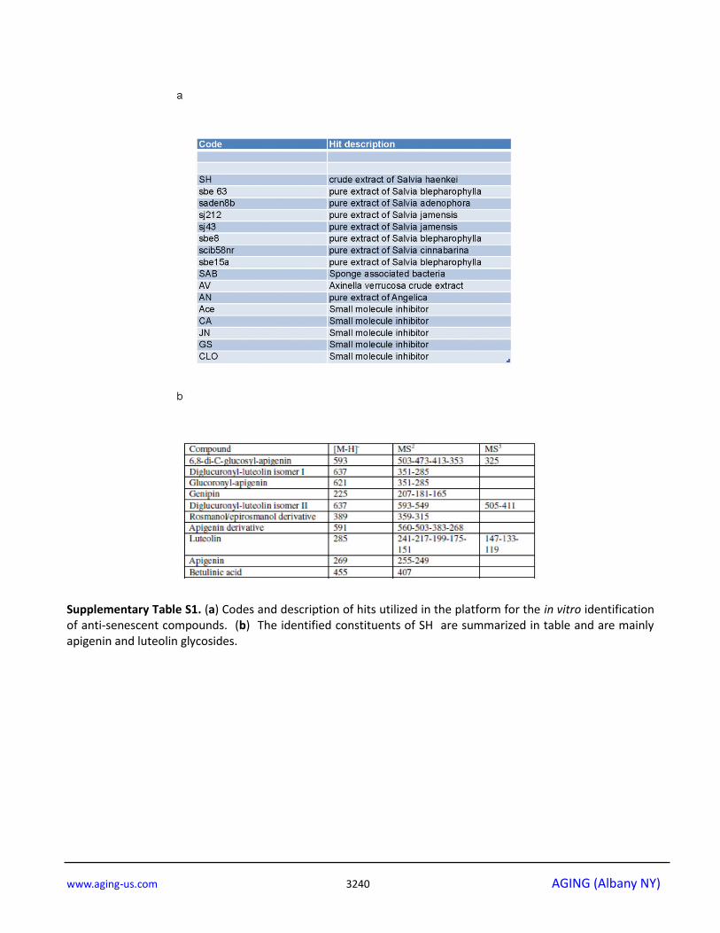
Supplementary Table S1. (a) Codes and description of hits utilized in the platform for the in vitro identification of anti-senescent compounds. (b) The identified constituents of SH are summarized in table and are mainly apigenin and luteolin glycosides.