Embed Size (px)
Citation preview
©2006 L
ANDES BIOSCI
ENCE.
DO NOT DIST
RIBUTE.
[Plant Signaling & Behavior 1:3, 122-133, May/June 2006]; ©2006 Landes Bioscience
122 Plant Signaling & Behavior 2006; Vol. 1 Issue 3
Markus Schlicht1
Miroslav Strnad2
Michael J. Scanlon3
Stefano Mancuso4,5
Frank Hochholdinger6
Klaus Palme7
Dieter Volkmann1
Diedrik Menzel1
Frantisek Baluska1,5,8,*1IZMB; Rheinische Friedrich-Wilhelms-Universität; Bonn, Germany
2Laboratory of Growth Regulators; Palacky University & Institute of ExperimentalBotany ASCR; Olomouc, Czech Republic
3Department of Botany; University of Georgia; Athens, Georgia USA
4Department of Horticulture; University of Florence; Florence, Italy
5International Plant Neurobiology Laboratory; Florence, Italy and Bonn, Germany
6University of Tuebingen; ZMBP—Center for Plant Molecular Biology; Departmentof General Genetics; Tuebingen, Germany
7University of Freiburg; Center for Applied Biosciences; Freiburg, Germany
8Institute of Botany; Slovak Academy of Sciences; Bratislava, Slovak Republic
*Correspondence to: Frantisek Baluska; Rheinische Friedrich-Wilhelms-Universität; Kirschallee 1; Bonn, D 53115 Germany; Tel.: +0049.228.734761;Fax: +0049.228.739004; Email: baluska @uni-bonn.de
Received 03/31/06; Accepted 04/03/06
Previously published online as a Plant Signaling & Behavior E-publication:http://www.landesbioscience.com/journals/psb/abstract.php?id=2759
KEY WORDS
actin, auxin, maize, secretion, vesicles, neuro-transmitter
ACKNOWLEDGEMENTS
See page 131.
NOTE
Supplemental material can be found at:http://www.landesbioscience.com/journals/psb/supplement/schlichtPSB1-3-sup.pdf
Research Paper
Auxin Immunolocalization Implicates Vesicular Neurotransmitter-LikeMode of Polar Auxin Transport in Root Apices
ABSTRACTImmunolocalization of auxin using a new specific antibody revealed, besides the
expected diffuse cytoplasmic signal, enrichments of auxin at end-poles (cross-walls), withinendosomes and within nuclei of those root apex cells which accumulate abundant F-actinat their end-poles. In Brefeldin A (BFA) treated roots, a strong auxin signal was scoredwithin BFA-induced compartments of cells having abundant actin and auxin at theirend-poles, as well as within adjacent endosomes, but not in other root cells. Importantly,several types of polar auxin transport (PAT) inhibitors exert similar inhibitory effects onendocytosis, vesicle recycling, and on the enrichments of F-actin at the end-poles. Thesefindings indicate that auxin is transported across F-actin-enriched end-poles (synapses) vianeurotransmitter-like secretion. This new concept finds genetic support from the sema-phore1, rum1 and rum1/lrt1 mutants of maize which are impaired in PAT, endocytosis andvesicle recycling, as well as in recruitment of F-actin and auxin to the auxin transportingend-poles. Although PIN1 localizes abundantly to the end-poles, and they also fail to sup-port the formation of in these mutants affected in PAT, auxin and F-actin are depletedfrom their end-poles which also fail to support formation of the large BFA-induced com-partments.
INTRODUCTIONPolar transcellular auxin transport (PAT) is important for both growth regulation and
control of polarity and pattern formation in plants.1-5 In the last few years, there has beena dramatic increase in our knowledge on several proteins which are known to be involvedin auxin transport, but we still lack information about how exactly auxin moves acrosscellular boundaries.4-6 Rather unexpectedly, putative auxin transporters of the PIN familywere shown to accomplish rapid vesicular recycling between the plasma membrane andendosomal compartments in Arabidopsis7 and in maize8 cells. There are three classes ofinhibitors, differing chemically and in their action mechanisms, which prevent PAT alongcell files of plant organs. These include TIBA, NPA and several morphactins all of whicheffectively inhibit PAT.9,10 Unfortunately, it is still unknown how any of these PATinhibitors act at the cellular level. Recent studies surprisingly revealed that TIBA and NPAact as unspecific inhibitors of endocytosis and vesicle recycling.11,12 Here we confirm thisfeature also for morphactins.
Apparently unrelated to these classical inhibitors of PAT, auxin efflux from cells is alsoeffectively and rapidly inhibited by two other inhibitors that block secretion from eukaryoticcells, monensin and brefeldin A (BFA).12-15 For instance, the potent exocytosis inhibitorBFA blocks PAT within a few minutes and also leads to the formation of endosomal BFAcompartments,16 within which recycling molecules of the PAT machinery are entrappedtogether with cell wall pectins.12,17 Importantly, although BFA inhibits PAT very rapidly,12,14
nearly two hours of BFA treatment are necessary to remove most, but still not all, of the PINmolecules from the plasma membrane and to trap them within the BFA-induced endo-cytic compartments.8,11,12,16 Similarly like cold treatment and depolymerization of theactin cytoskeleton,7,11,16 TIBA and auxin treatments inhibited endocytosis to such an extentthat larger BFA-induced compartments could not form in the TIBA/ auxin-BFA doubletreated cells.7,11,12
Up to now, all published auxin visualization procedures have drawbacks. First of all,auxin antibodies used in these previous studies are not monospecific. Secondly, the cross-linking agent, EDC, used to immobilize the small auxin molecules3,18 has drastic effectson F-actin in cells of maize root apices (Suppl. Fig. 1). Thus, EDC might alter significantlythe localization of other antigens too. In addition, although auxin responsive promoter
www.landesbioscience.com Plant Signaling & Behavior 123
elements are perfect tools to visualize auxin signaling activities, theyare less useful to localize both extra- and intra-cellular auxin as theyonly show the activity of these diverse auxin responsive promoters.So it is not surprising that the three most often used auxin-responsivepromoters (DR5, BA3, GH3) show different, often contrasting,expression patterns. For instance, the DR5 reporter shows maximumactivity in the root cap columella and quiescent centre cells,19-21
whereas the BA3 has its maximum in root cells embarking on rapidcell elongation22-24 (see also Suppl. Fig. 3). Moreover, these auxin-responsive promoter reporters are not absolutely specific for IAA.For example, the DR5-reporter is activated by brassinolides as well25
and, in fact, it requires exposure to brassinolides to reach full activi-ties.26 Therefore, it is not entirely correct, though often practiced, ifthe DR5 reporter maximum is interpreted as a so-called ‘auxinmaximum’, implying that it would correspond to the highestamount of free auxin in cells.19-21,27
BFA inhibits PAT rapidly,14,15,28 due to a block in auxin effluxafter some 10 minutes.14,28 Importantly, wash-out of BFA restoresauxin efflux also quite rapidly, within some 20 min.28 How is itpossible that such short treatments with a secretion inhibitor manip-ulates auxin efflux so rapidly? At these short exposure times, all thenecessary proteins of the auxin efflux apparatus are still located atthe plasma membrane as their plasma membrane presentation timesare typically above ten minutes.11,12,28 Currently, the most acceptedmodel is that PIN proteins show their auxin efflux activity at theplasma membrane. Although this simple model is appealing, it isunable to explain the rapidity with which BFA inhibits polar trans-port of auxin.14,15,28
As a plausible alternative, a neurotransmitter-like secretory modelhas been proposed in which auxin is sequestered within endosomesvia vesicular transporters and then might be secreted via a BFA-sensitive process out of the cells.4,29,30 If PIN proteins had theiractivities at membranes delimiting vesicles and recycling endo-somes,29,31 they would enrich these vesicular compartments withIAA. Endosomes and recycling vesicles could fuse with the plasmamembrane via a stimulus-activated BFA-sensitive process.32 This
would lead to a secretion of IAA out of cellsin a quantal manner, resembling the processof synaptic vesicle secretion. A neurotrans-mitter-like nature of auxin transport couldexplain the fast inhibitory effects of BFA onPAT as well as the high PIN1 turnover atthe plasma membrane. For this and othercompelling reasons, we have recently intro-duced the term “plant synapse”.29,33 Ourconcept would also explain a second mystery34 concerning Arabidopsis embryos,which generate a so-called auxin maximumat the emerging root pole as well as auxingradients across the developing embryo.3,20
This phenomenon is very difficult toexplain by the classical version of thechemiosmotic theory, because it lacks anymechanisms that would restrict the freediffusion of auxin through the cytoplasm, aswell as from cell-to-cell by the means ofplant plasmodesmata connections.34,35 Theattractive feature of the neurotransmitter-like mode of auxin transport is that endo-
somes could both exclude auxin from the plasmodesmata orificesand transport it effectively towards those subcellular sites which arerelevant for auxin signalling and transport. For instance towards thenuclei, or for the localized secretion in those cells which are part ofthe PAT pathway.
For auxin visualization on the cellular level, a specific antibody isthe tool of choice. However, all auxin antibodies in use are not specificenough. Here we introduce a new highly specific auxin antibody,recognizing only IAA, which allows us to address critical questionswith respect to auxin cell biology. We report here that IAA accumu-lates at the end-poles and adjacent endosomes only in those root cellswhich are active in transcellular transport of auxin.
MATERIALS AND METHODSPlant material and inhibitor treatments. Maize grains (Zea mays L.),
Semaphore1, lrt1, rum1 and lrt1-rum1 mutants were soaked for sixhours and germinated in well moistened rolls of filter paper for fourdays in darkness at 20˚C. Young seedlings with straight primaryroots, either 50–70 mm long (wildtype, lrt1 and rum1), or 25–60 mm(of the slower growing Semaphore1 and lrt1-rum1) were selected forinhibitor treatments and subsequent immunolabeling studies. Unlessstated otherwise, all chemicals were obtained from Sigma Chemicals(St. Louis, MO, USA). For pharmacological experiments, root apiceswere submerged into appropriate solutions at room temperature. Forbrefeldin A treatment, we used a 10-2 M stock solution (made inDMSO) further diluted in distilled water to achieve effective workingsolution of 10-4 M immediately before submergence of root apicesfor ten minutes or two hours. Latrunculin B, NPA, TIBA, Flurenol,Chlorflurenol, Chlorflurenolmethyl and IAA were used at 10-5 Mfor two hours.
Immunization schedule and purification of IAA-N-antibodies.The immunization schedule and purification of antibodies aredescribed in details in previous papers.70-72 The antibodies have beenpurified by ammonium sulphate precipitation. However, for theimmunocytochemical labeling protein A73 and IAA-N1-specific
Vesicular Neurotransmitter-Like Auxin Transport in Roots
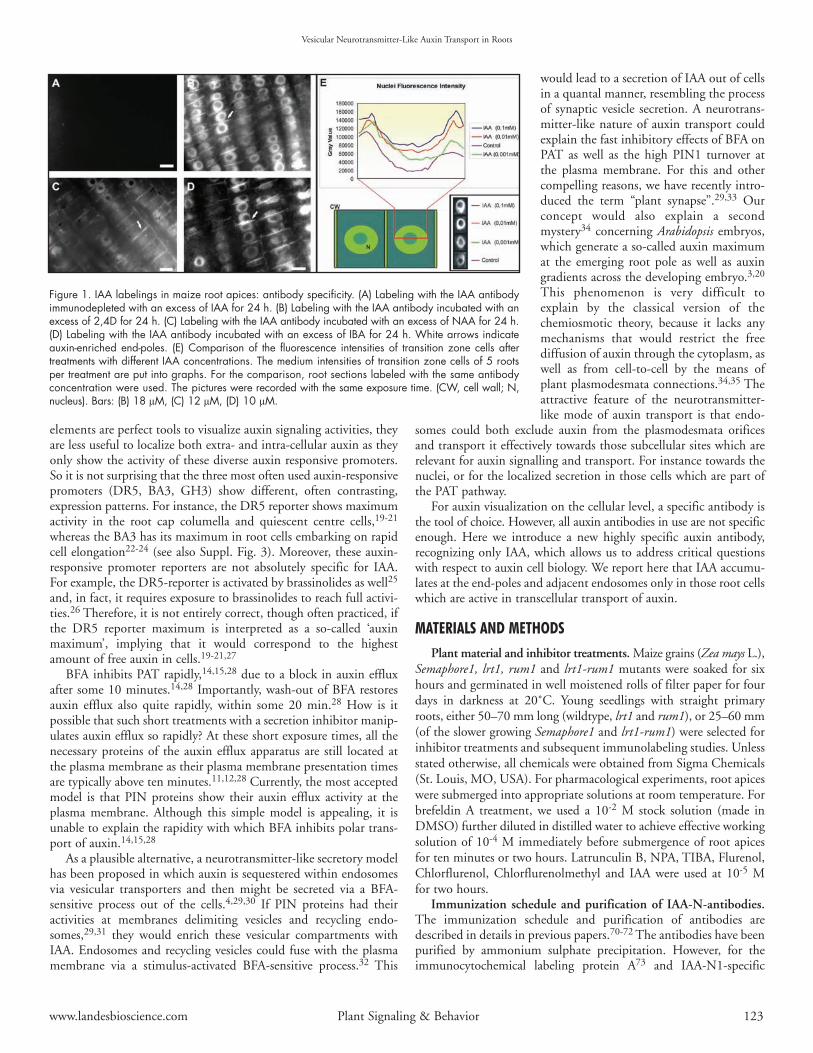
Figure 1. IAA labelings in maize root apices: antibody specificity. (A) Labeling with the IAA antibodyimmunodepleted with an excess of IAA for 24 h. (B) Labeling with the IAA antibody incubated with anexcess of 2,4D for 24 h. (C) Labeling with the IAA antibody incubated with an excess of NAA for 24 h.(D) Labeling with the IAA antibody incubated with an excess of IBA for 24 h. White arrows indicateauxin-enriched end-poles. (E) Comparison of the fluorescence intensities of transition zone cells aftertreatments with different IAA concentrations. The medium intensities of transition zone cells of 5 rootsper treatment are put into graphs. For the comparison, root sections labeled with the same antibodyconcentration were used. The pictures were recorded with the same exposure time. (CW, cell wall; N,nucleus). Bars: (B) 18 µM, (C) 12 µM, (D) 10 µM.
affinity chromatography were also used, as described in more detailspreviously.37,38
Characterization of antibodies by enzyme-linked immunosorbentassay. The IAA-N1-antibodies were characterized using a modificationof the ELISA protocol described by Weiler et al.74 The microtiter-plates (Gama, Ceské Budejovice, Czech Republic) were coated with150 µl of rabbit antibodies (5 µg.ml-1 50 mM NaHCO3, pH 9.6).The wells were washed with distilled water, filled with 200 µl ofbovine serum albumin solution (0.04 g.L-1) and incubated for 1 h at25˚C. After decanting and two washes with dist. H2O the wells werefilled in the following sequence: 50 µl TBS, 50 µl of standard orsample in TBS and 50 µl of IAA-alkaline phosphatase tracer dilutedin TBS-bovine serum albumin buffer (0.04 g.L-1). Non-specificbinding was determined by adding an excess (200 pmol) of a standard;for maximum tracer binding, TBS was used instead of standard.After one minute shaking, the plates were incubated for one hour at25˚C. The decanted plates were then washed four times with TBSand filled immediately with 150 µl of a p-nitrophenylphosphatesolution (1 mg.ml-1 50 mM NaHCO3, pH 9.6). The reaction wasstopped after one hour incubation at 25˚C by adding 50 µl 3M KOHand the absorbance measured at 405 nm in a Titertek MultiscanMCC 340 (Flow Labs, Irvine, UK). Sigmoidal curves for standardsand cross-reacting compounds were linearized by log-logit transfor-mation as follows: logit B/Bo = ln (B/Bo)/(100-B/Bo).74
Indirect immunofluorescence labeling. Fixation consisted ofexcising of apical root segments (7 mm) encompassing the majorgrowth zones into 3.7% formaldehyde prepared in stabilizing buffer(SB; 50 mM PIPES, 5 mM MgSO4 and 5 mM EGTA, pH 6.9) forone hour at room temperature. Following rinsing in SB, the rootapices were dehydrated in a graded ethanol series diluted withphosphate buffered saline (PBS). Then they were embedded inSteedman’s wax and processed for immunofluorescence (for detailssee ref. 39). To enable efficient penetration of antibodies, sectionswere dewaxed in absolute ethanol, passed through a graded ethanolseries diluted with PBS and then kept in PBS for 20 min. After thatthe sections were transferred to PBS containing 2% BSA for 15 minat room temperature.
They were then incubated with the following primary antibodies:JIM5 monoclonal antibodies diluted 1:20, anti-RGII polyclonal anti-bodies16 diluted 1:100, anti-PIN1 polyclonal antibodies diluted 1:40,anti-IAA polyclonal antibodies diluted 1:20. All primary antibodieswere diluted in PBS and the buffers were supplemented with 1%BSA. Sections were incubated in primary antibody for one hour atroom temperature. After rinsing in PBS, the sections were incubatedfor one hour either with FITC/TRITC-conjugated anti-rat IgGs(JIM5), or with anti-rabbit IgGs (Actin, Auxin, PIN1, RGII), each
raised in goat and diluted 1:100 in appropriate buffer containing1% BSA. A further PBS rinse (ten minutes) preceded ten minutestreatment with 0.01% Toluidine Blue to diminished autofluorescenceof root tissues. The sections were then mounted using an anti-fademounting medium containing p-phenylenediamine39 and examinedwith an Axiovert 405M inverted microscope (Zeiss, Oberkochen,Germany) equipped with epifluorescence and standard FITCexcitation and barrier filters. Identical microscope settings were usedto compare the labelings of different treatments.
Double immunofluorescence labeling. After the labeling of theIAA with TRITC-conjugated anti-rabbit IgGs preceded a 45 minpost fixation with 3.7% formaldehyde prepared in PBS, followed bya second blocking step with PBS containing 2% BSA for 15 min atroom temperature. The labeling of PIN1 and JIM5 followed ourstandard protocol.
Sucrose density gradient, aqueous two-phase system andimmunoblotting. Root tissue of four days treated or untreated maizewas collected and grinded in TE-buffer: 10 mM TRIS (pH 7.2),1 mM EDTA and 20% sucrose (w/v), 1 mM DTT and proteaseinhibitors (we use “Complete, EDTA-free” tablets) at 4˚C. Per 1 gfresh weight 2–3 ml buffer were used. All following working stepswere done on ice at 4˚C. The homogenate was cleared by spinningat 2,500 x g for five minutes at 4˚C. The pellet was discarded andthe supernatant was split into the cytosolic and microsomal fractionswith spinning at 100,000 x g for 45 min at 4˚C. The cytosolic super-natant was discarded and the microsomal pellet was resuspended in2 ml TE-buffer for the sucrose density gradient or in 330/5 buffer(330 mM Sucrose, 5 mm potassium phosphate (pH 7.8) for thetwo-phase system.
Vesicular Neurotransmitter-Like Auxin Transport in Roots
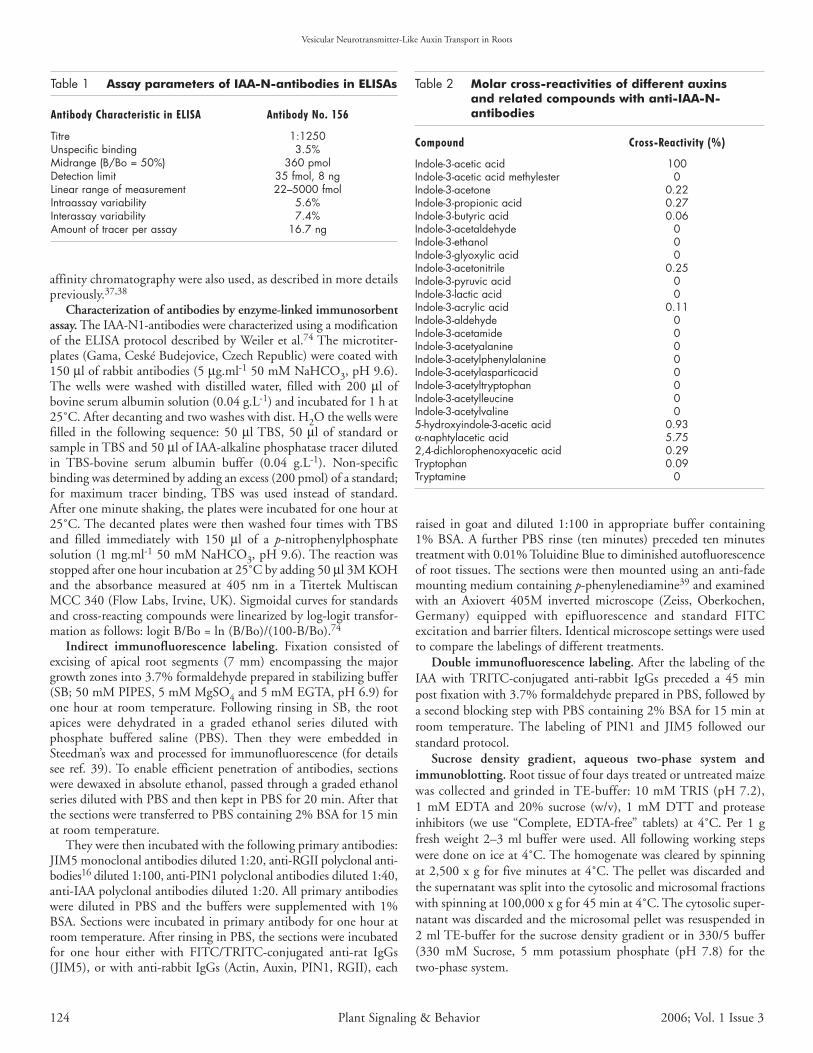
Table 1 Assay parameters of IAA-N-antibodies in ELISAs
Antibody Characteristic in ELISA Antibody No. 156
Titre 1:1250Unspecific binding 3.5%Midrange (B/Bo = 50%) 360 pmolDetection limit 35 fmol, 8 ngLinear range of measurement 22–5000 fmolIntraassay variability 5.6%Interassay variability 7.4%Amount of tracer per assay 16.7 ng
Table 2 Molar cross-reactivities of different auxins and related compounds with anti-IAA-N-antibodies
Compound Cross-Reactivity (%)
Indole-3-acetic acid 100Indole-3-acetic acid methylester 0Indole-3-acetone 0.22Indole-3-propionic acid 0.27Indole-3-butyric acid 0.06Indole-3-acetaldehyde 0Indole-3-ethanol 0Indole-3-glyoxylic acid 0Indole-3-acetonitrile 0.25Indole-3-pyruvic acid 0Indole-3-lactic acid 0Indole-3-acrylic acid 0.11Indole-3-aldehyde 0Indole-3-acetamide 0Indole-3-acetyalanine 0Indole-3-acetylphenylalanine 0Indole-3-acetylasparticacid 0Indole-3-acetyltryptophan 0Indole-3-acetylleucine 0Indole-3-acetylvaline 05-hydroxyindole-3-acetic acid 0.93α-naphtylacetic acid 5.752,4-dichlorophenoxyacetic acid 0.29Tryptophan 0.09Tryptamine 0
124 Plant Signaling & Behavior 2006; Vol. 1 Issue 3
Sucrose density gradient. From 20% to 60% sucrose in TE-buffer a step sucrose gradient was set up with five 2 ml steps. Theresuspended microsomal fraction was laid over the gradient. Thesamples were spun for 18 hours in a SW41 swinging bucket rotor at
35,000 rpm at 4˚C. Twelve 1 ml fractionswere collected from the top of the gradient.The sucrose concentration in each fractionwas measured by using a refractometer. Inaddition, the protein concentration ofeach fraction were measured with themethod following Bradford.
Aqeous two-phase system. The aqueoustwo-phase partitioning method was doneaccording to the batch procedure asdescribed.75 Phase separations were carriedout in a series of 10-g phase systems with afinal composition of 6.2% (w/w) dextranT500, 6.2% (w/w) polyethylene glycol3350, 330 mM sucrose and 5 mm potassi-um phosphate (pH 7.8), 3 mm KCl andprotease inhibitors. Three successive roundsof partitioning yielded the final upper phasesand lower phases. The combined upperphase was enriched in plasma membranesvesicles and the lower phase containedintracellular membranes.
The final upper and lower phases werediluted 5- and 10-fold, respectively, inice-cold Tris-HCl dilution buffer (10 mM,pH 7.4) containing 0.25 M Suc, 3 mMEDTA, 1 mM DTT, 3.6 mM l-Cys, 0.1 mMMgCl and the protease inhibitors. The
fractions were centrifuged at 100,000 g for 60 min. The pellets werethen resuspended in TE buffer and used for the protein concentrationmeasured with the method following Bradford. All procedures werecarried out at 4˚C.
The samples were precipitated with methanoland chloroform. Five-hundred microliters of afraction was combined with 500 µl MetOHand 125 µl chloroform in a eppendorf tube andvortexed. After centrifugation for ten minutes at13.000 rpm at 4˚C resulted in a two phasedsample. The upper phase was discarded. Afterthe addition of 500 µl MetOH, a secondcentrifugation of ten minutes with 13.000 rpmat 4˚C was done. The resulting pellets had todry completely and were resuspended in 1xSDS-PAGE sample buffer (final protein concen-tration of 1 µg per µl). Each sample was loadedonto an 15% SDS- PAGE gel which was blottedonto nitrocellulose.
All working steps of the immunoblottingwere done at room temperature. The nitrocellu-lose was washed in TBS buffer (10 mM TRIS(pH 7.4), 150 mM NaCl) and then blockedwith 4% BSA in TBS for one hour. After fiveminutes washing in TBS, incubation with thefirst antibodies (1:1000 or 1:2000) in TBS wascarried out. The nitrocellulose was washed threetimes in TBS with 0,05% Tween 20 (TTBS) to getrid of unspecific antibody binding. The secondaryantibodies are alkalic phosphatase conjugated andwere used 1:10.000 in TBS. After one hour
Vesicular Neurotransmitter-Like Auxin Transport in Roots
www.landesbioscience.com Plant Signaling & Behavior 125
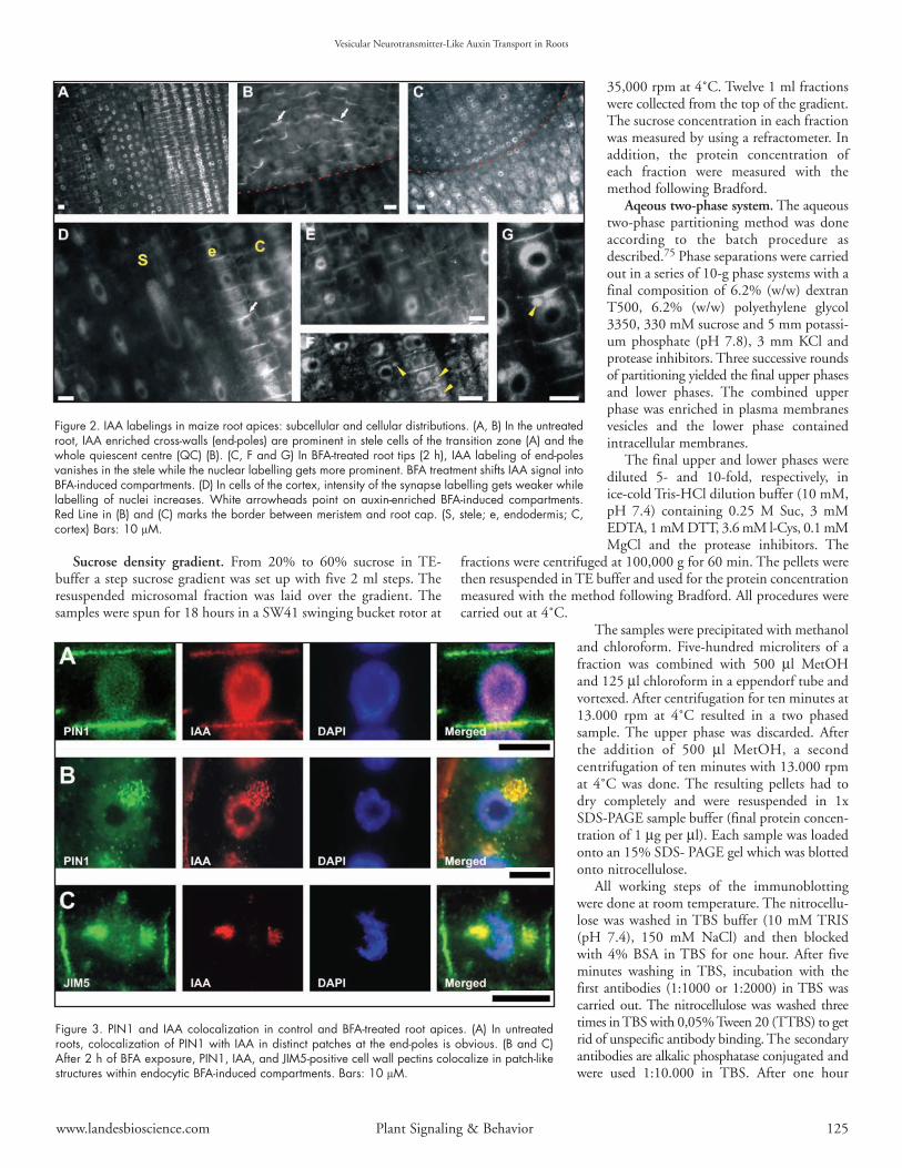
Figure 2. IAA labelings in maize root apices: subcellular and cellular distributions. (A, B) In the untreatedroot, IAA enriched cross-walls (end-poles) are prominent in stele cells of the transition zone (A) and thewhole quiescent centre (QC) (B). (C, F and G) In BFA-treated root tips (2 h), IAA labeling of end-polesvanishes in the stele while the nuclear labelling gets more prominent. BFA treatment shifts IAA signal intoBFA-induced compartments. (D) In cells of the cortex, intensity of the synapse labelling gets weaker whilelabelling of nuclei increases. White arrowheads point on auxin-enriched BFA-induced compartments.Red Line in (B) and (C) marks the border between meristem and root cap. (S, stele; e, endodermis; C,cortex) Bars: 10 µM.
Figure 3. PIN1 and IAA colocalization in control and BFA-treated root apices. (A) In untreatedroots, colocalization of PIN1 with IAA in distinct patches at the end-poles is obvious. (B and C)After 2 h of BFA exposure, PIN1, IAA, and JIM5-positive cell wall pectins colocalize in patch-likestructures within endocytic BFA-induced compartments. Bars: 10 µM.
incubation time, a washing with TTBS was repeated three times.The antibody detection was done with BCIP-NPT (SigmaChemical). The staining was stopped with 1% acetic acid in water.
Real time recordings of auxin uptake into root apices. Real timerecordings of auxin uptake into root cells with a vibrating micro-electrode system was described in detail previously.15
Image processing and fluorescence measurements. Imageprocessing was done with Photoshop 7 from Adobe and fluorescencemeasurement was done with the open source software Image-J(http://rsb.info.nih.gov/ij/).
RESULTSPreparation and characterization of the IAA-specific antibody.
IAA was coupled to BSA using the Mannich reaction.36 The immu-nization schedule and purification of antibodies were done as describedpreviously.37,38 All immunized rabbits produced antisera to theIAA-N1-BSA conjugate, but serum titres and affinity of antibodies
differed considerably and reflect differences in the responses of theindividual animals. Therefore, the IAA antibody was well character-ized in ELISA tests for several parameters (Table 1).
A number of structurally related indoles and IAA metabolitesmay be present in plant tissues. Antibody specificity is, therefore, acrucial point in the immunocytochemical assay of IAA. Thecompounds were tested for antibody binding over a range from0.1 up to 50 nmol per assay. Data for auxins and related compoundsproducing zero molar cross-reactivities are not shown. Namely, IAAprecursors such as tryptophan, tryptamine, tryptophol, indole-3-aldehyde, indole-3-acetonitrile, indole-3-acetamide were not reactive.All IAA conjugates were tested and shown to be inactive. IAAhomologues like indole-3-acrylic acid and indole-3-propionic acidwere weak competitors with cross-reactivity below 0.25%; significantimmunoreaction was achieved only after using 500-times higherconcentrations in plant tissues. Of the 25 compounds tested(Table 2), only α-naphthylacetic acid has relevant reactivity but thiscompound is a synthetic auxin and does not occur naturally.
Several controls were utilized in order to confirm the specificityof the IAA antibody at the level of immunolabeling: (1) labeling withthe preimmune rabbit IgG instead of IAA antibody producing onlyblack images (not shown), (2) labeling with the IAA antibodyimmunodepleted with an excess of IAA for 24 h (Fig. 1A), (3) labelingwith the IAA antibody incubated with an excess of 2,4D, NAA andIBA for 24 h (Figs. 1B–D), (4) labeling only with the anti-rabit IgG,omitting the first antibody step producing only black images (notshown). Labelings with the IAA antibody of root apices treated withdifferent concentrations of IAA show corresponding increases in thefluorescence signal (Fig. 1E). All these cytological controls unequiv-ocally confirmed the specificity of the IAA antibody, as alreadydocumented at the biochemical level.
IAA immunolocalization in cells of control, BFA-, IAA- andTIBA-treated root apices. The localization of IAA in untreatedmaize roots showed that the most prominent signal was scored incells of the root apex, especially in the transition zone (Fig. 2A) andin the quiescent centre (Fig. 2B). In these cells, a prominent auxinsignal was visible at the cross-walls (end-poles), (Fig. 2A, D and E).In BFA-treated roots, IAA was still localized within nuclei while slightlyweaker signal was scored at the end-poles (Fig. 2C, F and G).Additionally, BFA-induced compartments were enriched with auxin(Fig. 2G).
The signal at the end-poles was not a continuous labeling, butwas composed of closely apposed spots at which IAA colocalizedwith PIN1 labeled with a maize specific PIN1-antibody (Fig. 3A).This colocalization was obvious also in BFA-treated cells when
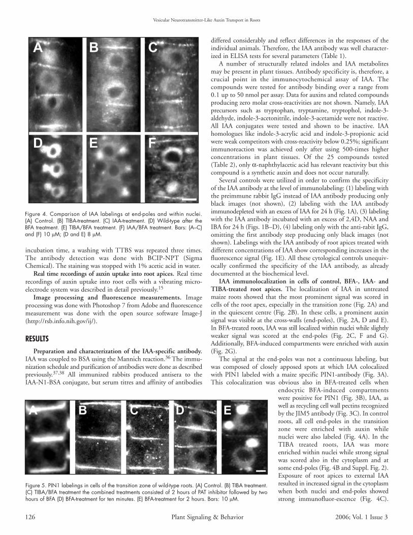
endocytic BFA-induced compartmentswere positive for PIN1 (Fig. 3B), IAA, aswell as recycling cell wall pectins recognizedby the JIM5 antibody (Fig. 3C). In controlroots, all cell end-poles in the transitionzone were enriched with auxin whilenuclei were also labeled (Fig. 4A). In theTIBA treated roots, IAA was moreenriched within nuclei while strong signalwas scored also in the cytoplasm and atsome end-poles (Fig. 4B and Suppl. Fig. 2).Exposure of root apices to external IAAresulted in increased signal in the cytoplasmwhen both nuclei and end-poles showedstrong immunofluor-escence (Fig. 4C).
Vesicular Neurotransmitter-Like Auxin Transport in Roots
Figure 4. Comparison of IAA labelings at end-poles and within nuclei.(A) Control. (B) TIBA-treatment. (C) IAA-treatment. (D) Wild-type after theBFA treatment. (E) TIBA/BFA treatment. (F) IAA/BFA treatment. Bars: (A–C)and (F) 10 µM; (D and E) 8 µM.
Figure 5. PIN1 labelings in cells of the transition zone of wild-type roots. (A) Control. (B) TIBA treatment.(C) TIBA/BFA treatment the combined treatments consisted of 2 hours of PAT inhibitor followed by twohours of BFA (D) BFA-treatment for ten minutes. (E) BFA-treatment for 2 hours. Bars: 10 µM.
126 Plant Signaling & Behavior 2006; Vol. 1 Issue 3
Vesicular Neurotransmitter-Like Auxin Transport in Roots
Importantly, BFA-treated wild-type roots showed IAA-enriched BFA-induced endocytic compartments (Fig. 4D). These compartmentswere smaller in TIBA and auxin pretreated roots (Fig. 4E and F).
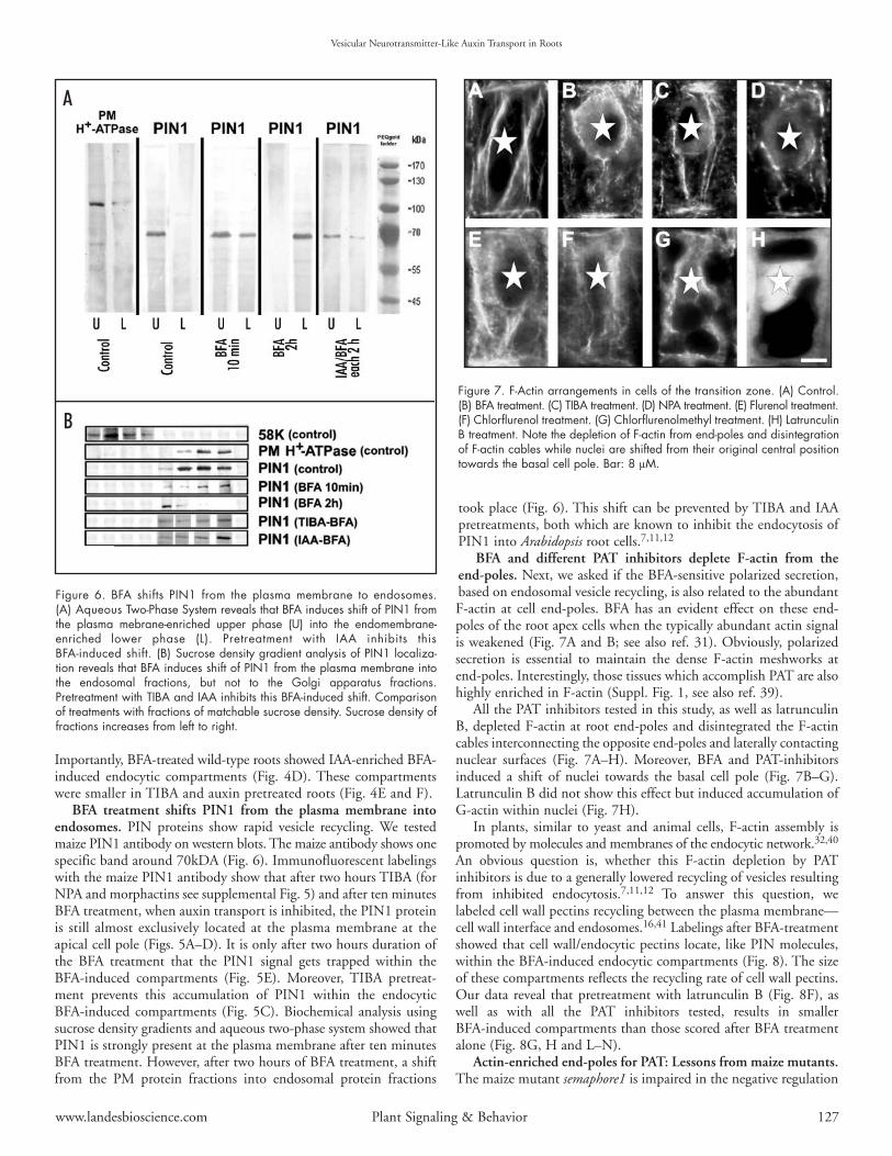
BFA treatment shifts PIN1 from the plasma membrane intoendosomes. PIN proteins show rapid vesicle recycling. We testedmaize PIN1 antibody on western blots. The maize antibody shows onespecific band around 70kDA (Fig. 6). Immunofluorescent labelingswith the maize PIN1 antibody show that after two hours TIBA (forNPA and morphactins see supplemental Fig. 5) and after ten minutesBFA treatment, when auxin transport is inhibited, the PIN1 proteinis still almost exclusively located at the plasma membrane at theapical cell pole (Figs. 5A–D). It is only after two hours duration ofthe BFA treatment that the PIN1 signal gets trapped within theBFA-induced compartments (Fig. 5E). Moreover, TIBA pretreat-ment prevents this accumulation of PIN1 within the endocyticBFA-induced compartments (Fig. 5C). Biochemical analysis usingsucrose density gradients and aqueous two-phase system showed thatPIN1 is strongly present at the plasma membrane after ten minutesBFA treatment. However, after two hours of BFA treatment, a shiftfrom the PM protein fractions into endosomal protein fractions
took place (Fig. 6). This shift can be prevented by TIBA and IAApretreatments, both which are known to inhibit the endocytosis ofPIN1 into Arabidopsis root cells.7,11,12
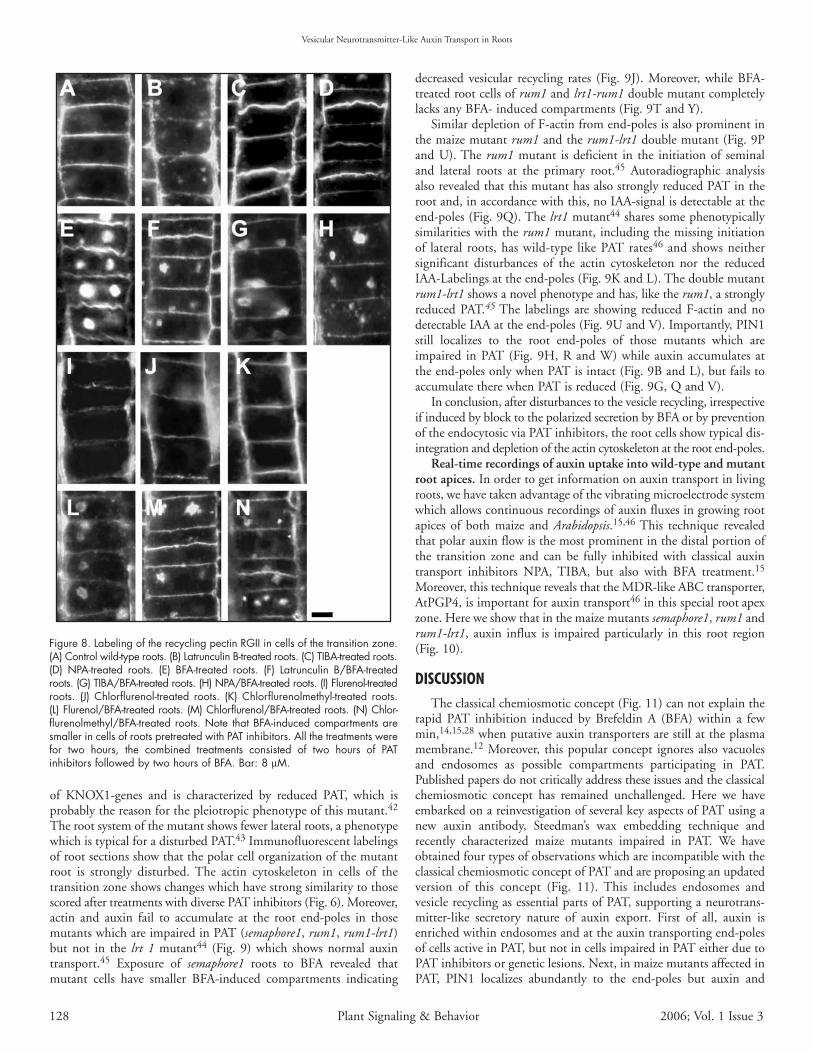
BFA and different PAT inhibitors deplete F-actin from theend-poles. Next, we asked if the BFA-sensitive polarized secretion,based on endosomal vesicle recycling, is also related to the abundantF-actin at cell end-poles. BFA has an evident effect on these end-poles of the root apex cells when the typically abundant actin signalis weakened (Fig. 7A and B; see also ref. 31). Obviously, polarizedsecretion is essential to maintain the dense F-actin meshworks atend-poles. Interestingly, those tissues which accomplish PAT are alsohighly enriched in F-actin (Suppl. Fig. 1, see also ref. 39).
All the PAT inhibitors tested in this study, as well as latrunculinB, depleted F-actin at root end-poles and disintegrated the F-actincables interconnecting the opposite end-poles and laterally contactingnuclear surfaces (Fig. 7A–H). Moreover, BFA and PAT-inhibitorsinduced a shift of nuclei towards the basal cell pole (Fig. 7B–G).Latrunculin B did not show this effect but induced accumulation ofG-actin within nuclei (Fig. 7H).
In plants, similar to yeast and animal cells, F-actin assembly ispromoted by molecules and membranes of the endocytic network.32,40
An obvious question is, whether this F-actin depletion by PATinhibitors is due to a generally lowered recycling of vesicles resultingfrom inhibited endocytosis.7,11,12 To answer this question, welabeled cell wall pectins recycling between the plasma membrane—cell wall interface and endosomes.16,41 Labelings after BFA-treatmentshowed that cell wall/endocytic pectins locate, like PIN molecules,within the BFA-induced endocytic compartments (Fig. 8). The sizeof these compartments reflects the recycling rate of cell wall pectins.Our data reveal that pretreatment with latrunculin B (Fig. 8F), aswell as with all the PAT inhibitors tested, results in smallerBFA-induced compartments than those scored after BFA treatmentalone (Fig. 8G, H and L–N).
Actin-enriched end-poles for PAT: Lessons from maize mutants.The maize mutant semaphore1 is impaired in the negative regulation
www.landesbioscience.com Plant Signaling & Behavior 127
Figure 7. F-Actin arrangements in cells of the transition zone. (A) Control.(B) BFA treatment. (C) TIBA treatment. (D) NPA treatment. (E) Flurenol treatment.(F) Chlorflurenol treatment. (G) Chlorflurenolmethyl treatment. (H) LatrunculinB treatment. Note the depletion of F-actin from end-poles and disintegrationof F-actin cables while nuclei are shifted from their original central positiontowards the basal cell pole. Bar: 8 µM.
Figure 6. BFA shifts PIN1 from the plasma membrane to endosomes.(A) Aqueous Two-Phase System reveals that BFA induces shift of PIN1 fromthe plasma mebrane-enriched upper phase (U) into the endomembrane-enriched lower phase (L). Pretreatment with IAA inhibits thisBFA-induced shift. (B) Sucrose density gradient analysis of PIN1 localiza-tion reveals that BFA induces shift of PIN1 from the plasma membrane intothe endosomal fractions, but not to the Golgi apparatus fractions.Pretreatment with TIBA and IAA inhibits this BFA-induced shift. Comparisonof treatments with fractions of matchable sucrose density. Sucrose density offractions increases from left to right.
A
B
of KNOX1-genes and is characterized by reduced PAT, which isprobably the reason for the pleiotropic phenotype of this mutant.42
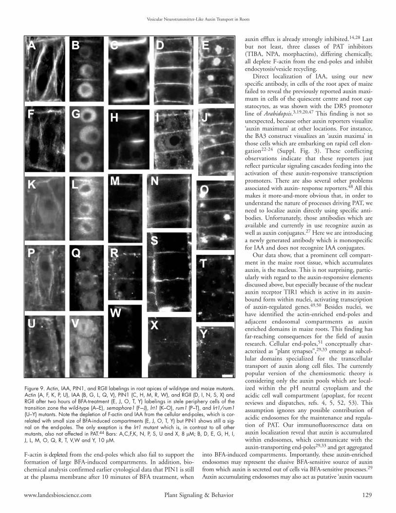
The root system of the mutant shows fewer lateral roots, a phenotypewhich is typical for a disturbed PAT.43 Immunofluorescent labelingsof root sections show that the polar cell organization of the mutantroot is strongly disturbed. The actin cytoskeleton in cells of thetransition zone shows changes which have strong similarity to thosescored after treatments with diverse PAT inhibitors (Fig. 6). Moreover,actin and auxin fail to accumulate at the root end-poles in thosemutants which are impaired in PAT (semaphore1, rum1, rum1-lrt1)but not in the lrt 1 mutant44 (Fig. 9) which shows normal auxintransport.45 Exposure of semaphore1 roots to BFA revealed thatmutant cells have smaller BFA-induced compartments indicating
decreased vesicular recycling rates (Fig. 9J). Moreover, while BFA-treated root cells of rum1 and lrt1-rum1 double mutant completelylacks any BFA- induced compartments (Fig. 9T and Y).
Similar depletion of F-actin from end-poles is also prominent inthe maize mutant rum1 and the rum1-lrt1 double mutant (Fig. 9Pand U). The rum1 mutant is deficient in the initiation of seminaland lateral roots at the primary root.45 Autoradiographic analysisalso revealed that this mutant has also strongly reduced PAT in theroot and, in accordance with this, no IAA-signal is detectable at theend-poles (Fig. 9Q). The lrt1 mutant44 shares some phenotypicallysimilarities with the rum1 mutant, including the missing initiationof lateral roots, has wild-type like PAT rates46 and shows neithersignificant disturbances of the actin cytoskeleton nor the reducedIAA-Labelings at the end-poles (Fig. 9K and L). The double mutantrum1-lrt1 shows a novel phenotype and has, like the rum1, a stronglyreduced PAT.45 The labelings are showing reduced F-actin and nodetectable IAA at the end-poles (Fig. 9U and V). Importantly, PIN1still localizes to the root end-poles of those mutants which areimpaired in PAT (Fig. 9H, R and W) while auxin accumulates atthe end-poles only when PAT is intact (Fig. 9B and L), but fails toaccumulate there when PAT is reduced (Fig. 9G, Q and V).
In conclusion, after disturbances to the vesicle recycling, irrespectiveif induced by block to the polarized secretion by BFA or by preventionof the endocytosic via PAT inhibitors, the root cells show typical dis-integration and depletion of the actin cytoskeleton at the root end-poles.
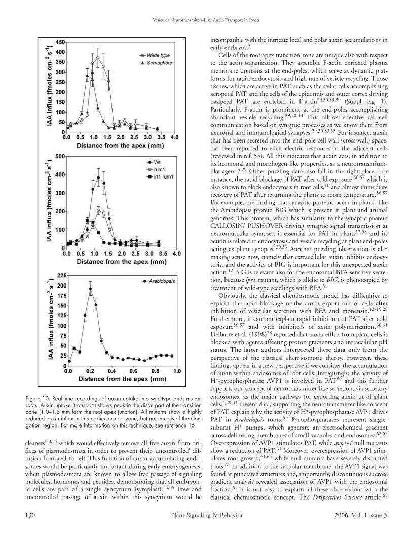
Real-time recordings of auxin uptake into wild-type and mutantroot apices. In order to get information on auxin transport in livingroots, we have taken advantage of the vibrating microelectrode systemwhich allows continuous recordings of auxin fluxes in growing rootapices of both maize and Arabidopsis.15,46 This technique revealedthat polar auxin flow is the most prominent in the distal portion ofthe transition zone and can be fully inhibited with classical auxintransport inhibitors NPA, TIBA, but also with BFA treatment.15
Moreover, this technique reveals that the MDR-like ABC transporter,AtPGP4, is important for auxin transport46 in this special root apexzone. Here we show that in the maize mutants semaphore1, rum1 andrum1-lrt1, auxin influx is impaired particularly in this root region(Fig. 10).
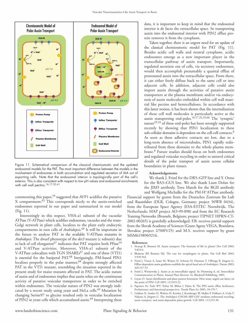
DISCUSSIONThe classical chemiosmotic concept (Fig. 11) can not explain the
rapid PAT inhibition induced by Brefeldin A (BFA) within a fewmin,14,15,28 when putative auxin transporters are still at the plasmamembrane.12 Moreover, this popular concept ignores also vacuolesand endosomes as possible compartments participating in PAT.Published papers do not critically address these issues and the classicalchemiosmotic concept has remained unchallenged. Here we haveembarked on a reinvestigation of several key aspects of PAT using anew auxin antibody, Steedman’s wax embedding technique andrecently characterized maize mutants impaired in PAT. We haveobtained four types of observations which are incompatible with theclassical chemiosmotic concept of PAT and are proposing an updatedversion of this concept (Fig. 11). This includes endosomes andvesicle recycling as essential parts of PAT, supporting a neurotrans-mitter-like secretory nature of auxin export. First of all, auxin isenriched within endosomes and at the auxin transporting end-polesof cells active in PAT, but not in cells impaired in PAT either due toPAT inhibitors or genetic lesions. Next, in maize mutants affected inPAT, PIN1 localizes abundantly to the end-poles but auxin and
Vesicular Neurotransmitter-Like Auxin Transport in Roots
Figure 8. Labeling of the recycling pectin RGII in cells of the transition zone.(A) Control wild-type roots. (B) Latrunculin B-treated roots. (C) TIBA-treated roots.(D) NPA-treated roots. (E) BFA-treated roots. (F) Latrunculin B/BFA-treatedroots. (G) TIBA/BFA-treated roots. (H) NPA/BFA-treated roots. (I) Flurenol-treatedroots. (J) Chlorflurenol-treated roots. (K) Chlorflurenolmethyl-treated roots.(L) Flurenol/BFA-treated roots. (M) Chlorflurenol/BFA-treated roots. (N) Chlor-flurenolmethyl/BFA-treated roots. Note that BFA-induced compartments aresmaller in cells of roots pretreated with PAT inhibitors. All the treatments werefor two hours, the combined treatments consisted of two hours of PATinhibitors followed by two hours of BFA. Bar: 8 µM.
128 Plant Signaling & Behavior 2006; Vol. 1 Issue 3
www.landesbioscience.com Plant Signaling & Behavior 129
F-actin is depleted from the end-poles which also fail to support theformation of large BFA-induced compartments. In addition, bio-chemical analysis confirmed earlier cytological data that PIN1 is stillat the plasma membrane after 10 minutes of BFA treatment, when
auxin efflux is already strongly inhibited.14,28 Lastbut not least, three classes of PAT inhibitors(TIBA, NPA, morphactins), differing chemically,all deplete F-actin from the end-poles and inhibitendocytosis/vesicle recycling.
Direct localization of IAA, using our new specific antibody, in cells of the root apex of maizefailed to reveal the previously reported auxin maxi-mum in cells of the quiescent centre and root capstatocytes, as was shown with the DR5 promoterline of Arabidopsis.3,19,20,47 This finding is not sounexpected, because other auxin reporters visualize‘auxin maximum’ at other locations. For instance,the BA3 construct visualizes an ‘auxin maxima’ inthose cells which are embarking on rapid cell elon-gation22-24 (Suppl. Fig. 3). These conflictingobservations indicate that these reporters justreflect particular signaling cascades feeding into theactivation of these auxin-responsive transcriptionpromoters. There are also several other problemsassociated with auxin- response reporters.48 All thismakes it more-and-more obvious that, in order tounderstand the nature of processes driving PAT, weneed to localize auxin directly using specific anti-bodies. Unfortunately, those antibodies which areavailable and currently in use recognize auxin aswell as auxin conjugates.27 Here we are introducinga newly generated antibody which is monospecificfor IAA and does not recognize IAA conjugates.
Our data show, that a prominent cell compart-ment in the maize root tissue, which accumulatesauxin, is the nucleus. This is not surprising, partic-ularly with regard to the auxin-responsive elementsdiscussed above, but especially because of the nuclearauxin receptor TIR1 which is active in its auxin-bound form within nuclei, activating transcriptionof auxin-regulated genes.49,50 Besides nuclei, wehave identified the actin-enriched end-poles andadjacent endosomal compartments as auxinenriched domains in maize roots. This finding hasfar-reaching consequences for the field of auxinresearch. Cellular end-poles,51 conceptually char-acterized as “plant synapses”,29,33 emerge as subcel-lular domains specialized for the transcellulartransport of auxin along cell files. The currentlypopular version of the chemiosmotic theory isconsidering only the auxin pools which are local-ized within the pH neutral cytoplasm and theacidic cell wall compartment (apoplast, for recentreviews and dispatches, refs. 4, 5, 52, 53). Thisassumption ignores any possible contribution ofacidic endosomes for the maintenance and regula-tion of PAT. Our immunofluorescence data onauxin localization reveal that auxin is accumulatedwithin endosomes, which communicate with theauxin-transporting end-poles29,33 and get aggregated
into BFA-induced compartments. Importantly, these auxin-enrichedendosomes may represent the elusive BFA-sensitive source of auxinfrom which auxin is secreted out of cells via BFA-sensitive processes.29
Auxin accumulating endosomes may also act as putative ‘auxin vacuum
Vesicular Neurotransmitter-Like Auxin Transport in Roots
Figure 9. Actin, IAA, PIN1, and RGII labelings in root apices of wild-type and maize mutants.Actin (A, F, K, P, U), IAA (B, G, L, Q, V), PIN1 (C, H, M, R, W), and RGII (D, I, N, S, X) andRGII after two hours of BFA-treatment (E, J, O, T, Y) labelings in stele periphery cells of thetransition zone the wild-type (A–E), semaphore1 (F–-J), lrt1 (K–O), rum1 (P–T), and lrt1/rum1(U–Y) mutants. Note the depletion of F-actin and IAA from the cellular end-poles, which is cor-related with small size of BFA-induced compartments (E, J, O, T, Y) but PIN1 shows still a sig-nal on the end-poles. The only exeption is the lrt1 mutant which is, in contrast to all othermutants, also not affected in PAT.44 Bars: A,C,F,K, N, P, S, U and X, 8 µM; B, D, E, G, H, I,J, L, M, O, Q, R, T, V,W and Y, 10 µM.
cleaners’30,54 which would effectively remove all free auxin from ori-fices of plasmodesmata in order to prevent their ‘uncontrolled’ dif-fusion from cell-to-cell. This function of auxin-accumulating endo-somes would be particularly important during early embryogenesis,when plasmodesmata are known to allow free passage of signalingmolecules, hormones and peptides, demonstrating that all embryon-ic cells are part of a single syncytium (symplast).34,35 Free anduncontrolled passage of auxin within this syncytium would be
incompatible with the intricate local and polar auxin accumulations inearly embryos.3
Cells of the root apex transition zone are unique also with respectto the actin organization. They assemble F-actin enriched plasmamembrane domains at the end-poles, which serve as dynamic plat-forms for rapid endocytosis and high rate of vesicle recycling. Thosetissues, which are active in PAT, such as the stelar cells accomplishingacropetal PAT and the cells of the epidermis and outer cortex drivingbasipetal PAT, are enriched in F-actin29,30,33,39 (Suppl. Fig. 1).Particularly, F-actin is prominent at the end-poles accomplishingabundant vesicle recycling.29,30,33 This allows effective cell-cellcommunication based on synaptic processes as we know them fromneuronal and immunological synapses.29,30,33,55 For instance, auxinthat has been secreted into the end-pole cell wall (cross-wall) space,has been reported to elicit electric responses in the adjacent cells(reviewed in ref. 55). All this indicates that auxin acts, in addition toits hormonal and morphogen-like properties, as a neurotransmitter-like agent.4,29 Other puzzling data also fall in the right place. Forinstance, the rapid blockage of PAT after cold exposure,56,57 which isalso known to block endocytosis in root cells,16 and almost immediaterecovery of PAT after returning the plants to room temperature.56,57
For example, the finding that synaptic proteins occur in plants, likethe Arabidopsis protein BIG which is present in plant and animalgenomes. This protein, which has similarity to the synaptic proteinCALLOSIN/ PUSHOVER driving synaptic signal transmission atneuromuscular synapses, is essential for PAT in plants12,58 and itsaction is related to endocytosis and vesicle recycling at plant end-polesacting as plant synapses.29,33 Another puzzling observation is alsomaking sense now, namely that extracellular auxin inhibits endocy-tosis, and the activity of BIG is important for this unexpected auxinaction.12 BIG is relevant also for the endosomal BFA-sensitive secre-tion, because lpr1 mutant, which is allelic to BIG, is phenocopied bytreatment of wild-type seedlings with BFA.58
Obviously, the classical chemiosmotic model has difficulties toexplain the rapid blockage of the auxin export out of cells afterinhibition of vesicular secretion with BFA and monensin.12-15,28
Furthermore, it can not explain rapid inhibition of PAT after coldexposure56,57 and with inhibitors of actin polymerization.60,61
Delbarre et al. (1998)28 reported that auxin efflux from plant cells isblocked with agents affecting proton gradients and intracellular pHstatus. The latter authors interpreted these data only from theperspective of the classical chemiosmotic theory. However, thesefindings appear in a new perspective if we consider the accumulationof auxin within endosomes of root cells. Intriguingly, the activity ofH+-pyrophosphatase AVP1 is involved in PAT59 and this furthersupports our concept of neurotransmitter-like secretion, via secretoryendosomes, as the major pathway for exporting auxin ut of plantcells.4,29,33 Present data, supporting the neurotransmitter-like conceptof PAT, explain why the activity of H+-pyrophosphatase AVP1 drivesPAT in Arabidopsis roots.59 Pyrophosphatases represent single-subunit H+ pumps, which generate an electrochemical gradientacross delimiting membranes of small vacuoles and endosomes.62,63
Overexpression of AVP1 stimulates PAT, while avp1-1 null mutantsshow a reduction of PAT.61 Moreover, overexpression of AVP1 stim-ulates root growth,61,64 while null mutants have severely disruptedroots.61 In addition to the vacuolar membrane, the AVP1 signal wasfound at punctated structures and, importantly, discontinuous sucrosegradient analysis revealed association of AVP1 with the endosomalfraction.61 It is not easy to explain all these observations with theclassical chemiosmotic concept. The Perspectives Science article,65
Vesicular Neurotransmitter-Like Auxin Transport in Roots
Figure 10. Real-time recordings of auxin uptake into wild-type and, mutantroots. Auxin uptake (transport) shows peak in the distal part of the transitionzone (1.0–1.5 mm form the root apex junction). All mutants show a highlyreduced auxin influx in this particular root zone, but not in cells of the elon-gation region. For more information on this technique, see reference 15.
130 Plant Signaling & Behavior 2006; Vol. 1 Issue 3
www.landesbioscience.com Plant Signaling & Behavior 131
commenting this paper,59 suggested that AVP1 acidifies the putativeX compartment.65 This corresponds nicely to the auxin-enrichedendosomes reported in our paper and summarized in our model(Fig. 11).
Interestingly in this respect, VHA-a1 subunit of the vacuolarATPase (V-ATPase) which acidifies endosomes, vacuoles and the trans-Golgi network in plant cells, localizes to the plant early endocyticcompartments in root cells of Arabidopsis.66 It will be important inthe future to analyze PAT in the available V-ATPases mutants inArabidopsis. The dwarf phenotype of the det3 mutant (c subunit) dueto lack of cell elongation67 indicates that PAT requires both PPase59
and V-ATPase activities. Moreover, VHA-a1 subunit of theV-ATPase colocalizes with TGN SNAREs67 and one of these (VTI1)is essential for the basipetal PAT.68 Intriguingly, PM-based PIN1localizes properly in the polar manner,68 despite strongly affectedPAT in the VTI1 mutant, resembling the situation reported in thepresent study for maize mutants affected in PAT. The acidic natureof auxin and of endosomes implies that auxin relies on the continuousactivity of putative vesicular transporters in order to be enrichedwithin endosomes. The vesicular nature of PIN2 was strongly indi-cated by a recent study using yeast and HeLa cells.69 Mutation bychanging Serine97 to glycine resulted only in vesicular localizationof PIN2 in yeast cells which accumulated auxin.69 Interpreting these
data, it is important to keep in mind that the endosomalinterior is de facto the extracellular space. So transportingauxin into the endosomal interior with PIN2 efflux pro-tein removes it from the cytoplasm.
Taken together, there is an urgent need for an update ofthe classical chemiosmotic model for PAT (Fig. 11).Besides acidic cell walls and neutral cytoplasm, acidicendosomes emerge as a new important player in thetranscellular pathway of auxin transport. Importantly,regulated secretion out of cells, via secretory endosomes,would then accomplish presumably a quantal efflux ofprotonated auxin into the extracellular space. From there,it can either freely diffuse back to the same cell or intoadjacent cells. In addition, adjacent cells could alsoimport auxin through the activities of putative auxintransporters at the plasma membrane and/or via endocy-tosis of auxin molecules embedded within cell wall mate-rial like pectins and hemicelluloses. In accordance withthis latter notion, it has been shown that the internalizationof these cell wall molecules is particularly active at theauxin transporting end-poles.16,17,32,33,66 The ‘synaptic’nature29,33 of these end-poles has been strongly supportedrecently by showing that PIN1 localization to thesesub-cellular domains is dependent on the cell-cell contacts.8
As soon as these adhesive contacts are lost, due to along-term absence of microtubules, PIN1 rapidly redis-tributed from these domains to the whole plasma mem-brane.8 Future studies should focus on both endosomesand regulated vesicular recycling in order to unravel criticaldetails of the polar transport of auxin across cellularboundaries in plant tissues.
AcknowledgementsWe thank J. Friml for the DR5::GFP line and Y. Oono
for the BA3::GUS line. We also thank Liam Dolan forthe JIM5 antibody, Toru Matoh for the RGII antibodyand Wolfgang Michalke for the PM-H+ATPase antibody.
Financial support by grants from the Deutsches Zentrum für Luftund Raumfahrt (DLR, Cologne, Germany; project 50WB 0434),from the European Space Agency (ESA-ESTEC Noordwijk, TheNetherlands; MAP project AO-99-098) and from the EC ResearchTraining Networks (Brussels, Belgium; project TIPNET HPRN-CT-2002-00265) is gratefully acknowledged. F.B. receives partial supportfrom the Slovak Academy of Sciences (Grant Ageny VEGA, Bratislava,Slovakia; project 2/5085/25) and M.S. receives support by grantMSM6198969216.
References1. Swarup R, Bennett M. Auxin transport: The fountain of life in plants? Dev Cell 2003;
5:824-6.2. Bhalerao RP, Bennett MJ. The case for morphogens in plants. Nat Cell Biol 2003;
5:939-943.3. Friml J, Vieten A, Sauer M, Weijers D, Schwarz H, Hamann T, Offringa R, Jürgens G.
Efflux-dependent auxin gradients establish the apical-basal axis of Arabidopsis. Nature 2003;426:147-53.
4. Friml J, Wisniewska J. Auxin as an intercellular signal. In: Flemming A, ed. IntercellularCommunication in Plants. Annual Plant Reviews 16, Blackwell Publishing, 2005.
5. Leyser O. Auxin distribution and plant pattern formation: How many angels can dance onthe point of PIN? Cell 2005; 121:819-22.
6. Paponov IA, Teale WT, Trebar M, Blilou I, Palme K. The PIN auxin efflux facilitators:Evolutionary and functional perspectives. Trends Plant Sci 2005; 10:170-7.
7. Geldner N, Anders N, Wolters H, Keicher J, Kornberger W, Muller P, Delbarre A, Ueda T,Nakano A, Jürgens G. The Arabidopsis GNOM ARF-GEF mediates endosomal recycling,auxin transport, and auxin-dependent plant growth. Cell 2003; 112:219-30.
Vesicular Neurotransmitter-Like Auxin Transport in Roots
Figure 11. Schematical comparison of the classical chemiosmotic and the updatedendosomal models for the PAT. The most important difference between the models is theinvolvement of endosomes in both accumulation and regulated secretion of IAA out ofexporting cells. Note that the endosomal interior is topologically part of the cell’sexterior. This is also consistent with respect to low pH values and endosomal enrichmentswith cell wall pectins.16,17,32,41
8. Boutté Y, Crosnier MT, Carraro N, Traas J, Satiat-Jeunemaitre B. The plasma membranerecycling pathway and cell polarity in plants: Studies on PIN proteins. J Cell Sci 2006;119:1255-65.
9. Schneider G. Morphactins: Physiology and performance. Annu Rev Plant Physiol 1970;21:499-536.
10. Katekaar JF, Geissler AE. Auxin transport inhibitors. IV. Evidence of a common mode ofaction for a proposed class of auxin transport inhibitors. Plant Physiol 1980; 66:1190-5.
11. Geldner N, Friml J, Stierfhoff Y, Jürgens G, Palme K. Auxin transport inhibitors blockPIN1 cycling and vesicle trafficking. Nature 2001; 413:425-8.
12. Paciorek T, Zazímalová E, Ruthhardt N, Petrásek J, Stierhof YD, Kleine-Vehn J, MorrisDA, Emans N, Jürgens G, Geldner N, Friml J. Auxin inhibits endocytosis and promotesits own efflux from cells. Nature 2005; 435:1251-6.
13. Wilkinson S, Morris DA. Targeting of auxin carriers to the plasma membrane: Effects ofmonensin on transmembrane auxin transport in Cucurbita pepo L. tissue. Planta 1994;193:194-202.
14. Delbarre A, Muller P, Imhoff Y, Guern J. Comparison of mechanisms controlling uptakeand accumulation of 2,4-dichlorophenoxy acetic acid, naphthalene-1-acetic acid, andindole-3-acetic acid in suspension-cultured tobacco cells. Planta 1996; 198:532-41.
15. Mancuso S, Marras AM, Volker M, Baluska F. Noninvasive and continuous recordings ofauxin fluxes in intact root apex with a carbon-nanotube-modified and self-referencingmicroelectrode. Anal Biochem 2005; 341:344-51.
16. Baluska F, Hlavacka A, Samaj J, Palme K, Robinson DG, Matoh T, McCurdy DW, MenzelD, Volkmann D. F-actin-dependent endocytosis of cell wall pectins in meristematic rootcells: Insights from brefeldin A-induced compartments. Plant Physiol 2002; 130:422-31.
17. Samaj J, Baluska F, Voigt B, Schlicht M, Volkmann D, Menzel D. Endocytosis, actincytoskeleton and signalling. Plant Physiol 2004; 135:1150-61.
18. Aloni R, Schwalm K, Langhans M, Ullrich C. Gradual shifts in sites of free-auxin produc-tion during leaf-primordium development and their role in vascular differentiation and leafmorphogenesis. Planta 2003; 216:841-53.
19. Sabatini S, Beis D, Wolkenfelt H, Murfett J, Guilfoyle T, Malamy J, Benfey P, Leyser O,Bechtold N, Weisbeek P, Scheres B. An auxin-dependent distal organizer of pattern andpolarity in the Arabidopsis root. Cell 1999; 99:463-72.
20. Friml J, Benková E, Blilou I, Wisniewska J, Hamann T, Ljung K, Woody S, Sandberg G,Scheres B, Jürgens G, Palme K. AtPIN4 mediates sink-driven auxin gradients and root pat-terning in Arabidopsis. Cell 2002; 108:661-73.
21. Blilou I, Xu J, Wildwater M, Willemsen V, Paponov I, Friml J, Heidstra R, Aida M, PalmeK, Scheres B. The PIN auxin efflux facilitator network controls growth and patterning inArabidopsis roots. Nature 2005; 433:39-44.
22. Oono Y, Chen QG, Overvoorde PJ, Köhler C, Theologis A. Age mutants of Arabidopsisexhibit altered auxin-regulated gene expression. Plant Cell 1998; 10:1649-62.
23. Armstrong JI, Yuan S, Dale JM, Tanner VN, Theologis A. Identification of inhibitors ofauxin transcriptional activation by means of chemical genetics in Arabidopsis. Proc NatlAcad Sci USA 2004; 101:14978-83.
24. Ramírez-Chávez E, López-Bucio J, Herrera-Estrella L, Molina-Torres J. Alkamides isolat-ed from plants promote growth and alter root development in Arabidopsis. Plant Physiol2004; 134:1058-68.
25. Nakamura A, Higuchi K, Goda H, Fujiwara MT, Sawa S. Brassinolide induces IAA5,IAA19, and DR5, a synthetic auxin response element in Arabidopsis implying a cross-talkpoint of brassinosteroid and auxin signaling. Plant Physiol 2003; 133:1-11.
26. Nemhauser JL, Mockler TC, Chory J. Interdependency of brassinosteroid and auxin sig-naling in Arabidopsis. PLOS Biol 2004; 2:1460-71.
27. Aloni R, Aloni E, Langhans M, Ullrich C. Role of auxin in regulating Arabidopsis flowerdevelopment. Planta 2005; 223:315-28.
28. Delbarre A, Muller P, Guern J. Short-lived and phosphorylated proteins contribute to car-rier-mediated efflux, but not to influx, of auxin in suspension-cultured tobacco cells. PlantPhysiol 1998; 116:833-44.
29. Baluska F, Samaj J, Menzel D. Polar transport of auxin: Carrier-mediated flux across theplasma membrane or neurotransmitter-like secretion? Trends Cell Biol 2003; 13:282-5.
30. Baluska F, Hlavacka A. Plant formins come to age: Something special about cross-walls.New Phytol 2005; 168:499-503.
31. Paciorek T, Friml J. Auxin signaling. J Cell Sci 2006; 119:1199-202.32. Samaj J, Read ND, Volkmann D, Menzel D, Baluska F. The endocytic network in plants.
Trends Cell Biol 2005; 15:425-33.33. Baluska F, Volkmann D, Menzel D. Plant synapses: Actin-based adhesion domains for
cell-to-cell communication. Trends Plant Sci 2005; 10:106-11.34. Stadler R, Lauterbach C, Sauer N. Cell-to-cell movement of green fluorescent protein
reveals post-phloem transport in the outer integument and identifies symplastic domainsin Arabidopsis seeds and embryos. Plant Physiol 2005; 139:701-12.
35. Kim I, Cho E, Crawford K, Hempel FD, Zambryski PC. Cell-to-cell movement of GFPduring embryogenesis and early seedling development in Arabidopsis. Proc Natl Acad SciUSA 2005; 102:2227-31.
36. Pengelly W, Meins F. A specific radioimmunoassay for nanogram quantities of the auxin,indole-3-acetic acid. Planta 1977; 136:173-80.
37. Dewitte W, Chiappetta A, Azmi A, Witters E, Strnad M, Rembur J, Noin M, Chriqui D,Van Onckelen H. Dynamics of cytokinins in apical shoot meristems of a day-neutral tobac-co during floral transition and flower formation. Plant Physiol 1999; 199:111-22.
38. Pence VC, Caruso JL. ELISHA determination of IAA using antibodies against ring-linkedIAA. Phytochemistry 1987; 26:1251-5.
39. Baluska F, Vitha S, Barlow PW, Volkmann D. Rearrangements of F-actin arrays in growingcells of intact maize root apex tissues: A major developmental switch occurs in the postmi-totic transition region. Eur J Cell Biol 1997; 72:113-21.
40. Samaj J, Baluska F, Voigt B, Volkmann D, Menzel D. Endocytosis and actomyosincytoskeleton. In: Samaj J, Baluska F, Menzel D, eds. Plant Endocytosis. Springer Verlag,2005.
41. Baluska F, Liners F, Hlavacka A, Schlicht M, Van Cutsem P, McCurdy D, Menzel D. Cellwall pectins and xyloglucans are internalized into dividing root cells and accumulate with-in cell plates during cytokinesis. Protoplasma 2005; 225:141-55.
42. Scanlon MJ, Henderson DC, Bernstein B. SEMAPHORE1 functions during the regulationof ancestrally duplicated knox genes and polar auxin transport in maize. Development2002; 129:2663-73.
43. Casimiro I, Beeckman T, Graham N, Bhalerao R, Zhang H, Casero P, Sandberg G, BennettMJ. Dissecting Arabidopsis lateral root development. Trends Plant Sci 2003; 8:165-71.
44. Hochholdinger F, Feix G. Early post-embryonic root formation is specifically affected in themaize mutant lrt1. Plant J 1998; 16:247-55.
45. Woll K, Borsuk LA, Stransky H, Nettleton D, Schnable PS, Hochholdinger F. Isolation,characterization, and pericycle-specific transcriptome analyses of the novel maize lateral andseminal root initiation mutant rum1. Plant Physiol 2005; 139:1255-67.
46. Santelia D, Vincenzetti V, Azzarello E, Bovet L, Fukao Y, Düchtig P, Mancuso S, MartinoiaE, Geisler M. MDR-like ABC transporter AtPGP4 is involved in auxin-mediated lateralroot and root hair development. FEBS Lett 2005; 579:5399-406.
47. Ottenschläger I, Wolff P, Wolverton C, Bhalerao RP, Sandberg G, Ishikawa H, Evans M,Palme K. Gravity-regulated differential auxin transport from columella to lateral root capcells. Proc Natl Acad Sci USA 2003; 100:2987-91.
48. Jenik PD, Barton MK. Surge and destroy: The role of auxin in plant embryogenesis.Development 2005; 132:3577-85.
49. Dharmasiri N, Dharmasiri S, Weijers D, Lechner E, Yamada M, Hobbie L, Ehrismann JS,Jürgens G, Estelle M. Plant development is regulated by a family of auxin receptor F boxproteins. Dev Cell 2005; 9:109-19.
50. Parry G, Estelle M. Auxin receptors: A new role for F-box proteins. Curr Opin Cell Biol2006; 18:152-6.
51. Baluska F, Wojtaszek P, Volkmann D, Barlow PW. The architecture of polarized cell growth:The unique status of elongating plant cells. BioEssays 2003; 25:569-76.
52. Moore I. Gravitropism: Lateral thinking in auxin transport. Curr Biol 2002; 12:R452-5.53. Blakeslee J, Peer WA, Murphy AS. Auxin transport. Curr Opin Cell Biol 2005; 8:494-500.54. Samaj J, Chaffey NJ, Tirlapur U, Jasik J, Volkmann D, Menzel D, Baluska F. Actin and
myosin VIII in plasmodesmata cell-cell channels. In: Baluska F, Volkmann D, Barlow PW,eds. Cell-Cell Channels. Landes Bioscience and Springer Verlag, 2006.
55. Baluska F, Mancuso S, Volkmann D, Barlow PW. Root apices as plant command centres:The unique ‘brain-like’ status of the root apex transition zone. Biologia 2004; 59:9-17.
56. Wyatt S, Rashotte A, Shipp M, Robertson D, Muday G. Mutations in the gravity persist-ence signal loci in Arabidopsis disrupts the perception and/or signal transduction of gravit-ropic stimuli. Plant Physiol 130:1426-35.
57. Nadella V, Shipp MJ, Muday GK, Wyatt SE. Evidence for altered polar and lateral auxintransport in the gravity persistent signal (gps) mutants of Arabidopsis. Plant Cell Environm2006; 29:682-90.
58. López-Bucio J, Hernández-Abreu E, Sánchez-Calderón L, Pérez-Torres A, Rampey RA,Bartel B, Herrera-Estrella L. An auxin transport independent pathway is involved in phos-phate stress-induced root architectural alterations in Arabidopsis. Identification of BIG as amediator of auxin in pericycle cell activation. Plant Physiol 2005; 137:681-91.
59. Hu S, Brady SR, Kovar D, Staiger CJ, Clarke GB, Roux SJ, Muday G. Identification ofplant F-actin-binding proteins by F-actin chromatography. Plant J 2000; 24:127-37.
60. Sun H, Basu S, Brady SR, Luciano RL, Muday GK. Interactions between auxin transportand the actin cytoskeleton in developmental polarity of Fucus distichus embryos in responseto light and gravity. Plant Physiol 2004; 135:266-78.
61. Li J, Yang H, Peer WA, Richter G, Blakeslee JJ, Bandyopadhyay A, Titapiwatanakun B,Undurraga S, Khodakovskaya M, Richards EL, Krizek B, Murphy AS, Gilroy S, Gaxiola R.Arabidopsis H+-PPase AVP1 regulates auxin-mediated organ development. Science 2005;310:121-5.
62. Rea PA, Poole RJ. Vacuolar H+-translocating pyrophosphatase. Annu Rev Plant PhysiolPlant Mol Biol 1999; 44:157-80.
63. Ratajczak R, Hinz G, Robinson DG. Localization of pyrophosphatase in membranes ofcauliflower inflorescence cells. Planta 1999; 208:205-11.
64. Park S, Li J, Pittman JK, Berkowitz GA, Yang H, Undurraga S, Morris J, Hirschi KD,Gaxiola RA. Up-regulation of a H+-pyrophosphatase (H+-PPase) as a strategy to engineerdrought-resistant crop plants. Proc Natl Acad Sci USA 2005; 102:18830-5.
65. Grebe M. Growth by auxin: When a weed needs acid. Science 2005; 310:60-1.66. Dettmer J, Hong-Hermesdorf A, Stierhof YD, Schumacher K. Vacuolar H+-ATPase activi-
ty is required for endocytic and secretory trafficking in Arabidopsis. Plant Cell 2006;18:715-30.
67. Schumacher K, Vafeados D, McCarthy M, Sze H, Wilkins T, Chory J. The Arabidopsis det3mutant reveals a central role for the vacuolar H+-ATPase in plant growth and development.Genes Dev 1999; 13:3259-79.
Vesicular Neurotransmitter-Like Auxin Transport in Roots
132 Plant Signaling & Behavior 2006; Vol. 1 Issue 3
www.landesbioscience.com Plant Signaling & Behavior 133
68. Surpin M, Zheng H, Morita MT, Saito C, Avila E, Blakeslee JJ, Bandyopadhyay A,Kovaleva V, Carter D, Murphy A, Tasaka M. The VTI family of SNARE proteins is nec-essary for plant viability and mediates different protein transport pathways. Plant Cell2003; 15:2885-99.
69. Petrasek J, Mravec J, Bouchard R, Blakeslee JJ, Abas M, Seifertova D, Wisniewska J, TadeleZ, Kubes M, Covanova M, Dhonukshe P, Skupa P, Benkova E, Perry L, Krecek P, Lee OR,Fink GR, Geisler M, Murphy AS, Luschnig C, Zazimalova E, Friml J. PIN proteins per-form a rate-limiting function in cellular auxin efflux. Science 2006; 312:914-8.
70. Strnad M, Vanek T, Binarová P, Kamínek M, Hanus J. Enzyme immunoassays forcytokinins and their use for immunodetection of cytokinins in alfalfa cell culture. In:Kutácek M, Elliott MC, Machácková I, eds. Molecular Aspects of Hormonal Regulationof Plant Development. The Hague: SPB Academic Publ., 1990:41-54.
71. Strnad M, Veres K, Hanus J, Siglerová V. Immunological methods for quantification andidentification of cytokinins. In: Kamínek M, Mok DWS, Zazímalová E, eds. Physiologyand Biochemistry of Cytokinins in Plants. The Hague: SPB Academic Publ,1992:437-446.
72. Strnad M, Peters W, Beck E, Kamínek M. Immunodetection and identification ofN6-(o-hydroxybenzylamino)purine as a naturally occurring cytokinin in Populus xcanadensis Moench cv. Robusta leaves. Plant Physiol 1992; 99:74-80.
73. Harlow E, Lane D. Antibodies - A laboratory manual. USA: Cold Spring HarborLaboratory, 1998.
74. Weiler EW, Jourdan PS, Conrad W. Levels of indole-3-acetic acid in intact and decapitat-ed coleoptiles as determined by a specific and sensitive solid-phase enzyme immunoassay.Planta 1981; 153:561-71.
75. Larsson C, Widell S, Kjellbom P. Preparation of high-purity plasma membranes. MethodsEnzymol 1987; 148:558-68.
Vesicular Neurotransmitter-Like Auxin Transport in Roots