Embed Size (px)
DESCRIPTION
Antimicriobial action of zinc oxaid nano
Citation preview
ANTIBACTERIAL ACTION OF ZINC OXIDE NANOPARTICLESAGAINST FOODBORNE PATHOGENSjfs_287 211..218
AHMED A. TAYEL1,4, WAEL F. EL-TRAS2, SHAABAN MOUSSA1, ASHRAF F. EL-BAZ1, HODA MAHROUS1,MOHAMMED F. SALEM1 and LEON BRIMER3
1Genetic Engineering and Biotechnology Research Institute, Minufiya University, El-Sadat City, 32897, Egypt2Department of Hygiene and Preventive Medicine (Zoonoses), Faculty of Veterinary Medicine, Kafrelsheikh University, Kafrelsheikh, Egypt3Department of Veterinary Disease Biology, Faculty of Life Sciences, University of Copenhagen, Copenhagen, Denmark
4Corresponding author. TEL: +2-010-1969909;FAX: +2-048-2601266-8; EMAIL:[email protected]
Accepted for Publication September 25, 2010
doi:10.1111/j.1745-4565.2010.00287.x
ABSTRACT
The current spreading of nanomaterial applications supports the search for furtherpossible functions of theses diminutive particles. The antibacterial potentiality ofzinc oxide (ZnO) nanoparticles (NPs), compared with conventional ZnO powder,against nine bacterial strains, mostly foodborne including pathogens, was evaluatedusing qualitative and quantitative assays. ZnO NP was more efficient as antibacterialagent than powder. Gram-positive bacteria were generally more sensitive to ZnOthan Gram negatives. The exposure of Salmonella typhimurium and Staphylococcusaureus to their relevant minimal inhibitory concentrations from ZnO NP reducedthe cell number to zero within 8 and 4 h, respectively. Scanning electron micro-graphs of the treated bacteria with NPs exhibited that the disruptive effect of ZnO onS. aureus was vigorous as all treated cells were completely exploded or lysed after only4 h from exposure. Promising results of ZnO NP antibacterial activity suggest itsusage in food systems as preservative agent after further required investigations andrisk assessments.
PRACTICAL APPLICATIONS
Foodborne pathogen invasion is still a recurrent serious problem facing researchersand food industry overseers. The introduction of novel powerful antimicrobialagents is of great importance for the control of pathogenic bacteria, especiallyantibiotic-resistant strains. Zinc oxide (ZnO) nanoparticle (NP) could be one ofthese potential alternatives. This study focused on ZnO NP because of its increasingpresence in many marketable products and that supports its application in foodindustries as a reasonably safe agent. The demonstrated antibacterial activity of ZnONP recommends its possible application in the food preservation field; otherwise itcan be applied as a potent sanitizing agent for disinfecting and sterilizing food indus-try equipment and containers against the attack and contamination with foodbornepathogenic bacteria.
INTRODUCTION
Nanotechnology offers possibilities of great advancements ina variety of industries by manipulating materials on theatomic or molecular level and thus obtain novel characteris-tics and functions of smaller constructed materials; thesesmaller materials are referred to as nanomaterials and definedas a particle less than 100 nm in at least one direction (Meyerand Kuusi 2002).
Nanotechnology is presently applied in electrical engineer-ing, chemistry, material sciences and cosmetics/sunscreens(Kumar 2006). Medicinal sciences are investigating the use ofnanotechnology to improve medical diagnosis and treat-ments (Bennet and Schuurbiers 2005; Howard and Kjems2007; Andersen et al. 2009).
Currently, metal oxide nanomaterials are among the mosthighly produced nanomaterials; their available applicationsinclude catalysis, sensors, environmental remediation and
Journal of Food Safety ISSN 1745-4565
211Journal of Food Safety 31 (2011) 211–218 © 2011 Wiley Periodicals, Inc.
personal care products (Kumar 2006). Thus, zinc oxide(ZnO) and copper oxide nanomaterials are incorporated intoa variety of medical and skin coatings because of their antimi-crobial and/or antifungal properties. ZnO nanoparticles(NPs) have been added as antimicrobials to wallpaper for usein hospitals (Richards 2006). ZnO and TiO2 NPs sunscreenformulations have an improved performance as they scatterultraviolet light than their respective bulk formulations(Serpone et al. 2007). While a metal oxide such as conven-tional titanium dioxide (E 171) is used already as a white foodcoloring (E171), e.g., to whiten skimmed milk (Phillips andBarbano 1997), ZnO has for decades been used in medicine asa mild topical astringent (and possible antibacterial agent) ineczema and slight excoriations, in wounds, and for hemor-rhoids (Sweetman 2005).
There are many supporters who believe that nanotechnol-ogy will provide great developments and advance the worldand quality of life, while there are many who regard nanotech-nology to be too dangerous and risky for continuing research(Woodhouse 2004). Jiang et al. (2009) reported that ZnO NPswere the most toxic antibacterial agent among the otherexamined metal oxide NPs (aluminum, silicon and titanium)against Bacillus subtilis, Escherichia coli and Pseudomonasfluorescens. ZnO NP could potentially be used as an effectiveantibacterial agent to protect agricultural and food safetyfrom foodborne pathogens, especially E. coli O157:H7 (Liuet al. 2009).
The antibacterial agents currently used in the food indus-try can be classified into two categories; organic and inor-ganic reagents. Inorganic antibacterial agents are more stableat high temperatures and pressures compared with theorganic materials, and the metallic oxide powders could besuggested as powerful antimicrobial agents in this field (Sawai2003).
Metal oxides such as ZnO have received increasing atten-tion as antibacterial materials in recent years because of theirstability under harsh processing conditions, and also becausethey are generally regarded as safe materials for human beingsand animals (Stoimenov et al. 2002; Fu et al. 2005). Manystudies have shown that some NPs made of metal oxides, suchas ZnO NP, have selective toxicity to bacteria and only exhibitminimal effect on human cells, which recommend their pro-spective uses in agricultural and food industries (Brayneret al. 2006; Thill et al. 2006; Reddy et al. 2007; Zhang et al.2007).
Today, Salmonella spp. has been considered as the mostimportant causal agents of a significant range of foodborneillnesses (Ryan and Ray 2004). Salmonella species have beenidentified and they are of major concern to nearly all sectorsof the food industry (Bell and Kyriakides 2002).
Staphylococcus is also a major group of the foodbornepathogens which mostly associated with community-acquired and nosocomial infections, and may be life threaten-
ing in immunodeficient conditions. The most importantspecies in the genus Staphylococcus is Staphylococcus aureus.They are natural inhabitants of human and animal skin, butcan sometimes cause infections that affect many organs(Bhunia 2008).
Therefore, the current study aimed to evaluate the antimi-crobial activity, qualitatively and quantitatively, of ZnO NPagainst many Gram-positive and -negative bacteria, mostlyfoodborne pathogens, in comparison with ZnO powderpotentiality. Furthermore, this article aims to study the effecton the cellular level using micrograph capturing of cells fromSalmonella typhimurium and Staphylococcus aureus treatedwith ZnO NP.
MATERIALS AND METHODS
ZnO Suspension Preparation
ZnO powder (~5 mm) and nano-scaled (�50 nm) particlesizes (PS) were purchased from Sigma-Aldrich Co. (St. Louis,MO). Equal weights from ZnO powder and nanoparticles(8.1 g) were initially sterilized at 160C for 3 h, and then dis-persed in ultrapure water (Milli-Q®, Millipore Corporation,Bedford, MA), vigorously vortexed for 10 min and addition-ally sonicated for 30 min to avoid aggregation and depositionof particles. The resulting suspensions (100 mL with concen-tration of 1 M) were considered as stock solution to be dilutedand used for bacterial susceptibility evaluation.
Bacterial Strains
Nine bacterial strains including pathogens, mostly food-borne, were examined for their susceptibility and sensitivitytoward the treatment with ZnO concentrations. The strains’name, origin and growth preference are illustrated in Table 1.The identity of isolated bacterial strains were recognized andconfirmed according to the reference microbiological guide-lines (Balows et al. 1991).
Antimicrobial Testing Assay
Two different assays (qualitative and quantitative) werecarried out to evaluate the antimicrobial activity of ZnOagainst examined bacterial strains. Bacterial cultures werekept in dark throughout the assays to avoid the possible effectof light on the antibacterial activity.
Paper Disc Diffusion Assay. Sterile paper discs(Whatman No.1, 6 mm in diameter) were placed on thesurface of suitable media plates, freshly inoculated with bac-terial cells, then 10 mL from ZnO stock solution was dis-pensed onto the surface of each disc. Plates were then
FOODBORNE PATHOGENS CONTROL WITH ZnO NANOPARTICLES A.A. TAYEL ET AL.
212 Journal of Food Safety 31 (2011) 211–218 © 2011 Wiley Periodicals, Inc.
incubated for 24 h at the optimum temperature for eachstrain, and diameters of the growth inhibition zones wererecorded in mm. Each experiment was made in triplicateand the inhibition zones are given as the mean � standarddeviation.
Determination of the Minimal Inhibitory Concentra-tions. The modified microdilution method of Eloff (1998)as described by Tayel et al. (2010) was applied for the determi-nation of minimal inhibitory concentrations (MIC) of ZnOsuspension against examined bacteria. Briefly, 20 mL of24-hour-old bacterial culture (~108 cfu/mL) was poured inmicroplate 96-wells as followed by 100 mL of a ZnO suspen-sion along with an equal volume of broth media. The ZnOsuspensions were produced by serial dilutions to give a finalZnO concentration in the range of 1–200 mM. ZnO-freesolution was used as a control. The microplates were thenincubated overnight. p-iodonitro-tetrazolium violet aqueoussolution (20 mL; INT, Sigma-Aldrich), with a concentrationof 4% w/v, was added to all wells as an indicator of bacterialgrowth by the formation of red-colored formazan producedby biologically active cells. MIC was defined as the lowest ZnOconcentration that completely inhibited bacterial growth, i.e.,a colorless well. As a confirmation test, after microplate incu-bation for 24 h at the optimum temperature for each strain,50 mL from each well were spread on solidified growth mediaplates and incubated for further 24 h. Growth-free plates vali-dated that the used concentration inhibited bacterial growth.
Effect of Exposure Time to ZnO NP on Bacterial Sur-vival. Two milliliter of 24-hour-old bacterial culture(~108 cfu/mL) from Salmonella typhimurium and Staphylo-coccus aureus were inoculated in 100 mL of nutrient brothmedia containing the MIC from ZnO NP and incubated at37C. Subsequently, aliquots (100 mL) from cell suspensionswere serially diluted and 100 mL of culture dilutions wasspread on nutrient agar plates. The latest step was repeated upto 12 h, with intervals of 1 h after treatment. Inoculated plateswere then incubated at 37C for 24 h and the colony forming
units (cfu) were counted. The survival percentage was calcu-lated as:
No. of cfu after treatment / No. of cfu for control cul-ture ¥ 100.
Morphological Test of the Bacterial Cells
Scanning electron microscope (SEM; S-500, Hitachi, Tokyo,Japan) was used to examine morphological characters/changes of bacterial cells before and after treatment with ZnONP (Marrie and Costerton 1984). First, cells were primarilyfixed with a fixative buffer (2.5% glutaraldehyde, 2%paraformaldehyde in 0.1 M Na-Cacodylate buffer, pH 7.35)for 30 min. The samples were then rinsed thrice with ultra-pure water, followed by dehydration with a series of ethanolsolutions (10, 30, 50, 70, 90 and 99%). The dehydratedsamples were dried immediately by critical point dryer (Auto-Samdri-815 Automatic Critical Point Dryer; Tousimis, Rock-ville, MD), followed by mounting onto SEM stubs andsputter-coated with gold/palladium using a cool-sputtercoater (E5100 II, Polaron Instruments Inc., Hatfield, PA). Sec-tions were then observed under SEM at 8 kV each hour aftertreatment. Captured areas were selected according to thealteration in the morphology of treated cells.
RESULTS
Different bacterial strains (two Gram positive and sevenGram negative) were examined for their susceptibility to ZnOpowder and nanoparticles (Table 1). Generally, the Gram-positive strains were more susceptible to ZnO forms usingeither qualitative or quantitative assay. The NP form (PS of�50 nm) of ZnO had a more effective antibacterial activitythan powder form (PS of ~5 mm) against all examined strains(Table 2). The diameter of growth inhibition zone increasedwith the decrement of required MIC from ZnO for eachstrain, but with no clear linear correlation between the usedassays. Bacillus cereus was the most sensitive strain among allof the examined strains toward ZnO forms. On the other
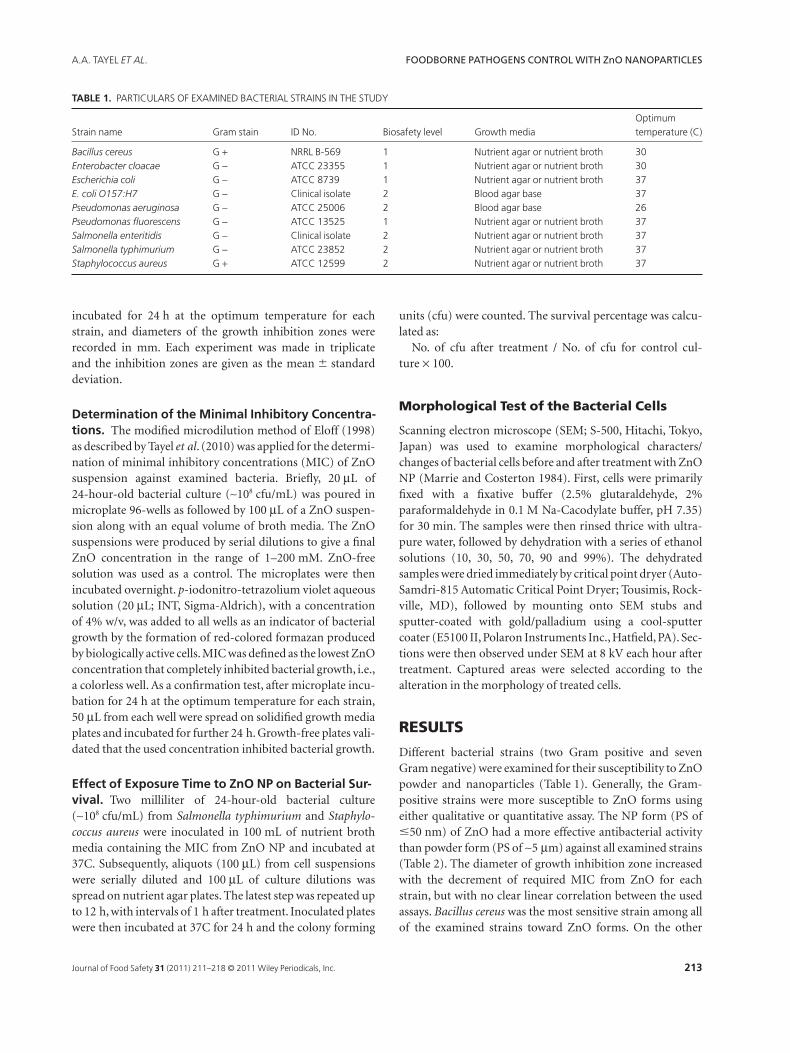
TABLE 1. PARTICULARS OF EXAMINED BACTERIAL STRAINS IN THE STUDY
Strain name Gram stain ID No. Biosafety level Growth mediaOptimumtemperature (C)
Bacillus cereus G + NRRL B-569 1 Nutrient agar or nutrient broth 30Enterobacter cloacae G - ATCC 23355 1 Nutrient agar or nutrient broth 30Escherichia coli G - ATCC 8739 1 Nutrient agar or nutrient broth 37E. coli O157:H7 G - Clinical isolate 2 Blood agar base 37Pseudomonas aeruginosa G - ATCC 25006 2 Blood agar base 26Pseudomonas fluorescens G - ATCC 13525 1 Nutrient agar or nutrient broth 37Salmonella enteritidis G - Clinical isolate 2 Nutrient agar or nutrient broth 37Salmonella typhimurium G - ATCC 23852 2 Nutrient agar or nutrient broth 37Staphylococcus aureus G + ATCC 12599 2 Nutrient agar or nutrient broth 37
A.A. TAYEL ET AL. FOODBORNE PATHOGENS CONTROL WITH ZnO NANOPARTICLES
213Journal of Food Safety 31 (2011) 211–218 © 2011 Wiley Periodicals, Inc.
hand, regarding their MICs, examined Pseudomonas spp.were the most resistant examined strains.
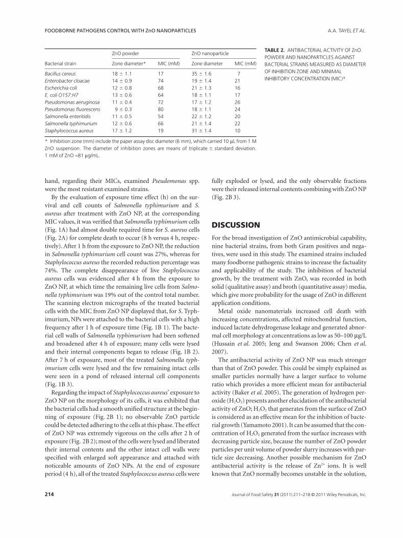
By the evaluation of exposure time effect (h) on the sur-vival and cell counts of Salmonella typhimurium and S.aureus after treatment with ZnO NP, at the correspondingMIC values, it was verified that Salmonella typhimurium cells(Fig. 1A) had almost double required time for S. aureus cells(Fig. 2A) for complete death to occur (8 h versus 4 h, respec-tively). After 1 h from the exposure to ZnO NP, the reductionin Salmonella typhimurium cell count was 27%, whereas forStaphylococcus aureus the recorded reduction percentage was74%. The complete disappearance of live Staphylococcusaureus cells was evidenced after 4 h from the exposure toZnO NP, at which time the remaining live cells from Salmo-nella typhimurium was 19% out of the control total number.The scanning electron micrographs of the treated bacterialcells with the MIC from ZnO NP displayed that, for S. Typh-imurium, NPs were attached to the bacterial cells with a highfrequency after 1 h of exposure time (Fig. 1B 1). The bacte-rial cell walls of Salmonella typhimurium had been softenedand broadened after 4 h of exposure; many cells were lysedand their internal components began to release (Fig. 1B 2).After 7 h of exposure, most of the treated Salmonella typh-imurium cells were lysed and the few remaining intact cellswere seen in a pond of released internal cell components(Fig. 1B 3).
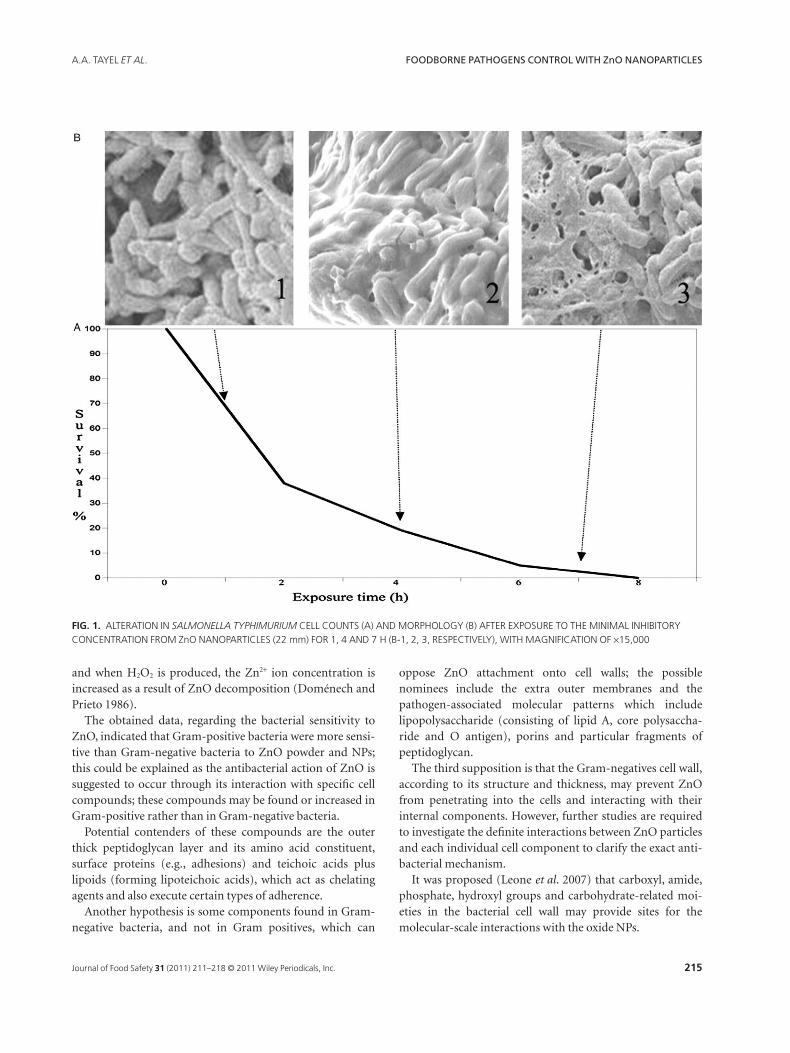
Regarding the impact of Staphylococcus aureus’ exposure toZnO NP on the morphology of its cells, it was exhibited thatthe bacterial cells had a smooth unified structure at the begin-ning of exposure (Fig. 2B 1); no observable ZnO particlecould be detected adhering to the cells at this phase. The effectof ZnO NP was extremely vigorous on the cells after 2 h ofexposure (Fig. 2B 2); most of the cells were lysed and liberatedtheir internal contents and the other intact cell walls werespecified with enlarged soft appearance and attached withnoticeable amounts of ZnO NPs. At the end of exposureperiod (4 h), all of the treated Staphylococcus aureus cells were
fully exploded or lysed, and the only observable fractionswere their released internal contents combining with ZnO NP(Fig. 2B 3).
DISCUSSION
For the broad investigation of ZnO antimicrobial capability,nine bacterial strains, from both Gram positives and nega-tives, were used in this study. The examined strains includedmany foodborne pathogenic strains to increase the factualityand applicability of the study. The inhibition of bacterialgrowth, by the treatment with ZnO, was recorded in bothsolid (qualitative assay) and broth (quantitative assay) media,which give more probability for the usage of ZnO in differentapplication conditions.
Metal oxide nanomaterials increased cell death withincreasing concentrations, affected mitochondrial function,induced lactate dehydrogenase leakage and generated abnor-mal cell morphology at concentrations as low as 50–100 mg/L(Hussain et al. 2005; Jeng and Swanson 2006; Chen et al.2007).
The antibacterial activity of ZnO NP was much strongerthan that of ZnO powder. This could be simply explained assmaller particles normally have a larger surface to volumeratio which provides a more efficient mean for antibacterialactivity (Baker et al. 2005). The generation of hydrogen per-oxide (H2O2) presents another elucidation of the antibacterialactivity of ZnO; H2O2 that generates from the surface of ZnOis considered as an effective mean for the inhibition of bacte-rial growth (Yamamoto 2001). It can be assumed that the con-centration of H2O2 generated from the surface increases withdecreasing particle size, because the number of ZnO powderparticles per unit volume of powder slurry increases with par-ticle size decreasing. Another possible mechanism for ZnOantibacterial activity is the release of Zn2+ ions. It is wellknown that ZnO normally becomes unstable in the solution,
TABLE 2. ANTIBACTERIAL ACTIVITY OF ZnOPOWDER AND NANOPARTICLES AGAINSTBACTERIAL STRAINS MEASURED AS DIAMETEROF INHIBITION ZONE AND MINIMALINHIBITORY CONCENTRATION (MIC)*
Bacterial strain
ZnO powder ZnO nanoparticle
Zone diameter* MIC (mM) Zone diameter MIC (mM)
Bacillus cereus 18 � 1.1 17 35 � 1.6 7Enterobacter cloacae 14 � 0.9 74 19 � 1.4 21Escherichia coli 12 � 0.8 68 21 � 1.3 16E. coli O157:H7 13 � 0.6 64 18 � 1.1 17Pseudomonas aeruginosa 11 � 0.4 72 17 � 1.2 26Pseudomonas fluorescens 9 � 0.3 80 18 � 1.1 24Salmonella enteritidis 11 � 0.5 54 22 � 1.2 20Salmonella typhimurium 12 � 0.6 66 21 � 1.4 22Staphylococcus aureus 17 � 1.2 19 31 � 1.4 10
* Inhibition zone (mm) include the paper assay disc diameter (6 mm), which carried 10 mL from 1 MZnO suspension. The diameter of inhibition zones are means of triplicate � standard deviation.1 mM of ZnO =81 mg/mL.
FOODBORNE PATHOGENS CONTROL WITH ZnO NANOPARTICLES A.A. TAYEL ET AL.
214 Journal of Food Safety 31 (2011) 211–218 © 2011 Wiley Periodicals, Inc.
and when H2O2 is produced, the Zn2+ ion concentration isincreased as a result of ZnO decomposition (Doménech andPrieto 1986).
The obtained data, regarding the bacterial sensitivity toZnO, indicated that Gram-positive bacteria were more sensi-tive than Gram-negative bacteria to ZnO powder and NPs;this could be explained as the antibacterial action of ZnO issuggested to occur through its interaction with specific cellcompounds; these compounds may be found or increased inGram-positive rather than in Gram-negative bacteria.
Potential contenders of these compounds are the outerthick peptidoglycan layer and its amino acid constituent,surface proteins (e.g., adhesions) and teichoic acids pluslipoids (forming lipoteichoic acids), which act as chelatingagents and also execute certain types of adherence.
Another hypothesis is some components found in Gram-negative bacteria, and not in Gram positives, which can
oppose ZnO attachment onto cell walls; the possiblenominees include the extra outer membranes and thepathogen-associated molecular patterns which includelipopolysaccharide (consisting of lipid A, core polysaccha-ride and O antigen), porins and particular fragments ofpeptidoglycan.
The third supposition is that the Gram-negatives cell wall,according to its structure and thickness, may prevent ZnOfrom penetrating into the cells and interacting with theirinternal components. However, further studies are requiredto investigate the definite interactions between ZnO particlesand each individual cell component to clarify the exact anti-bacterial mechanism.
It was proposed (Leone et al. 2007) that carboxyl, amide,phosphate, hydroxyl groups and carbohydrate-related moi-eties in the bacterial cell wall may provide sites for themolecular-scale interactions with the oxide NPs.
FIG. 1. ALTERATION IN SALMONELLA TYPHIMURIUM CELL COUNTS (A) AND MORPHOLOGY (B) AFTER EXPOSURE TO THE MINIMAL INHIBITORYCONCENTRATION FROM ZnO NANOPARTICLES (22 mm) FOR 1, 4 AND 7 H (B-1, 2, 3, RESPECTIVELY), WITH MAGNIFICATION OF ¥15,000
A.A. TAYEL ET AL. FOODBORNE PATHOGENS CONTROL WITH ZnO NANOPARTICLES
215Journal of Food Safety 31 (2011) 211–218 © 2011 Wiley Periodicals, Inc.
Also, it was demonstrated (Tam et al. 2008) that ZnO wasmore effective for killing Gram-positive than Gram-negativebacteria because they have simpler cell membrane structure.
Tsao et al. (2002) stated that the exposure of Gram-positivebacteria to carboxyfullerene NP resulted in the puncturing ofthe bacteria and cell death. Another proposed way in whichthe membrane can be compromised is the alteration of mem-brane lipid components (Koch et al. 2005).
The majority of previous studies suggested that the mecha-nism of NP toxicity may be related to its photosensitivity andproduction of reactive oxygen species (ROS) under specificwavelength high-intensity light; therefore, nanomaterials thatgenerate ROS can indirectly damage cell membranes. The cellmembrane architecture could be impaired through lipidperoxidation by ROS. Damage and disorganization in thecell wall were observed in the bacteria exposed to MgO(Stoimenov et al. 2002) and ZnO (Brayner et al. 2006) NPs.
In the current investigation, all of the assay steps werecarried out under dark conditions to avoid the possible effects
of released ROS from ZnO NP. This indicates the probableproduction of free radicals under dark conditions as was pre-viously suggested (Green and Howman 2005), or other toxic-ity mechanisms additional to ROS production.
Two different assays were applied in the current investiga-tion; growth inhibition zone assay expressed both the bacte-riostatic and bacteriocidal actions of ZnO, whereas thedetermination of MIC accompanied by INT staining focusedon the bactericidal action of the NPs.
Following the general evaluation of ZnO antibacteriallevel, Salmonella typhimurium and Staphylococcus aureuswere chosen as models to investigate the antibacterial mode ofaction of ZnO NP, because they exhibited relatively high resis-tance to ZnO concentrations and because of their significantrisk as serious foodborne and zoonotic pathogens.
In the current study, the high MIC applied from ZnOto inhibit Salmonella typhimurium (22 mM), comparedto the concentration applied for Staphylococcus aureus(10 mM),increasedNPsprobabilityof attachingtoSalmonella
FIG. 2. ALTERATION IN STAPHYLOCOCCUS AUREUS CELL COUNTS (A) AND MORPHOLOGY (B) AFTER EXPOSURE TO THE MINIMAL INHIBITORYCONCENTRATION FROM ZnO NANOPARTICLES (10 mm) FOR 0, 2 AND 4 H (B-1, 2, 3, RESPECTIVELY), WITH MAGNIFICATION OF ¥12,000
FOODBORNE PATHOGENS CONTROL WITH ZnO NANOPARTICLES A.A. TAYEL ET AL.
216 Journal of Food Safety 31 (2011) 211–218 © 2011 Wiley Periodicals, Inc.
typhimurium further than Staphylococcus aureus cells, at thebeginning of exposure.
It was proposed that nanomaterials that can physicallyattach to a cell can be bactericidal if they come into contactwith this cell (Jeng and Swanson 2006). If the membrane of abacterium is compromised, the cell may repair itself or, if thescratch is severe, the cell component may release and eventu-ally the cell will die (Lyon et al. 2007).
It was demonstrated that ZnO NPs possess antimicrobialactivities against Listeria monocytogenes and Salmonella enter-itidis in liquid egg white and culture media, and E. coliO157:H7 in culture media (Jin et al. 2009); Jin et al. (2009)recommended several approaches (powder, PVP capped, filmand coating) for the incorporation of ZnO into food system.Tam et al. (2008) revealed that ZnO nanorod in the liquidphase leads to bacterial cell death which occurred because ofcell membrane damage.
The impact of nanomaterials on living cells, including bac-teria, can also be elucidated by the interactions between thenanomaterial and the individual cell components. The firstinteraction between a material and a cell is at the membraneinterface; some nanoparticles were suggested to embed them-selves in the cell membrane (Jang et al. 2003).
Liu et al. (2009) indicated that ZnO NP may distort anddamage bacterial cell membrane, resulting in a leakage ofintracellular contents and eventually the death of bacterialcells; the inhibitory effects against E. coli O157:H7 increase asthe concentration of ZnO NP increased. However, there is stilla current lack of exact information regarding the interactionof NPs with the bacterial cell wall and possible permeation ofthe NPs into the bacterial cells (Jiang et al. 2009).
We can conclude, regarding the obtained results, that ZnONP could be proposed as an effective and powerful antibacte-rial agent against both Gram-positive and -negative bacteria.The treatment with ZnO NP resulted in bacterial cell explo-sion or lysis, and its antibacterial action was more vigorousin Gram-positive bacteria. The application of ZnO NP couldbe recommended as a preservative agent against foodbornepathogens, in food production and processing, after confirm-ing either its biosafety or toxicity.
REFERENCES
ANDERSEN, S., DONG, M., NIELSEN, M.M., JAHN, K., SUBRA-MANI, R., MAMDOUH, W., GOLAS, M.M., SANDER, B.,STARK, H., OLIVEIRA, C.L.P. ET AL. 2009. Self-assenbly of anano-scale DNA box with a controllable lid. Nature 459, 73–76.
BAKER, C., PRADHAN, A., PAKSTIS, L., POCHAN, D.J. andISMAT, S.S. 2005. Synthesis and antibacterial properties ofsilver nanoparticles. J. Nanosci. Nanotechnol. 5, 244–249.
BALOWS, A., HAUSLER, W.J., HERRMANN, K.L., ISENBERG,H.D. and SHADOMY, H.J. (eds.) 1991. Manual of Clinical
Microbiology, 5th Ed., American Society for Microbiology,Washington, DC.
BELL, C. and KYRIAKIDES, A. 2002. Salmonella: A PracticalApproach to the Organism and Its Control in Foods, BlackwellPublishing Ltd, Oxford, U.K.
BENNET, D.J. and SCHUURBIERS, D. 2005.Nanobiotechnology: Responsible action on issue in society andethics. NSTI-Nanotech. 2, 765–768.
BHUNIA, A.K. (ed.) 2008. Foodborne Microbial Pathogens:Mechanisms and Pathogenesis, Springer LLC, New York,NY.
BRAYNER, R., FERRARI-LLIOU, R., BRIVOIS, N., DJEDIAT, S.,BEBEDETTI, M.F. and FIEVET, F. 2006. Toxicology impactstudies based on Escherichia coli bacteria in ultrafine ZnOnanoparticles colloidal medium. Nano Lett. 6, 866–870.
CHEN, J., ZHU, J., CHO, H.-H., CUI, K., LI, F., ZHOU, X.,ROGERS, J.T., WONG, S.T.C. and HUANG, X. 2007.Differential cytotoxicity of metal oxide nanoparticles. In NSTINanotech 2007; May 20–24, Santa Clara, CA.
DOMÉNECH, J. and PRIETO, A. 1986. Stability of zinc oxideparticles in aqueous suspensions under UV illumination.J. Phys. Chem. 90, 1123–1126.
ELOFF, J.N. 1998. A sensitive and quick microplate method todetermine the minimal inhibitory concentration of plantextracts for bacteria. Planta Med. 64, 711–713.
FU, G., VARY, P.S. and LIN, C.T. 2005. Anatase TiO2
nanocomposites for antimicrobial coatings. J. Phys. Chem. B109, 8889–8898.
GREEN, M. and HOWMAN, E. 2005. Semiconductor quantumdots and free radical induced DNA nicking. Chem. Commun.(Camb). 7, 121–123.
HOWARD, K.A. and KJEMS, J. 2007. Polycation-basednanoparticle delivery for improved RNAi therapeutics. ExpertOpin. Biol. Ther. 7, 1811–1822.
HUSSAIN, S.M., HESS, K.L., GEARHART, J.M., GEISS, K.T. andSCHLAGER, J.J. 2005. In vitro toxicity of nanoparticles in BRL3A rat liver cells. Toxicol. In Vitro 19, 975–983.
JANG, H., PELL, L.E., KORGEL, B.A. and ENGLISH, D.S. 2003.Photoluminescence quenching of silicon nanoparticles inphospholipid vesicle bilayers. J. Photochem. Photobiol.A Chem. 158, 111–117.
JENG, H.A. and SWANSON, J. 2006. Toxicity of metal oxidenanoparticles in mammalian cells. J. Environ. Sci. Health A Tox.Hazard. Subst. Environ. Eng. 41, 2699–2711.
JIANG, W., MASHAYEKHI, H. and XING, B. 2009. Bacterialtoxicity comparison between nano- and micro-scaled oxideparticles. Environ. Pollut. 157, 1619–1625.
JIN, T., SUN, D., SU, J.Y., ZHANG, H. and SUE, H.-J. 2009.Antimicrobial efficacy of zinc oxide quantum dots againstListeria monocytogenes, Salmonella enteritidis, and Escherichiacoli O157:H7. J. Food Sci. 74, M46–M52.
KOCH, A.M., REYNOLDS, F., MERKLE, H.P., WEISSLEDER, R.and JOSEPHSON, L. 2005. Transport of surface-modifiednanoparticles through cell monolayers. Chembiochem 6,337–345.
A.A. TAYEL ET AL. FOODBORNE PATHOGENS CONTROL WITH ZnO NANOPARTICLES
217Journal of Food Safety 31 (2011) 211–218 © 2011 Wiley Periodicals, Inc.
KUMAR, C. (ed.) 2006. Nanomaterials: Toxicity, Health, andEnvironmental Issues, 1st Ed., Nanotechnologies for the LifeSciences Vol 5. Wiley-VHC, Weinheim, Germany.
LEONE, L., FERRI, D., MANFREDI, C., PERSSON, P.,SHCHUKAREV, A., SJOBERG, S. and LORING, J. 2007.Modeling the acid–base properties of bacterial surfaces: Acombined spectroscopic and potentiometric study of theGram-positive bacterium Bacillus subtilis. Environ. Sci.Technol. 41, 6465–6471.
LIU, Y., HE, L., MUSTAPHA, A., LI, H., HU, Z.Q. and LIN, M.2009. Antibacterial activities of zinc oxide nanoparticles againstEscherichia coli O157:H7. J. Appl. Microbiol. 107, 1193–1201.
LYON, D.Y., THILL, A., ROSE, J. and ALVAREZ, P.J.J. 2007.Ecotoxicological impacts of nanomaterials. In EnvironmentalNanotechnology, Applications and Impacts of Nanomaterials(M.R. WIESNER and J. BOTTERO, eds.) pp. 445–479, TheMcGraw-Hill, New York, NY.
MARRIE, T.J. and COSTERTON, J.W. 1984. Scanning andtransmission electron microscopy of in situ bacterialcolonization of intravenous and intraarterial catheters. J. Clin.Microbiol. 19, 687–693.
MEYER, M. and KUUSI, O. 2002. Nanotechnology:Generalizations in an interdisciplinary field of science andtechnology. Int. J. Philos. Chem. 10, 153–168.
PHILLIPS, L.G. and BARBANO, D.M. 1997. The influence of fatsubstitutes based on protein and titanium dioxide on thesensory properties of lowfat milk. J. Dairy Sci. 80,2726–2731.
REDDY, K.M., FERIS, K., BELL, J., WINGETT, D.G., HANLEY, C.and PUNNOOSE, A. 2007. Selective toxicity of zinc oxidenanoparticles to prokaryotic and eukaryotic systems. Appl.Phys. Lett. 90, 2139021–2139023.
RICHARDS, E. 2006. Antibacterial wallpaper. Chemtech. 2006,11.
RYAN, K.J. and RAY, C.G. (eds.) 2004. Sherris MedicalMicrobiology, 4th Ed., McGraw-Hill, Columbus, OH.
SAWAI, J. 2003. Quantitative evaluation of antibacterial activities
of metallic oxide powders (ZnO, MgO and CaO) byconductimetric assay. J. Microbiol. Methods 54, 177–182.
SERPONE, N., DONDI, D. and ALBINI, A. 2007. Inorganic andorganic UV filters: Their role and efficacy in sunscreens andsuncare products. Inorganica Chim. Acta 360, 794–802.
STOIMENOV, P.K., KLINGER, R.L., MARCHIN, G.L. andKLABUNDE, K.J. 2002. Metal oxide nanoparticles asbactericidal agents. Langmuir 18, 6679–6686.
SWEETMAN, S.C. (ed.) 2005. Martindale, The Complete DrugReference, p. 1163, 34th Ed., Pharmaceutical Press, London,U.K.
TAM, K.H., DJURIŠIC, A.B., CHAN, C.M.N., XI, Y.Y., TSE, C.W.,LEUNG, Y.H., CHAN, W.K., LEUNG, F.C.C. and AU, D.W.T.2008. Antibacterial activity of ZnO nanorods prepared by ahydrothermal method. Thin Solid Films 516, 6167–6174.
TAYEL, A.A., MOUSSA, S., OPWIS, K., KNITTEL, D.,SCHOLLMEYER, E. and NICKISCH- HARTFIEL, A. 2010.Inhibition of microbial pathogens by fungal chitosan. Int. J.Biol. Macromol. 47, 10–14.
THILL, A., ZEYONS, O., SPALLA, O., CHAUVAT, F., ROSE, J.,AUFFAN, M. and FLANK, A.M. 2006. Cytotoxicity of CeO2
nanoparticles for Escherichia coli. Physico-chemical insight ofthe cytotoxicity mechanism. Environ. Sci. Technol. 40,6151–6156.
TSAO, N., LUH, T., CHOU, C., CHANG, T., WU, J., LIU, C. andLEI, H. 2002. In vitro action of carboxyfullerene. J. Antimicrob.Chemother. 49, 641–649.
WOODHOUSE, E.J. 2004. Nanotechnology controversies.Technol. Soc. Mag. IEEE 23, 6–8.
YAMAMOTO, O. 2001. Influence of particle size on theantibacterial activity of zinc oxide. Int. J. Inorg. Mater. 3,643–646.
ZHANG, L.L., JIANG, Y.H., DING, Y.L., POVEY, M. and YORK,D. 2007. Investigation into the antibacterial behaviour ofsuspensions of ZnO nanoparticles (ZnO nanofluids).J. Nanopart. Res. 9, 479–489.
FOODBORNE PATHOGENS CONTROL WITH ZnO NANOPARTICLES A.A. TAYEL ET AL.
218 Journal of Food Safety 31 (2011) 211–218 © 2011 Wiley Periodicals, Inc.