Embed Size (px)
Citation preview

Yutin et al. Virology Journal 2013, 10:158http://www.virologyj.com/content/10/1/158
RESEARCH Open Access
Virophages, polintons, and transpovirons: acomplex evolutionary network of diverse selfishgenetic elements with different reproductionstrategiesNatalya Yutin1, Didier Raoult2 and Eugene V Koonin1*
Abstract
Background: Recent advances of genomics and metagenomics reveal remarkable diversity of viruses and otherselfish genetic elements. In particular, giant viruses have been shown to possess their own mobilomes that includevirophages, small viruses that parasitize on giant viruses of the Mimiviridae family, and transpovirons, distinct linearplasmids. One of the virophages known as the Mavirus, a parasite of the giant Cafeteria roenbergensis virus, sharesseveral genes with large eukaryotic self-replicating transposon of the Polinton (Maverick) family, and it has beenproposed that the polintons evolved from a Mavirus-like ancestor.
Results: We performed a comprehensive phylogenomic analysis of the available genomes of virophages andtraced the evolutionary connections between the virophages and other selfish genetic elements. The comparisonof the gene composition and genome organization of the virophages reveals 6 conserved, core genes that areorganized in partially conserved arrays. Phylogenetic analysis of those core virophage genes, for which a sufficientdiversity of homologs outside the virophages was detected, including the maturation protease and the packagingATPase, supports the monophyly of the virophages. The results of this analysis appear incompatible with the originof polintons from a Mavirus-like agent but rather suggest that Mavirus evolved through recombination between apolinton and an unknownvirus. Altogether, virophages, polintons, a distinct Tetrahymena transposable element Tlr1,transpovirons, adenoviruses, and some bacteriophages form a network of evolutionary relationships that is heldtogether by overlapping sets of shared genes and appears to represent a distinct module in the vast total networkof viruses and mobile elements.
Conclusions: The results of the phylogenomic analysis of the virophages and related genetic elements arecompatible with the concept of network-like evolution of the virus world and emphasize multiple evolutionaryconnections between bona fide viruses and other classes of capsid-less mobile elements.
BackgroundThe rapid advances of genomics and metagenomics leadnot only to the rapid growth of sequence databases butto discovery of fundamentally novel types of geneticelements. The discovery and characterization of giantviruses that infect unicellular eukaryotes, in particularmembers of the family Mimiviridae infecting amoeba,over the last decade revealed a remarkable new class of
* Correspondence: [email protected] Center for Biotechnology Information, National Library of Medicine,National Institutes of Health, Bethesda, MD 20894, USAFull list of author information is available at the end of the article
© 2013 Yutin et al.; licensee BioMed Central LCommons Attribution License (http://creativecreproduction in any medium, provided the or
agents that are typical viruses by structure and repro-duction strategy but exceed many parasitic bacteria insize and genomic complexity [1-6]. Much like bacteria,the giant viruses (sometimes called giruses) possess theirown parasites and their own mobilomes, i.e. communi-ties of associated mobile genetic elements [7]. The firstvirus infecting a giant virus, the Sputnik virophage, wasisolated from a mimivirus-infected acanthamoeba andshown to replicate within the mimivirus factories andpartially inhibit the reproduction of the host mimivirus[8,9]. The second isolated virophage, named Mavirus, isa parasite of the Cafeteria roenbergensis virus (CroV), a
td. This is an Open Access article distributed under the terms of the Creativeommons.org/licenses/by/2.0), which permits unrestricted use, distribution, andiginal work is properly cited.

Yutin et al. Virology Journal 2013, 10:158 Page 2 of 15http://www.virologyj.com/content/10/1/158
distant relative of the mimiviruses [10,11]. The thirdvirophage genome was isolated from the AntarcticOrganic Lake (hence OLV, Organic Lake Virophage)where it apparently controls the reproduction of its virushost that originally has been classified as a distinctphycodnavirus [12] but according to a more detailedrecent phylogenetic study, is actually more closelyrelated to the family Mimiviridae [13]. Very recently, 5additional genomes of putative virophages have beenassembled from metagenomic sequences [14]. Four com-plete genomes, those of Yellowstone Lake Virophages(YLSV1-4), appeared to be related to OLV, whereas thefifth, nearly complete one, the Ace Lake Mavirus (ALM),appeared to be a relative of the Mavirus [14].The three well-characterized virophages possess small
isocahedral virions and genomes of 20 to 25 kilobaseencoding 21 to 26 proteins each. Although the viro-phages are similar in genome size and structure and aregenerally construed as related, only a minority of thevirophage genes are homologous. The rest of the genesshow diverse phylogenetic affinities suggestive of chime-ric origins of the virophages [8,11,12].Analysis of the Mavirus genome [11] resulted in the
unexpected discovery that this virophage shared 5 hom-ologous genes with the large, self-replicating eukaryotictransposable elements of the Maverick/Polinton class(hereinafter Polintons). The Polintons that are scatteredamong genome of diverse eukaryotes and reach highabundance in some protists, such as Trichomonasvaginalis, have long been considered ‘virus-like’ transpo-sons because of their large size (20 kb and larger) andthe presence of several genes that are common in virusesbut not in other transposable elements such as B familyDNA polymerase (PolB), packaging ATPase (ATPase) andprotease (PRO) [15-18]. The Mavirus shows by far theclosest affinity with the Polintons among the currentlyknown viruses, and accordingly, it has been proposed thatthe Polintons evolved from the virophages [11].In addition to the virophages, the giant viruses host
several other groups of mobile elements. These includeself-splicing introns, inteins, putative bacterial-typetransposons and the most recently discovered novellinear plasmids named transpovirons [7]. The transpo-virons are highly abundant genetic elements associatedwith several giant viruses of the Mimiviridae family thatcontain only 6 to 8 genes two of which are homologous togenes of the Sputnik virophage, indicating multiple geneexchanges within the giant virus mobilome.We sought to decipher the evolutionary relationships
between the three known virophages, the Polintons,transpovirons and possibly other genetic elements andviruses. We come up with a complex network of evolu-tionary relationships that connect many of these diverseelements through overlapping sets of homologous genes.
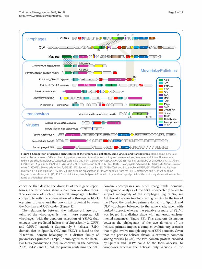
Results and discussionOrigin and evolution of the virophagesTo our knowledge, the evolutionary relationships bet-ween the virophages so far have not been analyzed in acomprehensive manner. Therefore we performed anexhaustive genomic comparison of the three well-characterized virophages that involved detailed sequenceanalysis for all predicted virophage proteins (seeMethods for details); at this stage, the 5 new virophagegenome sequences [14] were not included given po-tential uncertainties in the genome assembly from me-tagenomic data. This was followed by phylogeneticanalysis of the proteins that showed sufficient evolution-ary conservation that, in addition to the three previouslycharacterized virophages, included the 5 new ones. Allvirophages share 6 homologous proteins or domains: 1)Primase-Superfamily 3 helicase (S3H), 2) packagingATPase (ATPase), 3) cysteine protease (PRO), 4) Zn-ribbon domain (ZnR), 5) major capsid protein (MCP), 6)minor capsid protein (mCP) (Figure 1 and Table 1). Theminor capsid protein initially has not been detected inthe Mavirus but direct sequence comparisons supportedby gene synteny suggest that MV17 is indeed a highlydiverged homolog of the minor capsid protein of thetwo other virophages (see Additional file 1). The twovirion proteins have no detectable homologs outside thevirophages (in particular, no environmental homologs;see discussion below) and therefore, per force, areinferred to have evolved from a common ancestor. Therecently solved near atomic structure of the Sputnikvirion shows that the major capsid protein assumes a di-verged double jelly roll structure shared with numerousicosahedral viruses [19].The virophage Zn-ribbon is a distinct version of this
module that is shared by the virophages and severalother groups of mobile elements (see below). In theSputnik virophage the Zn-ribbon is a stand-alone pro-tein whereas in Mavirus and OLV it is fused to a GIY-YIG endonuclease (GIY), a domain architecture that wasdetected also in environmental homologs. Conceivably,the ZnR-nuclease fusion is the ancestral version of thisprotein, with the nuclease lost in the Sputnik lineage.The ZnR domain is too small for reliable phylogeneticanalysis (see Additional file 1 for multiple alignments).The phylogenetic trees for the cysteine protease and
the packaging ATPase strongly support the monophylyof the virophages along with the related environmentalsequences (Figure 2A,B). In both these trees, Sputnikforms a clade with OLV-YSLV, and the Mavirus-ALMclade is an outgroup to this clade. Taken together, theexistence of 5 signature genes including two genes forstructural proteins, along with the apparently mono-phyletic ATPase and the protease required for virionmorphogenesis, seems to present sufficient evidence to
Dictyostelium fasciculatum
1319 2017 18
1 2 3 4 6 7 8 9 10
2325 1
3 4 5 6 7 8 9 10 11 12
13 14 1720 21
1 2 36
12 13 14 15 16 17 18 19 9
Polysphondylium pallidum PN500
Polinton-1_CB of C. briggsae
Mavirus
Sputnik
OLV
Mimivirus lentille transpoviron Lentilletv_L4 tv_R6
tv_L5
DFA_00301
Ppl_12642
PY PC1 ATP POLB PC2PRO
INT
Tlr1 element of T. thermophilaTlr 6F
DNA helicaseINT
Bovine Adenovirus A pol pTP 52K pIIIA penton hexon DBP 100K VIII fiber IVa2
Polinton-1_TV of T. vaginalis POLB INT ATP1 ATP
Bacteriophage Bam35 COAT
Bacteriophage PRD1 COAT
Acyrthosiphon pisum RT
NS1 VP1 Minute virus of mice (parvovirus)
Cotesia congregata bracovirus
Tribolium castaneum
Mavericks/Polintons
virophages
transpoviron
viruses
18-19
S3HINTATPasePROGIYZnRMCPmCPTVpolPolBV21/OLV5 LipaseTlr 6FTlr helicaseTransposasePLA2
Figure 1 Comparison of genome architectures of the virophages, polintons, some viruses, and transpovirons. Homologous genes aremarked by same colors. Different hatching patterns are used to mark non-orthologous primase-helicase, integrase, and lipase. Homologousregions are shaded. Reference sequences were extracted from GenBank (D. fasciculatum, GI:328871053; P. palladium, GI: 281202948; T. castaneum,GI:58197573; A. pisum, GI:156713484; Mimivirus lentille transpoviron Lentille, GI: 374110342; C. congregate bracovirus, GI: 326937614 Minute virus ofmice, GI:9626993; Bovine adenovirus A, GI:52801677; Bacteriophage Bam35, GI:38640293; and Bacteriophage PRD1, GI:159192286) and Repbase(Polinton-1_CB and Polinton-1_TV [15,20]). The genome organization of Tlr1was adopted from ref. [18]. T. castaneum and A. pisum genomefragments are shown as in [21]. PLA2 stands for the phospholipase A2 domain of parvovirus capsid protein. Other color key abbreviations are thesame as throughout the text.
Yutin et al. Virology Journal 2013, 10:158 Page 3 of 15http://www.virologyj.com/content/10/1/158
conclude that despite the diversity of their gene reper-toires, the virophages share a common ancestral virus.The existence of such an ancestral virophage is furthercompatible with the conservation of a three-gene block(cysteine protease and the two virion proteins) betweenthe Mavirus and OLV clades (Figure 1).The relationship between the helicase-primase pro-
teins of the virophages is much more complex. Allvirophages (with the apparent exception of YSLV2 thatencodes two predicted helicases of Superfamily 2, ORF5and ORF10) encode a Superfamily 3 helicase (S3H)domain that in Sputnik, OLV and YSLV1 is fused to theN-terminal domain belonging to a distinct family ofpolymerases-primases (TVpol) homologous to the bacte-rial DNA polymerase I [22]. By contrast, in the Mavirus,ALM, YSLV3 and YSLV4, the protein containing the S3H
domain encompasses no other recognizable domains.Phylogenetic analysis of the S3H unexpectedly failed tosupport monophyly of the virophages (Figure 3A; seeAdditional file 2 for topology testing results). In the tree ofthe TVpol, the predicted primase domains of Sputnik andOLV virophages belonged to the same clade, albeit withlimited support, whereas the putative primase of YSLV1was lodged in a distinct clade with numerous environ-mental sequences (Figure 3B). This apparent distinctionbetween the phylogenies of the two domains of thehelicase-primase implies a complex evolutionary scenariothat might involve multiple origins of S3H domains. Giventhat the primase-helicase fusion is extremely commonamong viruses [23,24], the two-domain protein encodedby Sputnik and OLPV could be the form ancestral tovirophages whereas the helicase only versions in the

Table 1 Evolutionary provenance of the genes of the three well-characterized virophages
Gene/protein Domain architecture Predicted activity/function Phylogenetic spread and affinity Representation in environmentalsequences
Proteins (domains) conserved in all three virophages
V9, OLV7, MV16 C5-family cysteine protease Protease, probably involvedin capsid protein maturation
Only distantly related to other proteases fromNCLDV, adenoviruses, eukaryotes andsome bacteria
No obvious homologs
V3, OLV4, MV15 P-loop ATPase, FtsK-like family Packaging ATPase Only distantly related to other ATPases of theFtsK-like family: NCLDV, adenoviruses,diverse phages, bacteria and archaea (DNApumping during cell divisionand conjugation)
Abundant moderately conservedhomologs
V20, OLV9, MV18 Predicted distorted jelly-roll domain Major capsid protein No homologs beyond virophages None
V18-19, OLV8, MV17 No detectable domains Minor capsid protein No homologs beyond virophages None
V14, V4, MV06(C-terminal), OLV1(C-terminal),
C2H2 Zn-ribbon; N-terminal GIY-YIGendonuclease domain in MV06and OLV1
Unknown Homologs in transpovirons (closest to V14,Zn-ribbon only), Phytophtora and Dictyosteliumpolintons, P.globosa virus
Moderately conserved homologs,mostly containing GIY-YIG nucleasedomain
V13 (C-terminal),OLV25 (C-terminal),MV01
S3H helicase; N-terminal TVpol inV13 and OLV25
primase-helicase Sputnik helicase is most similar to bacterialand bacteriophage homologs; the MV01helicase is most similar to the NCLDV homolog;the OLV helicase is most similar to homologsfrom bacteriophages and polintons
Numerous conserved homologsincluding proteins with both TVpoland helicase domains
Proteins (domains) shared between Sputnik and OLV
V13 (N-terminal),OLV25 (N-terminal)
TVpol Primase and DNA polymerase ([22]) Related to Micromonas pusilla anddifferent bacteria
Numerous conserved homologsincluding proteins with both TVpoland helicase domains
V21, OLV5 No detectable domains Unknown No other homologs None
V6(part), V7(part)OLV13 (part), OLV19 (part),OLV20 (part)
Collagen-like repeats Adsorbtion on host virus? V6 is highly similar to mimiviruses, OLV13 - tobacteria; OLV19 has regions similar to OLPV,T.vaginalis (phage protein)
Abundant homologs mostlycontaining collagen domain
Proteins (domains) shared between OLV and Mavirus
OLV1 (N-terminal),OLV24 and MV06(N- terminal
GIY-YIG endonuclease, fused toC2H2 Zn-ribbon in OLV1 and MV06
Unknown Close homologs in Phytophtora polintons andP. globosa virus
Moderately conserved homologs
OLV12 (C- terminal),MV13 (C- terminal)
Lipase 3 domain Unknown Homologs in all cellular organisms; Mavirusclosest homolog is a Physcomitrella patensprotein; OLV12 isclose to bacterial proteins
Few moderately conservedhomologs for each of the proteins
Proteins (domains) shared between Sputnik and Mavirus
V10, MV02 Integrase Mavirus interase is related to Polintons, Sputnik - toarchaeal and bacterial proviruses
Very few homologs
Yutinet
al.VirologyJournal2013,10:158
Page4of
15http://w
ww.virologyj.com
/content/10/1/158

Table 1 Evolutionary provenance of the genes of the three well-characterized virophages (Continued)
Sputnik genes with homologs outside virophages
V17 Transposase, DNA-binding domain DNA-binding protein Closest homologs in transpovirons Numerous moderately conservedhomologs
V16 No detectable domains Unknown Homologs in moumouvirus: mv_L1152 none
V12 No detectable domains Unknown Highly conserved homologs in Mimiviridae none
V10 XerD family integrase Integrase Closest homologs in archaeal proviruses Only distant integrases
OLV genes with homologs outside virophages
OLV23 N6 A-specific methylase DNA methylase Numerous bacterial homolog Numerous homologs
OLV16, OLV21 Proline-rich, mucien –like repeats Unknown (adsorption on virushost?)
Similar repeats in bacteria and eukaryotes Numerous similar repeats
OLV18, OLV19 Phage Tail Collar Domain Unknown (adsorption on virushost?)
Closely related to a family of OLPV proteins Numerous close homologs
OLV2 Uncharacterized domain Unknown Homologs in many phycodnaviruses andin Tlr1 element (6Fp)
Abundant homologs with widerange of similarity including veryclose ones
OLV22 Uncharacterized domain Unknown Highly similar to OLPV2, GI:322510937 A few close homologs
OLV12(N-terminal) Uncharacterized domain fused to Lipase 3 Unknown Highly similar to Chloroviruses Numerous close homologs
Mavirus genes with homologs outside virophages
MV20 FNIP repeats Unknown Closely related homologs in mimiviruses Numerous moderately similarhomologs
MV04 C2H2 Zn finger Unknown No close homologs None
MV02 RVE family integrase Integration of Mavirus genomeinto the virus host genome?
Numerous homologs, closest in Polintons Numerous moderately similarhomologs
MV19, M09 S74 family peptidase (C-terminal), N-terminalglycosylase (?); MV09 has only the N-terminaldomain
Unknown Numerous homologs in phages andbacteria (prophages?); homologs in Marseillevirus,Lausannevirus, Paramecium virus, and Polintons(N-terminal only).
Numerous moderately similarhomologs
MV13 Lipase (a/b hydrolase superfamily) Unknown Homologs in all cellular organisms, closestin plants
Several moderately similarhomologs
MV03 B family DNA polymerase Genome replication Homologs in all cellular organisms andnumerous viruses, the closest homologs in Polintons
No close homologs
Yutinet
al.VirologyJournal2013,10:158
Page5of
15http://w
ww.virologyj.com
/content/10/1/158

BA ALM ORF10Mavirus MV16 326439164
OLV OLV7 322510453YSLV1 ORF23
YSLV2 ORF12YSLV3 ORF05
YSLV4 ORF16Sputnik V9 195982540
El Caere308465343PRO-1_Cip*PRO-2_Cip*
PRO-1_Dyp*PRO-1_TCp*
Polinton-8_NVi_3p*Polinton-2_NVi_3p*
Polinton-5_NVi_6p*PRO-1_SM*PRO-2_SM*
adenovirusesEl Caebr341887159El Caere308458158
El Caere308458160PRO-1_CBp*PRO-2_CBp*
PRO-2_TCp*PRO-3_TCp*
Polinton-4_NVi_2p*Polinton-6_NVi_3p*
Ea Dicfa328864869Ea Polpa281203227
Ea Dicfa328869865Ea Dicfa328866525
NCLDVNCLDV (poxviruses)
Eukaryotes
0.97
0.99
0.64
0.51
0.63
0.81
0.94
0.58
0.86
0.80
0.66
0.53
0.83
0.99
0.980.79
0.56
0.50
1.00
0.63
0.92
0.900.71
0.78
0.99
0.54
0.540.55
0.95
0.68
0.68
0.54
0.82
0.89
0.5
n2 Acaca351737817n2 Acapo311978049
n2 MouMo371944625n2 Megch363540482
YSLV1 ORF01YSLV4 ORF01
env139742087YSLV2 ORF01
YSLV3 ORF01env137739104
env136179552env136472150
OLV OLV4 322510450Sputnik V3 195982534
env136185112Mavirus MV15 326439163
ALM ORF11El Caebr268568730PC2-1_CBp*
u2 Amsmo9964464u2 Melsa9631462
Afrsw229891993Afrsw9628186
uncvi189036186
Maverics/Polintons
0.99
0.76
0.69
0.720.97
0.76
0.84
0.64
0.73
0.71
0.64
0.96
1.00
0.90
0.91
0.61
1.00
0.990.67
0.93
1.00
0.74
0.80
0.93
0.51
0.5
NCLDV (Mimi-, Asco-, Irido-,Phycodna-, Marseilleviruses)
T.thermophila Tlr6Rp 18481472
Chordopoxviruses
Mimiviridae (2nd copy)
Figure 2 Phylogenetic trees of conserved virophage proteins. A, maturation protease. B, packaging ATPase. Branches with bootstrap support(expected-likelihood weights) less than 0.5 were collapsed. Sequences marked with an asterisks (*) were taken from Repbase [20]. For othersequences, the species name abbreviation and the GenBank identification numbers are indicated; env stands for “marine metagenome.” Speciesabbreviations: Acaca, Acanthamoeba castellanii mamavirus; Acapo, Acanthamoeba polyphaga mimivirus; Afrsw, African swine fever virus; Amsmo,Amsacta moorei entomopoxvirus 'L'; Caebr, Caenorhabditis brenneri; Caere, Caenorhabditis remanei; Crovi, Crocodilepox virus; Dicfa, Dictyosteliumfasciculatum; Fowvi, Fowlpox virus isolate HP-438/Munich; Megch, Megavirus chiliensis; Melsa, Melanoplus sanguinipes entomopoxvirus; MouMo,Moumouvirus Monve; Orfvi, Orf virus; Popla, Polysphondylium pallidum PN500; Tanvi, Tanapox virus; uncvi, uncultured virus; Vacvi, Vaccinia virusTian Tan. Taxa abbreviations: Ea, Amoebozoa; El, Opisthokonta; n2, mimiviruses; u1, Chordopoxvirinae; u2, Entomopoxvirinae. Color code: Red,virophages; blue, (predicted) polintons and related elements; light brown, NCLDV; gray, unassigned environmental sequences.
Yutin et al. Virology Journal 2013, 10:158 Page 6 of 15http://www.virologyj.com/content/10/1/158
Mavirus, ALM, YSLV3 and YSLV4 could have evolved viadegradation of the primase domain, perhaps occurringindependently in different lineages. This scenario thenimplies displacement of the helicase domain with homolo-gous domains from different sources (Figure 3A). A recentexhaustive phylogenomic study of the NCLDV has shownthat such xenologous gene displacement is common inthe evolution of this class of viruses [25].Sputnik and OLV share two proteins (or domains) that
are missing in Mavirus including the primase domaindiscussed above and an uncharacterized protein V21/OLV5 (Figure 1 and Table 1). In addition, both Sputnikand OLV encode collagen-like repeat-containing pro-teins that, however, probably were acquired from diffe-rent sources (Table 1).The Mavirus and OLV share two homologous proteins
(domains) that are missing in Sputnik. One of these isthe GIY-YIG endonuclease domain that is encoded in
two genes in OLV and in a single gene in the Mavirus(Figure 1 and Table 1) and is fused to the conservedZnR that is encoded also in the Sputnik genome, with-out the endonuclease domain (Figure 1). Phylogeneticanalysis of the GIY-YIG endonuclease domain (Figure 4)once again suggests a non-trivial evolutionary scenario.The single endonuclease of the Mavirus belongs to astrongly supported cluster with the OLV homolog thatlacks the ZnR (OLV24) whereas the OLV domain fusedwith ZnR (OLV1) belongs in a well-separated clusterwith homologs from some NCLDV and polintons as wellas environmental sequences (Figure 4). Thus, the com-mon ancestor of the virophages most likely encoded aGIY-YIG-ZnR fusion. The subsequent evolution in theSputnik lineage involved loss of the nuclease domainwhereas evolution of OLV apparently involved a swap ofthe two endonuclease domains after acquisition of thesecond endonuclease gene. The second pair of homo-

BA
NCLDV (Irido-, mimi-, Marseilleviruses)
ALM ORF02El Monbr167518862
Bacteria/phagesE8 Physo348669711
env138744489YSLV1 ORF04
env136809399env136191113env142468211
env139600514OLV OLV25 322510469
f3 Steph213163899YSLV4 ORF11
Marse284504072Marse284504468
Ea Dicfa328871349Ea Dicfa328871692
Ea Polpa281203223Ea Dicfa328871395
q1 Parbu157952927q4 Micpu356980095
q3 Ectsi13242580env134315999
env142268062Sputnik V13 195982544
env136323252env136325966
env138349983Bacteria/phages
Afrsw162849454env134498612
env135304572env140751950
env143939493YSLV3 ORF11
env136817672Mavirus MV01 326439149
env139724783env144003915
0.82
0.93
0.79
0.99
0.55
0.63
0.98
0.92
0.70
0.88
0.51
0.980.85
0.58
0.98
1.000.81
0.72
0.80
0.98
0.60
1.00
0.95
0.84
0.52
0.84
0.99
0.56
0.66
0.99
0.62
0.73
0.82
0.75
0.74
0.67
0.86
0.81
0.99
0.2
environmental sequences
Poxviruses
YSLV1 ORF04env136739784
env134598377
env136524194env136886919
env138107926
env135704010**
env139598671env140259028
env141338644
env141240753env135226759
env139788933
Micromonas pusilla 303273892
env136323252env140911297
env142572870
Bacteria**
0.86
0.68
0.92
1.00
0.50
0.63
0.99
0.990.99
0.80
0.67
0.63
0.60
0.81
0.99
0.99
0.780.53
0.70
0.72
0.88
0.5
env139270174
Sputnik V13 195982544OLV OLV25 322510469
env138467983env144053052**
env138062967
env138130470
env144043289**
env144050931Tlr 6F TVpol
env144046755S3H
env138384984
Figure 3 Phylogenetic trees of virophage S3H helicase: A, helicase domain; B, TVpol domain. Branches with bootstrap support(expected-likelihood weights) less than 0.5 were collapsed. TVpol domain genome contexts are shown by same color scheme as in Figure 1.TVpol domain reference sequences (marked with two asterisks) were taken from [22]. For other sequences, the species name abbreviation andthe GenBank identification numbers are indicated; env stands for “marine metagenome.” Species abbreviations: Afrsw, African swine fever virus;Dicfa, Dictyostelium fasciculatum; Ectsi, Ectocarpus siliculosus virus 1; Marse, Marseillevirus; Micpu, Micromonas pusilla virus PL1; Monbr, Monosigabrevicollis MX1; Mycph, Mycobacterium phage; Parbu, Paramecium bursaria Chlorella virus NY2A; Physo, Phytophthora sojae; Popla, Polysphondyliumpallidum PN500; Steph, Stenotrophomonas phage S1. Taxa abbreviations: E8, stramenopiles; Ea, Amoebozoa; El, Opisthokonta; f3, Siphoviridae; q1,Chlorovirus; q3, Phaeovirus; q4, Prasinovirus. The color code is as in Figure 2.
Yutin et al. Virology Journal 2013, 10:158 Page 7 of 15http://www.virologyj.com/content/10/1/158
logous genes specific to OLV and the Mavirus encodeslipases; apparently, these genes have been acquired bythe two virophages independently.Sputnik and Mavirus exclusively share only one pair of
homologous genes that encode a catalytic subunit ofintegrase with homologs in numerous bacterial andeukaryotic transposons. The Sputnik integrase appearsto share a common ancestry with bacteriophage inte-grases [8], whereas the Mavirus integrase groups withhomologs from polintons [11]. Thus, the two virophageintegrases, although homologous, are not orthologousand might have been acquired in parallel from elementsof different type.The conservation and the demonstrable monophyly of
the two capsid protein genes and the key proteinsinvolved in the virion maturation, the protease and thepackaging ATPase, imply that the virophages evolvedfrom a common ancestor that was a bona fide virus. Inaddition to the genes that are conserved in all viro-
phages, the parsimony principle combined with thephylogenetic tree topologies dictates that those genesthat are shared by the Mavirus and either Sputnik orOLV are tentatively assigned to the ancestral virophageas well. In practice, there seems to be only one suchgene, the GIY-YIG endonuclease containing a ZnRdomain (Figure 1).Beyond the conclusion on the existence of an ancestral
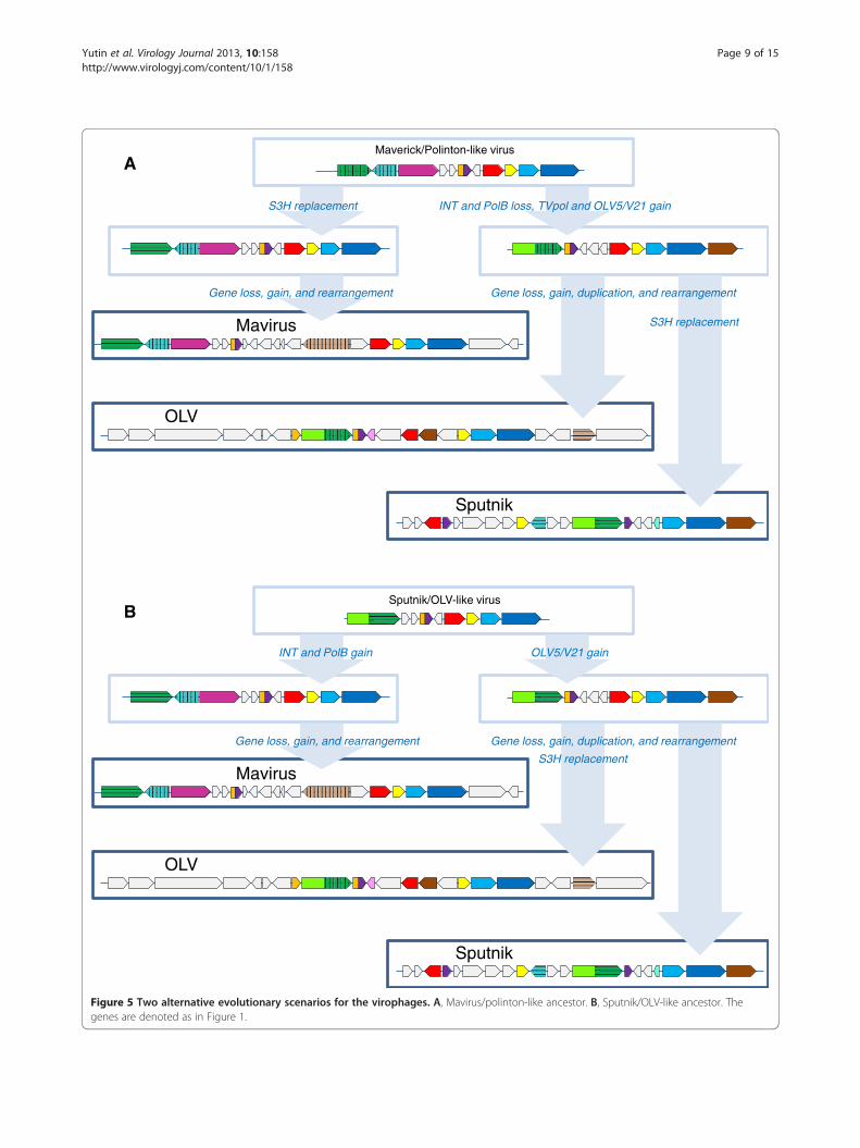
virophage, the comparative analysis of the 3 virophagegenomes, and in particular the complex history of thehelicase-primase gene (see above), seem to be compa-tible with either of two distinct evolutionary scenarios(Figure 5). Taking into account that in the phylogenetictrees of the conserved virophage genes, the Mavirusconsistently forms the outgroup to the Sputnik-OLVclade (Figure 2), the first scenario postulates thatthe Mavirus resembles the ancestral virophage form(Figure 5A). The ancestral virophage genome wouldencompass a phage or polinton-like S3H, RVE family

Figure 4 Phylogenetic tree of the GIY-YIG endonuclease. Branches with bootstrap support (expected-likelihood weights) less than 0.5 werecollapsed. The species name abbreviation and the GenBank identification numbers are indicated; env stands for “marine metagenome.” Speciesabbreviations: Aciph, Acinetobacter phage 133; Bacam, Bacillus amyloliquefaciens subsp. plantarum YAU B9601-Y2; Invir, Invertebrate iridescentvirus 6; Phagl, Phaeocystis globosa virus 12T; Phyin, Phytophthora infestans T30-4; Physo, Phytophthora sojae; Psyto, Psychroflexus torquis ATCC700755; Taxa abbreviations: Bb, Bacteriodetes/Chlorobi group; Bf, Firmicutes; l2, Iridovirus. The color code is as in Figure 2.
Yutin et al. Virology Journal 2013, 10:158 Page 8 of 15http://www.virologyj.com/content/10/1/158
integrase (INT), PolB, ZnR, and ATPase, and one or twocapsid proteins of probable viral origin. Under this sce-nario of virophage evolution, the Sputnik-OLV lineagelost the PolB and INT genes and acquired the TVpoldomain that became fused to the helicase gene, whereasthe Mavirus lineage has undergone replacement of theancestral helicase gene. After the Sputnik-OLV specia-tion, the Sputnik helicase domain was replaced as welland a distinct integrase gene was acquired (Figure 5A).The second evolutionary scenario postulates that
the Sputnik-OLV genome architecture including the
primase-helicase fusion gene is ancestral to virophageswhereas the PolB and INT genes were acquired by theMavirus lineage along with the loss of the TVpoldomain; under this scenario, the displacement of theprimase-helicase with a distinct helicase domain occur-red in the Mavirus lineage (Figure 5B). This scenario iscompatible with the fact that fusion of primase andhelicase domain is a common feature of diverse viruses(and related plasmids) of both prokaryotes and euka-ryotes that apparently evolved in parallel on multiple oc-casions [23,24]. A hybrid scenario of virophage evolution
Maverick/Polinton-like virusA
Mavirus
Sputnik
OLV
S3H replacement INT and PolB loss, TVpol and OLV5/V21 gain
Gene loss, gain, and rearrangement Gene loss, gain, duplication, and rearrangement
S3H replacement
Sputnik/OLV-like virus
Mavirus
Sputnik
OLV
INT and PolB gain OLV5/V21 gain
Gene loss, gain, and rearrangement Gene loss, gain, duplication, and rearrangement
S3H replacement
B
Figure 5 Two alternative evolutionary scenarios for the virophages. A, Mavirus/polinton-like ancestor. B, Sputnik/OLV-like ancestor. Thegenes are denoted as in Figure 1.
Yutin et al. Virology Journal 2013, 10:158 Page 9 of 15http://www.virologyj.com/content/10/1/158

Yutin et al. Virology Journal 2013, 10:158 Page 10 of 15http://www.virologyj.com/content/10/1/158
whereby the ancestral form possessed both the PolB-INT gene block and the primase-helicase cannot beruled out either although the combination of a PolB ofthe protein-primed subfamily with a primase-helicasedoes not seem to be common.Regardless of the exact evolutionary scenario, the
virophages clearly combine genes from several differentsources as noticed in the original report on Sputnik [8](Table 1). Modularity is a general feature of virus gen-ome evolution [26] but even against this background,the patchiness of the virophages is notable. The contri-butions of distinct modules with different biologicalprovenances are implied by the fact that closely relatedenvironmental homologs (primarily, from marine envi-ronments) are readily detectable for some virophagegenes, in particular the OLV and Sputnik primase-helicase, but not for those that encode the two virionproteins or the maturation protease (Table 1). As men-tioned above, a recent broad survey of metagenomicdata from diverse environments yielded homologs ofvarious virophage genes including those for the majorand minor capsid proteins that were used as an anchorto assemble the putative new virophage genomes [14],thus revealing limited presence of virophages in specifichabitats. It nevertheless seems likely that most of the en-vironmental homologs of the virophage genes do notcome from typical virophages but rather from distinct,still poorly characterized mobile elements, (possiblyplasmids) that encode primase-helicases homologous tothose of Sputnik and OLV [22]. By contrast, the “viral”module of the virophages, with the capsid proteins andthe protease, might have come from a group of eukar-yotic viruses that is not widely represented in marineenvironments.Remarkably, each of the virophages possesses genes
that are closely related to homologs from their specificgiant virus hosts (Table 1). Moreover, all these apparenthost-derived genes encode different repetitive proteins(distinct forms of collagen-like repeats in Sputnik andOLV, and FNIP repeats in the Mavirus) that could beimplicated in the interaction of the virophages withtheir giant virus hosts [9]. The presence of these genesseems to be a striking case of parallelism in virusevolution.
The evolutionary connections between virophages andpolintonsThe Polintons show notable variability of the gene reper-toire but possess a conserved core of 4 genes that con-sists of PolB, integrase, a C5-family protease and apackaging ATPase (Figure 1). All these core genes havehomologs in the Mavirus whereas only the latter two arealso found in the Sputnik- OLV branch of virophages.The phylogenetic trees of PolB and INT unequivocally
cluster the Mavirus-ALM clade within the Polintons as-suming the monophyly of the latter (Figure 6A,B). Inthe tree of the C5 family proteases, Mavirus forms astrongly supported clade with the other virophages, andthis clade again is nested within the polinton-adenovirus clade (the internal branches within thisclade are associated with relatively low ELW values butthe position of the virophages inside the polintons-adenoviruses is supported by several such branches)(Figure 2A). Finally, the tree of the fourth core gene ofthe Polintons, the packaging ATPase, includes thevirophage clade but fails to retrieve the monophyly ofthe virophages and the Polintons (Figure 2A). The sizeof the alignable domain in this case is small, and thereliability of the deep branches in the tree is low. SomePolintons also encode a S3H that falls within thebranch of the tree that includes OLV, YSLV1,YSLV4 and ALM along with numerous bacteriophagehomologs, but not the Mavirus (Figure 3A). This phylo-genetic affinity is compatible with the complex evolu-tionary scenario for the S3H that became apparentthrough the comparison of the virophage genomes(see above).The phylogenetic tree topologies of the virophage
genes show much uncertainty, presumably caused by thesmall size of the conserved domains and their highsequence divergence that probably reflects high andnon-uniform evolutionary rates in virophages, otherviruses and polintons. Nevertheless, the key observationin the phylogenetic analysis of the genes that are sharedby Mavirus with polintons seems to be that Mavirus (orall virophages in cases when they come across as a clade)does not cluster with the polintons as a group but ratherfalls inside the polinton subtree. This topology of thephylogenetic trees appears incompatible with the originof the polintons from a Mavirus-like ancestor as pre-viously proposed [11]. Instead, it suggests that either theancestral virophage evolved via recombination between apolinton and a yet unknown virus (under the scenario inFigure 5A) or perhaps more likely the common ances-tor of the Mavirus and ALM evolved via recombinationbetween a polinton and an ancestral virophage (underthe scenario in Figure 5B). Of special interest is thestrongly supported clade formed by the Mavirus-ALMand a distinct group of polintons from diverse protistsin the PolB tree (Figure 6A) that potentially mightpinpoint the specific origin of the Mavirus group ofvirophages.Under each of the two distinct scenarios shown in
Figure 5, the ancestral form is represented as a bona fidevirus. The ultimate origin of this virus is not illuminatedby the present analysis due to the insufficient resolutionof the phylogenetic trees and the extreme divergence ofthe virophage capsid proteins.

A B
Eukaryotesphages
El Caere308470751
El Strpu72077346
POLB-1_TCp*
El Nasvi345496918
El Xentr301606879
El Danre27802860
El Caeel379657271
Ea Polpa281203225
Ea Dicfa328871352
El Capow320170864
Mavirus MV03 326439151ALM ORF05
El Debha74626904
El Klula1352308
El Klula118830
El Lackl74627258
adenoviruses
0.91
0.80
0.89
0.90
0.77
0.99
0.910.93
0.52
1.00
0.950.82
0.99
1.00
1.00
0.99
0.54
0.98
0.68
0.5
Polintons group 2
Polintons group 1 El Trica189241946El Acypi328712673
A. pisum integrase 2**
ALM ORF04Mavirus MV02 326439150
Ea Dicfa328871350Ea Polpa281203224env138709430
OPIE2 I 1p*ZMCOPIA I 1p*
Ginger2-1_MG_1p*El Acypi328705992
ERV3 MD I 2p*HERV4 I 2p*ERV1-3-EC_I_1p*ERV1-1_FCa-I_2p*
Gypsy transposons*bacterial transposons
0.89
0.69
0.95
0.92
0.89
INT-1_Gip*INT-2_SM*
El Sacko291227659INT-2_Cip*
INT-1_TCp*El Xenop301617876El Danre189537622
N. vitripennis integrase**
0.82
0.600.51
0.72
0.98
0.54
0.85
0.76
0.79
0.61
0.95
0.99
0.99
0.73
0.99
0.2
T.thermophila Tlr1 integrase
Figure 6 Phylogenetic trees of Mavirus genes shared with Polintons but not with other virophages. A, B family DNA polymerase.B, Catalytic domain of RVE integrase. Branches with the bootstrap support (expected-likelihood weights) less than 0.5 were collapsed. Sequencesmarked with an astericks (*) were taken from Repbase [20]. For other sequences, the species name abbreviation and the GenBank identificationnumbers are indicated; env stands for “marine metagenome.” Species abbreviations: Acypi, Acyrthosiphon pisum; Caeel, Caenorhabditis elegans;Caere, Caenorhabditis remanei; Capow, Capsaspora owczarzaki ATCC 30864; Danre, Danio rerio; Debha, Debaryomyces hansenii; Dicfa, Dictyosteliumfasciculatum; Klula, Kluyveromyces lactis; Lackl, Lachancea kluyveri; Nasvi, Nasonia vitripennis; Polpa, Polysphondylium pallidum PN500; Sacko,Saccoglossus kowalevskii; Strpu, Strongylocentrotus purpuratus; Trica, Tribolium castaneum; Xentr, Xenopus (Silurana) tropicalis. Taxa abbreviations:Ea, Amoebozoa; El, Opisthokonta. The color code is as in Figure 2.
Yutin et al. Virology Journal 2013, 10:158 Page 11 of 15http://www.virologyj.com/content/10/1/158
Bringing in transpovirons and viruses: the virophage-polinton network moduleTranspovirons represent a novel class of mobile ele-ments, apparently linear plasmids that so far have beenidentified only in association with mimiviruses [7].Remarkably, of the four genes that are shared by diffe-rent transpovirons, two (ZnR and DNA-binding subunitof transposase) are homologous to genes of Sputnik, theonly known virophage parasite of mimiviruses (Figure 1).The ZnR in Sputnik and transpoviron is a stand-aloneprotein unlike the other two virophages in which it isfused to the GIY-YIG endonuclease (Figure 1). Thetransposase subunits of Sputnik and the transpovironsform a distinct clade in the phylogenetic tree [7]. Theseobservations imply a direct evolutionary connectionbetween Sputnik-like virophages and the transpovirons,most likely acquisition of the respective genes by theancestral transpoviron from a virophage.The Superfamily 1 helicase of the transpovirons has a
distinct evolutionary provenance being nested within a
branch of the respective phylogenetic tree that includesmostly bacterial and bacteriophage proteins (Figure 7A).Remarkably, however, other than environmental homo-logs, the closest neighbor of the transpovirons in thistree is the polinton-like transposable element Tlr1 fromT. thermophila [27], in which the helicase is fused to adistinct GIY-YIG endonuclease. A helicase of the samefamily is encoded in the unique terminal genomic regionof a single mimivirus, Megavirus chiliensis [28], in whichthe adjacent gene encodes a Zn-finger protein homolo-gous to proteins found in some polintons (Figure 7A). Inaddition to the transpoviron-like helicase, Tlr1 encodes ahomolog of OLV2 protein that, upon detailed analysis,was shown to belong to a family of uncharacterized smallproteins represented, additionally, in some phycodnavi-ruses, namely, Chloroviruses and Prasinoviruses as well asthe cryptomonad Guillardia theta (Figure 7B).The evolutionary relationship between virophages,
polintons and transpovirons is best represented as a net-work in which the edges correspond to shared genes

BAtr Megco374110340
tr Mimle374110347
tr Acaca374110354
tr MouMo374110362
Tlr helicase 18481459
n2 Megch363540213
f3 Lacph327197845
Bacteria/phages
q0 Phagl357289968
q0 OrgLa322510684
env136181623
Polinton-2 HM 12p*
El Hydma221121357
l2 Wisir339906026
l2 Invir15078743
E8 Ectsi298713171
Ea Entdi167389099
q2 Emihu347481596
El Homsa78482994
Ea Polpa281207229
Ea Dicdi66828369
Polinton-3 HM 1p*
Helitrons*
Bacteria/phages0.92
1.00
0.98
0.70
0.74
0.53
0.93
1.00
0.94
0.84
0.86
0.990.59
0.89
1.00
0.70
1.00
0.93
0.68
0.73
0.5
YSLV2 ORF20env135711014
env138995804env139490358
env137500756env141758703
env142157681q0 Ostta314055291
q0 Ostta378706322q4 Micsp313768418
q4 Ostlu313844178q0 Ostvi163955205
q0 Ostta378706309q0 Ostvi163955196q4 Micpu356980051
q4 Batsp312599327q1 Acatu155370963
q1 Parbu155121836q1 Parbu340025713
q1 Parbu157952472
OLV OLV2 322510448YSLV4 ORF26
env1422213270.840.86
0.56
0.77
0.69
0.86
0.58
0.71
0.50
1.00
0.71
0.82
0.76
0.88
0.55
0.76
0.98
0.82
0.2
Tlr 6Fp 18481473ATP
S3H
Guith428162484YSLV1 ORF28
env136814635env135293100
env136846524env139600241
env140123504env144051830
Guith428167288Guith428169541
Guith428173929
0.990.90
0.89
0.69
0.56
0.54
0.63
0.51
0.75
ubiquitin
GIY|ZnR
ATP
INT
Figure 7 Phylogenetic trees of Tlr1 proteins shared with transpovirons or virophages. A, Superfamily 1 helicase. B, Tlr1 6F protein.Branches with the bootstrap support (expected-likelihood weights) less than 0.5 were collapsed. Genome contexts are shown by same colorscheme as on Figure 1. Sequences marked with an astericks (*) were taken from Repbase [20]. For other sequences, the species nameabbreviation and the GenBank identification numbers are indicated; tr stands for transpoviron; env stands for environmental (metagenomic)sequence. Species abbreviations: Acaca, Acanthamoeba castellanii mamavirus; Acatu, Acanthocystis turfacea Chlorella virus 1; Batsp, Bathycoccus sp.RCC1105 virus BpV2; Dicdi, Dictyostelium discoideum AX4; Ectsi, Ectocarpus siliculosus; Emihu, Emiliania huxleyi virus 84; Entdi, Entamoeba disparSAW760; Guith, Guillardia theta; Homsa, Homo sapiens; Hydma, Hydra magnipapillata; Invir, Invertebrate iridescent virus 6; Lacph, Lactococcusphage 949; Megch, Megavirus chiliensis; Megco, Megavirus courdo7; Micpu, Micromonas pusilla virus PL1; Micsp, Micromonas sp. RCC1109 virusMpV1; Mimle, Mimivirus lentille; MouMo, Moumouvirus Monve; OrgLa, Organic Lake phycodnavirus 1; Ostlu, Ostreococcus lucimarinus virus OlV1;Ostta, Ostreococcus tauri virus RT-2011; Ostvi, Ostreococcus virus OsV5; Parbu, Paramecium bursaria Chlorella virus NY2A; Phagl, Phaeocystisglobosa virus 12T; Polpa, Polysphondylium pallidum PN500; Wisir, Wiseana iridescent virus. Taxa abbreviations: E8, stramenopiles; Ea, Amoebozoa; El,Opisthokonta; f3, Siphoviridae; l2, Iridovirus; n2, mimiviruses; q0, unassigned Phycodnaviridae; q1, Chlorovirus; q2, Coccolithovirus; q4, Prasinovirus.The color code is as in Figure 2.
Yutin et al. Virology Journal 2013, 10:158 Page 12 of 15http://www.virologyj.com/content/10/1/158
(Figure 8). This network also includes at least three dis-tinct groups of viruses, the NCLDV, adenoviruses andan assemblage of bacteriophages. The network is tightlyconnected, with the edges typically linking the nodesthrough multiple genes (Figure 8). Clearly, this networkis a module of a much large network that connects mostof the virus world, primarily through the virus hallmarkgenes such as S3H, the icosahedral capsid protein or theintegrase [26,29]. By its very nature, the network repre-sentation of evolutionary relationships lacks directiona-lity. While we concluded that the evolution of theMavirus branch of virophages involved a major contri-bution from polintons (see above), it is unclear whether
the polintons themselves originated as capsid-less, self-replicating elements or, perhaps more likely on generalgrounds, evolved from an unknown ancestral virus thatlost the capsid.
ConclusionsThe results of the phylogenomic analysis of thevirophages, polintons and other related genetic elementsreinforce the network character of the evolution of thevirus world [26,29]. The distinct groups of elements inthis network are connected through different, overlap-ping sets of shared genes (Figure 8) resulting in ablurred distinction between monophyly and polyphyly.

Figure 8 The virophage-polinton evolutionary network. Specific groups of bacteriophages that are involved in the network connections:Tectiviridae (PolB); Caudovirales (tailed bacteriophages: S3H and GIY-YIG); cyanophages (MV19 peptidase). Specific groups of NCLDV that areinvolved in the network connections: Irido-, Mimi-, Pox-, Marseilleviruses (Mavirus S3H helicase); Marseillevirus (OLV S3H helicase and MV19peptidase); Phaeocystis globosa virus and Invertebrate iridescent virus 6 (GIY-YIG); Phycodnaviridae (Tlr 6F); Pox- and Asfarviridae (ATPase), andMimiviridae (MV20 FNIP repeats).
Yutin et al. Virology Journal 2013, 10:158 Page 13 of 15http://www.virologyj.com/content/10/1/158
Certain groups, such as the virophages or the NCLDV(recently proposed to be recognized as the orderMegavirales [30]), can be considered monophyletic inthe sense that their common ancestor apparently sharedmany properties with the current representatives of therespective groups. Nevertheless, even in these groups,subsequent evolution involved acquisition, loss andreplacement of a large fraction of genes as demonstratedhere for the virophages. Notably, it has been recentlyshown that the virophages of the Mimiviridae have abroad host range and thus can serve as vectors for geneexchanges among the three different groups of mimi-viruses [31,32]. The virophage-polinton network (Figure 8)is not isolated from the rest of the virus world but ratheris connected to other groups of viruses and virus-likeelements through hallmark genes. However, it seemsto be a distinct module in the overall network of virusevolution.Another important outcome of this analysis is the
demonstration of multiple connections between bonafide viruses that encode capsid proteins and form infec-tious viruses and non-viral mobile elements such astransposons. It appears that viruses evolved from non-viral genetic elements and vice versa on more than oneoccasion even within this relatively small module of thevirus evolution networks. These findings imply thatcapsid-centric concepts of virus evolution [33,34] cap-ture only one, even if important, facet of the virus worldhistory.
MethodsThe protein sequences were extracted from the RefSeqdatabase (NCBI, NIH, Bethesda) [35]. The non-redundant
database of protein sequences at the NCBI was searchedusing the PSI-BLAST program [36]; for proteins of unclearprovenance the PSI-BLAST iterations were run until con-vergence with the E-value cut-off of 0.01 [37]. A separateBLASTP search was run against the environmental proteinsequence database (env_nr) at the NCBI. Reference euka-ryotic repetitive DNA elements were downloaded from theRepbase database [20], and each virophage protein wassearched against the Repbase proteins using BLASTP [36]with the E-value cut-off of 0.1. Nearly identical sequenceswere eliminated using blastclust (http://www.ncbi.nlm.nih.gov/Web/Newsltr/Spring04/blastlab.html); a representative(the longest) sequence from each cluster was taken. Pro-tein sequences were aligned using MUSCLE [38]; gappedcolumns (more than 30% of gaps) and columns with lowinformation content were removed from the alignment[39]. A preliminary tree was constructed using theFastTree program with default parameters (JTT evo-lutionary model, discrete gamma model with 20 ratecategories) [40]; the best-fit substitution model was identi-fied using ProtTest [41]; and the final maximum likeli-hood tree was calculated using TreeFinder [42], with thesubstitution model found to be the best for a given align-ment in the first-round analysis. The following substi-tution models were identified by ProtTest as the best fitfor individual genes for which phylogenetic analysis isreported: protease - WAG+G + F; ATPase - LG +G + F;S3H helicase - Blosum62 +G + F; TVpol - LG +G + F;GIY-YIG endonuclease - RTrev +G+ F; PolB - LG +G+ F;RVE integrase - Blosum62 +G; transpoviron helicase -LG +G; OLV2/Tlr6F - LG +G.The branch support values were expressed in
Expected-Likelihood Weights (ELW). For S3H helicase,

Yutin et al. Virology Journal 2013, 10:158 Page 14 of 15http://www.virologyj.com/content/10/1/158
alternative tree topologies were tested with TreeFinderusing the approximately unbiased (AU) test [43]. Inaddition to the TreeFinder, maximum likelihood treeswere also computed using the PhyML program [44] withthe same alignments and substitution models. Thetopologies of the PhyML trees were generally compatiblewith those obtained with TreeFinder but with lessresolution and weaker support (see Additional file 3).
Additional files
Additional file 1: Multiple alignments of conserved virophageproteins/domains: MCP, mCP, GIY, ZnR, and Mavirus peptidase mv19.
Additional file 2: Primase-helicase topology testing.
Additional file 3: Multiple sequence alignments and phylogenetictrees computed using TreeFinder and PhyML (Newic format).
Competing interestsThe authors declare that they have no competing interests.
Authors’ contributionsEVK initiated and designed the study; NY and EVK collected and analyzeddata; EVK and DR wrote the manuscript that was read, edited and approvedby all authors.
Author details1National Center for Biotechnology Information, National Library of Medicine,National Institutes of Health, Bethesda, MD 20894, USA. 2URMITE, CentreNational de la Recherche Scientifique UMR IRD 6236, Faculté de Médecine,Université de la Méditerranée, 27 Boulevard Jean Moulin, Marseille, Cedex 513385, France.
Received: 9 January 2013 Accepted: 19 April 2013Published: 23 May 2013
References1. Raoult D, Audic S, Robert C, Abergel C, Renesto P, Ogata H, La Scola B,
Suzan M, Claverie JM: The 1.2-megabase genome sequence of Mimivirus.Science 2004, 306(5700):1344–1350.
2. Raoult D, Forterre P: Redefining viruses: lessons from Mimivirus.Nat Rev Microbiol 2008, 6:315–319.
3. Claverie JM, Abergel C: Mimivirus: the emerging paradox of quasi-autonomous viruses. Trends Genet 2010, 26(10):431–437.
4. Claverie JM, Abergel C, Ogata H: Mimivirus. Curr Top Microbiol Immunol2009, 328:89–121.
5. Claverie JM, Ogata H, Audic S, Abergel C, Suhre K, Fournier PE: Mimivirusand the emerging concept of "giant" virus. Virus Res 2006, 117(1):133–144.
6. Van Etten JL, Lane LC, Dunigan DD: DNA viruses: the really big ones(giruses). Annu Rev Microbiol 2010, 64:83–99.
7. Desnues C, La Scola B, Yutin N, Fournous G, Robert C, Azza S, Jardot P,Monteil S, Campocasso A, Koonin EV, et al: Provirophages andtranspovirons as the diverse mobilome of giant viruses. Proc Natl AcadSci USA 2012, 109(44):18078–18083.
8. La Scola B, Desnues C, Pagnier I, Robert C, Barrassi L, Fournous G,Merchat M, Suzan-Monti M, Forterre P, Koonin E, et al: The virophage as aunique parasite of the giant mimivirus. Nature 2008, 455(7209):100–104.
9. Desnues C, Boyer M, Raoult D: Sputnik, a virophage infecting the viraldomain of life. Adv Virus Res 2012, 82:63–89.
10. Fischer MG, Allen MJ, Wilson WH, Suttle CA: Giant virus with a remarkablecomplement of genes infects marine zooplankton. Proc Natl Acad Sci USA2010, 107(45):19508–19513.
11. Fischer MG, Suttle CA: A virophage at the origin of large DNAtransposons. Science 2011, 332(6026):231–234.
12. Yau S, Lauro FM, DeMaere MZ, Brown MV, Thomas T, Raftery MJ,Andrews-Pfannkoch C, Lewis M, Hoffman JM, Gibson JA, et al: Virophage
control of antarctic algal host-virus dynamics. Proc Natl Acad Sci USA2011, 108(15):6163–6168.
13. Yutin N, Colson P, Raoult D, Koonin EV: Mimiviridae: clusters oforthologous genes, reconstruction of gene repertoire evolution andproposed expansion of the giant virus family. Virol J 2013. in press.
14. Zhou J, Zhang W, Yan S, Xiao J, Zhang Y, Li B, Pan Y, Wang Y: Diversity ofvirophages in metagenomic data sets. J Virol 2013, 87(8):4225–4236.
15. Kapitonov VV, Jurka J: Self-synthesizing DNA transposons in eukaryotes.Proc Natl Acad Sci USA 2006, 103(12):4540–4545.
16. Jurka J, Kapitonov VV, Kohany O, Jurka MV: Repetitive sequences incomplex genomes: structure and evolution. Annu Rev Genomics HumGenet 2007, 8:241–259.
17. Feschotte C, Pritham EJ: DNA transposons and the evolution of eukaryoticgenomes. Annu Rev Genet 2007, 41:331–368.
18. Pritham EJ, Putliwala T, Feschotte C: Mavericks, a novel class of gianttransposable elements widespread in eukaryotes and related to DNAviruses. Gene 2007, 390(1–2):3–17.
19. Zhang X, Sun S, Xiang Y, Wong J, Klose T, Raoult D, Rossmann MG:Structure of Sputnik, a virophage, at 3.5-A resolution. Proc Natl Acad SciUSA 2012, 109(45):18431–18436.
20. Jurka J, Kapitonov VV, Pavlicek A, Klonowski P, Kohany O, Walichiewicz J:Repbase Update, a database of eukaryotic repetitive elements.Cytogenet Genome Res 2005, 110(1–4):462–467.
21. Dupuy C, Periquet G, Serbielle C, Bezier A, Louis F, Drezen JM: Transfer of achromosomal Maverick to endogenous bracovirus in a parasitoid wasp.Genetica 2011, 139(4):489–496.
22. Iyer LM, Abhiman S, Aravind L: A new family of polymerases related tosuperfamily A DNA polymerases and T7-like DNA-dependent RNApolymerases. Biol Direct 2008, 3:39.
23. Ilyina TV, Gorbalenya AE, Koonin EV: Organization and evolution ofbacterial and bacteriophage primase-helicase systems. J Mol Evol 1992,34(4):351–357.
24. Iyer LM, Koonin EV, Leipe DD, Aravind L: Origin and evolution of thearchaeo-eukaryotic primase superfamily and related palm-domainproteins: structural insights and new members. Nucleic Acids Res 2005,33(12):3875–3896.
25. Yutin N, Koonin EV: Hidden evolutionary complexity of nucleo-cytoplasmic large DNA viruses of eukaryotes. Virol J 2012, 9(1):161.
26. Koonin EV, Senkevich TG, Dolja VV: The ancient virus world and evolutionof cells. Biol Direct 2006, 1(1):29.
27. Wuitschick JD, Gershan JA, Lochowicz AJ, Li S, Karrer KM: A novel family ofmobile genetic elements is limited to the germline genome inTetrahymena thermophila. Nucleic Acids Res 2002, 30(11):2524–2537.
28. Arslan D, Legendre M, Seltzer V, Abergel C, Claverie JM: Distant Mimivirusrelative with a larger genome highlights the fundamental features ofMegaviridae. Proc Natl Acad Sci USA 2011, 108(42):17486–17491.
29. Koonin EV: The logic of chance: the nature and origin of biological evolution.Upper Saddle River, NJ: FT press; 2011.
30. Colson P, de Lamballerie X, Fournous G, Raoult D: Reclassification of giantviruses composing a fourth domain of life in the new order Megavirales.Intervirology 2012, 55(5):321–332.
31. Gaia M, Pagnier I, Campocasso A, Fournous G, Raoult D, La Scola B: Broadspectrum of Mimiviridae allows its isolation using a Mimivirus reporter.PLoS One 2013. in press.
32. Loftus B, Clarke M, Lohia A, et al: Genome of the environmental hostAcanthamoeba castellanii highlights extensive lateral gene transfer andearly evolution of pattern recognition and tyrosine kinase signalling.Genome Res 2013. in press.
33. Bamford DH, Grimes JM, Stuart DI: What does structure tell us about virusevolution? Curr Opin Struct Biol 2005, 15(6):655–663.
34. Krupovic M, Bamford DH: Virus evolution: how far does the doublebeta-barrel viral lineage extend? Nat Rev Microbiol 2008, 6:941–948.
35. Sayers EW, Barrett T, Benson DA, Bolton E, Bryant SH, Canese K, ChetverninV, Church DM, Dicuccio M, Federhen S, et al: Database resources of theNational Center for Biotechnology Information. Nucleic Acids Res 2012,40(Database issue):D13–25.
36. Altschul SF, Madden TL, Schaffer AA, Zhang J, Zhang Z, Miller W, Lipman DJ:Gapped BLAST and PSI-BLAST: a new generation of protein databasesearch programs. Nucleic Acids Res 1997, 25(17):3389–3402.
37. Altschul SF, Koonin EV: PSI-BLAST - a tool for making discoveries insequence databases. Trends Biochem Sci 1998, 23:444–447.

Yutin et al. Virology Journal 2013, 10:158 Page 15 of 15http://www.virologyj.com/content/10/1/158
38. Edgar RC: MUSCLE: multiple sequence alignment with high accuracy andhigh throughput. Nucleic Acids Res 2004, 32(5):1792–1797.
39. Yutin N, Makarova KS, Mekhedov SL, Wolf YI, Koonin EV: The deep archaealroots of eukaryotes. Mol Biol Evol 2008, 25(8):1619–1630.
40. Price MN, Dehal PS, Arkin AP: FastTree 2–approximately maximum-likelihood trees for large alignments. PLoS One 2010, 5(3):e9490.
41. Abascal F, Zardoya R, Posada D: ProtTest: selection of best-fit models ofprotein evolution. Bioinformatics 2005, 21(9):2104–2105.
42. Jobb G, von Haeseler A, Strimmer K: TREEFINDER: a powerful graphicalanalysis environment for molecular phylogenetics. BMC Evol Biol 2004,4:18.
43. Shimodaira H: An approximately unbiased test of phylogenetic treeselection. Syst Biol 2002, 51(3):492–508.
44. Guindon S, Gascuel O: A simple, fast, and accurate algorithm to estimatelarge phylogenies by maximum likelihood. Syst Biol 2003, 52(5):696–704.
doi:10.1186/1743-422X-10-158Cite this article as: Yutin et al.: Virophages, polintons, and transpovirons:a complex evolutionary network of diverse selfish genetic elementswith different reproduction strategies. Virology Journal 2013 10:158.
Submit your next manuscript to BioMed Centraland take full advantage of:
• Convenient online submission
• Thorough peer review
• No space constraints or color figure charges
• Immediate publication on acceptance
• Inclusion in PubMed, CAS, Scopus and Google Scholar
• Research which is freely available for redistribution
Submit your manuscript at www.biomedcentral.com/submit


![DISCOVERY NOTES Open Access Conservation of major and ...lished between eukaryotic transposons of the Polinton/ Maverick family (hereafter Polintons) and the virophages [1,2], a group](https://img.pdfslide.us/doc/110x75/6137dc640ad5d2067648e5eb/discovery-notes-open-access-conservation-of-major-and-lished-between-eukaryotic.jpg)