Embed Size (px)
Citation preview

Rahma et al. Journal of Translational Medicine 2014, 12:55http://www.translational-medicine.com/content/12/1/55
RESEARCH Open Access
The immunological and clinical effects of mutatedras peptide vaccine in combination with IL-2,GM-CSF, or both in patients with solid tumorsOsama E Rahma1,7, J Michael Hamilton2, Malgorzata Wojtowicz3, Omar Dakheel1, Sarah Bernstein4, David J Liewehr5,Seth M Steinberg5 and Samir N Khleif1,6*
Abstract
Background: Mutant Ras oncogenes produce proteins that are unique to cancer cells and represent attractivetargets for vaccine therapy. We have shown previously that vaccinating cancer patients with mutant ras peptides isfeasible and capable of inducing a specific immune response against the relevant mutant proteins. Here, we testedthe mutant ras peptide vaccine administered in combination with low dose interleukin-2 (IL-2) or/andgranulocyte-macrophage colony-stimulating factor (GM-CSF) in order to enhance the vaccine immune response.
Methods: 5000 μg of the corresponding mutant ras peptide was given subcutaneously (SQ) along with IL-2(Arm A), GM-CSF (Arm B) or both (Arm C). IL-2 was given SQ at 6.0 million IU/m2/day starting at day 5, 5 days/weekfor 2 weeks. GM-CSF was given SQ in a dose of 100 μg/day one day prior to each ras peptide vaccination for 4 days.Vaccines were repeated every 5 weeks on arm A and C, and every 4 weeks on arm B, for a maximum of 15 cyclesor until disease progression.
Results: We treated 53 advanced cancer patients (38 with colorectal, 11 with pancreatic, 1 with common bile ductand 3 with lung) on 3 different arms (16 on arm A, 18 on arm B, and 19 on arm C). The median progression freesurvival (PFS) and overall survival (OS) was 3.6 and 16.9 months, respectively, for all patients evaluable for clinicalresponse (n = 48). There was no difference in PFS or OS between the three arms (P = 0.73 and 0.99, respectively).Most adverse events were grade 1-2 toxicities and resolved spontaneously. The vaccine induced an immuneresponse to the relevant ras peptide in a total of 20 out of 37 evaluable patients (54%) by ELISPOT, proliferativeassay, or both. While 92.3% of patients on arm B had a positive immune response, only 31% of patients on arm Aand 36% of patients on arm C had positive immune responses (P = 0.003, Fisher’s exact test).
Conclusions: The reported data showed that IL-2 might have a negative effect on the specific immune responseinduced by the relevant mutant ras vaccine in patients with advanced cancer. This observation deserves furtherinvestigations.
Trial registration: NCI97C0141
Keywords: Ras, Peptide, Vaccine, IL-2, GM-CSF, Immune response
* Correspondence: [email protected] Vaccine Branch, CCR, NCI, 10 Center Drive, Bethesda, MD 20892, USA6Georgia Regents University Cancer Center, 1411 Laney Walker Blvd, Augusta,GA 30912, USAFull list of author information is available at the end of the article
© 2014 Rahma et al.; licensee BioMed Central Ltd. This is an Open Access article distributed under the terms of the CreativeCommons Attribution License (http://creativecommons.org/licenses/by/2.0), which permits unrestricted use, distribution, andreproduction in any medium, provided the original work is properly credited. The Creative Commons Public DomainDedication waiver (http://creativecommons.org/publicdomain/zero/1.0/) applies to the data made available in this article,unless otherwise stated.

Rahma et al. Journal of Translational Medicine 2014, 12:55 Page 2 of 12http://www.translational-medicine.com/content/12/1/55
BackgroundRas oncogenes are extensively characterized mutatedgenes in human cancers [1,2]. With a single amino acidsubstitution, the ras protein can potentiate transformingcapabilities in human cells [3]. Such point mutated Rasgenes have been found in a broad spectrum of humanmalignancies, notably at codons 12, 13, and 61 [4].Codon 12 mutations account for more than 90% of allRas mutations in human cancers [5]. Ras mutations areprevalent in many types of tumors including pancreatic(90%) [6], colorectal (50%) [7] and lung cancer (30%) [8].Mutant ras peptides are processed and presented as for-eign antigens by both MHC class I or II molecules[9,10]. The products of mutant ras antigens represent at-tractive targets for therapeutic cancer vaccines due totheir distinctive expression in tumor tissues as comparedto normal tissues. We and others have shown that vac-cinating patients with mutant ras peptides could elicitspecific immune responses against the correspondingantigens [11-14]. In a previously reported phase I clinicaltrial, we demonstrated the safety of vaccinating advancedcancer patients with the corresponding mutated ras pep-tides [12]. In another study where patients were vac-cinated in the adjuvant setting, the correspondingmutated ras vaccines were capable of generating specificimmune responses with encouraging clinical outcomesin colorectal and pancreatic cancer patients [15]. There-fore, in an attempt to enhance the immune responsegenerated with our mutated ras peptide vaccine, we con-ducted the current study where we combined this vac-cine with interleukin-2 (IL-2), granulocyte-macrophagecolony-stimulating factor (GM-CSF) or both. This iswith the hope that the enhanced vaccine-induced im-mune response may translate to an improved clinicalefficacy.IL-2 plays a major role in enhancing the cytolytic ac-
tivity of T lymphocytes [16,17]. In addition, many inves-tigators have shown that IL-2 can improve the immuneeffect of cancer vaccines by potentiating the effect oftumor-specific lymphocytes [18-20]. Based on this evi-dence, we used low dose subcutaneous (SQ) IL-2 alongwith the mutant ras peptide vaccine on one arm of thestudy. GM-CSF is known to be an important element instimulating the growth of the antigen presenting cellssuch as dendritic cells (DCs) [21]. In addition, GM-CSFhas been found to enhance the vaccine efficacy by in-creasing the number of immature DCs (iDCs) at the vac-cination site [22] and enhancing their maturation andmigration [23]. Accordingly, we used GM-CSF SQ alongwith the ras vaccine in the second arm (arm B) of thistrial. Finally, patients in the third study arm (arm C) re-ceived the ras vaccine in combination with both GM-CSF and low dose SQ IL-2, which was supported by ourpre-clinical data showing a synergistic effect of this
combination by inducing a larger number of cytotoxic Tlymphocytes (CTLs) and a greater cytokine release re-sponse [24].
MethodsStudy objectivesThe primary endpoint of this pilot study was to evaluatethe immune response generated with our ras peptidevaccine admixed with Detox TM PC adjuvant when ad-ministered with IL-2, GM-CSF or the combination ofboth (IL-2 and GM-CSF). The secondary objectives wereto evaluate toxicities observed on each treatment arm,and to explore clinical responses noted with our vaccin-ation strategy.
Patient selectionPatients were assigned to three groups. All groups re-ceived tumor-specific mutated ras peptide vaccine withDetox™ PC admixture. The vaccine was given in combin-ation with Il-2 (SQ) in arm A, GM-CSF (SQ) in arm B,and both Il-2 and GM-CSF (SQ) in arm C. All study pa-tients had histologically proven advanced solid tumorsexpressing different Ras mutations and received multiplelines of therapy. All enrolled patients met the protocoleligibility criteria, including ECOG performance statusof 0-1 and life expectancy of more than 3 months. Themain exclusion criteria included evidence of brain me-tastasis, history of autoimmune disease, and history ofother malignancies except basal carcinoma of the skin.Both the National Cancer Institute (NCI) and NationalNaval Medical Center (NNMC) Institutional ReviewBoards (IRBs) approved the protocol, and the patients’consent was obtained prior to enrollment.
Peptide selectionThe peptides used in this study were 13-mer peptides(residues 5-17) corresponding to the tumor Ras muta-tions (Table 1). The Ras DNA mutations were deter-mined by Restriction Fragment Length Polymorphism(RFLP) and/or sequencing analysis of PCR amplifiedDNA extracted from paraffin embedded tumor and/orfresh tumor biopsy.
Peptide manufacturing and vaccine preparationSynthesis of the peptides was done under contract withMultiple Peptide Systems (San Diego, CA) for clinicaluse. The vaccine contained the peptide and Detox™ PCas an adjuvant. The vaccine was prepared in the follow-ing manner: the pharmacist calculated the volume corre-sponding to 120% of the dose of ras peptide. Sterilewater (0.48 ml) was then added to a vial containing 0.12ml Detox PC to bring the final vaccine volume to 0.6 ml.The sterile water/Detox PC emulsion was shaken. Theappropriate ras peptide was added to the resulting

Table 1 Ras peptides used for vaccination
Raspeptides
Amino acid sequence Vaccinated patients by arm
5 6 7 8 9 10 11 12 13 14 15 16 17
ras 5-17 Lys- Leu-Val- Val- Val- Gly- Ala- Gly- Gly- Val- Gly- Lys- Ser Nome
(wild type)
ras 5-17 Lys- Leu-Val- Val- Val- Gly- Ala- Asp- Gly- Val- Gly- Lys- Ser A: 1-4, 9-12, 14, 16
(Gly→Asp) B: 3-6, 8-10, 14-16
C: 1-5, 7, 9-11, 13-19
ras 5-17 Lys- Leu-Val- Val- Val- Gly- Ala- Val- Gly- Val- Gly- Lys- Ser A: 5, 6, 15
(Gly→Val) B: 1, 7, 11, 12, 17, 18
C: 6, 12
ras 5-17 Lys- Leu-Val- Val- Val- Gly- Ala- Cys- Gly- Val- Gly- Lys- Ser A: 7, 8, 13
(Gly→Cys) B: 2, 13
C: 8
The peptides used in the study were 13-mer peptides (residues 5-17) corresponding to the tumor Ras mutations. Corresponding mutant part of the peptideis bolded.
Rahma et al. Journal of Translational Medicine 2014, 12:55 Page 3 of 12http://www.translational-medicine.com/content/12/1/55
emulsion. The final admixture (1.8 ml) was gently vor-texed (not shaken) for 15-30 seconds. The product waslabeled with the peptide, concentration, time of prepar-ation, and “one hour expiration”. 1.5 ml of the admixturewas used for the patient’s vaccination.
Vaccine administrationAll study patients were assigned to one of three treat-ment arms. Patients in all treatment groups receivedspecific mutated ras peptide vaccine with admixture ofDetox™ PC adjuvant administered SQ. In addition, armA patients received IL-2 (SQ), arm B patients receivedGM-CSF (SQ), and arm C patients received both IL-2and GM-CSF (SQ).All patients received 1.5 ml of the vaccine solution
with one half of the total dose administered into each oftwo sites, over the deltoids, the thighs, or the abdomen(0.75 ml/site). Vaccination was repeated every 5 weekson arm A and C, and every 4 weeks on arm B for a totalof 3 vaccinations. Patients continued to receive sets of 3additional vaccinations for a total of 15 vaccinations aslong as they demonstrated lack of disease progression.After receiving the vaccine, patients were observed inthe outpatient clinic for any acute hypersensitivity reac-tion. IL-2 was administered SQ in a dose of 6.0 millionIU/m2/day starting 4 days after vaccination, 5 days/weekfor 2 weeks in the arms, abdomen, or thighs. The initialIL-2 dose was administered in the outpatient clinic withsubsequent injections were done via self-administrationby the patient on an outpatient basis. GM-CSF was givenSQ starting one day prior to the vaccination and contin-ued for 4 days at a dose of 100 μg/day. GM-CSF was ad-ministered at two separate sites, 50 μg per site, at thesame sites of the vaccine injection. The GM-CSF wasadministered in the outpatient clinic directly after the
peptide injection but with a separate syringe. Patientswere monitored for any hypersensitivity reaction.
Clinical monitoringPatients were evaluated for toxicity and tumor responseduring treatment. Tumor response was assessed byphysical exam and CT scan according to RECIST criteriaat baseline, after each set of 3 vaccinations, and every 2months during follow-up. Patients were taken off studydue to either deterioration in performance status, diseaseprogression or request to withdraw from the study. Dis-ease progression was defined according to the modifiedWHO criteria of progression as the appearance of newlesions and/or a 25% increase of measurable lesions asevident by CT scan. Once patients had progressed,follow-up was not required except to document late tox-icities and death. Adverse events and toxicities were de-fined and graded according to the NCI CommonToxicity Criteria.
Immune monitoringPeripheral blood mononuclear cells (PBMCs) were col-lected within 1 hour prior to every vaccination and every 2months during follow-up. PBMCs were isolated from hep-arinized venous blood by Ficoll Hypaque centrifugation,washed, and cryopreserved in 2-mL vials, using a CryoMedfreezer. The immunological response was assessed byin vitro T cell proliferation assay and enzyme-linked im-munosorbent spot (ELISPOT) assay before and after eachvaccination.
In vitro T cell proliferation assayThe patient’ PBMCs were incubated in vitro with the ap-propriate tumor-specific ras peptide and evaluated forpeptide-induced proliferation following up to 5 days of

Rahma et al. Journal of Translational Medicine 2014, 12:55 Page 4 of 12http://www.translational-medicine.com/content/12/1/55
incubation. Cells were pulsed with [3H]-thymidine for thefinal 18-24 hours of their culture. Proliferation was mea-sured and quantified by the incorporation of [3H]-thymi-dine. Positive control included cells pulsed with a recallantigen peptide (influenza matrix 58-66, GILGFVFTL). Aproliferation of more than two fold above the control ofthe wild-type ras peptide at 2 time points was consideredas a positive response.
ELISPOT assayAll ELISPOT assays were performed at the Laboratory ofCell-Mediated Immunity, SAIC-Frederick (CLIA-certifiedlab). Two frozen normal donor controls with known re-sponsive values were run with each assay to assure qualitycontrol of the assay results. For all assays, at least one ofthe two controls was within 2 standard deviations of thelaboratory-generated means for CMV and CEF. All assayswere performed on 7-8 day in vitro stimulated PBMCs(100 K/well) as the effectors and peptide-pulsed autolo-gous PBMCs (100 K/well) as the antigen presenting cells(APCs). When possible, PBMCs from the earliest timepoint were used as the APCs. However, if this was notpossible, the pulsed PBMCs were assayed alone to makesure they were not producing any spots. Briefly, theday before assay setup, 96-well polyvinylidene fluoride(PVDF) membrane, HTS opaque plates (Millipore, Billerica,Massachusetts, MSIPS4W10) were coated overnight witha 1:100 dilution of anti-human IFN-γ capture antibody(1mg/mL, Mabtech Inc., Mariemont, OH, Cat# 3420-3-1000) in Dulbecco's phosphate buffered saline (DPBS) atroom temperature. Antibody-coated plates were washedfour times in DPBS the next day and blocked with 5% hu-man AB ELISPOT medium at 37°C for approximately 2hours. 1 × 10⁵ in vitro stimulated PBMCs and 1 × 10⁵ au-tologous, peptide-pulsed PBMCs were plated per well.The plates were incubated for 18-20 hours at 37°C. Thenext day, the plates were manually washed six times with0.05% Tween 20 in DPBS, followed by a 2-hour incubationat room temperature with a 1:2000 dilution of the biotiny-lated secondary antibody, anti-human IFN-γ (1 mg/mLMabtech Inc., Mariemont, OH, Cat# 3420-6-1000) inDPBS/1% bovine serum albumin/0.05% Tween. After incu-bation and four washes in DPBS to remove excess anti-body, a 1:3000 dilution of streptavidin alkaline phosphatase(Mabtech, Mariemont, OH, Cat# 3310-10) in DPBS/1%bovine serum albumin, was added to each well for 1 hourat room temperature followed by 4 manual washes inDPBS. Finally, the BCIP/NPT substrate, 100 μl/well,(KPL, Gaithersburg, Maryland, Cat# 50-81-08) wasadded for 7-10 minutes, resulting in the development ofspots. The reaction was stopped by washing three timesin distilled water. Plates were dried overnight and thespots were visualized and counted using the Immuno-Spot Imaging Analyzer system (Cellular Technology
Ltd., Cleveland, OH). ELISPOT results were expressedas the “number of spots per 106 responder cells” aftersubtracting background spots obtained in wells of effec-tors with non-pulsed PBMCs. For each subject, PBMCsobtained before and after vaccination were analyzed inthe same assay to avoid inter-assay variability. An in-crease of number of spots to more than two fold abovethe control of the wild-type ras peptide or the irrelevantTAX peptide at 2 time points was considered as a posi-tive response.
Regulatory T cells (T-regs)Cryopreserved PBMCs were thawed rapidly at 37°C. Thecells were transferred into 15 mL conical tubes (Corning,Lowell, MA) and diluted to 10 mL by dropwise addition ofRPMI medium containing 20% FBS. The cells were pel-leted by low-speed centrifugation at 250 × g for 10 min at25°C. Supernatants were discarded and cell pellets resus-pended in 5 mL of DPBS containing 2% (huAB) serum toblock cell surface Fc receptors. The samples were mixedbriefly and incubated on ice for 15 minutes. Following in-cubation the cells were pelleted by centrifugation as de-scribed before, washed two times with DPBS containing2% bovine serum albumin (BSA; DPBS/2% BSA) and re-suspended in 1 mL of DPBS/2% BSA. The cells werecounted in a Coulter counter and adjusted to a final con-centration of 10 × 106/mL in DPBS/2% BSA. The cells(1 × 10⁶/tube) were stained for surface markers (CD25,CD3, and CD4) for 20 minutes at room temperature (RT)in the dark and washed two times with DPBS/2% BSA.Intracellular staining for FoxP3 was carried out using hu-man FoxP3 buffer prepared as described by the manufac-turer (BD BioSciences, San Jose, CA). Briefly, followingstaining of surface antigens, cells were resuspended in 2mL of fixing solution (buffer A) and incubated for 10 mi-nutes at RT in the dark. Cells were washed two times withPBS/2% BSA, resuspended in 0.5 mL permeabilization so-lution (buffer C) and incubated for 30 minutes at RT inthe dark. Cells were washed two times in PBS/2% BSAand stained with anti-human FoxP3 antibody for 30 mi-nutes at RT in the dark. Cells were then washed two timesand resuspended in 0.5 mL of PBS/2% BSA for four-colorflow cytometric analysis using the FACSCanto cytometer(BD BioSciences, San Jose, CA) running FACS Diva acqui-sition software (version 6.0). Each assay contained a paral-lel set of cells stained with relevant isotype controls (AlexaFluor 488 IgG1 and PE IgG1). Flow cytometric data ana-lysis was carried out using FlowJo Software. T cells wereidentified by plotting CD3 by side scatter. CD4+ T cellswere identified by further gating the CD3+ subset by for-ward and side scatter and by CD4. The regulatory CD4+T cell subset was identified by plotting CD25 versus FoxP3with the quadstat setting determined based on the isotypecontrol tube. The quadrant markers of the CD25 versus

Rahma et al. Journal of Translational Medicine 2014, 12:55 Page 5 of 12http://www.translational-medicine.com/content/12/1/55
FoxP3 dot plot were set based on the isotype controls. Ineach case the pre-vaccination sample and the post 4 or8-vaccination sample (based on how many vaccines thepatient received) were tested side by side in the same ex-periment and were done from frozen samples. This testingstrategy was used to minimize variability from day to dayin staining or thawing. The samples were tested in 4 inde-pendent setups over 3 days. We have included 2 internalcontrols in each experiment, one of those being a frozenleukapheresis sample that was included in each test run asa measure of interassay reproducibility.
Statistical analysisActuarial analyses were performed on the survival andprogression free survival (PFS) data, starting at the on-study date, or the date of progression when determiningprobability of survival following progression, using theKaplan-Meier method. Curves were compared using thelog-rank test. Survival and PFS times were censored ifthe patient was alive and/or without progression as ap-propriate at the last follow-up date. The fractions of pa-tients with immune responses on the three arms werecompared using Mehta’s modification to Fisher’s exacttest [25]. All reported P values are two-tailed, and un-adjusted for multiple comparisons.
Table 2 Arm A (vaccine + IL-2): patient profiles, clinical, and im
Pt Age Cancer Stage onenrollment
# ofcycles
Off-study reason PF(m
1A 56 Colon IV 2 PD 0.5
2A 57 Colon IV 3 PD 3.9
3A 62 Colon IV 3 PD 5.8
4A 50 Pancreatic IV 3 PD 3.6
5A 60 Lung IV 1 PD 1
6A 59 Colon IV 3 PD 3.5
7A 52 Colon NED 11 Completed 12
8A 68 Lung IV 3 PD 3.6
9A 56 Colon NED 10 PPS 18
10A 63 Colon IV 3 PD 3.3
11A 42 Pancreatic NED 6 PD 7.5
12A 39 Colon NED 3 PD 6.2
13A 67 Colon IV 3 PD 3.5
14A 51 Colon NED 3 PD 7.1
15A 60 Pancreatic IV 3 PPS/Lost to follow-up 2.7
16A 61 Colon IV 3 PD 3.6#Progression free survival was calculated as time from the date the consent was sig(+). *Overall Survival was calculated as time from consent date until death or last fosample not available. Immune response was marked as negative (-) or positive (+) aAbbreviations: NED No Evidence of Disease, PD Progression of Disease, PPS Poor PerSurvival, ms Months.
ResultsPatient profilesFifty-three patients were enrolled on this trial (16 onarm A, 18 on arm B, and 19 on arm C). Patient charac-teristics are summarized in Table 2, Table 3, Table 4. Ageof the patients ranged from 33 to 79 years with a meanof 55.3 years (standard deviation of 10.7 years). The ma-jority of the patients, 38 out of the 53 enrolled, had colo-rectal cancers (11 on arm A, 12 on arm B and 15 onarm C), eleven patients had pancreatic cancer (3 on armA, 5 on arm B and 3 on arm C), three patients had lungcancer (2 on arm A and 1 on arm C), and one patienton arm B had cancer of the common bile duct. All pa-tients had adenocarcinoma except for one who hadpoorly differentiated non-small cell carcinoma of thelung (3C). All patients’ tumors harbored Ras mutationsas follows: the majority of patients (36 patients) had aglycine to aspartic acid substitution and the rest had ei-ther a glycine to valine (11 patients) or a glycine to cyst-eine (6 patients) substitution. All patients were heavilypretreated. Most underwent at least one surgical resec-tion except for two patients on each arm (5A, 6A, 4B,18B, 3C, 19C). At least one chemotherapy regimen wasadministered to all patients except for one (16C). Inaddition, 39 of the 53 patients received 2 or morechemotherapy regimens prior to enrollment on the
munological outcomes
S#
s)OS*(ms)
Immune response
Proliferative assay Elispot
Pre-vaccine Post-vaccine Pre-vaccine Post-vaccine
5.5 ND
16.8 NA - -
21.5 - - - -
6.2 - - - -
2.8 ND
8.9 NA
9+ 129+ - + - +
13.1 - - - -
.8 37.2 - + - +
19.9 - - - -
24.1 - - - -
23 - - - -
4.8 - + + +
41.3 - - - -
+ 5.3 NA - -
17.3 - + - -
ned until evidence of disease progression or last follow-up without progressionllow-up (+). ND, not done because patient received 2 or less vaccines; NA,s described in the manuscript.formance Status, SD Stable Disease, PFS Progression Free Survival, OS Overall

Table 3 Arm B (vaccine + GM-CSF): patient profiles, clinical data, and immunological outcomes
Pt Age Cancer Stage onenrollment
# ofcycles
Off-study reason PFS#
(ms)OS*(ms)
Immune response
Proliferative assay Elispot
Pre-vaccine Post-vaccine Pre-vaccine Post-vaccine
1B 55 Biliary NED 3 PD 2.9 8.7 + + + +
2B 33 Colon IV 3 PD 2.9 16.9 - + NA
3B 51 Colon IV 5 PPS/Lost to follow-up 9.2+ 52.6 + + - +
4B 48 Pancreatic NED 13 Completed 35.4+ 35.4 - + - -
5B 51 Colon IV 1 PD 1 1.5 ND
6B 38 Colon IV 1 Refused further Tx 0.2+ 8.3 ND
7B 60 Pancreatic IV 3 PD 4.6 21 - - NA
8B 57 Colon IV 3 PD 3.1 6.8 NA
9B 52 Colon IV 3 PD 3.3 28.3 - + + +
10B 58 Pancreatic IV 3 PD 2.9 20.9 + + - +
11B 45 Pancreatic IV 2 PD 1 7.3 ND
12B 79 Colon IV 3 PPS 12.9 42.1 - + - +
13B 43 Colon IV 3 PD 2.6 6.3 - - - +
14B 78 Colon NED 8 Left voluntarily 16.8 69 - + - +
15B 63 Colon IV 3 PD 2.8 11.6 + + - +
16B 64 Colon IV 6 PD 5.7 20.7 - + - -
17B 71 Colon IV 6 PD 5.6 25.9 - + - +
18B 58 Pancreatic IV 2 PD 2.6 3.3 ND#Progression free survival was calculated as time from the date the consent was signed until evidence of disease progression or last follow-up without progression(+). *Overall Survival was calculated as time from consent date until death or last follow-up (+). ND, not done because patient received 2 or less vaccines; NA,sample not available. Arm B (Vaccine + GM-CSF): Patient Profiles, Clinical Data, and Immunological Outcomes.Abbreviations: NED No Evidence of Disease, PD Progression of Disease, PPS Poor Performance Status, SD Stable Disease, PFS Progression Free Survival, OS OverallSurvival, ms Months.
Rahma et al. Journal of Translational Medicine 2014, 12:55 Page 6 of 12http://www.translational-medicine.com/content/12/1/55
protocol. A total of 21 patients received radiation ther-apy. Twelve patients were enrolled on the trial with noevidence of disease following resection of their primaryor metastatic disease: 5 patients on arm A (7A, 9A, 11A,12A, 14A), 3 patients on arm B (1B, 4B, 14B) and 4 pa-tients on arm C (2C, 11C, 13C, 15C) (Table 2, Table 3,Table 4).
Safety and toxicityThe vaccine was well tolerated for all arms. The majorityof toxicities were grade 1 or 2, with fatigue, fever andlocal site reaction being the most common, accountingfor 77%, 71%, and 69% of patients, respectively. Themost common treatment-related grade 3 toxicities werefatigue (5.7%), followed by diarrhea (3.8%), vomiting(3.8%), and transaminitis (3.8%) (Table 5). The majorityof these toxicities were determined to be related to IL-2or GM-CSF and resulted in dose reduction in somecases. IL-2 was dose reduced to 50% in 3 patients, 1 pa-tient on arm A (7A) and 2 patients on arm C (5C, 6C).On the other hand, only two patients had 50% dose re-duction for GM-CSF, both on arm B (7B, 9B). Grade 4treatment-related toxicity occurred in only one patient
(3A) who developed myocardial infarction that was de-termined to be possibly related to IL-2. Treatment wasdiscontinued for that patient.
Clinical responseFive patients were excluded from the clinical responseanalyses since they received less than 2 vaccines. Theywere removed from the study due to early disease pro-gression (5A, 5B, and 7C), poor performance status(3C), or refusal of further treatment (6B). Therefore, theclinical response was evaluable in only 48 of the 53treated patients. Of these, 37 had progression of diseaseduring the course of treatment (12 patients on each ofarm A and B, and 13 on arm C). Five patients had stabledisease (one patient on arm A and 2 patients on each ofarm B and C). Six patients remained with no evidence ofdisease (2 on each arm); interestingly, 4 out of these 6patients completed the study after they received 11-15vaccines (7A, 4B, 2C, 11C) (Table 2, Table 3, Table 4).For the full cohort (n = 48), the median progression freesurvival (PFS) and overall survival (OS) was 3.6 and16.9 months, respectively (Figure 1). Patients on arm Ahad a median PFS and OS of 3.9 and 17.3 months,

Table 4 Arm C (vaccine + IL-2 + GM-CSF): patient profiles, clinical data, and immunological outcomes
Pt Age Cancer Stage onenrollment
# ofcycles
Off-study reason PFS#
(ms)OS*(ms)
Immune response
Elispot
Pre-vaccine Post-vaccine
1C 40 Rectal IV 15 PD 26.5 80.4 NA +
2C 59 Colon NED 14 Completed 110.2+ 110.2+ - -
3C 58 Lung III 1 PPS 8 72.8 ND
4C 35 Colon IV 6 PD 6.9 18.7 - -
5C 52 Rectal IV 3 PD 3.2 6.4 - -
6C 36 Rectal IV 2 PPS/Lost to follow-up 2+ 9.2 ND
7C 75 Colon IV 1 PD 0.9 5.2 ND
8C 49 Colon IV 3 PD 3.5 5.5 - -
9C 42 Colon IV 3 PD 4 7.6 - -
10C 48 Colon IV 2 PD 1.7 4.7 ND
11C 48 Rectal NED 15 Completed 120.4+ 120.4+ - +
12C 54 Colon IV 3 PD 3.6 26.6 + +
13C 57 Colon NED 3 PD 3.6 9.1 NA
14C 66 Pancreatic IV 2 PD 2.4 2.8 ND
15C 56 Rectal NED 4 PD 3.6 28.5 - +
16C 73 Pancreatic IV 3 PD 3.6 6.6 - -
17C 51 Colon IV 2 PD 1.9 7.9 ND
18C 57 Colon IV 3 PPS 4.4 9.8 - -
19C 66 Pancreatic IV 2 PD 2.1 2.7 ND
#Progression free survival was calculated as time from the date the consent was signed until evidence of disease progression or last follow-up without progression(+). *Overall Survival was calculated as time from consent date until death or last follow-up (+). ND, not done because patient received 2 or less vaccines; NA,sample no available. Immune response was marked as negative (-) or positive (+) as described in the manuscript.Abbreviations: NED No Evidence of Disease, PD Progression of Disease, PPS Poor Performance Status, SD Stable Disease, PFS Progression Free Survival, OS OverallSurvival, ms Months.
Rahma et al. Journal of Translational Medicine 2014, 12:55 Page 7 of 12http://www.translational-medicine.com/content/12/1/55
respectively. Patients on arm B had a median PFS andOS of 3.2 and 20.8 months, respectively, and patients onarm C had a median PFS and OS of 3.6 and 9.1 months,respectively (Figure 1). The difference in PFS or OS be-tween arms A, B and C was not statistically significant(P = 0.74 and 0.99, respectively). Finally, we conducted
Table 5 Grade 3-4 vaccine-related toxicities
Toxicity grade 3-4 Number of patients full cohort Number of pat
Fatigue 3 2
Diarrhea 2 0
Vomiting 2 0
Increased transaminase 2 0
Local site reaction 1 0
Fever 1 0
Myalgia 1 0
Generalized rash 1 0
Myocardial infarction* 1 1
*Grade 4 Toxicity.All grade 3 toxicities were reported for the full cohort and per arm. Only one grade
an unplanned subgroup analysis for patients with stageIV advanced colorectal cancer (n = 26) since they repre-sent the majority of patients treated on the three arms.We found that patients with advanced colorectal cancerhad a median PFS and OS of 3.5 and 14.2 months,respectively.
ients arm A Number of patients arm B Number of patients arm C
0 1
0 2
1 1
1 1
1 0
0 1
0 1
1 0
0 0
4 toxicity of myocardial infarction was reported in one patient (3A).
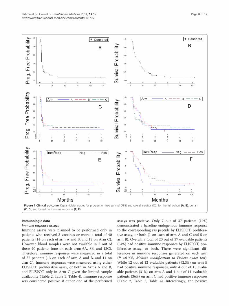
Figure 1 Clinical outcome. Kaplan-Meier curves for progression free survival (PFS) and overall survival (OS) for the full cohort (A, B), per arm(C, D), and based on immune response (E, F).
Rahma et al. Journal of Translational Medicine 2014, 12:55 Page 8 of 12http://www.translational-medicine.com/content/12/1/55
Immunologic dataImmune response assaysImmune assays were planned to be performed only inpatients who received 3 vaccines or more, a total of 40patients (14 on each of arm A and B, and 12 on Arm C).However, blood samples were not available in 3 out ofthese 40 patients (one on each arm: 6A, 8B, and 13C).Therefore, immune responses were measured in a totalof 37 patients (13 on each of arm A and B, and 11 onarm C). Immune responses were measured using eitherELISPOT, proliferative assay, or both in Arms A and B,and ELISPOT only in Arm C given the limited sampleavailability (Table 2, Table 3, Table 4). Immune responsewas considered positive if either one of the performed
assays was positive. Only 7 out of 37 patients (19%)demonstrated a baseline endogenous immune responseto the corresponding ras peptide by ELISPOT, prolifera-tive assay, or both (1 on each of arm A and C and 5 onarm B). Overall, a total of 20 out of 37 evaluable patients(54%) had positive immune responses by ELISPOT, pro-liferative assay, or both. There were significant dif-ferences in immune responses generated on each arm(P =0.003, Mehta’s modification to Fishers exact test).While 12 out of 13 evaluable patients (92.3%) on arm Bhad positive immune responses, only 4 out of 13 evalu-able patients (31%) on arm A and 4 out of 11 evaluablepatients (36%) on arm C had positive immune responses(Table 2, Table 3, Table 4). Interestingly, the positive
Rahma et al. Journal of Translational Medicine 2014, 12:55 Page 9 of 12http://www.translational-medicine.com/content/12/1/55
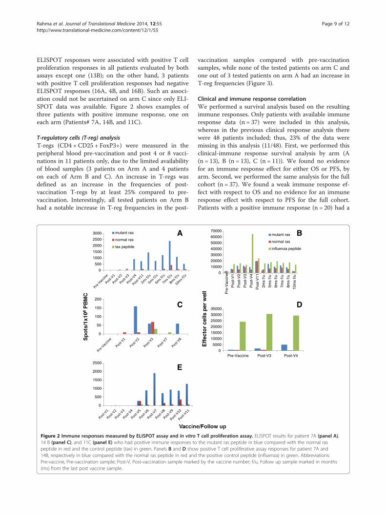
ELISPOT responses were associated with positive T cellproliferation responses in all patients evaluated by bothassays except one (13B); on the other hand, 3 patientswith positive T cell proliferation responses had negativeELISPOT responses (16A, 4B, and 16B). Such an associ-ation could not be ascertained on arm C since only ELI-SPOT data was available. Figure 2 shows examples ofthree patients with positive immune response, one oneach arm (Patients# 7A, 14B, and 11C).
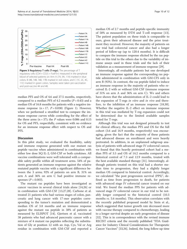
T-regulatory cells (T-reg) analysisT-regs (CD4 + CD25 + FoxP3+) were measured in theperipheral blood pre-vaccination and post 4 or 8 vacci-nations in 11 patients only, due to the limited availabilityof blood samples (3 patients on Arm A and 4 patientson each of Arm B and C). An increase in T-regs wasdefined as an increase in the frequencies of post-vaccination T-regs by at least 25% compared to pre-vaccination. Interestingly, all tested patients on Arm Bhad a notable increase in T-reg frequencies in the post-
0
500
1000
1500
2000
2500
3000 mutant ras
normal ras
tax peptide
0
50
100
150
200
0
500
1000
1500
2000
2500
A
C
E
Figure 2 Immune responses measured by ELISPOT assay and In vitro14 B (panel C), and 11C (panel E) who had positive immune responses topeptide in red and the control peptide (tax) in green. Panels B and D show14B, respectively in blue compared with the normal ras peptide in red andPre-vaccine, Pre-vaccination sample; Post-V, Post-vaccination sample marke(ms) from the last post vaccine sample.
vaccination samples compared with pre-vaccinationsamples, while none of the tested patients on arm C andone out of 3 tested patients on arm A had an increase inT-reg frequencies (Figure 3).
Clinical and immune response correlationWe performed a survival analysis based on the resultingimmune responses. Only patients with available immuneresponse data (n = 37) were included in this analysis,whereas in the previous clinical response analysis therewere 48 patients included; thus, 23% of the data weremissing in this analysis (11/48). First, we performed thisclinical-immune response survival analysis by arm (A(n = 13), B (n = 13), C (n = 11)). We found no evidencefor an immune response effect for either OS or PFS, byarm. Second, we performed the same analysis for the fullcohort (n = 37). We found a weak immune response ef-fect with respect to OS and no evidence for an immuneresponse effect with respect to PFS for the full cohort.Patients with a positive immune response (n = 20) had a
0
10000
20000
30000
40000
50000
60000
70000P
re-V
acci
ne
Pos
t-V
1
Pos
t-V
2
Pos
t-V
3
Pos
t-V
4
Pos
t-V
11
2ms
f/u
5ms
f/u
6ms
f/u
7ms
f/u
8ms
f/u
10m
s f/u
mutant ras
normal ras
influenza peptide
0
5000
10000
15000
20000
25000
30000
35000
Pre-Vaccine Post-V3 Post-V4
Eff
ecto
r ce
lls p
er w
ell
B
D
T cell proliferation assay. ELISPOT results for patient 7A (panel A),the mutant ras peptide in blue compared with the normal raspositive T cell proliferative assay responses for patient 7A andthe positive control peptide (influenza) in green. Abbreviations:d by the vaccine number; f/u, Follow up sample marked in months
% T
-reg
s (C
D4+
CD
25+F
oxP
3+)
0
5
10
15
20
25
30
35
Pre-Vaccine Post-V4 Post-V8
Patient 7A
Patient 9A
Patient 11A
Patient 4B
Patient 14B
Patient 16B
Patient 17B
Patient 1C
Patient 2C
Patient 4C
Patient 11C
Figure 3 Regulatory T cells (T-regs). The percentage of Tregulatory cells (CD4 + CD25 + FoxP3+) measured in the peripheralblood of selected patients on Arm A (7A, 9A, 11A) marked in bluecolors, B (4B, 14B, 16B, 17B) marked in red colors, and C (1C, 2C, 4C,11C) marked in green colors. Samples were taken pre-vaccinationand post-vaccine #4 and #8.
Rahma et al. Journal of Translational Medicine 2014, 12:55 Page 10 of 12http://www.translational-medicine.com/content/12/1/55
median PFS and OS of 4.6 and 27.5 months, respectively,compared to a median PFS of 4.2 months (P = 0.43) and amedian OS of 16.8 months for patients with a negative im-mune response (n = 17, P = 0.038) (Figure 1). However,when we performed a stratified test to compare the im-mune response curves while controlling for the effect ofthe three arms (n = 37); the P values were 0.086 and 0.15for OS and PFS, respectfully, consistent with no evidencefor an immune response effect with respect to OS andPFS.
DiscussionIn this pilot study, we evaluated the feasibility, safetyand immune response generated with our mutant raspeptide vaccine when administered in combination witheither low dose SQ IL-2, GM-CSF or both cytokines. Allvaccine combinations were well tolerated with a compar-able safety profile within all treatment arms. 54% of pa-tients generated an immune response against the relativemutated peptide vaccine with a significant difference be-tween the 3 arms; 92% of patients on arm B, 31% onarm A and 36% on arm C had positive immune re-sponses (P = 0.003).The mutant ras peptides have been investigated as
cancer vaccines in several clinical trials alone [14,26] orin combination with GM-CSF [13,27,28]. Carbone et al.treated 21 patients who had various stages of colon, pan-creatic and lung cancer with 17-mer peptides corre-sponding to the tumor’s mutation and demonstrated amedian OS of 3.8 months and an immune responseagainst the mutant ras peptide in 38% of patients asmeasured by ELISPOT [14]. Gjertsen et al. vaccinated38 patients who had advanced pancreatic cancer with amixture of 4 mutant ras peptides reflecting the substitu-tion of Gly at position 12 with an Asp, Cys, Val or Argresidue in combination with GM-CSF and reported a
median OS of 2.7 months and peptide-specific immunityof 58% as measured by DTH and T-cell response [13].The patient population on these trials is comparable toours, given their advanced disease and the prior treat-ment they received. However, the majority of patients onour trial had colorectal cancer and also had a longerperiod of follow-up (up to 120.4 months). It is difficultto compare the immune response elicited by the ras pep-tide on this trial to the others due to the variability of im-mune assays used in these trials and the lack of theirvalidation as a measurement of immune response [29].Interestingly, all evaluable patients but one developed
an immune response against the corresponding ras pep-tide administered in combination with GM-CFS only inarm B (92%). In contrast, the ras peptide failed to inducean immune response in the majority of patients who re-ceived IL-2 with or without GM-CSF (immune responseof 31% on arm A and 36% on arm C). We and othershave shown that the administration of IL-2 could lead tothe expansion of T-regs in vitro and in vivo and there-fore, to the inhibition of an immune response [18,30].Whether the negative IL-2 effect on immune responsein this trial was mediated by T-reg expansion could notbe determined due to the limited available samplestested for T-regs.Although this trial was not designed primarily to test
for clinical efficacy, the median PFS and OS for the fullcohort (3.6 and 16.9 months, respectively) was encour-aging, given the fact that the majority of these patientshad advanced disease on enrollment and were heavilypretreated. In addition, in an unplanned subgroup ana-lysis of patients with advanced stage IV colorectal cancer,we found that this heavily pretreated cohort had a me-dian PFS of 3.5 and OS of 14.2 months compared to ahistorical control of 7.3 and 12.9 months, treated withthe best available standard therapy [31]. Interestingly, al-though patients treated on this trial had a shorter me-dian time to progression, they had a slightly longermedian OS compared to historical control. Accordingly,we calculated “the post progression survival (PPS)”, de-fined as time from progression to death, for patientswith advanced stage IV colorectal cancer treated on ourtrial. We found the median PPS for patients with ad-vanced stage IV colorectal cancer in our trial to be not-ably longer compared to the historical control (10.3months vs. 5.6 months). This observation correlates withthe recently published proposed model by Stein et al.,which suggested that tumor growth rate decreased morewith vaccine therapy compared to chemotherapy, leadingto a longer survival despite an early progression of disease[32]. This is in correspondence with the revised immuneRECIST criteria and the recently published “FDA Guid-ance for Industry Clinical Considerations for TherapeuticCancer Vaccines” [33,34]. Indeed, the long follow-up time

Rahma et al. Journal of Translational Medicine 2014, 12:55 Page 11 of 12http://www.translational-medicine.com/content/12/1/55
that patients had on this trial (up to 120.4 months)allowed us to perform such analysis, although we realizethat our analysis is limited by the small number of patientsample and the various therapies that the patients receivedpost-vaccination. We and others have shown that patientswho generate a T-cell immune response are more likely tohave longer survival compared to non-immune responders[18,35,36]. In this trial, although we found a weak evidencefor an immune response effect with respect to OS (P =0.038), this effect was weaker when the effect of the threearms was taken into consideration (P = 0.086). Indeed, theamount of missing immunological data precludes a mean-ingful conclusion of the association between immunologicand clinical data in this trial.
ConclusionsIn summary, our trial confirmed the feasibility and safetyof using mutant ras peptide vaccine as a personalizedtreatment for patients with advanced cancers. Indeed,these mutant ras vaccines were shown to be capable ofgenerating a specific immune response against the rele-vant peptide. Further studies are needed to test whetherIL-2 is detrimental to cancer vaccines, given the lowerimmune response rate in patients who received it. None-theless, although not powered to test for clinical efficacy,our study finding correlates with the observations ofothers indicating that cancer vaccines could lead to lon-ger survival, despite early progression of disease.
AbbreviationsSQ: Subcutaneously; IL-2: Interleukin-2; GM-CSF: Granulocyte-macrophagecolony-stimulating factor; PFS: Progression free survival; OS: Overall survival;PPS: Post progression survival; DCs: Dendritic cells; iDCs: Immature dendriticcells; CTLs: Cytotoxic T lymphocytes; NCI: National Cancer Institute;NNMC: National Naval Medical Center; IRB: Institutional Review Board;RFLP: Restriction Fragment Length Polymorphism; ELISPOT: Enzyme-linkedimmunosorbent spot assay; T-regs: T-regulatory cells.
Competing interestsThe authors declare that they have no competing interest.
Authors’ contributionsOER collected the data and drafted the manuscript, JMH participated inpatient care, evaluated patients for eligibility, safety endpoints, and efficacyincluding follow-up assessments. MW participated in patient care, evaluatedpatients for eligibility, safety endpoints, and efficacy including follow-upassessments. OD collected the data, SB participated in patient care, SMSperformed the statistical analysis, DJL performed the statistical analysis, SNKconceived of the study and participated in its design and coordination. Allauthors read and approved the final manuscript.
AcknowledgmentsThis research was supported by the Intramural Research Program of the NIH,National Cancer Institute, Center for Cancer Research. The content of thispublication does not necessarily reflect the views or policies of theDepartment of Health and Human Services, nor does mention of tradenames, commercial products, or organizations imply endorsement by the USGovernment.
Author details1Cancer Vaccine Branch, CCR, NCI, 10 Center Drive, Bethesda, MD 20892,USA. 2OD, CCR, NCI, 9030 Old Georgetown Rd, Bethesda, MD 20892, USA.
3LUACR, DCP, NCI, 9609 Medical Center Drive, Rockville, MD 20850, USA.4Walter Reed National Military Medical Center, 8901 Wisconsin Ave, Bethesda,MD 20814, USA. 5Biostatistics and Data Management Section, CCR, NCI, 9609Medical Center Drive, Rockville, MD 20850, USA. 6Georgia Regents UniversityCancer Center, 1411 Laney Walker Blvd, Augusta, GA 30912, USA. 7Universityof Virginia, Charlottesville, VA 22908, USA.
Received: 25 September 2013 Accepted: 11 February 2014Published: 24 February 2014
References1. Fernandez-Medarde A, Santos E: Ras in cancer and developmental
diseases. Genes Cancer 2011, 2:344–358.2. Malumbres M, Barbacid M: RAS oncogenes: the first 30 years. Nat Rev
Cancer 2003, 3:459–465.3. Reddy EP, Reynolds RK, Santos E, Barbacid M: A point mutation is
responsible for the acquisition of transforming properties by the T24human bladder carcinoma oncogene. Nature 1982, 300:149–152.
4. Bos JL: ras oncogenes in human cancer: a review. Cancer Res 1989,49:4682–4689.
5. Kiaris H, Spandidos D: Mutations of ras genes in human tumors (review).Int J Oncol 1995, 7:413–421.
6. Hruban RH, van Mansfeld AD, Offerhaus GJ, van Weering DH, Allison DC,Goodman SN, Kensler TW, Bose KK, Cameron JL, Bos JL: K-ras oncogeneactivation in adenocarcinoma of the human pancreas. A study of 82carcinomas using a combination of mutant-enriched polymerase chainreaction analysis and allele-specific oligonucleotide hybridization. Am JPathol 1993, 143:545–554.
7. Vaughn CP, Zobell SD, Furtado LV, Baker CL, Samowitz WS: Frequency ofKRAS, BRAF, and NRAS mutations in colorectal cancer. GenesChromosomes Cancer 2011, 50:307–312.
8. Slebos RJ, Kibbelaar RE, Dalesio O, Kooistra A, Stam J, Meijer CJ, Wagenaar SS,Vanderschueren RG, van Zandwijk N, Mooi WJ, Bos JL, Rodenhuis S: K-rasoncogene activation as a prognostic marker in adenocarcinoma of thelung. N Engl J Med 1990, 323:561–565.
9. Jardetzky TS, Lane WS, Robinson RA, Madden DR, Wiley DC: Identificationof self peptides bound to purified HLA-B27. Nature 1991, 353:326–329.
10. Weiss S, Bogen B: MHC class II-restricted presentation of intracellularantigen. Cell 1991, 64:767–776.
11. Abrams SI, Khleif SN, Bergmann-Leitner ES, Kantor JA, Chung Y, Hamilton JM,Schlom J: Generation of stable CD4+ and CD8+ T cell lines from patientsimmunized with ras oncogene-derived peptides reflecting codon 12mutations. Cell Immunol 1997, 182:137–151.
12. Khleif SN, Abrams SI, Hamilton JM, Bergmann-Leitner E, Chen A, Bastian A,Bernstein S, Chung Y, Allegra CJ, Schlom J: A phase I vaccine trial with peptidesreflecting ras oncogene mutations of solid tumors. J Immunother 1999,22:155–165.
13. Gjertsen MK, Buanes T, Rosseland AR, Bakka A, Gladhaug I, Soreide O,Eriksen JA, Møller M, Baksaas I, Lothe RA, Saeterdal I, Gaudernack G:Intradermal ras peptide vaccination with granulocyte-macrophagecolony-stimulating factor as adjuvant: Clinical and immunologicalresponses in patients with pancreatic adenocarcinoma. Int J Cancer 2001,92:441–450.
14. Carbone DP, Ciernik IF, Kelley MJ, Smith MC, Nadaf S, Kavanaugh D, Maher VE,Stipanov M, Contois D, Johnson BE, Pendleton CD, Seifert B, Carter C, Read EJ,Greenblatt J, Top LE, Kelsey MI, Minna JD, Berzofsky JA: Immunization withmutant p53- and K-ras-derived peptides in cancer patients: immuneresponse and clinical outcome. J Clin Oncol: Off J Am Soc Clin Oncol 2005,23:5099–5107.
15. Toubaji A, Achtar M, Provenzano M, Herrin VE, Behrens R, Hamilton M,Bernstein S, Venzon D, Gause B, Marincola F, Khleif SN: Pilot study of mutantras peptide-based vaccine as an adjuvant treatment in pancreatic andcolorectal cancers. Cancer Immunol Immunother 2008, 57:1413–1420.
16. Zhang J, Scordi I, Smyth MJ, Lichtenheld MG: Interleukin 2 receptorsignaling regulates the perforin gene through signal transducer andactivator of transcription (Stat)5 activation of two enhancers. J Exp Med1999, 190:1297–1308.
17. Antony GK, Dudek AZ: Interleukin 2 in cancer therapy. Curr Med Chem2010, 17:3297–3302.
18. Rahma OE, Ashtar E, Czystowska M, Szajnik ME, Wieckowski E, Bernstein S,Herrin VE, Shams MA, Steinberg SM, Merino M, Gooding W, Visus C, Deleo AB,

Rahma et al. Journal of Translational Medicine 2014, 12:55 Page 12 of 12http://www.translational-medicine.com/content/12/1/55
Wolf JK, Bell JG, Berzofsky JA, Whiteside TL, Khleif SN: A gynecologic oncologygroup phase II trial of two p53 peptide vaccine approaches: subcutaneousinjection and intravenous pulsed dendritic cells in high recurrence riskovarian cancer patients. Cancer Immunol Immunother 2012, 61(3):373–384.
19. Rosenberg SA, Yang JC, Schwartzentruber DJ, Hwu P, Marincola FM, Topalian SL,Restifo NP, Dudley ME, Schwarz SL, Spiess PJ, Wunderlich JR, Parkhurst MR,Kawakami Y, Seipp CA, Einhorn JH, White DE: Immunologic and therapeuticevaluation of a synthetic peptide vaccine for the treatment of patients withmetastatic melanoma. Nat Med 1998, 4:321–327.
20. Lotem M, Shiloni E, Pappo I, Drize O, Hamburger T, Weitzen R, Isacson R, Kaduri L,Merims S, Frankenburg S, Peretz T: Interleukin-2 improves tumour response toDNP-modified autologous vaccine for the treatment of metastatic malignantmelanoma. Br J Cancer 2004, 90:773–780.
21. Disis ML, Bernhard H, Shiota FM, Hand SL, Gralow JR, Huseby ES, Gillis S,Cheever MA: Granulocyte-macrophage colony-stimulating factor: aneffective adjuvant for protein and peptide-based vaccines. Blood 1996,88:202–210.
22. Weber J, Sondak VK, Scotland R, Phillip R, Wang F, Rubio V, Stuge TB, GroshenSG, Gee C, Jeffery GG, Sian S, Lee PP: Granulocyte-macrophage-colony-stimulating factor added to a multipeptide vaccine for resected Stage IImelanoma. Cancer 2003, 97:186–200.
23. Dranoff G, Jaffee E, Lazenby A, Golumbek P, Levitsky H, Brose K, Jackson V,Hamada H, Pardoll D, Mulligan RC: Vaccination with irradiated tumor cellsengineered to secrete murine granulocyte-macrophage colony-stimulatingfactor stimulates potent, specific, and long-lasting anti-tumor immunity.Proc Natl Acad Sci USA 1993, 90:3539–3543.
24. Toubaji A, Hill S, Terabe M, Qian J, Floyd T, Simpson RM, Berzofsky JA, Khleif SN:The combination of GM-CSF and IL-2 as local adjuvant shows synergy inenhancing peptide vaccines and provides long term tumor protection. Vaccine2007, 25:5882–5891.
25. Mehta CR, Patel NR: A network algorithm for performing Fisher’s exacttest in r x c contingency Tables. J Am Stat Assoc 1983, 78:427–434.
26. Gjertsen MK, Bakka A, Breivik J, Saeterdal I, Gedde-Dahl T 3rd, Stokke KT,Sølheim BG, Egge TS, Søreide O, Thorsby E, Gaudernack G : Ex vivo raspeptide vaccination in patients with advanced pancreatic cancer: resultsof a phase I/II study. Int J Cancer 1996, 65:450–453.
27. Meyer RG, Korn S, Micke P, Becker K, Huber C, Wolfel T, Buhl R: An open-label, prospective phase I/II study evaluating the immunogenicity andsafety of a ras peptide vaccine plus GM-CSF in patients with non-smallcell lung cancer. Lung Cancer 2007, 58:88–94.
28. Hunger RE, Brand CU, Streit M, Eriksen JA, Gjertsen MK, Saeterdal I, BraathenLR, Gaudernack G: Successful induction of immune responses againstmutant ras in melanoma patients using intradermal injection ofpeptides and GM-CSF as adjuvant. Exp Dermatol 2001, 10:161–167.
29. Disis ML: Immunologic biomarkers as correlates of clinical response tocancer immunotherapy. Cancer Immunol Immunother 2011, 60:433–442.
30. Lemoine FM, Cherai M, Giverne C, Dimitri D, Rosenzwajg M, Trebeden-Negre H, Chaput N, Barrou B, Thioun N, Gattegnio B, Selles F, Six A, Azar N,Lotz JP, Buzyn A, Sibony M, Delcourt A, Boyer O, Herson S, Klatzmann D,Lacave R: Massive expansion of regulatory T-cells followinginterleukin 2 treatment during a phase I-II dendritic cell-basedimmunotherapy of metastatic renal cancer. Int J Oncol 2009, 35:569–581.
31. Giantonio BJ, Catalano PJ, Meropol NJ, O'Dwyer PJ, Mitchell EP, Alberts SR,Schwartz MA, Benson AB 3rd; Eastern Cooperative Oncology Group StudyE3200: Bevacizumab in combination with oxaliplatin, fluorouracil, andleucovorin (FOLFOX4) for previously treated metastatic colorectal cancer:results from the Eastern Cooperative Oncology Group Study E3200.J Clin Oncol: Off J Am Soc Clin Oncol 2007, 25:1539–1544.
32. Stein WD, Gulley JL, Schlom J, Madan RA, Dahut W, Figg WD, Ning YM,Arlen PM, Price D, Bates SE, Fojo T: Tumor regression and growth ratesdetermined in five intramural NCI prostate cancer trials: the growth rateconstant as an indicator of therapeutic efficacy. Clin Cancer Res: Off J AmAssoc Cancer Res 2011, 17:907–917.
33. Wolchok JD, Hoos A, O'Day S, Weber JS, Hamid O, Lebbe C, Maio M, BinderM, Bohnsack O, Nichol G, Humphrey R, Hodi FS: Guidelines for theevaluation of immune therapy activity in solid tumors: immune-relatedresponse criteria. Clin Cancer Res 2009, 15:7412–7420.
34. FDA: Guidance for Industry Clinical Considerations for Therapeutic CancerVaccines. FDA; 2011. http://www.fda.gov/downloads/biologicsbloodvaccines/guidancecomplianceregulatoryinformation/guidances/vaccines/ucm278673.pdf.
35. Slingluff CL Jr, Petroni GR, Olson W, Czarkowski A, Grosh WW, Smolkin M,Chianese-Bullock KA, Neese PY, Deacon DH, Nail C, Merrill P, Fink R, Patterson JW,Rehm PK: Helper T-cell responses and clinical activity of a melanoma vaccinewith multiple peptides from MAGE and melanocytic differentiationantigens. J Clin Oncol: Off J Am Soc Clin Oncol 2008, 26:4973–4980.
36. Kirkwood JM, Lee S, Moschos SJ, Albertini MR, Michalak JC, Sander C,Whiteside T, Butterfield LH, Weiner L: Immunogenicity and antitumoreffects of vaccination with peptide vaccine+/-granulocyte-monocytecolony-stimulating factor and/or IFN-alpha2b in advanced metastaticmelanoma: Eastern Cooperative Oncology Group Phase II Trial E1696.Clin Cancer Res: Off J Am Assoc Cancer Res 2009, 15:1443–1451.
doi:10.1186/1479-5876-12-55Cite this article as: Rahma et al.: The immunological and clinical effects ofmutated ras peptide vaccine in combination with IL-2,GM-CSF, or both in patients with solid tumors. Journal of Translational Medi-cine 2014 12:55.
Submit your next manuscript to BioMed Centraland take full advantage of:
• Convenient online submission
• Thorough peer review
• No space constraints or color figure charges
• Immediate publication on acceptance
• Inclusion in PubMed, CAS, Scopus and Google Scholar
• Research which is freely available for redistribution
Submit your manuscript at www.biomedcentral.com/submit