Embed Size (px)
Citation preview
RESEARCH Open Access
The HIV-1 gp120/V3 modifies the response ofuninfected CD4 T cells to antigen presentation:mapping of the specific transcriptional signatureAntigone K Morou1, Filippos Porichis2, Elias Krambovitis3, George Sourvinos1, Demetrios A Spandidos1 andAlexandros Zafiropoulos4*
Abstract
Background: The asymptomatic phase of HIV-1 infection is characterized by a progressive depletion of uninfectedperipheral effector/memory CD4+ T cells that subsequently leads to immune dysfunction and AIDS symptoms. Wehave previously demonstrated that the presence of specific gp120/V3 peptides during antigen presentation canmodify the activation of normal T-cells leading to altered immune function. The aim of the present study was tomap the specific transcriptional profile invoked by an HIV-1/V3 epitope in uninfected T cells during antigenpresentation.
Methods: We exposed primary human peripheral blood monocytes to V3 lipopeptides using a liposome deliverysystem followed by a superantigen-mediated antigen presentation system. We then evaluated the changes in theT-cell transcriptional profile using oligonucleotide microarrays and performed Ingenuity Pathway Analysis (IPA) andDAVID analysis. The results were validated using realtime PCR, FACS, Western blotting and immunofluorescence.
Results: Our results revealed that the most highly modulated transcripts could almost entirely be categorized asrelated to the cell cycle or transcriptional regulation. The most statistically significant enriched categories andnetworks identified by IPA were associated with cell cycle, gene expression, immune response, infectionmechanisms, cellular growth, proliferation and antigen presentation. Canonical pathways involved in energy andcell cycle regulation, and in the co-activation of T cells were also enriched.
Conclusions: Taken together, these results document a distinct transcriptional profile invoked by the HIV-1/V3epitope. These data could be invaluable to determine the underlying mechanism by which HIV-1 epitopes interferewith uninfected CD4+ T-cell function causing hyper proliferation and AICD.
BackgroundThe asymptomatic phase of HIV-1 infection is charac-terized by the progressive depletion of uninfected per-ipheral effector/memory (CD45RO+) CD4+ T cells [1]that leads to subsequent immunodeficiency and AIDSsymptoms. One of the potential implications of this dys-function involves the mechanism of activation-inducedcell death (AICD) that becomes enhanced and acceler-ated in uninfected CD45RO+/CD4+ T cells by the pre-sence of the virus [2]. The interaction of the HIV viral
envelope glycoprotein, gp120, with CD4 on the host cellsurface induces conformational changes in the gp120that allows the V3 domain of gp120 to interact with thehost cell chemokine receptors, CCR5 or CXCR4 [3,4].Although the functional importance of V3 in HIV infec-tion has been well established [5], the effects of V3 onthe host cell coreceptor signaling cascade have remainedelusive through the past decade [6,7]. In CCR5-tropicHIV isolates (R5 strains), participation of the gp120 V3domain (V3 loop) in the interaction with CCR5 is cru-cial for binding and cell entry [8-10]. R5 strains predo-minate in the asymptomatic phase, whereas isolates thatutilize both CCR5 and CXCR4 (R5X4 strains) or onlyCXCR4 emerge much later in 40-50% of infected
* Correspondence: [email protected] of Histology-Embryology, Medical School, University of Crete,Heraklion, Crete, Greece, Voutes, Heraklion, 71409 Crete, GreeceFull list of author information is available at the end of the article
Morou et al. Journal of Translational Medicine 2011, 9:160http://www.translational-medicine.com/content/9/1/160
© 2011 Morou et al; licensee BioMed Central Ltd. This is an Open Access article distributed under the terms of the Creative CommonsAttribution License (http://creativecommons.org/licenses/by/2.0), which permits unrestricted use, distribution, and reproduction inany medium, provided the original work is properly cited.
individuals and this often indicates the commencementto the clinical phase [11]. The persistence of an exclu-sive R5 viral population in vivo is not rare and is suffi-cient to cause immunodeficiency in the majority ofHIV-1 infected individuals who progress to AIDS[12,13].We have shown previously that antigen presentation
can be deregulated by the presence of V3 epitopes onthe surface of macrophages. CCR5 is one of the mainmediators of V3-induced intracellular signaling duringantigen presentation which leads to AICD, the V3-CCR5 interaction itself being of ionic nature [14,15].Microarray studies utilizing the whole gp120 haveshown increased expression of genes belonging to mito-gen-activated protein kinase signal transduction path-ways and genes regulating cell cycle in PBMCs [13,16].In view of the potential involvement of V3 in the
abnormal AICD process of uninfected CD4+ T cells, weaddressed in this study the effects of V3 on the intracel-lular signaling of CD4+ T cells. We investigated thetranscriptional differences in primary human CD4+ Tcells attributed to the presence of V3 during antigenpresentation signaling. We exposed macrophages to lin-ear synthetic lipopeptides from the crown of V3 pre-sented on liposomes and then we induced antigenpresentation complex formation with CD4+ T cells via asuperantigen presentation system [17]. Implementingoligonucleotide microarray mRNA analysis on CD4+ Tcells, we assessed the impact of the V3 crown on thetranscriptional state of the responding CD4+ T cells.Functional classification of significantly modulated genesand identification of canonical pathways and functionalgene networks analysis were performed by an IngenuityPathways Analysis (IPA) platform and overrepresenta-tion of functional ontologies by DAVID BioinformaticsResources [18,19].
MethodsPeptides and liposomesThe HIV-1 gp120/V3 peptide RKSIRIQRGPGRAFY(LAI strain, a.a. 304-318) was synthesised using F-moc/tBu chemistry (15). Lipopeptides were produced bycovalent binding of serine-S-[2,3-bis (palmitoyloxy)-(2RS)-propyl]-n-palmitoyl- (R)cysteine (BoehringerMannheim Biochemica, Germany) to the V3 peptide,according to the manufacturer’s instructions. Liposomeswere constructed by the dehydration-rehydrationmethod and were reconstituted with 100 μl distilledwater. Non-entrapped material was removed by washingwith PBS [15].
Cell isolationBuffy coats from healthy, HIV-1/Hepatitis b sero-nega-tive blood donors were obtained from Venizelio
Hospital Blood Transfusion Service, Heraklion, Crete.Informed consent was obtained from all the participat-ing volunteers. Peripheral blood mononuclear cells(PBMC) were isolated by density gradient centrifugationusing ficoll-paque (Amersham-Pharmacia, Uppsala, Swe-den) according to the manufacturer’s instructions andcultured in RPMI-1640 medium supplemented withantibiotics and 5% human serum. Depletion of CD8+cells was carried out with the magnetic cell sorting(MACS) system (Miltenyi Biotech, Germany) using PEanti-human CD8 (Mouse IgG1, k, RPA-T8, BD Phar-mingen) and anti-PE microbeads (Mouse IgG1, MiltenyiBiotech, Germany), according to the manufacturer’sinstructions. After the completion of the cell cultureexperiment, the cells harvested for the microarray analy-sis consisted of 86% CD3+CD4+ and 11% CD3+CD8+double positives. This cell population will be referred toas “CD4-enriched”.
Cell culture and oligonucleotide microarray analysisPrimary cell cultures were cultivated in triplicate for 3days with either plain liposomes (control) or liposomeswith lipoV3 peptides incorporated on the liposome sur-face [15]. 8 hours after the addition of 1 ng/ml of sta-phylococcal enterotoxin A [17] (Sigma-AldrichChemical Co.), total RNA extraction was performedaccording to the manufacturer’s instructions usingsilica-based spin columns (Qiagen RNeasy kit, Hilden,Germany). The RNA concentration was determined bymeasuring the absorbance at 260 nm. The purity wasestimated by the 260/280 nm absorbance ratio and theintegrity was assessed by agarose gel electophoresis.High quality RNA was obtained from three indepen-dently treated cell cultures from the same donor. TheRNA samples were subjected to oligonucleotide micro-arrays by Aros Applied Biotechnology (Denmark) usingthe Human Genome U133 Plus 2.0 Array (Affymetrix,Santa Clara, CA, USA) according to the manufacturer’sinstructions. These chips contained 54,000 probe setswhich could recognize transcripts from 38,500 humangenes. The V3 and control RNA samples were hybri-dized onto three separate microarray chips each, andthe values were calculated using the student’s t-test toidentify genes significantly modulated by V3 (p < 0.05,fold change > 2.0). Significantly modulated genes weredefined as those with an absolute fold change of > 2.0and a P value of < 0.05.
Gene expression dataThe primary microarray gene expression data discussedin this publication have been deposited in the ArrayEx-press (European Bioinformatics Institute, EBI) databasethrough NCBI website under accession number E-MEXP-1586, EBI http://www.ebi.ac.uk/microarray.
Morou et al. Journal of Translational Medicine 2011, 9:160http://www.translational-medicine.com/content/9/1/160
Page 2 of 16
BioinformaticsWe used two different bioinformatic tools to explore thefunctional relationships among the genes identified: theIngenuity Pathway Analysis tool version 8.0 (IPA; Inge-nuity® Systems, Inc, Redwood City, CA http://www.ingenuity.com and DAVID (Database for Annotation,Visualization, and Integrated Discovery; http://david.abcc.ncifcrf.gov/) as well as extensive examination ofpublished literature. IPA and DAVID provided comple-mentary pathway analysis. IPA uses a proprietary knowl-edge base while DAVID considers ontologies from theGene Ontology (GO) project and pathways from theKyoto Encyclopedia of Genes and Genomes (KEGG).Functional classification of statistically significant geneexpression changes was performed with IPA. This soft-ware analyzes RNA expression data in the context ofknown biological response and regulatory networks aswell as other canonical pathways using as a referenceset the Ingenuity Pathways Knowledge Base (GenesOnly), filtering for molecules and relationships that areassociated with the immune system. For all analyses, theright-tailed Fisher’s exact test was used to calculate a P-value determining the probability that each biologicalfunction assigned to that data set was due to chancealone. Corrected p-values based on the Benjamini-Hoch-berg method of accounting for multiple testing werealso calculated for significantly enriched Functions andCanonical pathways. Networks of highly interconnectedgenes were identified by statistical likelihood using thefollowing equation:
Score = −log10
⎛⎝1−
f−1∑i=0
C (G, i)C (N − G, s− i)C (N, s)
⎞⎠
Where N is the number of genes in the network ofwhich G are central node genes, for a pathway of sgenes of which f are central node genes. C(n, s) is thebinomial coefficient.We also implemented DAVID gene functional classifi-
cation to obtain biologically enriched functional genegroups with reference to our background list containing54,000 probe sets, applying the criteria of high strin-gency with kappa similarity threshold set to 0.40.
Cell cycle analysisProgression through different cell cycle phases wasmonitored by flow cytometric analysis of the DNA con-tent of cell populations stained with propidium iodideand was carried out with a fluorescence-activated cellsorter. Briefly, 48 h after the addition of SEA, cells wereharvested, fixed in 70% cold ethanol for 2 hours at 4°Cand stained with 20 ug/ml propidium iodide solutioncontaining 100 ug/ml Rnase A at room temperature for
30 min. Flow cytometric analysis was performed on aFACSCalibur (Becton Dickinson) using Modfit LT ana-lysis software (Verity Software House, Inc., Topsham,ME). A minimum of 20,000 cells was counted for eachsample.
Immunofluorescence2*106 cells were prepared for staining using a Cytospincentrifuge (Aero spray, Wescor, USA) for 5 min at 300rpm. Attached cells were then fixed and permeabilized,blocked in PBS containing 10% FBS, and stained withMKi67 rabbit polyclonal antibody (Abcam, ab15580) ata dilution 1:200 for 1 hour at room temperature. Cellsunderwent secondary staining for 1 h with Alexa488-conjugated rabbit antibody (Invitrogen, USA) at a dilu-tion of 1:500. To-Pro 3 iodide (Invitrogen, USA) wasalso used for the visualization of the nuclei. The sampleswere observed under a confocal microscope (TCS SP2;Leica).
ResultsPBMCs were isolated from the buffy coats of volunteerhealthy blood donors [20] using ficoll-paque. CD8+ Tcells were negatively depleted yielding over 90% purityof CD4+ T cells. The resultant cells were aliquoted fortriplicate stimulation experiments. The cells wereexposed for three days to liposome constructs for V3lipopeptides to be incorporated into macrophage cellmembranes, followed by pulsing 1 ng/ml SEA [21].Non-adherent CD4-enriched cells from each experimentwere harvested 8 hours after SEA treatment and werepooled in equal proportions for RNA extraction. Tran-scription profiles were generated (Aros Applied Biotech-nology, Denmark) with Affymetrix U133 Plus 2.0 Arraychips. These chips contained 54,000 probe sets whichcould recognize transcripts from 38,500 human genes.The V3 and control RNA samples were hybridized ontothree separate microarray chips each, and the resultantvalues were calculated using the student’s t-test to iden-tify genes significantly modulated by V3 (fold change >2.0, p < 0.05). The data from the microarray study weredeposited in the ArrayExpress (European BioinformaticsInstitute, EBI) database through the NCBI website(accession number E-MEXP-1586, EBI). The objective ofour analysis was (a) to assess genes exhibiting an exten-sive modulation, and (b) to identify the biologicalthemes and functional gene clusters as a coordinatedchange among many gene products, while the effect ofeach individual gene may be subtle.The validity of microarray results was confirmed (a) by
quantitative real-time PCR (NOC2L, IFI6, NFAT5,PI3KR1, CCNB1, TP53, MAP3K8, H-RAS); (b) by westernblotting (NFAT5, NOC2L); (c) by immunofluorescence(NFAT5, MKi67); and (d) by FACS (NFAT5, CD38).
Morou et al. Journal of Translational Medicine 2011, 9:160http://www.translational-medicine.com/content/9/1/160
Page 3 of 16
Genes with highly modulated expression are associatedwith regulation of cell cycle and proliferationAnalysis of the microarray data revealed that 440 geneswere affected; 378 genes were up-regulated, 18 of whichfor at least 10-fold, and 62 genes were down-modulated,as compared to the control treatment. The top rankedup- and down-regulated genes are presented in Table 1.The most striking result was the 70-fold up-regulationof NOC2L mRNA, a novel gene of the HDAC-indepen-dent inhibitor for histone acetyltransferase (INHAT)[22]. A 26-fold up-regulation of the mRNA level ofSEPT9 and SPIN encoding for septin 9 and spindlin 1were also observed. NFAT5, a transcription factor thatfacilitates cell proliferation under hypertonic conditionswas increased 18-fold due to V3 treatment. Finally, a30-fold up-regulation of IFI6, an ISG with largelyunknown function, was observed, though this result didnot reach the statistical significance cut-off (p = 0.065).Regarding down-regulated genes, the gene ATP-bindingcassette transporter G1 (ABCG1) that regulates choles-terol homeostasis in peripheral CD4 T cells showed themost significant down-regulation (-34.51 and -10.73-fold). Other interesting genes that were down-regulatedincluded PRKAR1B (-5.61-fold), which encodes the reg-ulatory subunit of cyclic AMP-dependent protein kinaseA (PKA), PPP2R1 (-2.89-fold), a constant regulatorysubunit of protein phosphatase 2 and ROCK1 (-2.56-fold), a cAMP-dependent protein kinase/protein kinaseG/protein kinase involved in the regulation of Fas-dependent induction [23]. As a general observation, themost significantly up- and down-regulated transcriptscould almost entirely be categorized as related to thecell cycle or transcriptional regulation.
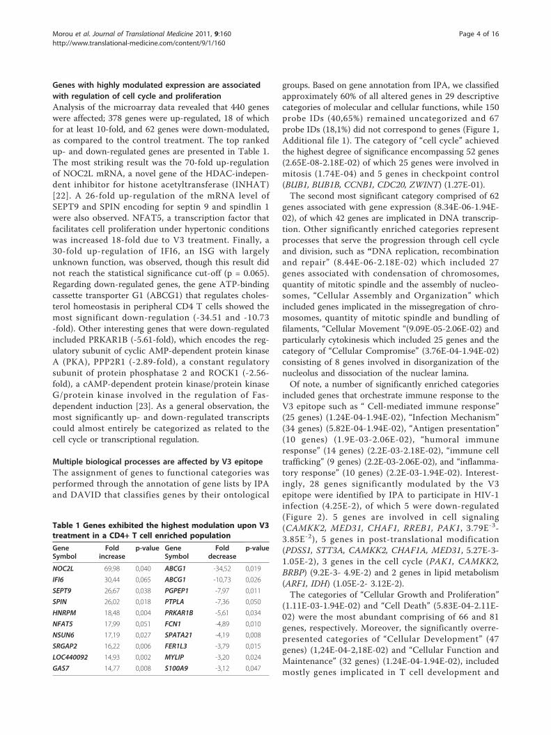
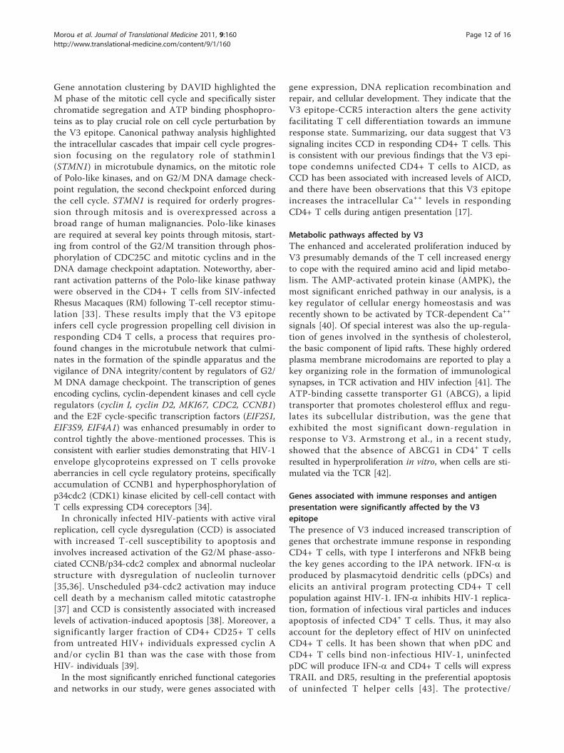
Multiple biological processes are affected by V3 epitopeThe assignment of genes to functional categories wasperformed through the annotation of gene lists by IPAand DAVID that classifies genes by their ontological
groups. Based on gene annotation from IPA, we classifiedapproximately 60% of all altered genes in 29 descriptivecategories of molecular and cellular functions, while 150probe IDs (40,65%) remained uncategorized and 67probe IDs (18,1%) did not correspond to genes (Figure 1,Additional file 1). The category of “cell cycle” achievedthe highest degree of significance encompassing 52 genes(2.65E-08-2.18E-02) of which 25 genes were involved inmitosis (1.74E-04) and 5 genes in checkpoint control(BUB1, BUB1B, CCNB1, CDC20, ZWINT) (1.27E-01).The second most significant category comprised of 62
genes associated with gene expression (8.34E-06-1.94E-02), of which 42 genes are implicated in DNA transcrip-tion. Other significantly enriched categories representprocesses that serve the progression through cell cycleand division, such as “DNA replication, recombinationand repair” (8.44E-06-2.18E-02) which included 27genes associated with condensation of chromosomes,quantity of mitotic spindle and the assembly of nucleo-somes, “Cellular Assembly and Organization” whichincluded genes implicated in the missegregation of chro-mosomes, quantity of mitotic spindle and bundling offilaments, “Cellular Movement “(9.09E-05-2.06E-02) andparticularly cytokinesis which included 25 genes and thecategory of “Cellular Compromise” (3.76E-04-1.94E-02)consisting of 8 genes involved in disorganization of thenucleolus and dissociation of the nuclear lamina.Of note, a number of significantly enriched categories
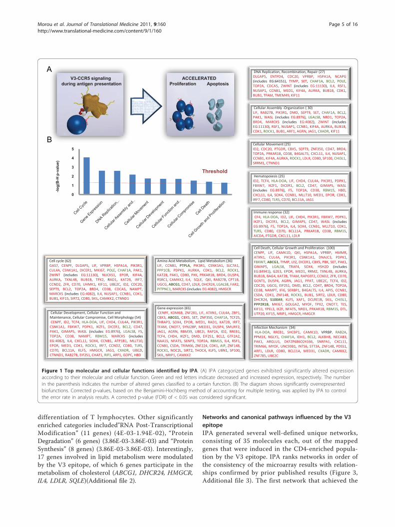
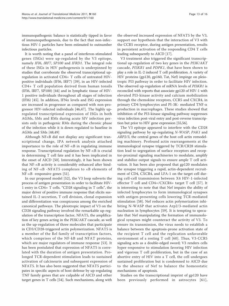
included genes that orchestrate immune response to theV3 epitope such as “ Cell-mediated immune response”(25 genes) (1.24E-04-1.94E-02), “Infection Mechanism”(34 genes) (5.82E-04-1.94E-02), “Antigen presentation”(10 genes) (1.9E-03-2.06E-02), “humoral immuneresponse” (14 genes) (2.2E-03-2.18E-02), “immune celltrafficking” (9 genes) (2.2E-03-2.06E-02), and “inflamma-tory response” (10 genes) (2.2E-03-1.94E-02). Interest-ingly, 28 genes significantly modulated by the V3epitope were identified by IPA to participate in HIV-1infection (4.25E-2), of which 5 were down-regulated(Figure 2). 5 genes are involved in cell signaling(CAMKK2, MED31, CHAF1, RREB1, PAK1, 3.79E-3-3.85E-2), 5 genes in post-translational modification(PDSS1, STT3A, CAMKK2, CHAF1A, MED31, 5.27E-3-1.05E-2), 3 genes in the cell cycle (PAK1, CAMKK2,BRBP) (9.2E-3- 4.9E-2) and 2 genes in lipid metabolism(ARF1, IDH) (1.05E-2- 3.12E-2).The categories of “Cellular Growth and Proliferation”
(1.11E-03-1.94E-02) and “Cell Death” (5.83E-04-2.11E-02) were the most abundant comprising of 66 and 81genes, respectively. Moreover, the significantly overre-presented categories of “Cellular Development” (47genes) (1,24E-04-2,18E-02) and “Cellular Function andMaintenance” (32 genes) (1.24E-04-1.94E-02), includedmostly genes implicated in T cell development and
Table 1 Genes exhibited the highest modulation upon V3treatment in a CD4+ T cell enriched population
GeneSymbol
Foldincrease
p-value GeneSymbol
Folddecrease
p-value
NOC2L 69,98 0,040 ABCG1 -34,52 0,019
IFI6 30,44 0,065 ABCG1 -10,73 0,026
SEPT9 26,67 0,038 PGPEP1 -7,97 0,011
SPIN 26,02 0,018 PTPLA -7,36 0,050
HNRPM 18,48 0,004 PRKAR1B -5,61 0,034
NFAT5 17,99 0,051 FCN1 -4,89 0,010
NSUN6 17,19 0,027 SPATA21 -4,19 0,008
SRGAP2 16,22 0,006 FER1L3 -3,79 0,015
LOC440092 14,93 0,002 MYLIP -3,20 0,024
GAS7 14,77 0,008 S100A9 -3,12 0,047
Morou et al. Journal of Translational Medicine 2011, 9:160http://www.translational-medicine.com/content/9/1/160
Page 4 of 16
differentiation of T lymphocytes. Other significantlyenriched categories included"RNA Post-TranscriptionalModification” (11 genes) (4E-03-1.94E-02), “ProteinDegradation” (6 genes) (3.86E-03-3.86E-03) and “ProteinSynthesis” (8 genes) (3.86E-03-3.86E-03). Interestingly,17 genes involved in lipid metabolism were modulatedby the V3 epitope, of which 6 genes participate in themetabolism of cholesterol (ABCG1, DHCR24, HMGCR,IL4, LDLR, SQLE)(Additional file 2).
Networks and canonical pathways influenced by the V3epitopeIPA generated several well-defined unique networks,consisting of 35 molecules each, out of the mappedgenes that were induced in the CD4-enriched popula-tion by the V3 epitope. IPA ranks networks in order ofthe consistency of the microarray results with relation-ships confirmed by prior published results (Figure 3,Additional file 3). The first network that achieved the
Cell cycle (62)GAS7, CENPF, DLGAP5, LIF, VPRBP, HSPA1A, PIK3R1,CUL4A, CSNK1A1, DICER1, MKI67, POLE, CHAF1A, PAK1,ZWINT (includes EG:11130), NUCKS1, EPOR, KIF4A,AURKA, TXNL4B, BUB1B, TPX2, RAD1, KAT2B, IRF7,CCND2, ZFR, CD70, UHMK1, KIF11, UBE2C, ID2, CDC20,SEPT9, BCL2, TOP2A, BRD4, CD38, CDCA5, NAMPT,MARCKS (includes EG:4082), IL4, NUSAP1, CCNB1, CDK1,BUB1, KIF15, SIRT2, CD80, SKIL, CAMKK2, CTNND1
Gene expression (65)CENPF, KDM6B, ZNF281, LIF, ATXN1, CUL4A, ZBP1,CBX3, ABCG1, CBX5, SET, ZNF350, CHAF1A, TCF25,THRAP3, SOX4, EPOR, MED1, RAD1, KAT2B, IRF7,TFAM, CNOT7, SYNJ2BP, MED31, DUSP4, SMURF2,JAG1, AGRN, RBM39, UBE2I, RAP2A, ID2, RREB1,TCF4, CHD4, IKZF1, DMD, EIF2S1, BCL2, GTF2A2,NAA15, NFAT5, SENP3, TOP2A, RBM15, IL4, RSF1,CCNB1, CSDA, TRIM66, ZNF224, CDK1, AIP, ZNF148,ROCK1, NOC2L, SIRT2, THOC4, KLF5, UBN1, SP100,SKIL, NRIP1, CAMKK2
DNA Replication, Recombination, Repair (27)DLGAP5, ENTPD4, CDC20, VPRBP, HSPA1A, NCAPG(includes EG:64151), TYMP, SET, CHAF1A, BCL2, POLE,TOP2A, CDCA5, ZWINT (includes EG:11130), IL4, RSF1,NUSAP1, CCNB1, MED1, KIF4A, AURKA, BUB1B, CDK1,BUB1, TFAM, TMEM49, KIF11
Cellular Assembly -Organization ( 30)LIF, RAB27B, PIK3R1, DMD, SEPT9, SET, CHAF1A, BCL2,PAK1, WASL (includes EG:8976), LGALS8, NRD1, TOP2A,BRD4, MARCKS (includes EG:4082), ZWINT (includesEG:11130), RSF1, NUSAP1, CCNB1, KIF4A, AURKA, BUB1B,CDK1, ROCK1, BUB1, ARF1, AGRN, JAG1, CXADR, KIF11
Hematopoiesis (25)ID2, TCF4, HLA-DOA, LIF, CHD4, CUL4A, PIK3R1, PDPK1,FBXW7, IKZF1, DICER1, BCL2, CD47, GIMAP5, WASL(includes EG:8976), F5, TOP2A, CD38, RBM15, HBD,CXCL11, IL4, SOX4, CCNB1, MLLT10, MED1, EPOR, CDK1,IRF7, CD80, TLR5, CD70, BCL11A, JAG1
Immune response (32)CF4, HLA-DOA, ID2, LIF, CHD4, PIK3R1, FBXW7, PDPK1,IKZF1, DICER1, BCL2, GIMAP5, CD47, WASL (includesEG:8976), F5, TOP2A, IL4, SOX4, CCNB1, MLLT10, CDK1,TLR5, CD80, CD70, BCL11A, PRKAR1B, CD38, RBM15,AICDA, PTGDR, CXCL11, LDLR
Cellular Development, Cellular Function and Maintenance, Cellular Compromise, Cell Morphology (54)CENPF, ID2, TCF4, HLA-DOA, LIF, CHD4, CUL4A, PIK3R1,CSNK1A1, FBXW7, PDPK1, IKZF1, DICER1, BCL2, CD47,PAK1, GIMAP5, WASL (includes EG:8976), LGALS8, F5,TOP2A, CD38, NAMPT, RBM15, MARCKS (includesEG:4082), IL4, CXCL11, SOX4, CCNB1, ATP1B1, MLLT10,EPOR, MED1, CDK1, ROCK1, IRF7, CCND2, CD80, TLR5,CD70, BCL11A, KLF5, HMGCR, JAG1, CXADR, UBE2I,CTNND1, RAB27B, EIF2S1, CHAF1, RIF1, ARF1, GOPC, HBD
Infection Mechanism (34)HLA-DOA, RREB1, SHCBP1, CAMK1D, VPRBP, RAB2A,PPM1K, NGLY1, CHAF1A, IDH1, BCL2, ALKBH8, NECAB3,PAK1, ARGLU1, DKFZP686O24166, SNRPA1, CXCL11,TRIM66, MYOF, UNC93B1, INTS6, STT3A, ZNF148, PDSS1,ARF1, LDLR, CD80, BCL11A, MED31, CXADR, CAMKK2,ZNF785, UBE2C
Cell Death, Cellular Growth and Proliferation (100)CENPF, LIF, CAMK1D, QKI, HSPA1A, VPRBP, HMMR,ATXN1, CUL4A, PIK3R1, CSNK1A1, DNAJC3, PDPK1,FBXW7, ABCG1, TYMP, LYZ, DICER1, CBX5, PBK, SET, PAK1,GIMAP5, LGALS8, TRAF4, SOX4, HSH2D (includesEG:84941), G2E3, EPOR, MED1, RRM2, TXNL4B, AURKA,BUB1B, BAG4, KAT2B, TFAM, RAPGEF2, CCND2, ZFR, CD70,GCNT1, DUSP4, AGRN, JAG1, PPAT, UBE2C, TCF4, ID2,CDC20, UGCG, EIF2S1, DMD, BCL2, CD47, BRD4, TOP2A,CD38, NAMPT, IFI6, SERBP1, B4GALT5, IL4, API5, CCNB1,CSDA, CDK1, ZNF148, ROCK1, BUB1, SIRT2, LDLR, CD80,DHCR24, S100A9, KLF5, XAF1, DCLRE1B, SKIL, CHI3L1,PPP2R1B, MKI67, GOLGA2, MYOF, TPX2, CNOT7, TES,KIF11, YPEL3, IKZF, NFAT5, NRD1, PRKAR1B, RBM15, DTL,UTP20, KIF15, NRIP1, HMGCR, HMGCR
Cellular Movement (25)ID2, CDC20, PTGDR, CBX5, SEPT9, ZNF350, CD47, BRD4,TOP2A, PRKAR1B, CD38, B4GALT5, CXCL11, IL4, NUSAP1,CCNB1, KIF4A, AURKA, ROCK1, LDLR, CD80, SP100, CHI3L1,SRRM1, CTNND1
Amino Acid Metabolism, Lipid Metabolism (36)LIF, CCNB1, PTPLA, PIK3R1, CSNK1A1, SLC7A1,PPP1CB, PDPK1, AURKA, CDK1, BCL2, ROCK1,KAT2B, PAK1, CD80, PXK, PRKAR1B, BRD4, DUSP4,RSRC1, CAMKK2, IL4, SQLE, QKI, RAB27B, CPT1B,UGCG, ABCG1, CD47, LDLR, DHCR24, LGALS8, FAR2,PITPNC1, MARCKS (includes EG:4082), HMGCR
B
A
B
AV3-CCR5 signaling
during antigen presentationACCELERATED
Proliferation Apoptosis
0
1
2
3
4
5
-log(
B-H
p-v
alue
)
Threshold
Figure 1 Top molecular and cellular functions identified by IPA. (A) IPA categorized genes exhibited significantly altered expressionaccording to their molecular and cellular function. Green and red letters indicate decreased and increased expression, respectively. The numberin the parenthesis indicates the number of altered genes classified to a certain function. (B) The diagram shows significantly overrepresentedbiofunctions. Corrected p-values, based on the Benjamini-Hochberg method of accounting for multiple testing, was applied by IPA to controlthe error rate in analysis results. A corrected p-value (FDR) of < 0.05 was considered significant.
Morou et al. Journal of Translational Medicine 2011, 9:160http://www.translational-medicine.com/content/9/1/160
Page 5 of 16
Extracellular space
Nucleus
Cytoplasm
Unknown
Figure 2 HIV-1 infection related genes modulated by V3 epitope during the process of antigen presentation. This diagram shows genesassociated with HIV-1 infection according to IPA Knowledge Base and their cellular location. Gene products are graphically displayed as nodes.Nodes are further displayed using various shapes that represent the functional class of the gene product. The intensity of the node colorindicates the degree of up- (red) or down- (green) regulation.
Morou et al. Journal of Translational Medicine 2011, 9:160http://www.translational-medicine.com/content/9/1/160
Page 6 of 16
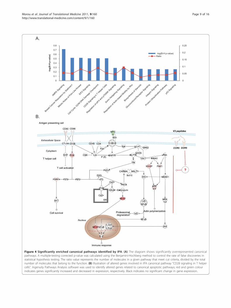
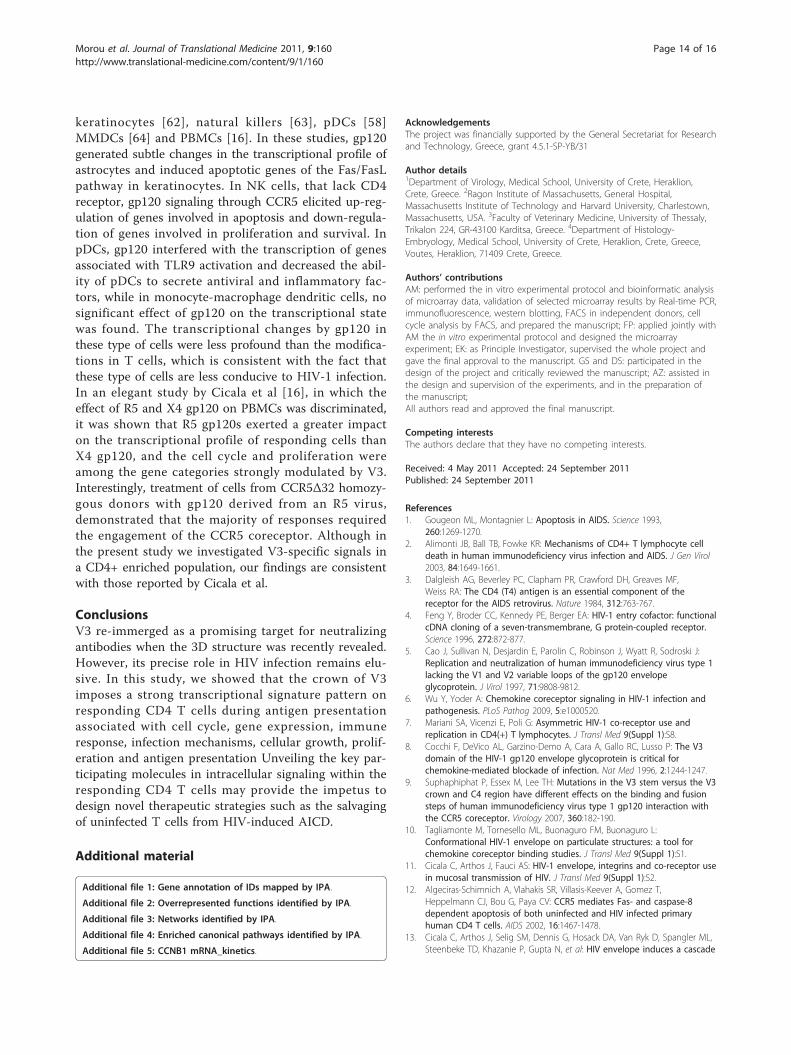
highest score (38), contained genes involved in geneexpression, cellular development and hematological sys-tem development and function. Among these, LDLR,HSPA1A, IKF1 and IDH2 are associated with immuneresponse and LYZ with HIV infection (Figure 3A). Thesecond network contained PP2A, PP1 protein complexgroup, CDK1, CCNB1 and VEGF as main hubs connect-ing cell cycle-promoting regulators, such as CDC20,BUB1, MKI67 and AURKA (Figure 3B). The third net-work consisted of a number of transcription regulatorsand enzymes implicated in cellular growth and prolifera-tion, and in the metabolism of lipids and nucleic acids.NOC2L, the gene that exhibited the most significant up-regulation was included in this network, as well asKAT2B, UBE2I and MED31 (Figure 3C). Interestingly,the IFNa and NF�B complex were the central hubs inthe fourth network, connecting genes related to antimi-crobial response, inflammation and the immuneresponse, most notably, IFI6, IRF7, AICDA, CXCL11 andthe down-regulated TLR5 (Figure 3D)Within IPA 23 statistically significant canonical path-
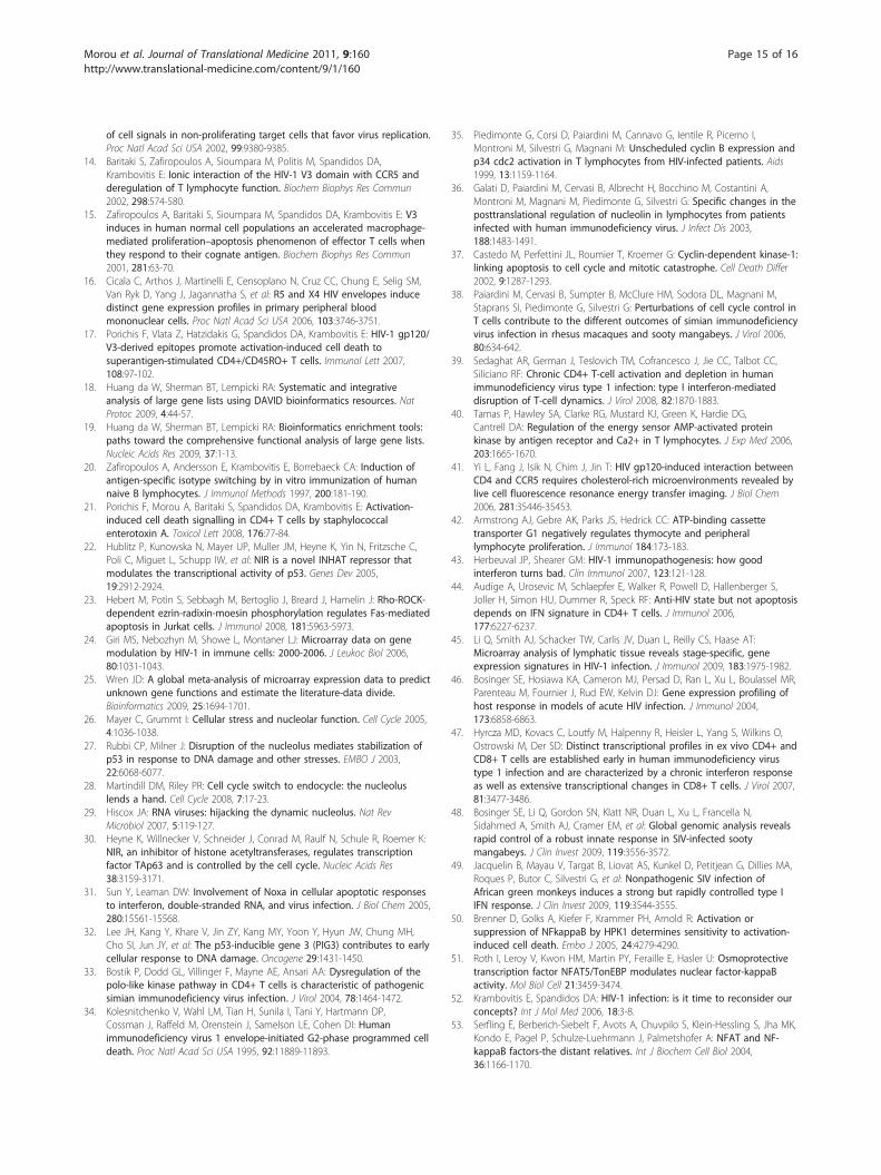
ways were enriched in the gene list when the correctedFischer’s exact test p-value (FDR) of < 0.05 was consid-ered significant (Figure 4A, Additional file 4). The mostsignificant canonical pathway identified was AMPK Sig-naling (FDR = 7.76E-4), the key pathway in energy regu-lation in which PIK3R1, AK2, PDPK1, HMGCR, KAT2B,CPT1B and PPAT were up-regulated, while PRKAR1Band PPP2R1B were down-regulated. Other significantoverrepresented canonical pathways were involved incell cycle progression and cell proliferation. The “BreastCancer Regulation by Stathmin1” IPA-pathway (2.63E-3)which includes the V3-modulated genes ROCK1, PAK1,PPP1R10, CAMK1D, PIK3R1, PRKAR1B, PPP1CB,UHMK1, PPP2R1B and CDK1, plays a regulatory role inmicrotubule dynamics. In the “Mitotic Roles of Polo-Like Kinase” IPA-pathway (5.24E-3), that regulates theentry and exit in mitosis as well as cytokinesis, CCNB1,KIF11 CDC20, CDK1 and PPP2R1B transcripts exhib-ited an altered expression and in “Cell Cycle: G2/MDNA Damage Checkpoint Regulation” IPA-pathway(5.49E-3), CCNB1, TOP2A, CDK1 and KAT2B were up-regulated. The “EIF2” IPA-pathway (6.76E-3), in whichthe differentially expressed transcripts EIF3B, PIK3R1,EIF4A1, PDPK1, PPP1CB and EIF2S1 participate, regu-lates protein synthesis in response to various types ofenvironmental stress and plays a crucial role in the con-trol of the cell cycle mediating both cell proliferationand p53-dependent/independent apoptosis. Interestingly,the “CD28 Signaling in T Helper Cells” IPA-pathway,the most prominent costimulatory pathway of TCR sig-naling, was also significantly enriched (7.41E-3), and keymolecules that participate in the pathway showed a sig-nificant transcriptional up-regulation of over 4-fold
(Figure 4B). Other significantly enriched canonical path-ways identified by IPA included the “Biosynthesis ofSteroids” (2.39E-2), “Integrin Signaling” (2.88E-2), “p53Signaling” (2.95E-2), “Role of Pattern RecognitionReceptors in Recognition of Bacteria and Viruses”(4.89E-2) and the “ERK/MAPK Signaling” (4.89E-2)pathways.We extended the analysis applying the DAVID Gene
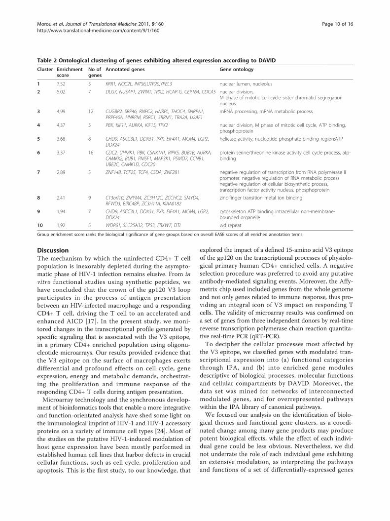
Functional Classification Tool, a module-centric methodthat takes into account the redundant and network nat-ure of biological annotation contents in order to con-centrate on the larger biological picture expressed inbiological modules rather than on individual genes.Under high stringency, DAVID clustered these genesinto 10 functional groups depicted in Table 2. Promi-nent within the list of gene clusters was the cluster of 5molecules of the nuclear lumen and specifically of thenucleolus. The second cluster contained 7 genes impli-cated in nuclear division and in particular sister chro-matide segregation. mRNA processing was the commonfunction of the 12 genes in the third cluster. 5 genesinvolved in nuclear division were also annotated asATP-binding phosphoproteins, and included PBK,KIF11 and AURKA, constituting the fourth cluster. Thefifth cluster contained 8 genes with helicase activity.Intriguingly, 16 ATP-binding serine/threonine kinasesthat regulate the cell-cycle process constituted the sixthcluster. Other clusters included classified genes relatedto negative regulation, zinc finger, cytoskeleton and WDrepeat.
The cell cycle state is influenced by the V3 epitopeThe “Cell cycle” was the most highly enriched categoryaccording our bioinformatic analysis. We validated theseresults with 3 independent approaches:(a) we characterised the cell cycle state of cell popula-
tions by FACS using 3 independent donors. Weobserved a V3-dependent increase of cells in the S andG2/M phases in all three donors. In donor 1, weobtained a 4.1-fold increase in the percentage of cellsentering the S phase and 2.01-fold increase entering theG2/M phase. The corresponding fold increases indonors 2 and 3 were 1.6 and 1.2 for the S phase, and2.46 and 3.26 for the G2/M phase, respectively;(b) we analysed the mRNA kinetics of CCNB1 by
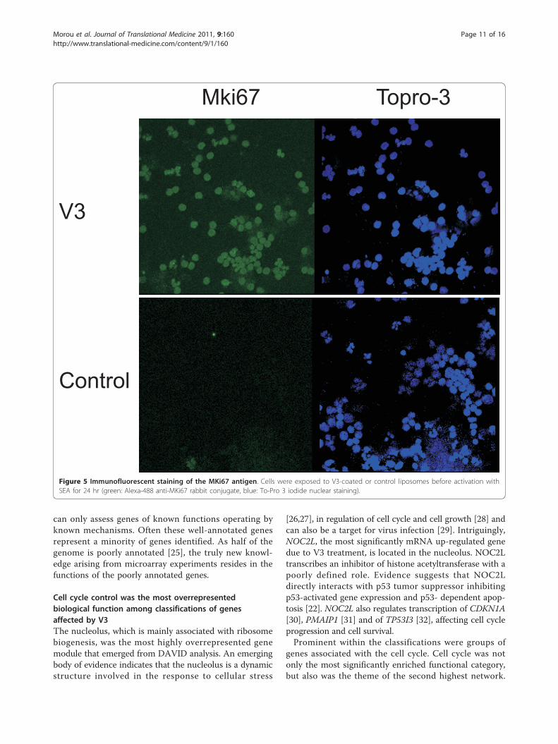
quantitative real-time PCR in 3 new donors. Weobserved a significant V3-dependent fold-increase whichis indicative of the anticipated changes in the progres-sion of cell cycle (additional file 5_CCNB1mRNA_ki-netics) (c) finally, we investigated the number of MKi67positive cells by immunofluorescence microscopy whichis indicative of their proliferation state. We observed adistinct increase that further supports the impact of V3on the cell cycle state (Figure 5).
Morou et al. Journal of Translational Medicine 2011, 9:160http://www.translational-medicine.com/content/9/1/160
Page 7 of 16
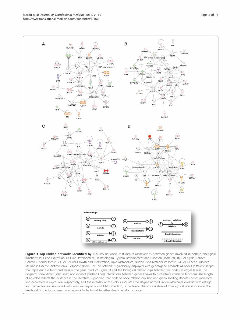
A
DC
B
Figure 3 Top ranked networks identified by IPA. IPA networks that depict associations between genes involved in certain biologicalfunctions: (a) Gene Expression, Cellular Development, Hematological System Development and Function (score 38), (b) Cell Cycle, Cancer,Genetic Disorder (score 36), (c) Cellular Growth and Proliferation, Lipid Metabolism, Nucleic Acid Metabolism (score 35), (d) Genetic Disorder,Metabolic Disease, Antimicrobial Response (score 32). The network is graphically displayed with genes/gene products as nodes (different shapesthat represent the functional class of the gene product, Figure 2) and the biological relationships between the nodes as edges (lines). Thediagrams show direct (solid lines) and indirect (dashed lines) interactions between genes known to orchestrate common functions. The lengthof an edge reflects the evidence in the literature supporting that node-to-node relationship. Red and green shading denotes genes increasedand decreased in expression, respectively, and the intensity of the colour indicates the degree of modulation. Molecules overlaid with orangeand purple line are associated with immune response and HIV-1 infection, respectively. The score is derived from a p value and indicates thelikelihood of the focus genes in a network to be found together due to random chance.
Morou et al. Journal of Translational Medicine 2011, 9:160http://www.translational-medicine.com/content/9/1/160
Page 8 of 16
A.
B.
0
0.05
0.1
0.15
0.2
0.25
0
0.1
0.2
0.3
0.4
0.5
0.6
0.7
0.8-lo
g(B
-H p
-val
ue) -log(B-H p-value)
Ratio
Figure 4 Significantly enriched canonical pathways identified by IPA. (A) The diagram shows significantly overrepresented canonicalpathways. A multiple-testing corrected p-value was calculated using the Benjamini-Hochberg method to control the rate of false discoveries instatistical hypothesis testing. The ratio value represents the number of molecules in a given pathway that meet cut criteria, divided by the totalnumber of molecules that belong to the function. (B) Illustration of altered genes involved in IPA canonical pathway “CD28 signaling in T helpercells”. Ingenuity Pathways Analysis software was used to identify altered genes related to canonical apoptotic pathways; red and green colourindicates genes significantly increased and decreased in expression, respectively. Black indicates no significant change in gene expression.
Morou et al. Journal of Translational Medicine 2011, 9:160http://www.translational-medicine.com/content/9/1/160
Page 9 of 16
DiscussionThe mechanism by which the uninfected CD4+ T cellpopulation is inexorably depleted during the asympto-matic phase of HIV-1 infection remains elusive. From invitro functional studies using synthetic peptides, wehave concluded that the crown of the gp120 V3 loopparticipates in the process of antigen presentationbetween an HIV-infected macrophage and a respondingCD4+ T cell, driving the T cell to an accelerated andenhanced AICD [17]. In the present study, we moni-tored changes in the transcriptional profile generated byspecific signaling that is associated with the V3 epitope,in a primary CD4+ enriched population using oligonu-cleotide microarrays. Our results provided evidence thatthe V3 epitope on the surface of macrophages exertsdifferential and profound effects on cell cycle, geneexpression, energy and metabolic demands, orchestrat-ing the proliferation and immune response of theresponding CD4+ T cells during antigen presentation.Microarray technology and the synchronous develop-
ment of bioinformatics tools that enable a more integrativeand function-orientated analysis have shed some light onthe immunological imprint of HIV-1 and HIV-1 accessoryproteins on a variety of immune cell types [24]. Most ofthe studies on the putative HIV-1-induced modulation ofhost gene expression have been mostly performed inestablished human cell lines that harbor defects in crucialcellular functions, such as cell cycle, proliferation andapoptosis. This is the first study, to our knowledge, that
explored the impact of a defined 15-amino acid V3 epitopeof the gp120 on the transcriptional processes of physiolo-gical primary human CD4+ enriched cells. A negativeselection procedure was preferred to avoid any putativeantibody-mediated signaling events. Moreover, the Affy-metrix chip used included genes from the whole genomeand not only genes related to immune response, thus pro-viding an integral icon of V3 impact on responding Tcells. The validity of microarray results was confirmed ona set of genes from three independent donors by real-timereverse transcription polymerase chain reaction quantita-tive real-time PCR (qRT-PCR).To decipher the cellular processes most affected by
the V3 epitope, we classified genes with modulated tran-scriptional expression into (a) functional categoriesthrough IPA, and (b) into enriched gene modulesdescriptive of biological processes, molecular functionsand cellular compartments by DAVID. Moreover, thedata set was mined for networks of interconnectedmodulated genes, and for overrepresented pathwayswithin the IPA library of canonical pathways.We focused our analysis on the identification of biolo-
gical themes and functional gene clusters, as a coordi-nated change among many gene products may producepotent biological effects, while the effect of each indivi-dual gene could be less obvious. Nevertheless, we didnot underrate the role of each individual gene exhibitingan extensive modulation, as interpreting the pathwaysand functions of a set of differentially-expressed genes
Table 2 Ontological clustering of genes exhibiting altered expression according to DAVID
Cluster Enrichmentscore
No ofgenes
Annotated genes Gene ontology
1 7,52 5 KRR1, NOC2L, INTS6,UTP20,YPEL3 nuclear lumen, nucleolus
2 5,02 7 DLG7, NUSAP1, ZWINT, TPX2, HCAP-G, CEP164, CDCA5 nuclear division,M phase of mitotic cell cycle sister chromatid segregationnucleus
3 4,99 12 CUGBP2, SRP46, RNPC2, HNRPL, THOC4, SNRPA1,PRPF40A, HNRPM, RSRC1, SRRM1, TRA2A, U2AF1
mRNA processing, mRNA metabolic process
4 4,37 5 PBK, KIF11, AURKA, KIF15, TPX2 nuclear division, M phase of mitotic cell cycle, ATP binding,phosphoprotein
5 3,68 8 CHD9, ASCC3L1, DDX51, PXK, EIF4A1, MCM4, LGP2,DDX24
helicase activity, nucleotide phosphate-binding region:ATP
6 3,37 16 CDC2, UHMK1, PBK, CSNK1A1, RIPK5, BUB1B, AURKA,CAMKK2, BUB1, PMSF1, MAP3K1, PSMD7, CCNB1,UBE2C, CAMK1D, CDC20
protein serine/threonine kinase activity cell cycle process, atp-binding
7 2,89 5 ZNF148, TCF25, TCF4, CSDA, ZNF281 negative regulation of transcription from RNA polymerase IIpromoter, negative regulation of RNA metabolic processnegative regulation of cellular biosynthetic process,transcription factor activity nucleus, phosphoprotein
8 2,41 9 C13orf10, ZMYM4, ZC3H12C, ZCCHC2, SMYD4,RFWD3, BIRC4BP, ZC3H11A, KIAA0182
zinc-finger transition metal ion binding
9 1,94 7 CHD9, ASCC3L1, DDX51, PXK, EIF4A1, MCM4, LGP2,DDX24
cytoskeleton ATP binding intracellular non-membrane-bounded organelle
10 1,92 5 WDR61, SLC25A32, TP53, FBXW7, DTL wd repeat
Group enrichment score ranks the biological significance of gene groups based on overall EASE scores of all enriched annotation terms.
Morou et al. Journal of Translational Medicine 2011, 9:160http://www.translational-medicine.com/content/9/1/160
Page 10 of 16
can only assess genes of known functions operating byknown mechanisms. Often these well-annotated genesrepresent a minority of genes identified. As half of thegenome is poorly annotated [25], the truly new knowl-edge arising from microarray experiments resides in thefunctions of the poorly annotated genes.
Cell cycle control was the most overrepresentedbiological function among classifications of genesaffected by V3The nucleolus, which is mainly associated with ribosomebiogenesis, was the most highly overrepresented genemodule that emerged from DAVID analysis. An emergingbody of evidence indicates that the nucleolus is a dynamicstructure involved in the response to cellular stress
[26,27], in regulation of cell cycle and cell growth [28] andcan also be a target for virus infection [29]. Intriguingly,NOC2L, the most significantly mRNA up-regulated genedue to V3 treatment, is located in the nucleolus. NOC2Ltranscribes an inhibitor of histone acetyltransferase with apoorly defined role. Evidence suggests that NOC2Ldirectly interacts with p53 tumor suppressor inhibitingp53-activated gene expression and p53- dependent apop-tosis [22]. NOC2L also regulates transcription of CDKN1A[30], PMAIP1 [31] and of TP53I3 [32], affecting cell cycleprogression and cell survival.Prominent within the classifications were groups of
genes associated with the cell cycle. Cell cycle was notonly the most significantly enriched functional category,but also was the theme of the second highest network.
Mki67 Topro-3
V3
Control
Figure 5 Immunofluorescent staining of the MKi67 antigen. Cells were exposed to V3-coated or control liposomes before activation withSEA for 24 hr (green: Alexa-488 anti-MKi67 rabbit conjugate, blue: To-Pro 3 iodide nuclear staining).
Morou et al. Journal of Translational Medicine 2011, 9:160http://www.translational-medicine.com/content/9/1/160
Page 11 of 16
Gene annotation clustering by DAVID highlighted theM phase of the mitotic cell cycle and specifically sisterchromatide segregation and ATP binding phosphopro-teins as to play crucial role on cell cycle perturbation bythe V3 epitope. Canonical pathway analysis highlightedthe intracellular cascades that impair cell cycle progres-sion focusing on the regulatory role of stathmin1(STMN1) in microtubule dynamics, on the mitotic roleof Polo-like kinases, and on G2/M DNA damage check-point regulation, the second checkpoint enforced duringthe cell cycle. STMN1 is required for orderly progres-sion through mitosis and is overexpressed across abroad range of human malignancies. Polo-like kinasesare required at several key points through mitosis, start-ing from control of the G2/M transition through phos-phorylation of CDC25C and mitotic cyclins and in theDNA damage checkpoint adaptation. Noteworthy, aber-rant activation patterns of the Polo-like kinase pathwaywere observed in the CD4+ T cells from SIV-infectedRhesus Macaques (RM) following T-cell receptor stimu-lation [33]. These results imply that the V3 epitopeinfers cell cycle progression propelling cell division inresponding CD4 T cells, a process that requires pro-found changes in the microtubule network that culmi-nates in the formation of the spindle apparatus and thevigilance of DNA integrity/content by regulators of G2/M DNA damage checkpoint. The transcription of genesencoding cyclins, cyclin-dependent kinases and cell cycleregulators (cyclin I, cyclin D2, MKI67, CDC2, CCNB1)and the E2F cycle-specific transcription factors (EIF2S1,EIF3S9, EIF4A1) was enhanced presumably in order tocontrol tightly the above-mentioned processes. This isconsistent with earlier studies demonstrating that HIV-1envelope glycoproteins expressed on T cells provokeaberrancies in cell cycle regulatory proteins, specificallyaccumulation of CCNB1 and hyperphosphorylation ofp34cdc2 (CDK1) kinase elicited by cell-cell contact withT cells expressing CD4 coreceptors [34].In chronically infected HIV-patients with active viral
replication, cell cycle dysregulation (CCD) is associatedwith increased T-cell susceptibility to apoptosis andinvolves increased activation of the G2/M phase-asso-ciated CCNB/p34-cdc2 complex and abnormal nucleolarstructure with dysregulation of nucleolin turnover[35,36]. Unscheduled p34-cdc2 activation may inducecell death by a mechanism called mitotic catastrophe[37] and CCD is consistently associated with increasedlevels of activation-induced apoptosis [38]. Moreover, asignificantly larger fraction of CD4+ CD25+ T cellsfrom untreated HIV+ individuals expressed cyclin Aand/or cyclin B1 than was the case with those fromHIV- individuals [39].In the most significantly enriched functional categories
and networks in our study, were genes associated with
gene expression, DNA replication recombination andrepair, and cellular development. They indicate that theV3 epitope-CCR5 interaction alters the gene activityfacilitating T cell differentiation towards an immuneresponse state. Summarizing, our data suggest that V3signaling incites CCD in responding CD4+ T cells. Thisis consistent with our previous findings that the V3 epi-tope condemns unifected CD4+ T cells to AICD, asCCD has been associated with increased levels of AICD,and there have been observations that this V3 epitopeincreases the intracellular Ca++ levels in respondingCD4+ T cells during antigen presentation [17].
Metabolic pathways affected by V3The enhanced and accelerated proliferation induced byV3 presumably demands of the T cell increased energyto cope with the required amino acid and lipid metabo-lism. The AMP-activated protein kinase (AMPK), themost significant enriched pathway in our analysis, is akey regulator of cellular energy homeostasis and wasrecently shown to be activated by TCR-dependent Ca++
signals [40]. Of special interest was also the up-regula-tion of genes involved in the synthesis of cholesterol,the basic component of lipid rafts. These highly orderedplasma membrane microdomains are reported to play akey organizing role in the formation of immunologicalsynapses, in TCR activation and HIV infection [41]. TheATP-binding cassette transporter G1 (ABCG), a lipidtransporter that promotes cholesterol efflux and regu-lates its subcellular distribution, was the gene thatexhibited the most significant down-regulation inresponse to V3. Armstrong et al., in a recent study,showed that the absence of ABCG1 in CD4+ T cellsresulted in hyperproliferation in vitro, when cells are sti-mulated via the TCR [42].
Genes associated with immune responses and antigenpresentation were significantly affected by the V3epitopeThe presence of V3 induced increased transcription ofgenes that orchestrate immune response in respondingCD4+ T cells, with type I interferons and NFkB beingthe key genes according to the IPA network. IFN-a isproduced by plasmacytoid dendritic cells (pDCs) andelicits an antiviral program protecting CD4+ T cellpopulation against HIV-1. IFN-a inhibits HIV-1 replica-tion, formation of infectious viral particles and inducesapoptosis of infected CD4+ T cells. Thus, it may alsoaccount for the depletory effect of HIV on uninfectedCD4+ T cells. It has been shown that when pDC andCD4+ T cells bind non-infectious HIV-1, uninfectedpDC will produce IFN-a and CD4+ T cells will expressTRAIL and DR5, resulting in the preferential apoptosisof uninfected T helper cells [43]. The protective/
Morou et al. Journal of Translational Medicine 2011, 9:160http://www.translational-medicine.com/content/9/1/160
Page 12 of 16
immunopathogenic balance is statistically tipped in favorof immunopathogenesis, due to the fact that non-infec-tious HIV-1 particles have been estimated to outnumberinfectious particles.It is worth noting that a panel of interferon-stimulated
genes (ISGs) were up-regulated by the V3 epitope,namely IFI6, IRF7, SP100 and IFRD1. The integral roleof these ISGs in HIV pathogenesis is underpinned bystudies that corroborate the observed transcriptional up-regulation in activated CD4+ T cells of untreated HIV-positive individuals (IFI6, IRF7) [39], in an HIV-infectedCD4+ T cell population derived from human tonsils(IFI6, IRF7, SP100) [44] and in lymphatic tissue of HIV-1 positive individuals throughout all stages of infection(IFI6) [45]. In addition, IFNa levels and ISG expressionare increased in progressor as compared with non-pro-gressor HIV-infected individuals [46,47]. The highly up-regulated transcriptional expression of ISGs in bothAGMs, SMs and RMs during acute SIV infection per-sists only in pathogenic RMs during the chronic phaseof the infection while it is down-regulated to baseline inAGMs and SMs [48,49]Although NF�B did not display any significant tran-
scriptional change, IPA network analysis attachedimportance to the role of NF-�Β in regulating immuneresponse. Transcriptional regulation by NF-�B is crucialin determining T cell fate and it has been implicated inthe onset of AICD [50]. Interestingly, it has been shownthat NF-�B activity is considerably enhanced after bind-ing of NF-�B -NFAT5 complexes to �B elements ofNF-�B -responsive genes [51].In our proposed model [52], the V3 loop subverts the
process of antigen presentation in order to facilitate HIV-1 entry in CD4+ T cells. “CD28 signaling in T cells”, themajor driver of positive immune response that elicits sus-tained IL-2 secretion, T cell division, clonal expansionand differentiation was conspicuous among the enrichedcanonical pathways. The pleiotropic impact of V3 on theCD28 signaling pathway involved the remarkable up-reg-ulation of the transcription factor, NFAT5, the amplifica-tion of key genes acting in the PI3K/AKT cascade, as wellas the up-regulation of three molecules that participatein CD3/CD28-triggered actin polymerization. NFAT5 isa member of the Rel family of transcription factors,which comprises of the NF-kB and NFAT proteins,which are major regulators of immune response [53]. Ithas been postulated that expression of NFAT5 is corre-lated with the duration of antigen presentation. Pro-longed TCR-dependent stimulation leads to sustainedactivation of calcineurin and subsequent expression ofNFAT5. It has also been suggested that NFAT5 partici-pates in specific aspects of host defense by up-regulatingTNF family genes that are culpable of AICD and othertarget genes in T cells [54]. Such mechanisms, along with
the observed increased expression of NFAT5 by the V3,support our hypothesis that the interaction of V3 withthe CCR5 receptor, during antigen presentation, resultsin persistent activation of the responding CD4 T cellsleading subsequently to AICD [17].V3 treatment also triggered the significant transcrip-
tional up-regulation of two key genes in the PI3K/AKTcascade, PI3KR1 and PDPK1, that have been shown toplay a role in IL-2 induced T cell proliferation. A variety ofHIV proteins (gp120, gp160, Tat, Nef) impinge on pleio-tropic PI3 pathway in order to facilitate HIV infection.The observed up-regulation of mRNA levels of PI3KR1 isreconciled with reports that associate gp120 of HIV-1 withelevated PI3-kinase activity and calcium mobilizationthrough the chemokine receptors, CCR5 and CXCR4, inprimary CD4 lymphocytes and PI-3K- mediated TNF-aproduction in macrophages. These studies showed thatinhibition of the PI3-kinase signaling pathway suppressesvirus infection post-viral entry and post-reverse transcrip-tion but prior to HIV gene expression [55,56].The V3 epitope appeared to interfere with the CD28
signaling pathway by up-regulating N-WASP, PAK1 andARP2/3, the central genes of the host cell actin remodel-ing machinery. Profound actin rearrangements at theimmunological synapse triggered by TCR/CR28 stimula-tion lead to segregation of surface receptors and recep-tor-proximal signaling machineries to modulate, amplifyand stabilize output signals to ensure ample T cell acti-vation. It has been also proposed that gp120 modulatesthe synapse triggering a rapid, actin-dependent recruit-ment of CD4, CXCR4, and LFA-1 on the target cell dur-ing cell-cell transmission between X4 HIV-1-infectedeffector T cell and CD4+/CXCR4+ target T cell [57]. Itis interesting to note that that Nef impairs the ability ofinfected lymphocytes to form immunological synapseswith antigen-presenting cells lowering TCR-mediatedstimulation [58]. Nef reduces actin polymerization inhi-biting N-WASP that activates Arp2/3-mediated actinnucleation in lymphocytes [59]. It is tempting to specu-late that Nef manipulating the formation of immunolo-gical synapses might counteract the activity of V3. Toensure its transmission, the virus needs to establish abalance between the apoptosis-prone activation state ofthe recipient T cell and the replication unfavorableenvironment of a resting T cell [60]. Thus, V3-CCR5signaling acts as a double-edged sword; V3 renders cellshyper-responsive to stimulation favoring HIV infectionand vigorous T cell proliferation, but in the case of anabortive entry of HIV into a T cell, the cell undergoessustained proliferation but is condemned to AICD dueto the absence of Nef to balance the homeostaticmechanisms of apoptosis.Studies on the transcriptional imprint of gp120 have
been previously performed in astrocytes [61],
Morou et al. Journal of Translational Medicine 2011, 9:160http://www.translational-medicine.com/content/9/1/160
Page 13 of 16
keratinocytes [62], natural killers [63], pDCs [58]MMDCs [64] and PBMCs [16]. In these studies, gp120generated subtle changes in the transcriptional profile ofastrocytes and induced apoptotic genes of the Fas/FasLpathway in keratinocytes. In NK cells, that lack CD4receptor, gp120 signaling through CCR5 elicited up-reg-ulation of genes involved in apoptosis and down-regula-tion of genes involved in proliferation and survival. InpDCs, gp120 interfered with the transcription of genesassociated with TLR9 activation and decreased the abil-ity of pDCs to secrete antiviral and inflammatory fac-tors, while in monocyte-macrophage dendritic cells, nosignificant effect of gp120 on the transcriptional statewas found. The transcriptional changes by gp120 inthese type of cells were less profound than the modifica-tions in T cells, which is consistent with the fact thatthese type of cells are less conducive to HIV-1 infection.In an elegant study by Cicala et al [16], in which theeffect of R5 and X4 gp120 on PBMCs was discriminated,it was shown that R5 gp120s exerted a greater impacton the transcriptional profile of responding cells thanX4 gp120, and the cell cycle and proliferation wereamong the gene categories strongly modulated by V3.Interestingly, treatment of cells from CCR5Δ32 homozy-gous donors with gp120 derived from an R5 virus,demonstrated that the majority of responses requiredthe engagement of the CCR5 coreceptor. Although inthe present study we investigated V3-specific signals ina CD4+ enriched population, our findings are consistentwith those reported by Cicala et al.
ConclusionsV3 re-immerged as a promising target for neutralizingantibodies when the 3D structure was recently revealed.However, its precise role in HIV infection remains elu-sive. In this study, we showed that the crown of V3imposes a strong transcriptional signature pattern onresponding CD4 T cells during antigen presentationassociated with cell cycle, gene expression, immuneresponse, infection mechanisms, cellular growth, prolif-eration and antigen presentation Unveiling the key par-ticipating molecules in intracellular signaling within theresponding CD4 T cells may provide the impetus todesign novel therapeutic strategies such as the salvagingof uninfected T cells from HIV-induced AICD.
Additional material
Additional file 1: Gene annotation of IDs mapped by IPA.
Additional file 2: Overrepresented functions identified by IPA.
Additional file 3: Networks identified by IPA.
Additional file 4: Enriched canonical pathways identified by IPA.
Additional file 5: CCNB1 mRNA_kinetics.
AcknowledgementsThe project was financially supported by the General Secretariat for Researchand Technology, Greece, grant 4.5.1-SP-YB/31
Author details1Department of Virology, Medical School, University of Crete, Heraklion,Crete, Greece. 2Ragon Institute of Massachusetts, General Hospital,Massachusetts Institute of Technology and Harvard University, Charlestown,Massachusetts, USA. 3Faculty of Veterinary Medicine, University of Thessaly,Trikalon 224, GR-43100 Karditsa, Greece. 4Department of Histology-Embryology, Medical School, University of Crete, Heraklion, Crete, Greece,Voutes, Heraklion, 71409 Crete, Greece.
Authors’ contributionsAM: performed the in vitro experimental protocol and bioinformatic analysisof microarray data, validation of selected microarray results by Real-time PCR,immunofluorescence, western blotting, FACS in independent donors, cellcycle analysis by FACS, and prepared the manuscript; FP: applied jointly withAM the in vitro experimental protocol and designed the microarrayexperiment; EK: as Principle Investigator, supervised the whole project andgave the final approval to the manuscript. GS and DS: participated in thedesign of the project and critically reviewed the manuscript; AZ: assisted inthe design and supervision of the experiments, and in the preparation ofthe manuscript;All authors read and approved the final manuscript.
Competing interestsThe authors declare that they have no competing interests.
Received: 4 May 2011 Accepted: 24 September 2011Published: 24 September 2011
References1. Gougeon ML, Montagnier L: Apoptosis in AIDS. Science 1993,
260:1269-1270.2. Alimonti JB, Ball TB, Fowke KR: Mechanisms of CD4+ T lymphocyte cell
death in human immunodeficiency virus infection and AIDS. J Gen Virol2003, 84:1649-1661.
3. Dalgleish AG, Beverley PC, Clapham PR, Crawford DH, Greaves MF,Weiss RA: The CD4 (T4) antigen is an essential component of thereceptor for the AIDS retrovirus. Nature 1984, 312:763-767.
4. Feng Y, Broder CC, Kennedy PE, Berger EA: HIV-1 entry cofactor: functionalcDNA cloning of a seven-transmembrane, G protein-coupled receptor.Science 1996, 272:872-877.
5. Cao J, Sullivan N, Desjardin E, Parolin C, Robinson J, Wyatt R, Sodroski J:Replication and neutralization of human immunodeficiency virus type 1lacking the V1 and V2 variable loops of the gp120 envelopeglycoprotein. J Virol 1997, 71:9808-9812.
6. Wu Y, Yoder A: Chemokine coreceptor signaling in HIV-1 infection andpathogenesis. PLoS Pathog 2009, 5:e1000520.
7. Mariani SA, Vicenzi E, Poli G: Asymmetric HIV-1 co-receptor use andreplication in CD4(+) T lymphocytes. J Transl Med 9(Suppl 1):S8.
8. Cocchi F, DeVico AL, Garzino-Demo A, Cara A, Gallo RC, Lusso P: The V3domain of the HIV-1 gp120 envelope glycoprotein is critical forchemokine-mediated blockade of infection. Nat Med 1996, 2:1244-1247.
9. Suphaphiphat P, Essex M, Lee TH: Mutations in the V3 stem versus the V3crown and C4 region have different effects on the binding and fusionsteps of human immunodeficiency virus type 1 gp120 interaction withthe CCR5 coreceptor. Virology 2007, 360:182-190.
10. Tagliamonte M, Tornesello ML, Buonaguro FM, Buonaguro L:Conformational HIV-1 envelope on particulate structures: a tool forchemokine coreceptor binding studies. J Transl Med 9(Suppl 1):S1.
11. Cicala C, Arthos J, Fauci AS: HIV-1 envelope, integrins and co-receptor usein mucosal transmission of HIV. J Transl Med 9(Suppl 1):S2.
12. Algeciras-Schimnich A, Vlahakis SR, Villasis-Keever A, Gomez T,Heppelmann CJ, Bou G, Paya CV: CCR5 mediates Fas- and caspase-8dependent apoptosis of both uninfected and HIV infected primaryhuman CD4 T cells. AIDS 2002, 16:1467-1478.
13. Cicala C, Arthos J, Selig SM, Dennis G, Hosack DA, Van Ryk D, Spangler ML,Steenbeke TD, Khazanie P, Gupta N, et al: HIV envelope induces a cascade
Morou et al. Journal of Translational Medicine 2011, 9:160http://www.translational-medicine.com/content/9/1/160
Page 14 of 16
of cell signals in non-proliferating target cells that favor virus replication.Proc Natl Acad Sci USA 2002, 99:9380-9385.
14. Baritaki S, Zafiropoulos A, Sioumpara M, Politis M, Spandidos DA,Krambovitis E: Ionic interaction of the HIV-1 V3 domain with CCR5 andderegulation of T lymphocyte function. Biochem Biophys Res Commun2002, 298:574-580.
15. Zafiropoulos A, Baritaki S, Sioumpara M, Spandidos DA, Krambovitis E: V3induces in human normal cell populations an accelerated macrophage-mediated proliferation–apoptosis phenomenon of effector T cells whenthey respond to their cognate antigen. Biochem Biophys Res Commun2001, 281:63-70.
16. Cicala C, Arthos J, Martinelli E, Censoplano N, Cruz CC, Chung E, Selig SM,Van Ryk D, Yang J, Jagannatha S, et al: R5 and X4 HIV envelopes inducedistinct gene expression profiles in primary peripheral bloodmononuclear cells. Proc Natl Acad Sci USA 2006, 103:3746-3751.
17. Porichis F, Vlata Z, Hatzidakis G, Spandidos DA, Krambovitis E: HIV-1 gp120/V3-derived epitopes promote activation-induced cell death tosuperantigen-stimulated CD4+/CD45RO+ T cells. Immunol Lett 2007,108:97-102.
18. Huang da W, Sherman BT, Lempicki RA: Systematic and integrativeanalysis of large gene lists using DAVID bioinformatics resources. NatProtoc 2009, 4:44-57.
19. Huang da W, Sherman BT, Lempicki RA: Bioinformatics enrichment tools:paths toward the comprehensive functional analysis of large gene lists.Nucleic Acids Res 2009, 37:1-13.
20. Zafiropoulos A, Andersson E, Krambovitis E, Borrebaeck CA: Induction ofantigen-specific isotype switching by in vitro immunization of humannaive B lymphocytes. J Immunol Methods 1997, 200:181-190.
21. Porichis F, Morou A, Baritaki S, Spandidos DA, Krambovitis E: Activation-induced cell death signalling in CD4+ T cells by staphylococcalenterotoxin A. Toxicol Lett 2008, 176:77-84.
22. Hublitz P, Kunowska N, Mayer UP, Muller JM, Heyne K, Yin N, Fritzsche C,Poli C, Miguet L, Schupp IW, et al: NIR is a novel INHAT repressor thatmodulates the transcriptional activity of p53. Genes Dev 2005,19:2912-2924.
23. Hebert M, Potin S, Sebbagh M, Bertoglio J, Breard J, Hamelin J: Rho-ROCK-dependent ezrin-radixin-moesin phosphorylation regulates Fas-mediatedapoptosis in Jurkat cells. J Immunol 2008, 181:5963-5973.
24. Giri MS, Nebozhyn M, Showe L, Montaner LJ: Microarray data on genemodulation by HIV-1 in immune cells: 2000-2006. J Leukoc Biol 2006,80:1031-1043.
25. Wren JD: A global meta-analysis of microarray expression data to predictunknown gene functions and estimate the literature-data divide.Bioinformatics 2009, 25:1694-1701.
26. Mayer C, Grummt I: Cellular stress and nucleolar function. Cell Cycle 2005,4:1036-1038.
27. Rubbi CP, Milner J: Disruption of the nucleolus mediates stabilization ofp53 in response to DNA damage and other stresses. EMBO J 2003,22:6068-6077.
28. Martindill DM, Riley PR: Cell cycle switch to endocycle: the nucleoluslends a hand. Cell Cycle 2008, 7:17-23.
29. Hiscox JA: RNA viruses: hijacking the dynamic nucleolus. Nat RevMicrobiol 2007, 5:119-127.
30. Heyne K, Willnecker V, Schneider J, Conrad M, Raulf N, Schule R, Roemer K:NIR, an inhibitor of histone acetyltransferases, regulates transcriptionfactor TAp63 and is controlled by the cell cycle. Nucleic Acids Res38:3159-3171.
31. Sun Y, Leaman DW: Involvement of Noxa in cellular apoptotic responsesto interferon, double-stranded RNA, and virus infection. J Biol Chem 2005,280:15561-15568.
32. Lee JH, Kang Y, Khare V, Jin ZY, Kang MY, Yoon Y, Hyun JW, Chung MH,Cho SI, Jun JY, et al: The p53-inducible gene 3 (PIG3) contributes to earlycellular response to DNA damage. Oncogene 29:1431-1450.
33. Bostik P, Dodd GL, Villinger F, Mayne AE, Ansari AA: Dysregulation of thepolo-like kinase pathway in CD4+ T cells is characteristic of pathogenicsimian immunodeficiency virus infection. J Virol 2004, 78:1464-1472.
34. Kolesnitchenko V, Wahl LM, Tian H, Sunila I, Tani Y, Hartmann DP,Cossman J, Raffeld M, Orenstein J, Samelson LE, Cohen DI: Humanimmunodeficiency virus 1 envelope-initiated G2-phase programmed celldeath. Proc Natl Acad Sci USA 1995, 92:11889-11893.
35. Piedimonte G, Corsi D, Paiardini M, Cannavo G, Ientile R, Picerno I,Montroni M, Silvestri G, Magnani M: Unscheduled cyclin B expression andp34 cdc2 activation in T lymphocytes from HIV-infected patients. Aids1999, 13:1159-1164.
36. Galati D, Paiardini M, Cervasi B, Albrecht H, Bocchino M, Costantini A,Montroni M, Magnani M, Piedimonte G, Silvestri G: Specific changes in theposttranslational regulation of nucleolin in lymphocytes from patientsinfected with human immunodeficiency virus. J Infect Dis 2003,188:1483-1491.
37. Castedo M, Perfettini JL, Roumier T, Kroemer G: Cyclin-dependent kinase-1:linking apoptosis to cell cycle and mitotic catastrophe. Cell Death Differ2002, 9:1287-1293.
38. Paiardini M, Cervasi B, Sumpter B, McClure HM, Sodora DL, Magnani M,Staprans SI, Piedimonte G, Silvestri G: Perturbations of cell cycle control inT cells contribute to the different outcomes of simian immunodeficiencyvirus infection in rhesus macaques and sooty mangabeys. J Virol 2006,80:634-642.
39. Sedaghat AR, German J, Teslovich TM, Cofrancesco J, Jie CC, Talbot CC,Siliciano RF: Chronic CD4+ T-cell activation and depletion in humanimmunodeficiency virus type 1 infection: type I interferon-mediateddisruption of T-cell dynamics. J Virol 2008, 82:1870-1883.
40. Tamas P, Hawley SA, Clarke RG, Mustard KJ, Green K, Hardie DG,Cantrell DA: Regulation of the energy sensor AMP-activated proteinkinase by antigen receptor and Ca2+ in T lymphocytes. J Exp Med 2006,203:1665-1670.
41. Yi L, Fang J, Isik N, Chim J, Jin T: HIV gp120-induced interaction betweenCD4 and CCR5 requires cholesterol-rich microenvironments revealed bylive cell fluorescence resonance energy transfer imaging. J Biol Chem2006, 281:35446-35453.
42. Armstrong AJ, Gebre AK, Parks JS, Hedrick CC: ATP-binding cassettetransporter G1 negatively regulates thymocyte and peripherallymphocyte proliferation. J Immunol 184:173-183.
43. Herbeuval JP, Shearer GM: HIV-1 immunopathogenesis: how goodinterferon turns bad. Clin Immunol 2007, 123:121-128.
44. Audige A, Urosevic M, Schlaepfer E, Walker R, Powell D, Hallenberger S,Joller H, Simon HU, Dummer R, Speck RF: Anti-HIV state but not apoptosisdepends on IFN signature in CD4+ T cells. J Immunol 2006,177:6227-6237.
45. Li Q, Smith AJ, Schacker TW, Carlis JV, Duan L, Reilly CS, Haase AT:Microarray analysis of lymphatic tissue reveals stage-specific, geneexpression signatures in HIV-1 infection. J Immunol 2009, 183:1975-1982.
46. Bosinger SE, Hosiawa KA, Cameron MJ, Persad D, Ran L, Xu L, Boulassel MR,Parenteau M, Fournier J, Rud EW, Kelvin DJ: Gene expression profiling ofhost response in models of acute HIV infection. J Immunol 2004,173:6858-6863.
47. Hyrcza MD, Kovacs C, Loutfy M, Halpenny R, Heisler L, Yang S, Wilkins O,Ostrowski M, Der SD: Distinct transcriptional profiles in ex vivo CD4+ andCD8+ T cells are established early in human immunodeficiency virustype 1 infection and are characterized by a chronic interferon responseas well as extensive transcriptional changes in CD8+ T cells. J Virol 2007,81:3477-3486.
48. Bosinger SE, Li Q, Gordon SN, Klatt NR, Duan L, Xu L, Francella N,Sidahmed A, Smith AJ, Cramer EM, et al: Global genomic analysis revealsrapid control of a robust innate response in SIV-infected sootymangabeys. J Clin Invest 2009, 119:3556-3572.
49. Jacquelin B, Mayau V, Targat B, Liovat AS, Kunkel D, Petitjean G, Dillies MA,Roques P, Butor C, Silvestri G, et al: Nonpathogenic SIV infection ofAfrican green monkeys induces a strong but rapidly controlled type IIFN response. J Clin Invest 2009, 119:3544-3555.
50. Brenner D, Golks A, Kiefer F, Krammer PH, Arnold R: Activation orsuppression of NFkappaB by HPK1 determines sensitivity to activation-induced cell death. Embo J 2005, 24:4279-4290.
51. Roth I, Leroy V, Kwon HM, Martin PY, Feraille E, Hasler U: Osmoprotectivetranscription factor NFAT5/TonEBP modulates nuclear factor-kappaBactivity. Mol Biol Cell 21:3459-3474.
52. Krambovitis E, Spandidos DA: HIV-1 infection: is it time to reconsider ourconcepts? Int J Mol Med 2006, 18:3-8.
53. Serfling E, Berberich-Siebelt F, Avots A, Chuvpilo S, Klein-Hessling S, Jha MK,Kondo E, Pagel P, Schulze-Luehrmann J, Palmetshofer A: NFAT and NF-kappaB factors-the distant relatives. Int J Biochem Cell Biol 2004,36:1166-1170.
Morou et al. Journal of Translational Medicine 2011, 9:160http://www.translational-medicine.com/content/9/1/160
Page 15 of 16
54. Lopez-Rodriguez C, Aramburu J, Jin L, Rakeman AS, Michino M, Rao A:Bridging the NFAT and NF-kappaB families: NFAT5 dimerizationregulates cytokine gene transcription in response to osmotic stress.Immunity 2001, 15:47-58.
55. Francois F, Klotman ME: Phosphatidylinositol 3-kinase regulates humanimmunodeficiency virus type 1 replication following viral entry inprimary CD4+ T lymphocytes and macrophages. J Virol 2003,77:2539-2549.
56. Lee C, Tomkowicz B, Freedman BD, Collman RG: HIV-1 gp120-inducedTNF-{alpha} production by primary human macrophages is mediated byphosphatidylinositol-3 (PI-3) kinase and mitogen-activated protein (MAP)kinase pathways. J Leukoc Biol 2005, 78:1016-1023.
57. Jolly C, Kashefi K, Hollinshead M, Sattentau QJ: HIV-1 cell to cell transferacross an Env-induced, actin-dependent synapse. J Exp Med 2004,199:283-293.
58. Thoulouze MI, Sol-Foulon N, Blanchet F, Dautry-Varsat A, Schwartz O,Alcover A: Human immunodeficiency virus type-1 infection impairs theformation of the immunological synapse. Immunity 2006, 24:547-561.
59. Haller C, Rauch S, Michel N, Hannemann S, Lehmann MJ, Keppler OT,Fackler OT: The HIV-1 pathogenicity factor Nef interferes with maturationof stimulatory T-lymphocyte contacts by modulation of N-Wasp activity.J Biol Chem 2006, 281:19618-19630.
60. Fackler OT, Alcover A, Schwartz O: Modulation of the immunologicalsynapse: a key to HIV-1 pathogenesis? Nat Rev Immunol 2007, 7:310-317.
61. Galey D, Becker K, Haughey N, Kalehua A, Taub D, Woodward J,Mattson MP, Nath A: Differential transcriptional regulation by humanimmunodeficiency virus type 1 and gp120 in human astrocytes. JNeurovirol 2003, 9:358-371.
62. Acheampong EA, Parveen Z, Muthoga LW, Wasmuth-Peroud V, Kalayeh M,Bashir A, Diecidue R, Mukhtar M, Pomerantz RJ: Molecular interactions ofhuman immunodeficiency virus type 1 with primary human oralkeratinocytes. J Virol 2005, 79:8440-8453.
63. Kottilil S, Shin K, Jackson JO, Reitano KN, O’Shea MA, Yang J, Hallahan CW,Lempicki R, Arthos J, Fauci AS: Innate immune dysfunction in HIVinfection: effect of HIV envelope-NK cell interactions. J Immunol 2006,176:1107-1114.
64. Harman AN, Kraus M, Bye CR, Byth K, Turville SG, Tang O, Mercier SK,Nasr N, Stern JL, Slobedman B, et al: HIV-1-infected dendritic cells show 2phases of gene expression changes, with lysosomal enzyme activitydecreased during the second phase. Blood 2009, 114:85-94.
doi:10.1186/1479-5876-9-160Cite this article as: Morou et al.: The HIV-1 gp120/V3 modifies theresponse of uninfected CD4 T cells to antigen presentation: mapping ofthe specific transcriptional signature. Journal of Translational Medicine2011 9:160.
Submit your next manuscript to BioMed Centraland take full advantage of:
• Convenient online submission
• Thorough peer review
• No space constraints or color figure charges
• Immediate publication on acceptance
• Inclusion in PubMed, CAS, Scopus and Google Scholar
• Research which is freely available for redistribution
Submit your manuscript at www.biomedcentral.com/submit
Morou et al. Journal of Translational Medicine 2011, 9:160http://www.translational-medicine.com/content/9/1/160
Page 16 of 16