Embed Size (px)
Citation preview

RESEARCH Open Access
Physiologic upper limits of pore size of differentblood capillary types and another perspective onthe dual pore theory of microvascularpermeabilityHemant Sarin
Abstract
Background: Much of our current understanding of microvascular permeability is based on the findings of classicexperimental studies of blood capillary permeability to various-sized lipid-insoluble endogenous and non-endogenous macromolecules. According to the classic small pore theory of microvascular permeability, which wasformulated on the basis of the findings of studies on the transcapillary flow rates of various-sized systemically orregionally perfused endogenous macromolecules, transcapillary exchange across the capillary wall takes placethrough a single population of small pores that are approximately 6 nm in diameter; whereas, according to thedual pore theory of microvascular permeability, which was formulated on the basis of the findings of studies onthe accumulation of various-sized systemically or regionally perfused non-endogenous macromolecules in thelocoregional tissue lymphatic drainages, transcapillary exchange across the capillary wall also takes place through aseparate population of large pores, or capillary leaks, that are between 24 and 60 nm in diameter. The classificationof blood capillary types on the basis of differences in the physiologic upper limits of pore size to transvascular flowhighlights the differences in the transcapillary exchange routes for the transvascular transport of endogenous andnon-endogenous macromolecules across the capillary walls of different blood capillary types.
Methods: The findings and published data of studies on capillary wall ultrastructure and capillary microvascularpermeability to lipid-insoluble endogenous and non-endogenous molecules from the 1950s to date werereviewed. In this study, the blood capillary types in different tissues and organs were classified on the basis of thephysiologic upper limits of pore size to the transvascular flow of lipid-insoluble molecules. Blood capillaries wereclassified as non-sinusoidal or sinusoidal on the basis of capillary wall basement membrane layer continuity or lackthereof. Non-sinusoidal blood capillaries were further sub-classified as non-fenestrated or fenestrated based on theabsence or presence of endothelial cells with fenestrations. The sinusoidal blood capillaries of the liver, myeloid(red) bone marrow, and spleen were sub-classified as reticuloendothelial or non-reticuloendothelial based on thephago-endocytic capacity of the endothelial cells.
Results: The physiologic upper limit of pore size for transvascular flow across capillary walls of non-sinusoidal non-fenestrated blood capillaries is less than 1 nm for those with interendothelial cell clefts lined with zona occludensjunctions (i.e. brain and spinal cord), and approximately 5 nm for those with clefts lined with macula occludensjunctions (i.e. skeletal muscle). The physiologic upper limit of pore size for transvascular flow across the capillarywalls of non-sinusoidal fenestrated blood capillaries with diaphragmed fenestrae ranges between 6 and 12 nm (i.e.exocrine and endocrine glands); whereas, the physiologic upper limit of pore size for transvascular flow across thecapillary walls of non-sinusoidal fenestrated capillaries with open ‘non-diaphragmed’ fenestrae is approximately15 nm (kidney glomerulus). In the case of the sinusoidal reticuloendothelial blood capillaries of myeloid bone
Correspondence: [email protected] Institute of Biomedical Imaging and Bioengineering, NationalInstitutes of Health, Bethesda, Maryland 20892, USA
Sarin Journal of Angiogenesis Research 2010, 2:14http://www.jangiogenesis.com/content/2/1/14 JOURNAL OF
ANGIOGENESIS RESEARCH
© 2010 Sarin; licensee BioMed Central Ltd. This is an Open Access article distributed under the terms of the Creative CommonsAttribution License (http://creativecommons.org/licenses/by/2.0), which permits unrestricted use, distribution, and reproduction inany medium, provided the original work is properly cited.

marrow, the transvascular transport of non-endogenous macromolecules larger than 5 nm into the bone marrowinterstitial space takes place via reticuloendothelial cell-mediated phago-endocytosis and transvascular release,which is the case for systemic bone marrow imaging agents as large as 60 nm in diameter.
Conclusions: The physiologic upper limit of pore size in the capillary walls of most non-sinusoidal blood capillariesto the transcapillary passage of lipid-insoluble endogenous and non-endogenous macromolecules ranges between5 and 12 nm. Therefore, macromolecules larger than the physiologic upper limits of pore size in the non-sinusoidalblood capillary types generally do not accumulate within the respective tissue interstitial spaces and theirlymphatic drainages. In the case of reticuloendothelial sinusoidal blood capillaries of myeloid bone marrow,however, non-endogenous macromolecules as large as 60 nm in diameter can distribute into the bone marrowinterstitial space via the phago-endocytic route, and then subsequently accumulate in the locoregional lymphaticdrainages of tissues following absorption into the lymphatic drainage of periosteal fibrous tissues, which is thelymphatic drainage of myeloid bone marrow. When the ultrastructural basis for transcapillary exchange across thecapillary walls of different capillary types is viewed in this light, it becomes evident that the physiologic evidencefor the existence of aqueous large pores ranging between 24 and 60 nm in diameter in the capillary walls ofblood capillaries, is circumstantial, at best.
IntroductionThe transvascular exchange of blood plasma water andlipid-insoluble small molecules such as electrolytes andnon-electrolytes, and in some cases, of endogenousmacromolecules such as peptides (i.e. hormones) andsmall globular proteins (i.e. albumin), between the tissueblood capillary microvasculature and tissue interstitium,takes place across water-filled channels, or aqueouspores, in the capillary wall. Since several different-sizedof aqueous pores exist in the endothelial cell lining layerof the capillary wall of all blood capillary types, thesedifferent pore populations constitute the potential paral-lel transport pathways for the transcapillary exchange ofendogenous substances across the capillary walls of allblood capillaries. One of these is the aqua(glycerol)porinaqueous small pore population within the cell mem-branes of endothelial cells that line the capillary walls ofseveral different capillary types [1-4]. However, sinceaqua(glycerol)porins are relatively narrow channels,being less 0.5 nm wide, only limited transcapillary flowof water and lipid-insoluble small molecules can takeplace through this sub-population of aqueous smallpores. Therefore, in all blood capillary types, the greatestproportion of water and endogenous lipid-insolublemolecule transcapillary exchange takes place acrossother larger, less restrictive, aqueous small pores in thecapillary wall, such as: (1) the pores in the interendothe-lial cell junctions of non-sinusoidal non-fenestratedblood capillaries with macula occludens interendothelialcell junctions, which permit the transcapillary passage ofendogenous lipid-insoluble molecules up to 5 nm in dia-meter; and (2) the pores in the fenestrated endothelialcell membranes (diaphragmed fenestrae) of non-sinusoi-dal fenestrated blood capillaries with diaphragmedfenestrae, which permit the transcapillary passage of
endogenous lipid-insoluble molecules between 6 and12 nm in diameter.Over the years, tissue blood capillary permeability to
water and lipid-insoluble molecules has been studied byseveral different methodologies. The investigations thathave yielded the most information on the ultrastructuraland physiologic basis for tissue blood capillary microvas-cular permeability include: (1) the transcapillary flowrates of perfused endogenous molecules of various sizesacross the capillary walls of isolated cat hind-limb capil-lary microvasculature with the isogravometric osmotictransient method [5-7], and single intestinal mesenterycapillaries with the micro-injection micro-occlusiontechnique [8-10]; and (2) the transcapillary accumulationof systemically administered non-endogenous moleculesof various sizes, including labeled denatured proteins(radio-iodinated albumin and immunoglobulin), dex-trans, and plastic nanoparticles, in the lymphatic drai-nage of various body regions [11-13]. Although thehydraulic permeability coefficient (Lp) of the capillarywall varies over a wide range across tissue blood capil-lary beds, the capillary wall osmotic reflection coefficient(s) to albumin (diameter ~7 nm [14]), which is the frac-tion of albumin reflected at the level of the capillarywall, is close to 1 for several different tissue capillarybeds. These findings suggest that the differences in themicrovascular permeabilities of different tissue bloodcapillaries are attributable primarily to differences in thetotal porous surface area available for transcapillaryexchange, and that there is a remarkable conservation inthe sizes of the pores in the capillary walls of differenttissue blood capillaries. The findings of these studies onmicrovascular permeability, taken altogether, providestrong evidence for the existence of a single populationof aqueous ‘small’ pores in the capillary walls of most
Sarin Journal of Angiogenesis Research 2010, 2:14http://www.jangiogenesis.com/content/2/1/14
Page 2 of 19
tissue blood capillaries that restrict the transvascularflow of endogenous macromolecules larger in size thanalbumin. However, the findings of the lymph flow stu-dies provide only circumstantial evidence for the exis-tence of an additional population of aqueous ‘large’pores, or ‘capillary leaks’, in the capillary walls of all tis-sue blood capillaries that range between 24 and 60 nmin diameter [12,13]. Furthermore, as of yet, there is noconclusive morphological evidence in support of theexistence of the large pore population in the capillarywalls of tissue blood capillaries [15], other than in thecase of the capillary walls of hepatic tissue blood capil-laries, in which there exist aqueous large pores upwardsof a 100 to 200 nm in diameter [16,17].In the absence of conclusive morphological evidence
for the existence of the aqueous large pore population,the ‘vesiculo-vacuolar organelles’ found in the cytoplasmof the endothelial cells of most tissue blood capillarytypes have been assigned this role [18,19], althoughthese organelles do not form bona fide transendothelialchannels through endothelial cells. Furthermore, sincethe endothelial cells of most blood capillaries do notactively phago-endocytose macromolecules at high rates[20], it is expected that there would be very limitedtranscapillary phago-endocytic transport and transvascu-lar release, or ‘spill-over’, of macromolecules into theinterstitial spaces of tissues supplied by such bloodcapillary types. However, it is important to note thatthis is not the case in myeloid (red) bone marrow andhepatic blood capillaries, since the capillary walls ofthese blood capillaries are lined by reticuloendothelialcells, which phago-endocytose non-endogenous mole-cules at high rates [20-22], particularly those non-endo-genous macromolecules that are not rapidly clearedfrom blood circulation via phagocytosis by hepatic Kupf-fer macrophages and splenic red pulp macrophages[20-33]. Non-endogenous macromolecules with lessimmunogenic surfaces, such as dextran and polyethyleneglycol coated nanoparticles, that are less than 60 nm indiameter can evade phagocytosis by hepatic Kupffermacrophages and splenic red pulp macrophages [34-38].For this reason, such non-endogenous macromoleculesremain in blood circulation for a sufficiently long timeto be phago-endocytosed efficiently by the capillary walllining reticuloendothelial cells of myeloid bone marrowblood capillaries to accumulate to high concentrationswithin the myeloid bone marrow interstitial spaces[34-39], which is the basis for their clinical use as bonemarrow imaging agents.Since the capillary walls of myeloid bone marrow
blood capillaries lack aqueous large pores [40-46], theprimary route by which non-endogenous macromole-cules larger than 5 nm in diameter can distribute intothe bone marrow interstitial spaces and enter the
locoregional lymphatic drainages is via the phago-endo-cytic transfer of particles into bone marrow interstitialspaces and then the absorption of macromolecules intothe lymphatic drainage of periosteal fibrous tissues[47-50], which is the lymphatic drainage of myeloidbone marrow. Therefore, the presence of intravenouslyadministered dextrans as large as approximately 24 nmin diameter in cervical and lower extremity lymphaticdrainages is attributable to the accumulation of the dex-tran nanoparticles, first, in the transcapillary filtrates ofmyeloid bone marrow interstitial spaces, and then, inthe lymphatic drainages of locoregional periostealfibrous tissues. Therefore, when non-endogenousmacromolecules are used as test substances to measurecapillary permeability to macromolecules in lymph flowstudies of microvascular permeability [11-13,51-54],these non-endogenous test substances accumulate in thelocoregional lymphatics of various tissues upon phago-endocytic transfer across myeloid bone marrow bloodcapillaries, and subsequent absorption into the initiallymphatics of the local periosteal fibrous tissues. Due toa paucity of specific data on myeloid bone marrow sinu-soidal capillary wall surface area, and on its hydraulicand osmotic reflection coefficients, at this time, it is notpossible to determine the overall contribution of thephago-endocytic transcapillary transport pathway ofmyeloid bone marrow blood capillaries. However, it hasbeen possible to formulate the proposed hypothesis fol-lowing critical appraisal of the currently available mor-phological data on sinusoidal blood capillary wallultrastructures in context of the available physiologicdata on the endogenous macromolecule and non-endo-genous nanoparticle uptake and distribution in tissuesand organs supplied by sinusoidal blood capillaries.In this study, the different tissue blood capillary types
were classified on the basis of differences in the physio-logic upper limits of pore size for transcapillaryexchange of lipid-insoluble molecules in order to high-light the differences in the transcapillary routes for thetransvascular transport of endogenous and non-endo-genous macromolecules across the capillary walls ofnon-sinusoidal and sinusoidal blood capillaries. Whenthe ultrastructural basis for transcapillary exchange isviewed in this light, it becomes evident that there is lit-tle physiologic evidence for the existence of the aqueouslarge pore population in the capillary walls of bloodcapillaries.
MethodsThe findings and published data of studies on bloodcapillary wall ultrastructure and capillary microvascularpermeability to lipid-insoluble endogenous and non-endogenous molecules from the 1950s to the presentwere reviewed. These studies included: (1) electron
Sarin Journal of Angiogenesis Research 2010, 2:14http://www.jangiogenesis.com/content/2/1/14
Page 3 of 19
microscopy studies on the morphologies of blood capil-lary microvasculatures in different tissues; (2) tracer-based electron microscopy studies on the permeabilityof blood capillary microvasculature to systemicallyinfused or perfused tracer molecules of various sizesincluding ionic lanthanum (diameter < 1 nm) [55-57],colloidal lanthanum (diameter ~2 nm) [55-57], horse-radish peroxidise (diameter ~4.6 nm) [58], hemoglobin(diameter ~6.4 nm) [14], ferritin (diameter ~12.2 nm)[59], and dextrans [14,60]; (3) physiology studies on thepermeability of isolated single capillaries and on themicrovascular permeability of body regions perfusedwith endogenous molecules of various sizes includingmyoglobin (diameter ~4 nm) [7] and albumin (diameter~7 nm) [14]; and (4) immunolocalization studies ofendogenous macromolecules of various sizes includingalbumin and immunoglobulins [14]. When pertinentphysiologic data on the upper limit of pore size for a tis-sue blood capillary bed was unavailable, the tissue bloodcapillary bed was primarily classified on the basis of thecapillary wall morphology.
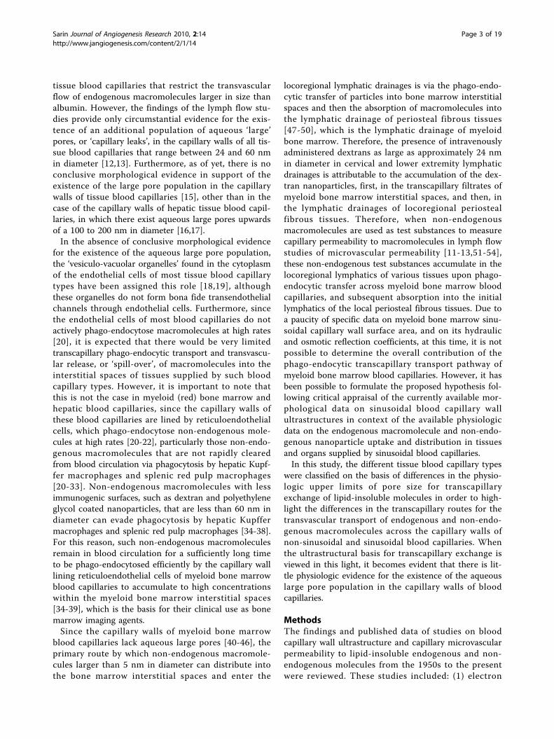
ResultsGeneralThe different types of tissue blood capillary microvascu-lature are classified in Tables 1, 2, 3 and 4: Table 1 isthe classification scheme for non-sinusoidal non-fene-strated blood capillary microvasculature, Table 2 is theclassification scheme for non-sinusoidal fenestratedblood capillary microvasculature, Table 3 is theclassification scheme for sinusoidal reticuloendothelialblood capillary microvasculature, and Table 4 is the
classification scheme for sinusoidal non-reticuloendothe-lial blood capillary microvasculature. The capillary wallultrastructures of different blood capillary microvascula-tures are schematically depicted in Figure 1 (Panels A,B, C, D and E; Individual panels and detailed descrip-tions in Additional files 1, 2, 3, 4 and 5). The physiolo-gic upper limit of pore size of the blood capillary, asdefined here, is the hydrodynamic diameter of the lar-gest lipid-insoluble molecule that is restricted from pas-sing through the pores in the capillary wall, and as such,constitutes the size of the molecule to which the poresin the capillary wall are impermeable. The blood capil-lary wall is a three-layered structure in most types of tis-sue blood capillaries, which consists of the endothelialglycocalyx layer (EGL) on the luminal face [61-63], thebasement membrane layer on the abluminal face[64-68], and the endothelial cell lining layer in betweenthe glycocalyx and the basement membrane [69-78](Figure 1, panels A, B, C, D and E; Additional files 1, 2,3, 4 and 5). The physiologic upper limit of pore size isfor any given capillary type is determined by the mostrestrictive layer of the capillary wall (Tables 1, 2, 3 and4; Figure 1, panels A, B, C, D and E; Additional files 1,2, 3, 4 and 5). For non-sinusoidal non-fenestrated bloodcapillaries, the pore size of the interendothelial cell junc-tion openings delineates the physiologic upper limit ofpore size [69,70,79-81] (Table 1; Figure 1, panel A;Additional file 1). For non-sinusoidal fenestrated bloodcapillaries with diaphragmed fenestrae, the pore size ofthe open spaces devoid of diaphragm membranous com-ponents delineates the physiologic upper limit of poresize [72-76,82] (Table 2; Figure 1 panel B; Additional
Table 1 Classification of non-sinusoidal non-fenestrated blood capillary microvasculature
NON-SINUSOIDALCAPILLARY TYPE
Primary anatomic sites oftransvascular flow
Determinants of physiologic pore size Physiologicupper limit of
pore size
Representativetissuemicrovascularbeds
NON-FENESTRATED •Non-fenestrated endothelial cells •Continuous anionic basement membrane rich in sulphated proteoglycans[64,86] •Anionicglycocalyx matrix layer on endothelial cell surfaces & interendothelial cell clefts rich in sialyated glycoproteins[69,142,152-154]
Non-fenestratedblood capillary with
tight junctions
Zona occludensinterendothelial junctions•Tight opposition of adjacentendothelial cell membranes atjunctions [70,92,93,155]
•Zona Occludens interendothelial cell junctions inseries constitute an absolute barrier to thetransvascular flow of macromolecules
< 1 nm •Retinal [156,157]•Brain-Spinal Cord[85,93,95,96,158,159]•NerveEndoneurium [160]•Enteric NervousSystem [91]•Lymphoid tissueCortex [161-163]
Non-fenestratedblood capillary with
loose junctions
Macula occludensinterendothelial junctions•Loose opposition of adjacentendothelial cell membranes atjunctions [79,80]
•Macula Occludens interendothelial cell junctions inseries constitute a relative barrier to thetransvascular flow of macromolecules
~5 nm •Skin* [164,165]•Muscle [7,79,80,90]•Cortical Bone [88]•Adipose tissue [87]•Lung [81]•IntestinalMesentary [15,71]•Develop. OvarianFollicle [94]
Sarin Journal of Angiogenesis Research 2010, 2:14http://www.jangiogenesis.com/content/2/1/14
Page 4 of 19

file 2). For non-sinusoidal fenestrated blood capillarieswith open ‘non-diaphragmed’ fenestrae, the fenestralopenings are bounded by a high-concentration of glyco-calyx matrix fibers; therefore, the pore size of the openspaces between the individual glycocalyx matrix fibers
delineates the physiologic upper limit of pore size [61](Table 2; Figure 1, panel C; Additional file 3). In case ofthe sinusoidal blood capillaries of the liver that haveopen fenestrae, the boundaries of the fenestral openingslack an appreciable concentration of glycocalyx matrix
Table 2 Classification of non-sinusoidal fenestrated blood capillary microvasculature
NON-SINUSOIDALCAPILLARY
TYPE
Primary anatomic sites oftransvascular flow
Determinants of physiologic poresize
Physiologicupper limitof pore size
Representative tissue microvascularbeds
FENESTRATED •Fenestrated endothelial cells •Continuous anionic basement membrane rich in sulphated proteoglycans[64,86] •Anionic glycocalyxmatrix of endothelial cell surfaces & clefts rich in sialyated glycoproteins & of fenestrated spaces rich in sulphated proteoglycans[166-170]
Fenestratedblood capillary
withdiaphragmedfenestrae
Diaphragmed fenestrae•Diameters of fenestrae rangebetween 60 and 80 nm•Widths of closed membranouscentral diaphragms rangebetween 10 and 30 nm•Eight to twelve, 2 to 7 nm wide,outwardly radiating membranousseptae from central diaphragm•Arc widths of fenestrated openspaces between ~6 and ~12 nm[72-76,82]
•Arc widths of open spaces devoid ofmembranous components (centraldiaphragm and septae) delineate theupper limits of pore size•Diaphragms of diaphragmedfenestrae constitute the barriers to thetransvascular flow of macromolecules•Anionic glycocalyx matrix overfenestrated spaces charge barrier tothe transvascular flow of anionicmacromolecules
Between 6& 12 nm
•Skin* [164,165]•Testis [171-173]•Connective tissue [174,175]•Eye Choriocapillaris[82,102-104,176,177]•Exocrine Glands [105-108]•Kidney Peritubular [72,178]•Endocrine Glands [73,106,179-185]•Intestinal Mucosa [186-191]•Peripheral Ganglia [158,192-194]•Nerve Epineurium [160]•Circumventricular Organs[109,110,195-199]•Choroid Plexus [109-113]•Pre-Ovulatory Follicle [114,115]•Eye Ciliary Process [116-120]
Fenestratedblood capillary
with openfenestrae
Open fenestrae•Open ‘non-diaphragmed’fenestrae with avg. diameters of65 nm devoid of the centraldiaphragm & septae [61,72,200]
•Narrow interspacing of glycocalyxmatrix fibers is the barrier to thetranscapillary flow of macromoleculeslarger than ~15 nm in diameter
~15 nm •Kidney Glomerulus[60,72,101,123-126,201,202]╬╬Slit diaphragms at the level ofpodocyte foot processes restrict thefiltration of plasma proteins larger than6 nm in diameter (i.e. hemoglobin,albumin)
Table 3 Classification of sinusoidal reticuloendothelial blood capillary microvasculature
SINUSOIDALCAPILLARY TYPE
Primary anatomic sites of transvascular flow Ultrastructural determinants of transvasculartransport
Physiologic upperlimit of pore size
RETICULO-ENDOTHELIAL
•Endothelial cells w/high levels of phago-endocytosis[130,131] •Basement membrane commonly absent[64,77,203] •Patchyanionic glycocalyx of sialyated glycoproteins at non-endocytic sites[132-134] deficient in hyaluronan[137-140]
Hepaticsinusoidal blood
capillary
Open fenestrae•Diameters of open fenestrae variable acrossmammalian species: 1)human & rabbit: avg.diameter ~105 nm (range 50 to 180 nm) [17,83]2) mouse & rat: avg. diameter ~135 nm (range 50to 280 nm) [16,83]•Absence of basement membrane underlyingfenestrae and lack of glycocalyx matrix fiberswithin fenestrae [204]•Phago-endocytic phenotype of endothelial cells[205-207]
•Absence of basement membrane underlyingfenestral openings and relative lack of glycocalyxmatrix fibers in the vicinity of the fenestral openingsrenders fenestrae permeable to macromolecules aslarge as the diameters of the fenestrae themselves[16,17]•Open fenestrae constitute the transvascular pathwayfor the passage of macromolecules into the interstitialhepatic Space of Disse•Phago-endocytosis and release of non-endogenousmacromolecules into the hepatic interstitium [141]•Wide size range of nanoparticles [136,208-210]*
Transvascular flow~180 nm (Human,
Rabbit)~280 nm (Mouse,
Rat)Phago-endocytic(Non-endogenous)
Wide Range*
Myeloid bonemarrow
sinusoidal bloodcapillary
Interendothelial junctions•Endothelial cells non-fenestrated except duringtranscellular passage of blood cells acrossendothelial cell membrane when cells transientlyfenestrated [40-42]•Macula occludens interendothelial junctions[42-44]•Phago-endocytic phenotype of endothelial cells[45,46,211]
•Transvascular flow of macromolecules smaller than~5 nm into the bone marrow interstitial space acrossmaculae occludens interendothelial junctions• Phago-endocytosis and release of non-endogenousmacromolecules into bone marrow interstitium[34,35]•Narrow size range of nanoparticles [36-38]**
Transvascular flow~5 nm
Phago-endocytic(Non-endogenous)Narrow Range**
Sarin Journal of Angiogenesis Research 2010, 2:14http://www.jangiogenesis.com/content/2/1/14
Page 5 of 19

fibers, and the capillary wall lacks a basement mem-brane; therefore, the pore size of the fenestral openingsdelineates the physiologic upper limit of pore size[16,17,83] (Table 3; Figure 1, panel E; Additional file 5).
Non-sinusoidal versus sinusoidal blood capillarymicrovasculatureBlood capillaries can be classified as non-sinusoidal orsinusoidal based on differences in the ultrastructure ofthe basement membrane layer of the capillary wall. Themain distinction between non-sinusoidal and sinusoidalcapillaries is the presence or absence of a continuousbasement membrane layer (Figure 1, panels A, B and C;Additional files 1, 2 and 3 versus Figure 1, panels D andE; Additional files 4 and 5). Non-sinusoidal blood capil-laries have traditionally been termed ‘continuous’ capil-laries due to the presence of a continuous basementmembrane layer [84]. These blood capillaries are classi-fied as ‘non-sinusoidal’ capillaries here is to minimizeambiguity, since these capillaries are further sub-classi-fied as either non-fenestrated or fenestrated based onthe absence or presence of fenestrations within theendothelial cells of the endothelial cell lining layer. Thebasement membrane layer of both types of non-sinusoi-dal capillaries is typically between 60 and 100 nm inthickness, and is composed of collagen type IV proteinsand glycoproteins interlinked by proteoglycans withheparan sulphate glycosaminoglycan side chains, whichare concentrated at the anionic sites of the basementmembrane [64,67,68].Although the blood capillaries of most tissues are non-
sinusoidal, the blood capillaries of certain tissues, the mye-loid bone marrow, liver, and spleen, are sinusoidal, as
these capillary types are deficient in the basement layer[16,17,24,25,27,29-31,41,42]. In the case of myeloid bonemarrow and hepatic sinusoidal capillaries, or sinusoids,the basement membrane layer is generally absent, or spar-sely distributed along the capillary path length, therefore,discontinuous [16,17,41,42,44] (Figure 1, panels D and E;Additional files 4 and 5). In the case of splenic sinusoidalcapillaries, the basement membrane of both splenic redpulp terminal arterial capillary endings and splenic redpulp venous capillaries (sinuses) is discontinuous, but fordifferent reasons. In the case of splenic red pulp terminalarterial capillary endings, the basement membrane is spar-sely distributed around the capillary ending [24,27,29,30];whereas, in the case of splenic red pulp venous capillaries(sinuses), the basement membrane of is comprised ofringed belts of basement membrane with individual belts 2to 3 μm apart [23,25,31,32]. The discontinuities in thebasement membrane layer of myeloid bone marrow sinu-soids and splenic venous sinuses render these sinusoidalcapillaries less restrictive to blood cell transmigration; assuch, the less restrictive capillary wall phenotype of thesesinusoidal capillaries is consistent with the functional rolesthat these tissues play in hematopoiesis and in theimmune response of mononuclear phagocytic system,which requires that monocytes and phagocytes residingwithin the respective interstitial spaces are efficientlymobilized when necessary.
Non-sinusoidal non-fenestrated blood capillaries:Ultrastructure and the physiologic upper limit of poresizeThe endothelial cell lining layer of non-sinusoidal non-fenestrated blood capillaries is continuous, as is the
Table 4 Classification of sinusoidal non-reticuloendothelial blood capillary microvasculature
SINUSOIDALCAPILLARY TYPE
Primary anatomic sites of transvascular flow Ultrastructural determinants of transvasculartransport
Physiologicupper limit of
pore size
NON-RETICULO-ENDOTHELIAL
•Endothelial cells w/low levels of phago-endocytosis[20-22] •Basement membrane discontinuous[23-25] •Thin anionicglycocalyx over endothelial cell surfaces of both capillary types[26]
Splenic red pulparterial blood
capillary (Terminal)
Terminal capillary ending•Terminal capillary ending openings ~5 microns(μm) in diameter [27,28]•Basement membrane sparse and intermittent•Macrophages in the terminal arterialpericapillary sheath and within the splenic redpulp reticular meshwork [29,30,212]
•Terminal arterial capillary network of the splenic red pulpreticulum constitutes the primary mode of splenicfiltration•Macromolecules as large as 5 μm pass into splenic redpulp reticulum through terminal capillary ending openings•Exogenous macromolecules phagocytosed bymacrophages in the terminal arterial pericapillary sheathand in the red pulp reticulum
~5 μm
Splenic red pulpvenous bloodcapillary (Sinus)
Interendothelial slits•Cuboidal endothelial cells•Interendothelial slits between apical and basaladherens junctions•Basement membrane ringed and belts ofbasement membrane rings 2-3 μm apart [25]•Slits closed except during active blood cellmigration [25,31] and macrophagephagocytosis [32]
•Few direct connections exist between splenic arteriolesand venous capillaries and constitutes the minor pathwayin splenic filtration•Exogenous macromolecules in sinus lumen phagocytosedat level of the interendothelial slits by finger-likepseudopodia of splenic pulp reticulum macrophages
__
Sarin Journal of Angiogenesis Research 2010, 2:14http://www.jangiogenesis.com/content/2/1/14
Page 6 of 19

basement membrane layer [70,80,81,85,86]. Since thereare no fenestrations within the endothelial cells of non-sinusoidal non-fenestrated capillaries, the interendothe-lial cell clefts constitute the primary pathways for trans-vascular flow across the capillary wall[15,71,79-81,85,87-94] (Table 1; Figure 1, panel A; Addi-tional file 1). Although the widths of the interendothelialcell clefts at the level of the endothelial cell surface areapproximately 20 nm, the diameters of pore openings inthe interendothelial cell junctions of the cleft are muchnarrower. In the case of zona occludens tight junctions,the endothelial cell membranes are tightly opposed atsites of most junctions, as there are no gaps of
measurable dimensions at the sites of these junctionswith electron microscopy [70,85,92,93]. Although therecan be occasional breaks in the junctional strand of oneor more zona occludens junctions within any giveninterendothelial cell cleft, this is almost never the casefor the entire series of junctions within the cleft; there-fore, a series of zona occludens junctions in a cleft con-stitute an absolute barrier to the transvascular passageof macromolecules [70]. Therefore, in the physiologicstate in vivo, the capillary walls of non-fenestrated capil-laries with zona occludens tight junctions only permitthe transvascular flow of small molecules, and comple-tely restrict the transvascular flow of macromolecules; as
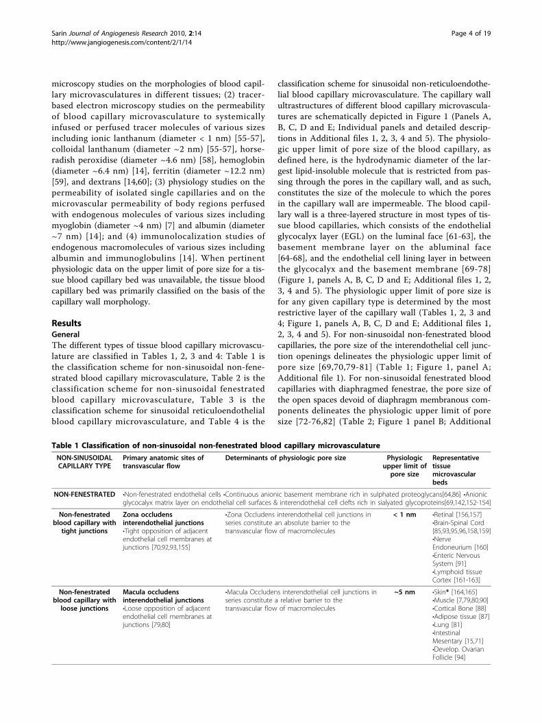
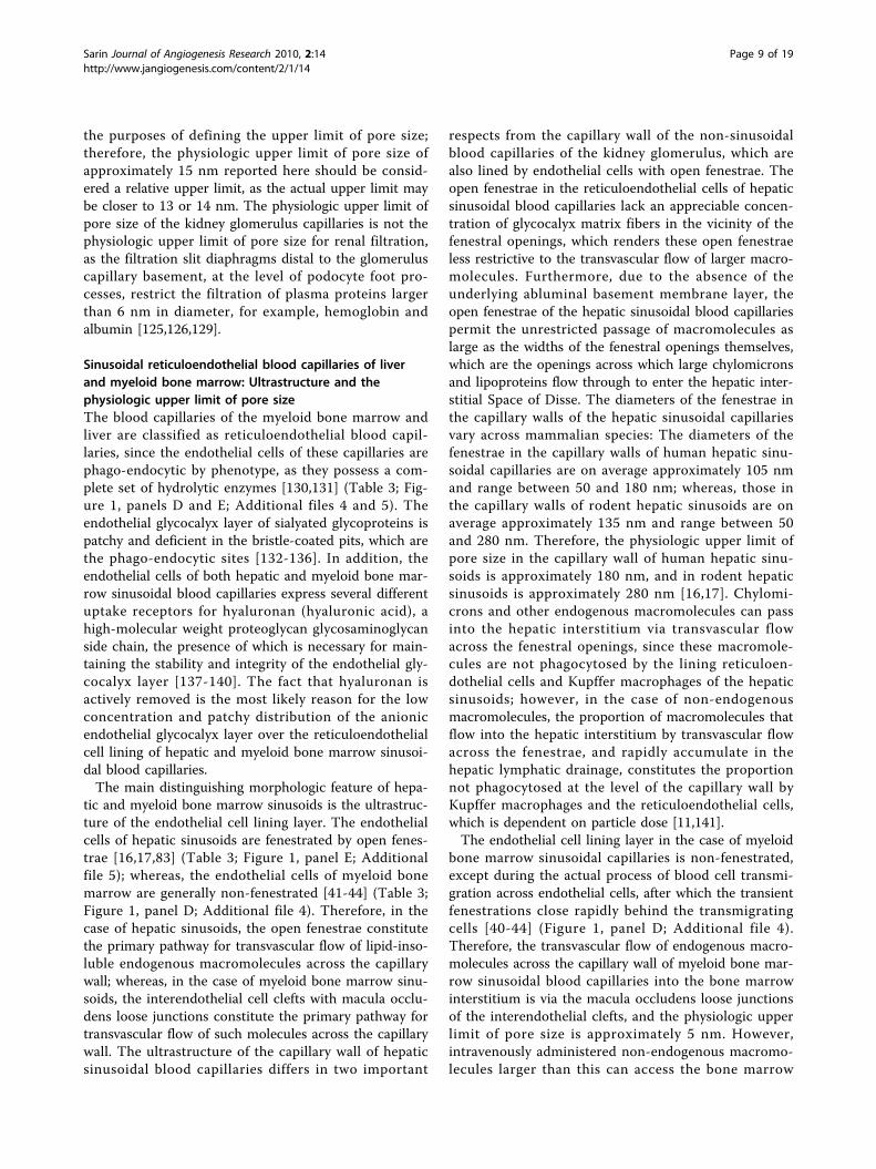
Figure 1 Schematic depictions of the capillary wall ultrastructure in different blood capillary microvasculatures. Shown in red are theanatomic sites in the capillary walls of the respective blood capillary types that are the primary pathways for transvascular flow and transportacross the capillary wall, and as such, constitute the ultrastructural determinants of the physiologic upper limit of pore size to transvascular flow.The green pillars that emanate from the luminal surface of the endothelial lining represent the individual mucopolysaccharide fibers of theendothelial glycocalyx layer (EGL), and the orange hatched region that encircles the abluminal surface of the endothelial cell lining representsthe collagenous basement layer (interna and externa). As depicted in the schematics, the capillary walls of the different types of non-sinusoidalblood capillaries are proficient in all three layers (panels A, B and C), which is not the case for the capillary walls of the sinusoidal bloodcapillaries of myeloid (red) bone marrow and the liver (panels D and E). Also depicted in panels D and E are the ‘bristle-coated pits’ of myeloidbone marrow and hepatic sinusoidal blood capillary the reticuloendothelial cells, which constitute the anatomic sites at which the phago-endocytosis of non-endogenous macromolecules occurs. A. Non-sinusoidal non-fenestrated blood capillaries B. Non-sinusoidal fenestrated bloodcapillaries with diaphragmed fenestrae C. Non-sinusoidal fenestrated blood capillaries with open ‘non-diaphragmed’ fenestrae D. Sinusoidalreticuloendothelial non-fenestrated blood capillaries of myeloid (red) bone marrow E. Sinusoidal reticuloendothelial fenestrated blood capillariesof the liver (Please view Additional files 1, 2, 3, 4 and 5 for individual Figure 1 panels A, B, C, D and E with detailed panel descriptions)
Sarin Journal of Angiogenesis Research 2010, 2:14http://www.jangiogenesis.com/content/2/1/14
Page 7 of 19

such, the physiologic upper limit of pore size of thesecapillaries is less than 1 nm [85,92,93,95,96]. Whereaszona occludens tight junctions are only permeable tosmall lipid-insoluble molecules, macula occludens loosejunctions are open junctions with pore diametersbetween 4 and 5 nm on electron micrographs [79,80],and are permeable to macromolecules as large as myo-globin (diameter ~4 nm) and horseradish peroxidise(diameter ~4.6 nm) [79-81,90,94]. Although there canoccasionally be significant breaks in the junctionalstrands of one or more macula occludens junctionswithin the interendothelial cell cleft [71], this is alsoalmost never the case for the entire series of maculaoccludens junctions in the cleft. Therefore, the physiolo-gic upper limit of pore size in the capillary walls ofnon-fenestrated capillaries with macula occludens loosejunctions is approximately 5 nm.
Non-sinusoidal fenestrated blood capillaries:Ultrastructure and the physiologic upper limit of poresizeThe basement membrane layer of non-sinusoidal fene-strated blood capillaries is continuous (Table 2; Figure1, panels B and C; Additional files 2 and 3), and there-fore, similar to that of non-sinusoidal non-fenestratedblood capillaries [86] (Table 1; Figure 1, panel A; Addi-tional file 1). However, the endothelial cells of theendothelial lining of non-sinusoidal fenestrated bloodcapillaries are fenestrated, either by diaphragmed fenes-trae, or by open ‘non-diaphragmed’ fenestrae, with thediameters of both types of fenestrae being within the 70nm range [72-76,82] (Table 2; Figure 1, panels B and C;Additional files 2 and 3). The induction of diaphragmedfenestrae within endothelial cells is known to bemediated primarily by vascular endothelial growth factor(VEGF), and results in the formation of fenestrae withmembranous components [97-100]; whereas, the specificmolecular pathways that result in the formation of theopen ‘non-diaphragmed’ fenestrae in the endothelialcells of the non-sinusoidal fenestrated blood capillariesof the kidney glomerulus have not yet been deciphered[101]. In the case of diaphragmed fenestrae, the dia-phragmed portion consists of a membranous centraldiaphragm between 10 and 30 nm wide, and the eightto twelve, 2 to 7 nm wide, membranous septae thatradiate outward from the central diaphragm to thefenestral rim [72,74-76,82]. The arc widths of the openspaces devoid of membranous components have beenmeasured to range between 6 and 12 nm [72,82]. Thephysiologic upper limit of pore size in the capillary wallsof non-sinusoidal blood capillaries with diaphragmedfenestrae ranges between 6 and 12 nm, with the upperlimit of pore size of eye choriocapillaris, exocrine glandsand kidney peritubules being closer to 6 nm [102-108],
and that of choroid plexus, pre-ovulatory follicle andeye ciliary process being closer to 12 nm [109-120].Even though in this particular study, only the bloodcapillary types found in different normal healthy tissueshave been formally classified, it deserves mention, thatthe VEGF-derived pathologic blood capillaries of solidcancers are also non-sinusoidal fenestrated blood capil-laries with diaphragmed fenestrae, and that the physiolo-gic upper limit of pore size in these blood capillaries hasrecently been defined as being approximately 12 nm[121,122]. The fact that the anatomically-measured arcwidths of the open spaces within the diaphragmedfenestrae of the non-sinusoidal fenestrated blood capil-laries from different tissues is similar to physiologically-measured sizes of the pores within these capillaries fromdifferent tissues strongly suggests that the primary phy-sical barriers to the transvascular flow of macromole-cules are the diaphragmed membranous components ofthe diaphragmed fenestrae. The highly anionic fibers ofthe endothelial glycocalyx that are rich in heparin andheparan sulphate proteoglycans are located in the vici-nity of the pores of the diaphragmed fenestrae; andtherefore, they constitute the primary electrostatic bar-riers to the transvascular flow of macromolecules withlow isoelectric points.Whereas non-sinusoidal fenestrated blood capillaries
with diaphragmed fenestrae are present in a wide-varietyof tissue types, non-sinusoidal fenestrated blood capil-laries with open fenestrae are only known to be presentin the kidney glomerulus. Even though the anatomicdiameters of open fenestrae are on average approxi-mately 65 nm, the physiologic upper limit of pore sizeof kidney glomeruli capillaries is approximately 15 nm[101,123-127]. This difference between the anatomicdiameters of the open fenestrae and the observed phy-siologic upper limit of pore size is attributable to pre-sence of narrowly interspaced endothelial glycocalyxmatrix fibers flanking over the open fenestrae (Table 2;Figure 1, panel C; Additional file 3). Since the interspa-cing of glycocalyx matrix fibers is a maximum of 20 nmin all directions, the physiologic upper limit of pore sizeof open fenestrae would be a maximum of 20 nm,which depends, of course, on the thickness of the indivi-dual fibers [61,128].The physiologic upper limit of pore size of approxi-
mately 15 nm for kidney glomeruli capillaries statedhere is based on the evidence that both endogenous andnon-endogenous macromolecules with hydrodynamicdiameters of approximately 12 nm (native ferritin,immunoglobulin G) traverse the open fenestrae[101,123-125], while dextrans with diameters of approxi-mately 15 nm are restricted at the level of the openfenestrae [60]. It is noted that dextrans are flexible poly-mers, and may not be the optimal macromolecules for
Sarin Journal of Angiogenesis Research 2010, 2:14http://www.jangiogenesis.com/content/2/1/14
Page 8 of 19
the purposes of defining the upper limit of pore size;therefore, the physiologic upper limit of pore size ofapproximately 15 nm reported here should be consid-ered a relative upper limit, as the actual upper limit maybe closer to 13 or 14 nm. The physiologic upper limit ofpore size of the kidney glomerulus capillaries is not thephysiologic upper limit of pore size for renal filtration,as the filtration slit diaphragms distal to the glomeruluscapillary basement, at the level of podocyte foot pro-cesses, restrict the filtration of plasma proteins largerthan 6 nm in diameter, for example, hemoglobin andalbumin [125,126,129].
Sinusoidal reticuloendothelial blood capillaries of liverand myeloid bone marrow: Ultrastructure and thephysiologic upper limit of pore sizeThe blood capillaries of the myeloid bone marrow andliver are classified as reticuloendothelial blood capil-laries, since the endothelial cells of these capillaries arephago-endocytic by phenotype, as they possess a com-plete set of hydrolytic enzymes [130,131] (Table 3; Fig-ure 1, panels D and E; Additional files 4 and 5). Theendothelial glycocalyx layer of sialyated glycoproteins ispatchy and deficient in the bristle-coated pits, which arethe phago-endocytic sites [132-136]. In addition, theendothelial cells of both hepatic and myeloid bone mar-row sinusoidal blood capillaries express several differentuptake receptors for hyaluronan (hyaluronic acid), ahigh-molecular weight proteoglycan glycosaminoglycanside chain, the presence of which is necessary for main-taining the stability and integrity of the endothelial gly-cocalyx layer [137-140]. The fact that hyaluronan isactively removed is the most likely reason for the lowconcentration and patchy distribution of the anionicendothelial glycocalyx layer over the reticuloendothelialcell lining of hepatic and myeloid bone marrow sinusoi-dal blood capillaries.The main distinguishing morphologic feature of hepa-
tic and myeloid bone marrow sinusoids is the ultrastruc-ture of the endothelial cell lining layer. The endothelialcells of hepatic sinusoids are fenestrated by open fenes-trae [16,17,83] (Table 3; Figure 1, panel E; Additionalfile 5); whereas, the endothelial cells of myeloid bonemarrow are generally non-fenestrated [41-44] (Table 3;Figure 1, panel D; Additional file 4). Therefore, in thecase of hepatic sinusoids, the open fenestrae constitutethe primary pathway for transvascular flow of lipid-inso-luble endogenous macromolecules across the capillarywall; whereas, in the case of myeloid bone marrow sinu-soids, the interendothelial cell clefts with macula occlu-dens loose junctions constitute the primary pathway fortransvascular flow of such molecules across the capillarywall. The ultrastructure of the capillary wall of hepaticsinusoidal blood capillaries differs in two important
respects from the capillary wall of the non-sinusoidalblood capillaries of the kidney glomerulus, which arealso lined by endothelial cells with open fenestrae. Theopen fenestrae in the reticuloendothelial cells of hepaticsinusoidal blood capillaries lack an appreciable concen-tration of glycocalyx matrix fibers in the vicinity of thefenestral openings, which renders these open fenestraeless restrictive to the transvascular flow of larger macro-molecules. Furthermore, due to the absence of theunderlying abluminal basement membrane layer, theopen fenestrae of the hepatic sinusoidal blood capillariespermit the unrestricted passage of macromolecules aslarge as the widths of the fenestral openings themselves,which are the openings across which large chylomicronsand lipoproteins flow through to enter the hepatic inter-stitial Space of Disse. The diameters of the fenestrae inthe capillary walls of the hepatic sinusoidal capillariesvary across mammalian species: The diameters of thefenestrae in the capillary walls of human hepatic sinu-soidal capillaries are on average approximately 105 nmand range between 50 and 180 nm; whereas, those inthe capillary walls of rodent hepatic sinusoids are onaverage approximately 135 nm and range between 50and 280 nm. Therefore, the physiologic upper limit ofpore size in the capillary wall of human hepatic sinu-soids is approximately 180 nm, and in rodent hepaticsinusoids is approximately 280 nm [16,17]. Chylomi-crons and other endogenous macromolecules can passinto the hepatic interstitium via transvascular flowacross the fenestral openings, since these macromole-cules are not phagocytosed by the lining reticuloen-dothelial cells and Kupffer macrophages of the hepaticsinusoids; however, in the case of non-endogenousmacromolecules, the proportion of macromolecules thatflow into the hepatic interstitium by transvascular flowacross the fenestrae, and rapidly accumulate in thehepatic lymphatic drainage, constitutes the proportionnot phagocytosed at the level of the capillary wall byKupffer macrophages and the reticuloendothelial cells,which is dependent on particle dose [11,141].The endothelial cell lining layer in the case of myeloid
bone marrow sinusoidal capillaries is non-fenestrated,except during the actual process of blood cell transmi-gration across endothelial cells, after which the transientfenestrations close rapidly behind the transmigratingcells [40-44] (Figure 1, panel D; Additional file 4).Therefore, the transvascular flow of endogenous macro-molecules across the capillary wall of myeloid bone mar-row sinusoidal blood capillaries into the bone marrowinterstitium is via the macula occludens loose junctionsof the interendothelial clefts, and the physiologic upperlimit of pore size is approximately 5 nm. However,intravenously administered non-endogenous macromo-lecules larger than this can access the bone marrow
Sarin Journal of Angiogenesis Research 2010, 2:14http://www.jangiogenesis.com/content/2/1/14
Page 9 of 19

interstitium via the phago-endocytic route, which formsthe basis of imaging myeloid bone marrow with systemi-cally administered dextran and polyethylene glycolcoated nanoparticle-based bone marrow imaging agents[34-38]. These non-endogenous macromolecules, whichare approximately 60 nm in diameter, are efficientlyphago-endocytosed by the reticuloendothelial cells ofthe myeloid bone marrow, and upon transvascularrelease, or spill-over, accumulate within myeloid bonemarrow interstitial spaces.
Sinusoidal non-reticuloendothelial blood capillaries of thespleen: Ultrastructure and the physiologic upper limit ofpore sizeTwo types of sinusoidal blood capillaries exist in thesplenic red pulp: the terminal arterial capillaries andvenous capillaries (sinuses) (Table 4; not illustratedschematically). Both types are classified as non-reticu-loendothelial sinusoidal blood capillaries since theendothelial cells of the endothelial lining of these splenicblood capillaries are of the non-phagocytic phenotype,as they lack a full complement of hydrolytic enzymes,and in this respect, are similar in phenotype to those ofthe endothelial cell lining of non-sinusoidal blood capil-laries [20-22]. The endothelial cell lining of both theterminal arterial capillaries and venous capillaries iscoated by a thin endothelial glycocalyx layer [26]. Thesplenic red pulp arterial capillaries terminate in the redpulp reticular interstitium. The segment of the terminalarterial capillary wall just proximal to the capillary end-ing in the red pulp is lined by fenestrated endothelialcells [24,27]; however, these endothelial cell fenestraeare covered by the pericapillary macrophage sheath, andtherefore, are not functionally open fenestrae [29,30].The terminal arterial capillary ending openings areapproximately 5 μm wide, which permit the passage ofplastic microspheres of this size into the red pulp reticu-lar interstitium [28]. Therefore, the physiologic upperlimit of pore size of terminal arterial capillary endingopenings is approximately 5 μm. The red pulp terminalarterial capillary network constitutes the ‘open’ slow cir-culation of the spleen, and is the primary route takenfor splenic filtration [28]. The splenic red pulp venouscapillaries originate in the splenic red pulp reticularmeshwork and drain into the splenic venous system[23,25]. There are very few direct connections betweensplenic arterial arterioles and splenic venous capillaries;the connections that do exist constitute the ‘closed’ fastcirculation of the spleen, which is the minor route takenfor splenic filtration [28]. The interendothelial cell junc-tions of the tall cuboidal endothelial cells of endotheliallining of splenic red pulp venous capillaries are the api-cal and basal adherens junctions located 2 to 3 μmapart [23,25]. The interendothelial slits between the
apical and basal adherens junctions constitute closed‘potential spaces’ across which blood cells migrate, andacross which reticulum macrophages extend pseudopo-dia to phagocytose, for example, nanoparticles withinthe lumens of venous capillaries [31,32], as belts of theringed basement membrane layer only exist at apicaland basal portions of the lining endothelial cells. It islikely that the closed potential space of the interen-dothelial slits permits the transvascular convective flowof macromolecules, which have not been phagocytosedat the level of the luminal face of the sinus wall, by thepseudopodia of reticulum macrophages; however, avail-able data is lacking on the subject, the physiologic upperlimit of pore size for the interendothelial slits of splenicred pulp venous sinuses has not been defined here.
DiscussionIn the 1940s, the possibility that a thin endothelial gly-cocalyx layer may exist on the luminal surface of theendothelial cell lining of the blood capillary microvascu-lature was suggested by Danielli [78], and Chambersand Zweifach [142]; however, at that time, the glycoca-lyx layer was difficult to visualize by conventional lightand electron microscopy staining techniques. Therefore,in 1959, when the morphological classification schemefor vertebrate blood capillaries was developed by Ben-nett and colleagues [84], the capillary wall was still con-sidered to be a two-layered structure, consisting of theendothelial cell lining and basement membrane layers.Although evidence for the existence of an approximately20 nm thick glycocalyx layer was provided by Luft in1966 [69] based on electron microscopy of fixed skeletalmuscle capillaries stained with ruthenium red, it is nowwell-known that the endothelial glycocalyx layer is a 150to 400 nm-thick polysaccharide-rich anionic matrix ofsialyated and sulphated proteoglycans and glycoproteinsin the physiologic state in vivo [61,128]. Since the indivi-dual fibers of the glycocalyx are circumferentially spaceda maximum of 20 nm apart [61,128], this relatively nar-row interspacing of the individual glycocalyx fiberswould restrict the transvascular passage of larger macro-molecules through the endothelial glycocalyx layer. This,indeed, appears to be case in the non-sinusoidal fene-strated blood capillaries of the kidney glomeruli thatpossess open ‘non-diaphragmed’ fenestrae covered by anintact glycocalyx layer, as the physiologic upper limit ofpore size in the capillary wall of this blood capillary typeis approximately 15 nm [60], and may be closer tobetween 13 and 14 nm if the upper limit of pore size ofkidney glomerulus capillaries is interrogated using non-flexible spherical macromolecules. This being the case,the barrier to the renal filtration of macromoleculessuch as hemoglobin (diameter ~6.4 nm) and albumin(diameter ~ 7 nm) are the slit-diaphragms of podocyte
Sarin Journal of Angiogenesis Research 2010, 2:14http://www.jangiogenesis.com/content/2/1/14
Page 10 of 19

foot processes on the abluminal face of the basementmembrane layer.In 1951, Pappenheimer and colleagues formulated the
classic small pore theory of microvascular permeabilityon the basis of experimental data on the restriction tothe transvascular flow of various-sized unlabeled endo-genous lipid-insoluble molecules across the walls of cathind-limb microvasculature [6]. By the measurement ofthe osmotic transients generated by the respective testmolecules using the isogravometric osmotic transienttechnique, Pappenheimer et al. determined that cathind-limb microvasculature was permeable to inulin(diameter ~3 nm), but not to hemoglobin (diameter~6.4 nm). On comparison of the experimental values forthe restriction to diffusion to theoretically predictedvalues for uniform water filled cylindrical pores 6 nm indiameter (less restrictive pores), or alternatively, for uni-form water-filled rectangular slit-pores approximately3.7 nm in width (more restrictive pores), it was observedthat the experimental data values fit better with the the-oretically predicted values for cylindrical pores 6 nm indiameter. However, in lieu of the polydisperse nature ofinulin, which was the smaller of the two macromolecu-lar test substances, it was noted that the range of theupper limit of pore size could only be established withadditional experimental data demonstrating therestricted transvascular flow of less polydisperse macro-molecular test substances similar in size to inulin. Insubsequent, similar perfused cat hind-limb osmotic tran-sient experiments with myoglobin (diameter ~4 nm), therestricted transvascular flow of myoglobin was demon-strated, and it was established that the physiologic upperlimit of pore size in cat hind-limb blood capillary micro-vasculature is between 4 and 6 nm [7]. The blood capil-lary microvasculature of the cat hind-limb is comprisedof the capillary microvasculatures of the several differenttissue types in the limb, which includes the capillarymicrovasculatures of skin, muscle, nerve, bone, myeloidbone marrow, adipose, and connective tissues; of theselimb tissues, muscle, bone, and adipose tissue capillarymicrovasculature constitutes one type (non-sinusoidalnon-fenestrated) (Table 1; Figure 1, panel A; Additionalfile 1); skin, nerve, and connective tissue another type(non-sinusoidal fenestrated) (Table 2; Figure 1, panel B;Additional file 2); and myeloid (red) bone marrow yetanother (sinusoidal reticuloendothelial) (Table 3; Figure1, panel D; Additional file 4). The physiologic upperlimit of pore size for skin, nerve, and connective tissueblood capillaries, being non-sinusoidal fenestrated capil-laries, is within the 6 to 12 nm range (Table 2). There-fore, the physiologic upper limit of pore size of cathind-limb blood capillary microvasculature, as measuredby the isogravometric osmotic transient method, wouldbe a slight over-estimation of the ‘actual’ physiologic
upper limit of pore size in skeletal muscle blood capil-lary microvasculature. This is the likely reason why theexperimental data values fit better with the theoreticallypredicted values for aqueous cylindrical pores 6 nm indiameter. This conclusion is supported by the ultra-structural evidence that the interendothelial cell clefts inthe capillary walls of the non-sinusoidal non-fenestratedskeletal muscle blood capillaries are lined by maculaoccludens interendothelial cell junctions, which, in ser-ies, restrict the transcapillary passage of macromoleculeslarger than horseradish peroxidase (diameter ~4.6 nm)across the interendothelial cell cleft [79](Table 1; Figure1, panel A; Additional file 1).The interplay of tissue blood capillary and interstitial
space hydrostatic and oncotic pressures favors the netfiltration of fluids and lipid-insoluble molecules acrossthe blood capillary walls, and this ultrafiltrate, is the tis-sue lymph [47,143-147]. Since the openings of the term-inal endings of tissue interstitium lymphatic capillaries,or initial lymphatics, are permeable to macromoleculesupwards of several hundred nanometers, the level of therestriction to the passage of systemically administeredmacromolecules is the tissue blood capillary wall[148-151]. These differences in the ultrastructure of thetissue blood capillary and lymphatic capillary walls isthe basis on which differences in macromolecule plasmaconcentration and regional lymph concentration havebeen used as indexes of regional differences in bloodcapillary permeability. Non-endogenous macromoleculesthat are not rapidly cleared from blood circulation accu-mulate in the myeloid bone marrow interstitium andthe periosteal fibrous tissue lymphatics [47-49]. Thisinformation is most pertinent to the interpretation oflower extremity and cervical region lymph flow data asthe presence of non-endogenous macromolecules largerthan approximately 5 nm in diameter within the lym-phatic drainage of these regions would be attributable tothe accumulation of these non-endogenous macromole-cules in the myeloid bone marrow sinusoidal transcapil-lary filtrate.Upon the intravenous administration of radiolabeled
native macromolecules, radio-iodinated albumin andimmunoglobulin, to dogs with cannulated thoraciclymph ducts, and the measurement of the changes inthe respective test molecule concentrations in plasmaand thoracic duct lymph, Wasserman and Mayerson, in1952, noted that: (1) the lymph concentration of radio-iodinated albumin increases approximately 1.6 times fas-ter than that of radio-iodinated immunoglobulin; andthat (2) when administered at high-doses, steady-statelymph levels of both radio-iodinated species areachieved within 90 minutes after administration;whereas, when administered at low-doses, steady-statelymph levels are achieved between 7 and 13 hours after
Sarin Journal of Angiogenesis Research 2010, 2:14http://www.jangiogenesis.com/content/2/1/14
Page 11 of 19
administration [11]. The faster rate of accumulation ofradio-iodinated albumin than immunoglobulin in thor-acic duct lymph is consistent with the fact that the poresizes in the diaphragmed fenestrae of non-sinusoidalfenestrated blood capillaries in many visceral organs andtissues are more restrictive to the transcapillary passageof immunoglobulin than albumin, as the physiologicupper limits of pore size in non-sinusoidal fenestratedblood capillaries with diaphragmed fenestrae varybetween 6 and 12 nm (Table 2). However, the fact thatthe test substances employed were radiolabeled nativemacromolecules is notable, as these radio-iodinated testsubstances [54] constitute non-endogenous macromole-cules that would be phago-endocytosed by reticuloen-dothelial cells of hepatic and myeloid bone marrowsinusoidal capillary walls. Based on the observed dose-related differences in the rates of radio-iodinated albu-min and immunoglobulin accumulation in the thoracicduct lymphatic drainage, which is primarily that of thehepatic region lymph, the great proportion of thesenon-endogenous macromolecules the administered atlow-doses are likely phago-endocytosed at the level ofthe hepatic sinusoidal blood capillary walls, and do nothave the opportunity to actually flow across the openfenestrae of the hepatic sinusoids to enter the hepaticinterstitium by the transvascular convective route. How-ever, at high doses, the phago-endocytic activity thresh-old of the lining reticuloendothelial cells of the hepaticsinusoid blood capillaries is reached; and as such, theproportion of the dose of the circulating macromole-cules above and beyond the phago-endocytic activitythreshold flows across the open fenestrae and enters thehepatic interstitial space via the transvascular convectiveroute. Therefore, at high doses, the radio-iodinated albu-min and immunoglobulin accumulate in the hepaticregion lymph and in thoracic lymphatic drainage at fas-ter rates, than when administered at low-doses.In 1956, Grotte et al. performed a series of additional
dog lymph flow studies by employing various-sized dex-tran and plastic nanoparticles, the findings of which werethe basis for the formulation of the dual pore hypothesisof microvascular permeability [12,13]. The experimentalfindings underlying the formulation of the dual porehypothesis of capillary permeability are discussed hereinfrom the physiologic perspective. When the findings areviewed in this light, it becomes apparent that the findingsare not per se a confirmation for the existence of a largepore population, but rather, are evidence for the role ofthe reticuloendothelial cells of the hepatic and myeloidbone marrow sinusoids in the phago-endocytosis non-endogenous macromolecules. Grotte employed dextrannanoparticles ranging in size from approximately 2.5 to 24nm in diameter, and fluorescent spherical plastic (methyl-methacrylate) nanoparticles ranging in size from 60 to
140 nm in diameter, and measured the steady-state con-centrations of the respective non-endogenous macromole-cules in the locoregional lymphatics; which, in case of thelymph flow studies with the dextrans were the hepatic,lower extremity, and cervical region lymphatics; and in thecase of the plastic nanoparticles were the hepatic, lowerextremity, cardiac, and bronchial region lymphatics. Thelymph concentrations of the dextrans smaller than 8 to 10nm in diameter were measured at 7 hours after the intra-venous infusion to animals with renal occlusion, and thoseof the larger dextrans were measured at 24 hours, whichwould have been sufficient periods of time for the smallerand larger dextrans to have reached steady-state lymphconcentrations. It is notable that the lymph concentrationsof the plastic nanoparticles were measured immediatelyfollowing infusions at various time points over a period ofbetween 3 and 4 hours, since these larger plastic nanopar-ticles were quickly cleared from systemic blood circula-tion, which is attributable to the rapid removal of theseimmunogenic particles by phago-endocytic uptake byhepatic Kupffer macrophages, and splenic red pulp macro-phages, as well as, the reticuloendothelial cells of the hepa-tic sinusoidal capillaries.On review of lymph flow study experimental data, it is
evident that the point at which the particle plasma:lymph concentration ratio first deviates from approxi-mately 1, or unity, represents the pore size cut-off of thecapillary ultrafiltrate and the lymphatic drainage of thecapillary population with the lowest upper limit of poresize, which constitutes that of non-sinusoidal non-fene-strated capillaries; and, several graded decreases in theparticle plasma:lymph concentration ratio over a rangeof particle sizes represent the pore size cut-offs of thecapillary ultrafiltrates of multiple capillary populationswith different upper limits of pore size, which constitu-tes that of fenestrated capillaries. In case of the lowerextremity and cervical region lymphatic drainage, thereare graded decreases in the dextran particle plasma:lymph concentration ratios between dextran particlesizes from 4 to 8 nm in diameter, from approximately 1to approximately 0.15 for lower extremity lymph, andfrom approximately 1 to approximately 0.25 for cervicallymph, which indicates the presence of populations ofnon-sinusoidal non-fenestrated capillaries with theupper limit of pore size closer to 4 nm, and indicatesthe presence of populations of non-sinusoidal fene-strated capillaries in the region with the upper limit ofpore size closer to 8 nm. In the case of the hepaticregion lymphatic drainage, the dextran particle plasma:lymph concentration ratio decreases from approximately1 to approximately 0.85 at a dextran particle size of8 nm, which indicates the presence of a population ofnon-sinusoidal fenestrated capillaries with a physiologicupper limit of pore size of approximately 8 nm,
Sarin Journal of Angiogenesis Research 2010, 2:14http://www.jangiogenesis.com/content/2/1/14
Page 12 of 19

consistent with the cut-off of pore size of the capillaryultrafiltrate of intestinal mucosal non-sinusoidal fene-strated blood capillaries (Table 2).The observation that the plasma:lymph concentration
ratio for the larger dextran particles between 8 and24 nm in diameter remains unchanged for various bodyregions has been cited as physiologic evidence for theexistence of another population of large pores, or ‘capil-lary leaks’, at least 24 nm in diameter in blood capillarymicrovasculature. Further, this observation, coupledwith the finding that the plastic particle plasma:lymphconcentration ratio for the lower extremity, cardiac, andbronchial region lymphs is approximately 0, or unmea-surable, has been cited as the physiologic evidence forthe existence of the large pore population in capillarymicrovasculature. Based on these findings, that particlesas large as 24 nm accumulate in the lower extremityand cervical lymph (dextran nanoparticles), and thatparticles 60 nm, and larger, do not accumulate in thelower extremity and cervical lymph (plastic nanoparti-cles), it has been concluded that the physiologic upperlimit of pore size of the large pore population in capil-lary microvasculature ranges between 24 and 60 nm.Furthermore, the differences in the regional dextran par-ticle plasma:lymph concentration ratios for the 8 to 24nm diameter dextran particles (approximately 0.15 forlower extremity region lymph, approximately 0.25 forcervical region lymph, and approximately 0.85 for hepa-tic region lymph) have been additionally cited as evi-dence for differences in the number of small and largepores in the blood capillary populations of the lowerextremity, cervical, and hepatic regions, with the small-to-large pore ratios for the respective regions being1:34,000, 1:18,000, and 1:340 [12].In case of the dextran nanoparticles between 8 and 24 nm
in diameter, the findings taken altogether, support the likeli-hood that the administered dose of the dextran nanoparti-cles was high enough to saturate the phago-endocyticcapacity of the reticuloendothelial cells of the hepatic andmyeloid bone marrow sinusoidal capillaries, resulting in theaccumulation of the dextrans nanoparticles in the respectiveinterstitial tissue spaces and regional lymph. With regardsto the accumulation in the hepatic interstitium and lymph,this would be via transvascular flow through open fenestraeand the phago-endocytic route; and with regards to themyeloid bone marrow, this would be via the phago-endocy-tic route. It is postulated here that the measured dextrannanoparticle concentrations in the lower extremity and cer-vical regional lymphatic drainages constitute the dextrannanoparticle concentrations of the myeloid bone marrowsinusoidal capillary filtrates of the respective regions. Therelatively high dextran particle plasma:lymph concentrationratio of the hepatic region lymph (approximately 0.85), ascompared to that of lower extremity and cervical region
lymphs (approximately 0.2), is consistent with the hepaticsinusoidal capillary filtrate constituting the major propor-tion of the lymphatic drainage of the hepatic region. In caseof the plastic nanoparticles between 60 and 140 nm, theparticle plasma:lymph concentration ratio for the hepaticregion lymph only reaches approximately 0.2 over 3 hours,and does not maintain steady-state levels over longer timeperiods; whereas particle plasma:lymph concentration ratiofor the lower extremity, cardiac, and bronchial regionlymph is approximately 0, or unmeasurable. The relativelyrapid clearance of the plastic particles from blood circula-tion is attributable to the rapid sequestration of these parti-cles in the splenic red pulp followed by phagocytosis ofparticles by the pulp macrophages. Therefore, the low levelof plastic particle accumulation in the hepatic interstitiumand associated lymphatic drainage, and virtually no plasticparticle accumulation in the lower extremity, cardiac, andbronchial region lymphatic drainages, reflects the low levelof phago-endocytic particle uptake at the level of the mye-loid bone marrow blood capillaries, secondary to the rapidclearance of plastic particles from systemic bloodcirculation.
ConclusionsTissue blood capillaries have been classified here on thebasis of the capillary type-specific differences in the phy-siologic upper limit of pore size to the transcapillary pas-sage of lipid-insoluble molecules. When tissue bloodcapillaries are classified on the basis of capillary wallultrastructural differences, as these differences relate tofunction, it becomes evident that the primary route forthe transcapillary exchange of lipid-insoluble small mole-cules and macromolecules across the capillary walls ofblood capillaries, other than those of myeloid bone mar-row blood capillaries, is via transvascular flow of themolecules through aqueous small pores, and that theupper limit of pore size in the capillary walls of theseblood capillaries is the physiologic upper limit of poresize for transcapillary transport of lipid-insoluble mole-cules. In case of myeloid bone marrow blood capillaries,the upper size limit to transvascular transport of lipid-insoluble molecules across the capillary wall is dependenton the route of the transvascular transport, since thetranscapillary passage of lipid-insoluble macromoleculessmaller than 5 nm in diameter can take place throughaqueous small pores in the interendothelial cell junctions;whereas, that of larger non-endogenous macromoleculestakes place upon phago-endocytosis of the macromole-cules by reticuloendothelial cells and then the release ofendocytosed molecules across the capillary wall into themyeloid bone marrow interstitium.The physiologic upper limit of pore size in the capil-
lary walls of most non-sinusoidal blood capillaries to thetranscapillary passage of lipid-insoluble endogenous and
Sarin Journal of Angiogenesis Research 2010, 2:14http://www.jangiogenesis.com/content/2/1/14
Page 13 of 19

non-endogenous macromolecules ranges between 5 and12 nm. Therefore, macromolecules larger than the phy-siologic upper limits of pore size in the non-sinusoidalblood capillary types generally do not accumulate withinthe respective tissue interstitial spaces and their lympha-tic drainages. In the case of reticuloendothelial sinusoi-dal blood capillaries of myeloid bone marrow, however,non-endogenous macromolecules as large as 60 nm indiameter can distribute into the bone marrow interstitialspace via the phago-endocytic route, and then subse-quently accumulate in the locoregional lymphatic drai-nages of tissues following absorption into the lymphaticdrainage of periosteal fibrous tissues, which is the lym-phatic drainage of myeloid bone marrow. When theultrastructural basis for transcapillary exchange acrossthe capillary walls of different capillary types is viewedin this light, it becomes evident that the physiologic evi-dence for the existence of aqueous large pores rangingbetween 24 and 60 nm in diameter in the capillary wallsof blood capillaries, is circumstantial, at best.
Additional material
Additional file 1: Figure 1 panel A with detailed descriptionFor non-sinusoidal non-fenestrated blood capillaries, the pore size of theinterendothelial cell junction openings delineates the physiologic upperlimit of pore size in the capillary wall, which is < 1 nm for non-sinusoidalnon-fenestrated tissue blood capillaries with zona occludens junctions (i.e. brain and spinal cord), and approximately 5 nm for non-sinusoidalnon-fenestrated tissue blood capillaries with macula occludens junctions(i.e. skeletal muscle).
Additional file 2: Figure 1 panel B with detailed descriptionFor non-sinusoidal fenestrated blood capillaries with diaphragmed fenestrae, thepore size of the open spaces within the fenestrae devoid ofmembranous components (central diaphragm [shown in red] and theseptae of the diaphragm that radiate outward to the fenestral rim [notshown]) delineates the physiologic upper limit of pore size, which rangesbetween 6 and 12 nm.
Additional file 3: Figure 1 panel C with detailed descriptionIn thecase of non-sinusoidal fenestrated blood capillaries with open ‘non-diaphragmed’ fenestrae, the only known healthy tissue with this bloodcapillary type is the kidney glomerulus. The pore size of the open spacesbetween the individual glycocalyx matrix fibers in the vicinity of thefenestrae (shown in red) delineates the physiologic upper limit of poresize, which is approximately 15 nm.
Additional file 4: Figure 1 panel D with detailed descriptionIn thecase of sinusoidal reticuloendothelial non-fenestrated blood capillaries ofmyeloid bone marrow, the lining reticuloendothelial cells of myeloidbone marrow sinusoidal blood capillaries are only fenestrated during theactual process of blood cell transmigration, as is depicted in panel D.Since these ‘cellular transmigration pores’ close immediately followingcellular transit, and the endothelial cells are not permanently fenestrated,the endothelial cells are non-fenestrated with respect to thetransvascular flow of macromolecules. The pore size of the openings inthe macula occludens interendothelial cell junctions is the primarydeterminant of the physiologic upper limit of pore size to thetransvascular flow of macromolecules, which is ~5 nm. Non-endogenousmacromolecules larger than 5 nm in diameter with long blood half-lives,which are not rapidly phagocytosed by macrophages (hepatic Kupfferand splenic red pup macrophages), accumulate in the bone marrowinterstitium upon the transvascular release of phago-endocytosedparticles into the marrow interstitium.
Additional file 5: Figure 1 panel E with detailed descriptionIn thecase of sinusoidal reticuloendothelial fenestrated blood capillaries of theliver, the capillary wall of hepatic sinusoidal blood capillaries is lined byreticuloendothelial cells with open fenestrae of relatively wide diameters,which can be on the order of 180 nm (humans) to 280 nm (rodents).Due to the lack of an appreciable concentration of glycocalyx matrixfibers in the vicinity of the fenestral openings, and an absence of thebasement membrane layer, the physiologic upper limit of pore size inthe hepatic sinusoidal capillary wall is approximately the pore size of thefenestral openings, which permit the unrestricted transvascular flow ofsmaller chylomicrons and lipoproteins into the hepatic interstitium. Non-endogenous macromolecules with long blood half-lives can access thehepatic interstitium either via transvascular flow across the openfenestrae or upon the transvascular release of macromolecules phago-endocytosed by capillary wall reticuloendothelial cells and hepaticKupffer macrophages.
AcknowledgementsThe research was funded by the National Institute of Biomedical Imagingand Bioengineering, National Institutes of Health, Bethesda, Maryland 20892,USA.
Authors’ contributionsHS conceptualized the work and wrote the manuscript.
Competing interestsThe authors declare that they have no competing interests.
Received: 18 March 2010 Accepted: 11 August 2010Published: 11 August 2010
References1. Verkman AS: Aquaporin water channels and endothelial cell function.
Journal of Anatomy 2002, 200:617-627.2. Kozono D, Yasui M, King LS, Agre P: Aquaporin water channels: atomic
structure molecular dynamics meet clinical medicine. The Journal ofClinical Investigation 2002, 109:1395-1399.
3. Oliva R, Calamita G, Thornton JM, Pellegrini-Calace M: Electrostatics ofaquaporin and aquaglyceroporin channels correlates with their transportselectivity. Proceedings of the National Academy of Sciences 2010,107:4135-4140.
4. Wilson AJ, Carati CJ, Gannon BJ, Haberberger R, Chataway TK: Aquaporin-1in blood vessels of rat circumventricular organs. Cell Tissue Res 2010,340:159-168.
5. Pappenheimer JR, Soto-Rivera A: Effective osmotic pressure of the plasmaproteins and other quantities associated with the capillary circulation inthe hindlimbs of cats and dogs. Am J Physiol 1948, 152:471-491.
6. Pappenheimer JR, Renkin EM, Borrero LM: Filtration, Diffusion andMolecular Sieving Through Peripheral Capillary Membranes: AContribution to the Pore Theory of Capillary Permeability. Am J Physiol1951, 167:13-46.
7. Pappenheimer JR: Passage of Molecules Through Capillary Walls. PhysiolRev 1953, 33:387-423.
8. Michel CC: The investigation of capillary permeability in single vessels.Acta Physiologica Scandinavica 1979, 106:67-74.
9. Michel CC: Filtration coefficients and osmotic reflexion coefficients of thewalls of single frog mesenteric capillaries. The Journal of Physiology 1980,309:341-355.
10. Michel CC, Mason JC, Curry FE, Tooke JE, Hunter PJ: A development of theLandis technique for measuring the filtration coefficient of individualcapillaries in the frog mesentery. Q J Exp Physiol Cogn Med Sci 1974,59:283-309.
11. Wasserman K, Mayerson HS: Dynamics of Lymph and Plasma ProteinExchange. Cardiology 1952, 21:296-307.
12. Grotte G: Passage of dextran molecules across the blood-lymph barrier.Acta Chir Scand Suppl 1956, 211:1-84.
13. Grotte G, Juhlin L, Sandberg N: Passage of solid spherical particles acrossthe blood-lymph barrier. Acta Physiologica Scandinavica 1960, 50:287-293.
Sarin Journal of Angiogenesis Research 2010, 2:14http://www.jangiogenesis.com/content/2/1/14
Page 14 of 19

14. Armstrong JK, Wenby RB, Meiselman HJ, Fisher TC: The hydrodynamic radiiof macromolecules and their effect on red blood cell aggregation.Biophysical Journal 2004, 87:4259-4270.
15. Levick JR, Michel CC: The permeability of individually perfused frogmesenteric capillaries to T1824 and T1824-albumin as evidence for alarge pore system. Quarterly journal of experimental physiology and cognatemedical sciences 1973, 58:67-85.
16. Naito M, Wisse E: Filtration effect of endothelial fenestrations onchylomicron transport in neonatal rat liver sinusoids. Cell and TissueResearch 1978, 190:371-382.
17. Wisse E, Jacobs F, Topal B, Frederik P, De Geest B: The size of endothelialfenestrae in human liver sinusoids: implications for hepatocyte-directedgene transfer. Gene Therapy 2008, 15:1193-1199.
18. Simionescu M, Simionescu N, Palade GE: Morphometric data on theendothelium of blood capillaries. Journal of Cell Biology 1974, 60:128-152.
19. Dvorak AM, Kohn S, Morgan ES, Fox P, Nagy JA, Dvorak HF: The vesiculo-vacuolar organelle (VVO): A distinct endothelial cell structure thatprovides a transcellular pathway for macromolecular extravasation.Journal of Leukocyte Biology 1996, 59:100-115.
20. Cotran RS: Endothelial phagocytosis: An electron-microscopic study.Experimental and Molecular Pathology 1965, 4:217-231.
21. Dorfman RF: Nature of the Sinus Lining Cells of the Spleen. Nature 1961,190:1021-1022.
22. Snodgrass MJ: A study of some histochemical and phagocytic reactionsof the sinus lining cells of the rabbit’s spleen. The Anatomical Record1968, 161:353-359.
23. Weiss L: A scanning electron microscopic study of the spleen. Blood1974, 43:665-691.
24. McCuskey RS, McCuskey PA: In vivo and electron microscopic studies ofthe splenic microvasculature in mice. Cellular and Molecular Life Sciences1985, 41:179-187.
25. Chen L-T, Weiss L: Electron microscopy of the red pulp of human spleen.American Journal of Anatomy 1972, 134:425-457.
26. Ueda H, Takehana K, Eerdunchaolu , Iwasa K, Fujimori O, Shimada S:Electron Microscopic Cytochemical Studies of Anionic Sites in the RatSpleen. The Journal of Veterinary Medical Science 2001, 63:287-291.
27. Suzuki T, Furusato M, Takasaki S, Shimizu S, Hataba Y: Stereoscopicscanning electron microscopy of the red pulp of dog spleen withspecial reference to the terminal structure of the cordal capillaries. Celland Tissue Research 1977, 182:441-453.
28. Chen L: Microcirculation of the spleen: and open or closed circulation?Science 1978, 201:157-159.
29. Blue J, Weiss L: Periarterial macrophage sheaths (Ellipsoids) in cat spleen- an electron microscope study. American Journal of Anatomy 1981,161:115-134.
30. Blue J, Weiss L: Electron microscopy of the red pulp of the dog spleenincluding vascular arrangements periarterial macrophage sheaths(ellipsoids), and the contractile innervated reticular meshwork. AmericanJournal of Anatomy 1981, 161:189-218.
31. MacDonald IC, Ragan DM, Schmidt EE, Groom AC: Kinetics of red bloodcell passage through interendothelial slits into venous sinuses in ratspleen analyzed by in vivo microscopy. Microvascular Research 1987,33:118-134.
32. Burke JS, Simon GT: Electron microscopy of the spleen. II. Phagocytosis ofcolloidal carbon. American Journal of Pathology 1970, 58:157-181.
33. Ishida T, Ichihara M, Wang X, Kiwada H: Spleen plays an important role inthe induction of accelerated blood clearance of PEGylated liposomes.Journal of Controlled Release 2006, 115:243-250.
34. Daldrup HE, Link TM, Blasius S, Strozyk A, Könemann S, Jürgens H,Rummeny EJ: Monitoring radiation-induced changes in bone marrowhistopathology with ultra-small superparamagnetic iron oxide (USPIO)-enhanced MRI. Journal of Magnetic Resonance Imaging 1999, 9:643-652.
35. Daldrup-Link HE, Link TM, Rummeny EJ, August C, Konemann S, Jurgens H,Heindel W: Assessing permeability alterations of the blood-bone marrowbarrier due to total body irradiation: In vivo quantification with contrastenhanced magnetic resonance imaging. Bone Marrow Transplantation2000, 25:71.
36. Illum L, Davis SS: Targeting of colloidal particles to the bone marrow. LifeSciences 1987, 40:1553-1560.
37. Davis SS, Ilium L, Moghimi SM, Davies MC, Porter CJH, Muir IS, Brindley A,Christy NM, Norman ME, Williams P, Dunn SE: Microspheres for targetingdrugs to specific body sites. Journal of Controlled Release 1993, 24:157-163.
38. Martindale AA, Papadimitriou JM, Turner JH: Technetium-99m AntimonyColloid for Bone-Marrow Imaging. J Nucl Med 1980, 21:1035-1041.
39. Huang TS: Passage of foreign particles through the sinusoidal wall of therabbit bone marrow–an electron microscopic study. Acta Pathol Jpn 1971,21:349-367.
40. Tavassoli M, Crosby WH: Fate of the nucleus of the marrow erythroblast.Science 1973, 179:912-913.
41. De Bruyn PP, Michelson S, Thomas TB: The migration of blood cells of thebone marrow through the sinusoidal wall. Journal of Morphology 1971,133:417-437.
42. Campbell FR: Ultrastructural studies of transmural migration of bloodcells in the bone marrow of rats mice and guinea pigs. American Journalof Anatomy 1972, 135:521-535.
43. Tavassoli M, Shaklai M: Absence of Tight Junctions in Endothelium ofMarrow Sinuses: Possible Significance for Marrow Cell Egress. BritishJournal of Haematology 1979, 41:303-307.
44. Campbell F: Fine structure of the bone marrow of the chicken andpigeon. Journal of Morphology 1967, 123:405-439.
45. De Bruyn PPH, Michelson S, Becker RP: Endocytosis, transfer tubules andlysosomal activity in myeloid sinusoidal endothelium. Journal ofUltrastructure Research 1975, 53:133-151.
46. De Bruyn PPH, Michelson S, Bankston PW: In-vivo endocytosis by bristle-coated pits and intracellular transport of endogenous albumin in theendothelium of the sinuses of liver and bone marrow. Cell and TissueResearch 1985, 240:1-7.
47. Fuller KM, Munro RR: Lymphatic drainage of bone marrow. ANZ Journal ofSurgery 1964, 34:11-14.
48. Kolodny A: The relation of the bone marrow to the lymphatic system: Itsrole in the spreading of carcinomatous metastases throughout theskeleton. Arch Surg 1925, 11:690-707.
49. Campbell E: Periosteal Lymphatics. Arch Surg 1929, 18:2099-2106.50. Suzuki T, Kurokawa K, Yamanaka H, Jimbo H: Lymphatico-venous
anastomosis after obstruction of abdominal lymphatics and lymphaticdrainage of the prostate in canines. Clinical Anatomy 1993, 6:87-93.
51. Wasserman K, Mayerson HS: Plasma, Lymph and Urine Studies AfterDextran Infusions. Am J Physiol 1952, 171:218-232.
52. Wasserman K, Loeb L, Mayerson HS: Capillary Permeability toMacromolecules. Circ Res 1955, 3:594-603.
53. Shirley HH JR, Wolfram CG, Wasserman K, Mayerson HS: CapillaryPermeability to Macromolecules: Stretched Pore Phenomenon. Am JPhysiol 1957, 190:189-193.
54. Freeman T: The biological behaviour of normal and denatured humanplasma albumin. Clinica Chimica Acta 1959, 4:788.
55. Shaklai M, Tavassoli M: Lanthanum as an electron microscopic stain. JHistochem Cytochem 1982, 30:1325-1330.
56. Sharov VG: Use of colloidal lanthanum as an electron-microscopic tracer.Bulletin of Experimental Biology and Medicine 1981, 92:1748-1750.
57. Schatzki PF, Newsome A: Neutralized Lanthanum Solution a LargelyNoncolloidal Ultrastructural Tracer. Biotechnic & Histochemistry 1975,50:171-178.
58. Vegge T, Winther FØ, Olsen BR: Horseradish peroxidase in plasma studiedby gel filtration. Histochemistry and Cell Biology 1971, 28:16-22.
59. Harrison PM: The structure of apoferritin: Molecular size shape andsymmetry from X-ray data. Journal of Molecular Biology 1963, 6:404-422,IN403-IN409.
60. Caulfield JP, Farquhar MG: The permeability of glomerular capillaries tograded dextrans: Identification of the Basement Membrane as thePrimary Filtration Barrier. J Cell Biol 1974, 63:883-903.
61. Squire JM, Chew M, Nneji G, Neal C, Barry J, Michel C: Quasi-PeriodicSubstructure in the Microvessel Endothelial Glycocalyx: A PossibleExplanation for Molecular Filtering? Journal of Structural Biology 2001,136:239-255.
62. Weinbaum S, Tsay R, Curry FE: A three-dimensional junction-pore-matrixmodel for capillary permeability. Microvascular Research 1992, 44:85-111.
63. Pries AR, Secomb TW, Gaehtgens P: The endothelial surface layer. PflügersArchiv European Journal of Physiology 2000, 440:653-666.
Sarin Journal of Angiogenesis Research 2010, 2:14http://www.jangiogenesis.com/content/2/1/14
Page 15 of 19

64. Charonis AS, Wissig SL: Anionic sites in basement membranes.Differences in their electrostatic properties in continuous andfenestrated capillaries. Microvascular Research 1983, 25:265.
65. Yurchenco PD, Schittny JC: Molecular architecture of basementmembranes. FASEB Journal 1990, 4:1577-1590.
66. Yurchenco PD, Patton BL: Developmental and pathogenic mechanisms ofbasement membrane assembly. Current Pharmaceutical Design 2009,15:1277-1294.
67. Kanwar YS, Farquhar MG: Presence of heparan sulfate in the glomerularbasement membrane. Proceedings of the National Academy of Sciences ofthe United States of America 1979, 76:1303-1307.
68. Leblond CP, Inoue S: Structure, composition and assembly of basementmembrane. American Journal of Anatomy 1989, 185:367-390.
69. Luft JH: Fine structures of capillary and endocapillary layer as revealedby ruthenium red. Federation Proceedings 1966, 25:1773-1783.
70. Brightman MW, Reese TS: Junctions between intimately apposed cellmembranes in the vertebrate brain. Journal of Cell Biology 1969,40:648-677.
71. Mason JC, Curry FE, White IF, Michel CC: The ultrastructure of frogmesenteric capillaries of known filtration coefficient. Quarterly Journal ofExperimental Physiology 1979, 64:217-224.
72. Bearer EL, Orci L, Sors P: Endothelial fenestral diaphragms: A quick-freeze,deep-etch study. Journal of Cell Biology 1985, 100:418-428.
73. Elfvin L-G: The ultrastructure of the capillary fenestrae in the adrenalmedulla of the rat. Journal of Ultrastructure Research 1965, 12:687-704.
74. Friederici HHR: The tridimensional ultrastructure of fenestrated capillaries.Journal of Ultrastructure Research 1968, 23:444-456.
75. Maul GG: Structure and formation of pores in fenestrated capillaries.Journal of Ultrastructure Research 1971, 36:768-782.
76. Rhodin JAG: The diaphragm of capillary endothelial fenestrations. Journalof Ultrastructure Research 1962, 6:171-185.
77. Yee A, Revel J: Endothelial cell junctions. J Cell Biol 1975, 66:200-204.78. Danielli JF, Stock A: The structure and permeability of blood capillaries.
Biological Reviews 1944, 19:81-94.79. Karnovsky MJ: The ultrastructural basis of capillary permeability studied
with peroxidase as a tracer. Journal of Cell Biology 1967, 35:213-236.80. Ward BJ, Bauman KF, Firth JA: Interendothelial junctions of cardiac
capillaries in rats: their structure and permeability properties. Cell andTissue Research 1988, 252:57-66.
81. Schneeberger EE: The permeability of the alveolar-capillary membrane toultrastructural protein tracers. Annals of the New York Academy of Sciences1974, 221:238-243.
82. Melamed S, Ben-Sira I, Ben-Shaul Y: Ultrastructure of fenestrations inendothelial choriocapillaries of the rabbit–a freeze-fracturing study.British Journal of Ophthalmology 1980, 64:537-543.
83. Snoeys J, Lievens J, Wisse E, Jacobs F, Duimel H, Collen D, Frederik P, DeGeest B: Species differences in transgene DNA uptake in hepatocytesafter adenoviral transfer correlate with the size of endothelial fenestrae.Gene Therapy 2007, 14:604-612.
84. Bennett HS, Luft JH, Hampton JC: Morphological classifications ofvertebrate blood capillaries. Am J Physiol 1959, 196:381-390.
85. Reese TS, Karnovsky MJ: Fine structural localization of a blood-brainbarrier to exogenous peroxidase. Journal of Cell Biology 1967, 34:207-217.
86. Carlson EC, Audette JL, Veitenheimer NJ, Risan JA, Laturnus DI, Epstein PN:Ultrastructural morphometry of capillary basement membrane thicknessin normal and transgenic diabetic mice. The Anatomical Record Part A:Discoveries in Molecular Cellular, and Evolutionary Biology 2003,271A:332-341.
87. Williamson JR: Adipose Tissue: Morphological Changes Associated withLipid Mobilization. J Cell Biol 1964, 20:57-74.
88. Takahashi S, Sugimoto M, Kotoura Y, Sasai K, Oka M, Yamamuro T: Long-term changes in the haversian systems following high-dose irradiation:An ultrastructural and quantitative histomorphological study. Journal ofBone and Joint Surgery - Series A 1994, 76:722-738.
89. Alvarez OA, Yudilevich DL: Heart capillary permeability to lipid-insolublemolecules. Journal of Physiology 1969, 202:45-58.
90. Trap-Jensen J, Lassen N: Restricted diffusion in skeletal muscle capillariesin man. Am J Physiol 1971, 220:371-376.
91. Gershon MD, Bursztajn S: Properties of the enteric nervous system:limitation of access of intravascular macromolecules to the myenteric
plexus and muscularis externa. Journal of Comparative Neurology 1978,180:467-474.
92. Fenstermacher JD, Johnson JA: Filtration and reflection coefficients of therabbit blood-brain barrier. The American journal of physiology 1966,211:341-346.
93. Sorensen SC: The permeability to small ions of tight junctions betweencerebral endothelial cells. Brain Research 1974, 70:174-178.
94. Anderson WA: Permeability of ovarian blood vessels and follicles ofjuvenile rats. Microvascular Research 1972, 4:348-373.
95. Bouldin TW, Krigman MR: Differential permeability of cerebral capillaryand choroid plexus to lanthanum ion. Brain Research 1975, 99:444-448.
96. Reese TS, Feder N, Brightman MW: Electron microscopic study of theblood-brain and blood-cerebrospinal fluid barriers with microperoxidase.Journal of Neuropathology and Experimental Neurology 1971, 30:137-138.
97. Kamba T, Tam BYY, Hashizume H, Haskell A, Sennino B, Mancuso MR,Norberg SM, O’Brien SM, Davis RB, Gowen LC, et al: VEGF-dependentplasticity of fenestrated capillaries in the normal adult microvasculature.Am J Physiol Heart Circ Physiol 2006, 290:H560-576.
98. Roberts WG, Delaat J, Nagane M, Huang S, Cavenee WK, Palade GE: Hostmicrovasculature influence on tumor vascular morphology andendothelial gene expression. American Journal of Pathology 1998,153:1239-1248.
99. Esser S, Wolburg K, Wolburg H, Breier G, Kurzchalia T, Risau W: Vascularendothelial growth factor induces endothelial fenestrations in vitro.Journal of Cell Biology 1998, 140:947-959.
100. Roberts WG, Palade GE: Neovasculature induced by vascular endothelialgrowth factor is fenestrated. Cancer Research 1997, 57:765-772.
101. Ichimura K, Stan RV, Kurihara H, Sakai T: Glomerular Endothelial Cells FormDiaphragms during Development and Pathologic Conditions. J Am SocNephrol 2008, 19:1463-1471.
102. Pino RM, Essner E: Structure and permeability to ferritin of thechoriocapillary endothelium of the rat eye. Cell and Tissue Research 1980,208:21-27.
103. Pino RM, Essner E: Permeability of rat choriocapillaris to hemeproteins.Restriction of tracers by a fenestrated endothelium. Journal ofHistochemistry and Cytochemistry 1981, 29:281-290.
104. Pino RM, Essner E, Pino LC: Permeability of the neonatal ratchoriocapillaris to hemeproteins and ferritin. American Journal of Anatomy1982, 164:333-341.
105. Matsuzawa T, Kubosumi K: The Ultrastructur Morphogenesis andHistochemistry of the Sweat Glands in the Rat Foot Pads as Revealed byElectron Microscopy. J Electron Microsc (Tokyo) 1963, 12:175-191.
106. Henderson JR, Moss MC: A morphometric study of the endocrine andexocrine capillaries of the pancreas. Quarterly Journal of ExperimentalPhysiology 1985, 70:347-356.
107. Mohamed AH: Ultrastructural permeability studies in capillaries of rabbitoral mucosa and salivary glands. Microvascular Research 1975, 9:287-303.
108. Clough G, Smaje LH: Exchange area and surface properties of themicrovasculature of the rabbit submandibular gland following ductligation. J Physiol 1984, 354:445-456.
109. Dermietzel R, Thürauf N, Kalweit P: Surface charges associated withfenestrated brain capillaries: II. In Vivo studies on the role of molecularcharge in endothelial permeability. Journal of Ultrastructure Research 1983,84:111-119.
110. Schmidley JW, Wissig SL: Anionic sites on the luminal surface offenestrated and continuous capillaries of the CNS. Brain Research 1986,363:265-271.
111. Fleury J, Bellon B, Bernaudin JF, Bouchaud C, Pinchon MC, Kuhn J, Poirier J:Electron-microscopic immunohistochemical study of the localization ofimmunoglobulin G in the choroid plexus of the rat. Cell and TissueResearch 1984, 238:177-182.
112. Peress NS, Roxburgh VA, Gelfand MC: Binding sites for immunecomponents in human choroid plexus. Arthritis and Rheumatism 1981,24:520-526.
113. Peress NS, Tompkins D: Effect of molecular charge on choroid-plexuspermeability: Tracer studies with cationized ferritins. Cell and TissueResearch 1981, 219:425-431.
114. Okuda Y, Okamura H, Kanzaki H, Takenaka A: Capillary permeability ofrabbit ovarian follicles prior to ovulation. Journal of Anatomy 1983,137:263-269.
Sarin Journal of Angiogenesis Research 2010, 2:14http://www.jangiogenesis.com/content/2/1/14
Page 16 of 19

115. Hess KA, Chen L, Larsen WJ: The ovarian blood follicle barrier is bothcharge- and size-selective in mice. Biology of Reproduction 1998,58:705-711.
116. Shabo AL, Maxwell DS: The blood-aqueous barrier to tracer protein: alight and electron microscopic study of the primate ciliary process.Microvascular Research 1972, 4:142-158.
117. Holmberg A: The Ultrastructure of the Capillaries in the Ciliary Body. AMAArch Ophthalmol 1959, 62:949-951.
118. Peress NS, Roxburgh VA, Gelfand MC: Binding sites for immunoglobulin Gin rabbit ciliary processes. Investigative Ophthalmology and Visual Science1982, 23:457-463.
119. Peress NS, Tompkins DC: Pericapillary permeability of the ciliaryprocesses: Role of molecular charge. Investigative Ophthalmology andVisual Science 1982, 23:168-175.
120. Dernouchamps JP, Heremans JF: Molecular sieve effect of the blood:aqueous barrier. Experimental Eye Research 1975, 21:289-297.
121. Sarin H, Kanevsky AS, Wu H, Brimacombe KR, Fung SH, Sousa AA, Auh S,Wilson CM, Sharma K, Aronova MA, et al: Effective transvascular deliveryof nanoparticles across the blood-brain tumor barrier into malignantglioma cells. Journal of Translational Medicine 2008, 6:80.
122. Sarin H, Kanevsky AS, Wu H, Sousa AA, Wilson CM, Aronova MA, Griffiths GL,Leapman RD, Vo HQ: Physiologic upper limit of pore size in the blood-tumor barrier of malignant solid tumors. Journal of Translational Medicine2009, 7:51.
123. Farquhar MG, Wissig SL, Palade GE: Glomerular permeability. I. Ferritintransfer across the normal glomerular capillary wall. The Journal ofexperimental medicine 1961, 113:47-66.
124. Rennke HG, Venkatachalam MA: Glomerular permeability: In vivo tracerstudies with polyanionic and polycationic ferritins. Kidney Int 1977,11:44-53.
125. Fujigaki Y, Nagase M, Kobayasi S, Hidaka S, Shimomura M, Hishida A: Intra-GBM site of the functional filtration barrier for endogenous proteins inrats. Kidney Int 1993, 43:567-574.
126. Ghitescu L, Desjardins M, Bendayan M: Immunocytochemical study ofglomerular permeability to anionic neutral and cationic albumins. KidneyInternational 1992, 42:25-32.
127. Graham RC Jr, Karnovsky MJ: Glomerular permeability. Ultrastructuralcytochemical studies using peroxidases as protein tracers. Journal ofExperimental Medicine 1966, 124:1123-1134.
128. Weinbaum S, Zhang X, Han Y, Vink H, Cowin SC: Mechanotransductionand flow across the endothelial glycocalyx. Proceedings of the NationalAcademy of Sciences of the United States of America 2003, 100:7988-7995.
129. Monke JV, Yuile CL: The renal clearance of hemoglobin in the dog. J ExpMed 1940, 72:149-165.
130. Gibaud S, Demoy M, Andreux JP, Weingarten C, Gouritin B, Couvreur P:Cells involved in the capture of nanoparticles in hematopoietic organs.Journal of Pharmaceutical Sciences 1996, 85:944-950.
131. Widmann JJ, Cotran RS, Fahimi HD: Mononuclear phagocytes (Kupffercells) and endothelial cells. Identification of two functional cell types inrat liver sinusoids by endogenous peroxidase activity. Journal of CellBiology 1972, 52:159-170.
132. De Bruyn P, Michelson S, Becker R: Nonrandom distribution of sialic acidover the cell surface of bristle-coated endocytic vesicles of thesinusoidal endothelium cells. J Cell Biol 1978, 78:379-389.
133. Ghitescu L, Fixman A: Surface charge distribution on the endothelial cellof liver sinusoids. Journal of Cell Biology 1984, 99:639-647.
134. Nakamura-Ishizu A, Morikawa S, Shimizu K, Ezaki T: Characterization ofsinusoidal endothelial cells of the liver and bone marrow using anintravital lectin injection method. Journal of Molecular Histology 2008,39:471-479.
135. Soda R, Tavassoli M: Transendothelial transport (transcytosis) of iron-transferrin complex in the bone marrow. J Ultrastruct Res 1984, 88:18-29.
136. Kishimoto T, Tavassoli M: Transendothelial transport (transcytosis) of iron-transferrin complex in the rat liver. American Journal of Anatomy 1987,178:241-249.
137. Qian H, Johansson S, McCourt P, Smedsrod B, Ekblom M: Stabilins areexpressed in bone marrow sinusoidal endothelial cells and mediatescavenging and cell adhesive functions. Biochemical and BiophysicalResearch Communications 2009, 390:883-886.
138. Falkowski M, Schledzewski K, Hansen B, Goerdt S: Expression of stabilin-2,a novel fasciclin-like hyaluronan receptor protein in murine sinusoidal
endothelia avascular tissues and at solid/liquid interfaces. Histochemistryand Cell Biology 2003, 120:361-369.
139. McCourt PAG, Smedsrød BH, Melkko J, Johansson S: Characterization of ahyaluronan receptor on rat sinusoidal liver endothelial cells and itsfunctional relationship to scavenger receptors. Hepatology 1999,30:1276-1286.
140. Ichida T, Sugitani S, Satoh T, Matsuda Y, Sugiyama M, Yonekura K,Ishikawa T, Asakura H: Localization of hyaluronan in human liversinusoids: a histochemical study using hyaluronan-binding protein. Liver1996, 16:365-371.
141. Schoenberg MD, Gilman PA, Mumaw V, Moore RD: Proliferation of thereticuloendothelial system and phagocytosis. Experimental and MolecularPathology 1963, 2:126-143.
142. Chambers R, Zweifach BW: Intercellular cement and capillarypermeability. Physiol Rev 1947, 27:436-463.
143. Starling EH: On the Absorption of Fluids from the Connective TissueSpaces. The Journal of Physiology 1896, 19:312-326.
144. Drinker CK, Field ME: The protein content of mammalian lymph and therelation of lymph to tissue fluid. Am J Physiol 1931, 97:32-39.
145. Field ME, Drinker CK: The permeability of the capillaries of the dog toprotein. Am J Physiol 1931, 97:40-51.
146. Drinker CK: The formation and movements of lymph. American HeartJournal 1939, 18:389-402.
147. Drinker CK, Yoffrey JM: Lymphatics, Lymph and Lymphoid Tissue CambridgeMA: Harvard University Press 1941.
148. Casley-Smith JR: The identification of chylomicra and lipoproteins intissue sections and their passage into jejunal lacteals. J Cell Biol 1962,15:259-277.
149. Casley-Smith JR: An electron microscopical study of the passage of ionsthrough the endothelium of lymphatic and blood capillaries andthrough the mesothelium. Quarterly journal of experimental physiology andcognate medical sciences 1967, 52:105.
150. Casley-Smith JR: The role of the endothelial intercellular junctions in thefunctioning of the initial lymphatics. Angiologica 1972, 9:106.
151. Casley Smith JR, Foldi Borcsok E, Foldi M: The prelymphatic pathways ofthe brain as revealed by cervical lymphatic obstruction and the passageof particles. British Journal of Experimental Pathology 1976, 57:179.
152. Vink H, Duling BR: Identification of distinct luminal domains formacromolecules erythrocytes, and leukocytes within mammaliancapillaries. Circulation Research 1996, 79:581-589.
153. Vorbrodt AW: Ultracytochemical characterization of anionic sites in thewall of brain capillaries. Journal of Neurocytology 1989, 18:359-368.
154. Simionescu M, Simionescu N, Santoro F, Palade GE: Differentiatedmicrodomains of the luminal plasmalemma of murine muscle capillaries:segmental variations in young and old animals. Journal of Cell Biology1985, 100:1396-1407.
155. Brightman MW, Hori M, Rapoport SI: Osmotic opening of tight junctionsin cerebral endothelium. Journal of Comparative Neurology 1973,152:317-325.
156. Olsson Y, Kristensson K: Permeability of blood vessels and connectivetissue sheaths in retina and optic nerve. Acta Neuropathologica 1973,26:147-156.
157. Raviola G: The structural basis of the blood-ocular barriers. ExperimentalEye Research 1977, 25:27-63.
158. Azzi G, Bernaudin JF, Bouchaud C, Bellon B, Fleury-Feith J: Permeability ofthe normal rat brain spinal cord and dorsal root gangliamicrocirculations to immunoglobulins G. Biology of the Cell 1990, 68:31-36.
159. Brightman MW: Morphology of blood-brain interfaces. Experimental EyeResearch 1977, 25:1-25.
160. Olsson Y, Reese TS: Permeability of vasa nervorum and perineurium inmouse sciatic nerve studied by fluorescence and electron microscopy.Journal of Neuropathology and Experimental Neurology 1971, 30:105-119.
161. Anderson AO, Anderson ND: Studies on the structure and permeability ofthe microvasculature in normal rat lymph nodes. American Journal ofPathology 1975, 80:387-418.
162. Raviola E, Karnovsky MJ: Evidence for a blood-thymus barrier usingelectron-opaque tracers. Journal of Experimental Medicine 1972,136:466-498.
163. Gretz JE, Norbury CC, Anderson AO, Proudfoot AEI, Shaw S: Lymph-BorneChemokines and Other Low Molecular Weight Molecules Reach HighEndothelial Venules via Specialized Conduits While a Functional Barrier
Sarin Journal of Angiogenesis Research 2010, 2:14http://www.jangiogenesis.com/content/2/1/14
Page 17 of 19

Limits Access to the Lymphocyte Microenvironments in Lymph NodeCortex. J Exp Med 2000, 192:1425-1440.
164. Takada M, Hattori S: Presence of fenestrated capillaries in the skin. TheAnatomical Record 1972, 173:213-219.
165. Imayama S: Scanning and Transmission Electron Microscope Study onthe Terminal Blood Vessels of the Rat Skin. J Investig Dermatol 1981,76:151-157.
166. Rostgaard J, Qvortrup K: Electron microscopic demonstrations offilamentous molecular sieve plugs in capillary fenestrae. MicrovascularResearch 1997, 53:1-13.
167. Simionescu N, Simionescu M, Palade GE: Differentiated microdomains onthe luminal surface of the capillary endothelium. I. Preferentialdistribution of anionic sites. Journal of Cell Biology 1981, 90:605-613.
168. Simionescu M, Simionescu N, Silbert JE, Palade GE: Differentiatedmicrodomains on the luminal surface of the capillary endothelium.II. Partial characterization of their anionic sites. Journal of Cell Biology1981, 90:614-621.
169. Pino RM: The cell surface of a restrictive fenestrated endothelium.I. Distribution of lectin-receptor monosaccharides on the choriocapillaris.Cell and Tissue Research 1986, 243:145-155.
170. Pino RM: The cell surface of a restrictive fenestrated endothelium.II. Dynamics of cationic ferritin binding and the identification of heparinand heparan sulfate domains on the choriocapillaris. Cell and TissueResearch 1986, 243:157-164.
171. Abe K, Takano H, Ito T: Microvasculature of the mouse epididymis withspecial reference to fenestrated capillaries localized in the initialsegment. The Anatomical Record 1984, 209:209-218.
172. Ergün S, Davidoff M, Holstein AF: Capillaries in the lamina propria ofhuman seminiferous tubules are partly fenestrated. Cell and TissueResearch 1996, 286:93-102.
173. Nakai M, Hashimoto Y, Kitagawa H, Kon Y, Kudo N: Microvasculature of theepididymis and ductus deferens of domestic fowls. Nippon juigaku zasshiThe Japanese journal of veterinary science 1988, 50:371-381.
174. Knight AD, Levick JR: Morphometry of the ultrastructure of the blood-joint barrier in the rabbit knee. Q J Exp Physiol 1984, 69:271-288.
175. Moxham BJ, Shore RC, Berkovitz BKB: Fenestrated capillaries in theconnective tissues of the periodontal ligament. Microvascular Research1985, 30:116-124.
176. Lutty GA, Hasegawa T, Baba T, Grebe R, Bhutto I, McLeod DS: Developmentof the human choriocapillaris. Eye 2010, 1-8.
177. Pino RM, Thouron CL: Vascular permeability in the rat eye toendogenous albumin and immunoglobulin G (IgG) examined byimmunohistochemical methods. Journal of Histochemistry andCytochemistry 1983, 31:411-416.
178. Deen W, Ueki I, Brenner B: Permeability of renal peritubular capillaries toneutral dextrans dextrans and endogenous albumin. Am J Physiol 1976,231:283-291.
179. Fujita H, Kataoka K: Capillary endothelial cells of the anterior pituitaryshould be excluded from the reticulo-endothelial system. Anatomy andEmbryology 1969, 128:318-328.
180. Farquhar MG: Fine structure and function in capillaries of the anteriorpituitary gland. Angiology 1961, 12:270-292.
181. Trier JS: The Fine Structure of the Parathyroid Gland. J Cell Biol 1958,4:13-22.
182. Murakami T, Tanaka T, Taguchi T, Ohtsuka A, Kikuta A: Blood vascular bedand pericapillary space in rat parathyroid glands. Microscopy Research andTechnique 1995, 32:112-119.
183. Apkarian RP: The fine structure of fenestrated adrenocortical capillariesrevealed by in-lens field-emission scanning electron microscopy andscanning transmission electron microscopy. Scanning 1997, 19:361-367.
184. Ekholm R, Sjöstrand FS: The ultrastructural organization of the mousethyroid gland. Journal of Ultrastructure Research 1957, 1:178-199.
185. Kvietys PR, Perry MA, Granger DN: Permeability of pancreatic capillaries tosmall molecules. The American journal of physiology 1983, 245.
186. Venkatachalam MA, Karnovsky MJ: Extravascular protein in the kidney. Anultrastructural study of its relation to renal peritubular capillarypermeability using protein tracers. Laboratory Investigation 1972,27:435-444.
187. Hart TK, Pino RM: Variations in capillary permeability from apex and cryptin the villus of the ileo-jejunum. Cell and Tissue Research 1985,241:305-315.
188. Perry MA, Granger DN: Permeability of intestinal capillaries to smallmolecules. American Journal of Physiology - Gastrointestinal and LiverPhysiology 1981, 4.
189. Clementi F, Palade GE: Intestinal capillaries. I. Permeability to peroxidaseand ferritin. Journal of Cell Biology 1969, 41:33.
190. Milici AJ, Bankston PW: Fetal and neonatal rat intestinal capillaries:Permeability to carbon ferritin, hemoglobin and myoglobin. AmericanJournal of Anatomy 1982, 165:165-186.
191. Milici AJ, Bankston PW: Fetal and neonatal rat intestinal capillaries: A TEMstudy of changes in the mural structure. American Journal of Anatomy1981, 160:435-448.
192. Jacobs JM, MacFarlane RM, Cavanagh JB: Vascular leakage in the dorsalroot ganglia of the rat studied with horseradish peroxidase. Journal ofthe Neurological Sciences 1976, 29:95-107.
193. Arvidsson B: Distribution of intravenously injected protein tracers inperipheral ganglia of adult mice. Experimental Neurology 1979, 63:388-410.
194. DePace DM: Morphologic Study of the Blood Vessels of the SuperiorCervical Ganglion of the Albino Rat. Cells Tissues Organs 1981,109:238-246.
195. Eurenius L: An electron microscope study of the differentiating capillariesof the mouse neurohypophysis. Anatomy and Embryology 1977,152:89-108.
196. Ugrumov MV, Ivanova IP, Mitskevich MS: Permeability of the blood-brainbarrier in the median eminence during the perinatal period in rats. Celland Tissue Research 1983, 230:649-660.
197. Gross PM, Blasberg RG, Fenstermacher JD, Patlak CS: The microcirculationof rat circumventricular organs and pituitary gland. Brain Research Bulletin1987, 18:73-85.
198. Gross PM, Sposito NM, Pettersen SE, Fenstermacher JD: Differences inFunction and Structure of the Capillary Endothelium in Gray MatterWhite Matter and a Circumventricular Organ of Rat Brain. Journal ofVascular Research 1986, 23:261-270.
199. Sposito NM, Gross PM: Topography and morphometry of capillaries inthe rat subfornical organ. The Journal of Comparative Neurology 1987,260:36-46.
200. Rostgaard J, Qvortrup K: Sieve plugs in fenestrae of glomerular capillaries- Site of the filtration barrier? Cells Tissues Organs 2001, 170:132-138.
201. Avasthi PS, Koshy V: The anionic matrix at the rat glomerular endothelialsurface. Anatomical Record 1988, 220:258-266.
202. Desjardins M, Bendayan M: Ultrastructural distribution of endogenousIgGs in the glomerular wall of control and diabetic rats. HistochemicalJournal 1989, 21:731-742.
203. Weiss L: The histophysiology of bone marrow. Clinical Orthopaedics andRelated Research 1967, 52:13-23.
204. Bankston PW, Milici AJ: A survey of the binding of polycationic ferritin inseveral fenestrated capillary beds: Indication of heterogeneity in theluminal glycocalyx of fenestral diaphragms. Microvascular Research 1983,26:36-48.
205. Wisse E: An ultrastructural characterization of the endothelial cell in therat liver sinusoid under normal and various experimental conditions as acontribution to the distinction between endothelial and Kupffer cells. JUltrastruct Res 1972, 38:528-562.
206. De Bruyn PPH, Cho Y, Michelson S: In vivo endocytosis by bristle coatedpits of protein tracers and their intracellular transport in the endothelialcells lining the sinuses of the liver. I. The endosomal disposition. Journalof Ultrastructure Research 1983, 85:272-289.
207. De Bruyn PPH, Cho Y, Michelson S: In vivo endocytosis by bristle coatedpits of protein tracers and their intracellular transport in the endothelialcells lining the sinuses of the liver. II. The endosomal-lysosomaltransformation. Journal of Ultrasructure Research 1983, 85:290-299.
208. Jansen RW, Molema G, Harms G, Kruijt JK, van Berkel TJC, Hardonk MJ,Meijer DKF: Formaldehyde treated albumin contains monomeric andpolymeric forms that are differently cleared by endothelial and kupffercells of the liver: Evidence for scavenger receptor heterogeneity.Biochemical and Biophysical Research Communications 1991, 180:23-32.
209. Lenaerts V, Nagelkerke JF, Van Berkel TJ, Couvreur P, Grislain L, Roland M,Speiser P: In vivo uptake of polyisobutyl cyanoacrylate nanoparticles byrat liver Kupffer endothelial, and parenchymal cells. J Pharm Sci 1984,73:980-982.
210. Ogawara K-i, Yoshida M, Higaki K, Toshikiro K, Shiraishi K, Nishikawa M,Takakura Y, Hashida M: Hepatic uptake of polystyrene microspheres in
Sarin Journal of Angiogenesis Research 2010, 2:14http://www.jangiogenesis.com/content/2/1/14
Page 18 of 19
rats: Effect of particle size on intrahepatic distribution. Journal ofControlled Release 1999, 59:15-22.
211. Bankston PW, De Bruyn PPH: The permeability to carbon of the sinusoidallining cells of the embryonic rat liver and rat bone marrow. AmericanJournal of Anatomy 1974, 141:281.
212. Sørby R, Wien TN, Husby G, Espenes A, Landsverk T: Filter Function andImmune Complex Trapping in Splenic Ellipsoids. Journal of ComparativePathology 2005, 132:313-321.
doi:10.1186/2040-2384-2-14Cite this article as: Sarin: Physiologic upper limits of pore size ofdifferent blood capillary types and another perspective on the dualpore theory of microvascular permeability. Journal of AngiogenesisResearch 2010 2:14.
Submit your next manuscript to BioMed Centraland take full advantage of:
• Convenient online submission
• Thorough peer review
• No space constraints or color figure charges
• Immediate publication on acceptance
• Inclusion in PubMed, CAS, Scopus and Google Scholar
• Research which is freely available for redistribution
Submit your manuscript at www.biomedcentral.com/submit
Sarin Journal of Angiogenesis Research 2010, 2:14http://www.jangiogenesis.com/content/2/1/14
Page 19 of 19