Embed Size (px)
Citation preview
Switzer et al. Retrovirology 2010, 7:57http://www.retrovirology.com/content/7/1/57
Open AccessR E S E A R C H
© 2010 Switzer et al; licensee BioMed Central Ltd. This is an Open Access article distributed under the terms of the Creative CommonsAttribution License (http://creativecommons.org/licenses/by/2.0), which permits unrestricted use, distribution, and reproduction inany medium, provided the original work is properly cited.
ResearchAbsence of evidence of Xenotropic Murine Leukemia Virus-related virus infection in persons with Chronic Fatigue Syndrome and healthy controls in the United StatesWilliam M Switzer*1, Hongwei Jia1, Oliver Hohn2, HaoQiang Zheng1, Shaohua Tang1, Anupama Shankar1, Norbert Bannert2, Graham Simmons3, R Michael Hendry1, Virginia R Falkenberg4, William C Reeves4 and Walid Heneine1
AbstractBackground: XMRV, a xenotropic murine leukemia virus (MuLV)-related virus, was recently identified by PCR testing in 67% of persons with chronic fatigue syndrome (CFS) and in 3.7% of healthy persons from the United States. To investigate the association of XMRV with CFS we tested blood specimens from 51 persons with CFS and 56 healthy persons from the US for evidence of XMRV infection by using serologic and molecular assays. Blinded PCR and serologic testing were performed at the US Centers for Disease Control and Prevention (CDC) and at two additional laboratories.
Results: Archived blood specimens were tested from persons with CFS defined by the 1994 international research case definition and matched healthy controls from Wichita, Kansas and metropolitan, urban, and rural Georgia populations. Serologic testing at CDC utilized a Western blot (WB) assay that showed excellent sensitivity to MuLV and XMRV polyclonal or monoclonal antibodies, and no reactivity on sera from 121 US blood donors or 26 HTLV-and HIV-infected sera. Plasma from 51 CFS cases and plasma from 53 controls were all WB negative. Additional blinded screening of the 51 cases and 53 controls at the Robert Koch Institute using an ELISA employing recombinant Gag and Env XMRV proteins identified weak seroreactivity in one CFS case and a healthy control, which was not confirmed by immunofluorescence. PCR testing at CDC employed a gag and a pol nested PCR assay with a detection threshold of 10 copies in 1 ug of human DNA. DNA specimens from 50 CFS patients and 56 controls and 41 US blood donors were all PCR-negative. Blinded testing by a second nested gag PCR assay at the Blood Systems Research Institute was also negative for DNA specimens from the 50 CFS cases and 56 controls.
Conclusions: We did not find any evidence of infection with XMRV in our U.S. study population of CFS patients or healthy controls by using multiple molecular and serologic assays. These data do not support an association of XMRV with CFS.
BackgroundChronic fatigue syndrome (CFS) is a complex illness thataffects between 0.5 and 2 percent of adults in the U.S.[1,2]. CFS is characterized by a severe debilitating fatiguelasting at least six consecutive months that is not allevi-
ated with rest. Individuals with CFS also report variouscognitive, sleep and musculoskeletal pain disturbances,and symptoms similar to those of infectious diseases [3].At least a quarter of those suffering from CFS are unem-ployed or receiving disability because of the illness; theaverage affected family forgoes $20,000 annually in lostearnings and wages; and, the annual value of lost produc-tivity in the United States is at least $9 billion [2,4-6].Diagnostic, treatment, and prevention strategies have
* Correspondence: [email protected] Laboratory Branch, Division of HIV/AIDS Prevention, National Center for HIV/AIDS, Viral Hepatitis, STD, and TB Prevention, Centers for Disease Control and Prevention, Atlanta, GA 30333, USAFull list of author information is available at the end of the article
1
Switzer et al. Retrovirology 2010, 7:57http://www.retrovirology.com/content/7/1/57
Page 2 of 13
proven difficult to devise because the etiology,pathophysiology and risk factors for CFS remain unclear[3,7].
Because the symptoms characterizing CFS resemblethose of infectious diseases, many studies have investi-gated a viral etiology in CFS. However, involvement ofseveral viruses including human herpes virus-6 (HHV-6),Epstein-Barr virus (EBV), various enteroviruses, and thehuman T-lymphotropic virus type 2 (HTLV-2) has notbeen conclusively proven [3,7-10]. In October 2009,Lombardi et al. reported finding a gammaretroviruscalled xenotropic murine leukemia virus-related virus(XMRV) in peripheral blood mononuclear cell (PBMC)DNA from about 67% (68/101) of CFS patients comparedto only 3.6% (5/218) of healthy persons using PCR testing[11]. Virus isolation and antibody detection were alsoreported in some CFS patients [11].
XMRV is phylogenetically related to the xenotropicmurine leukemia viruses (MuLV) sharing about 94%nucleotide identity across the viral genome [12]. XMRVwas initially identified in prostate tissues from about 10%of prostate cancer patients using microarray and PCRanalysis [12]. XMRV prevalence in this study was higherin patients with an inherited mutation in the RNase Lgene [12]. More recent studies examining XMRV preva-lence in prostate tissues of patients with prostate cancerfrom the US and Europe have reported both negative andpositive findings [13-15], highlighting the need for morestudies to assess the role of XMRV in prostate cancer.
Confirmation of an association and etiologic role ofXMRV in CFS is important because it could provide auseful diagnostic test and might lead to new treatmentinterventions. However, two recent studies of CFSpatients from the United Kingdom using PCR testingalone or together with serologic testing reported negativeXMRV results in 186 and 170 CFS patients, respectively[16,17]. XMRV was also not found by PCR testing of 32CFS patients and 43 matched controls from the Nether-lands [18]. Additional studies of different patient cohorts,including those from the US, are critical to better evalu-ate both a possible association of XMRV with CFS and apotential geographic link.
We describe here results from the first US study follow-ing the initial report by Lombardi et al. [11]. Testing of 51specimens from CFS patients and 56 matched andhealthy controls from the US was performed indepen-dently in three laboratories for XMRV DNA by using sev-eral PCR tests and for anti-XMRV antibodies usingdifferent serological assays.
ResultsAbsence of XMRV antibodies in persons with CFS and healthy controlsSerologic testing at CDC was performed with a newlydeveloped WB assay using a strategy employed success-
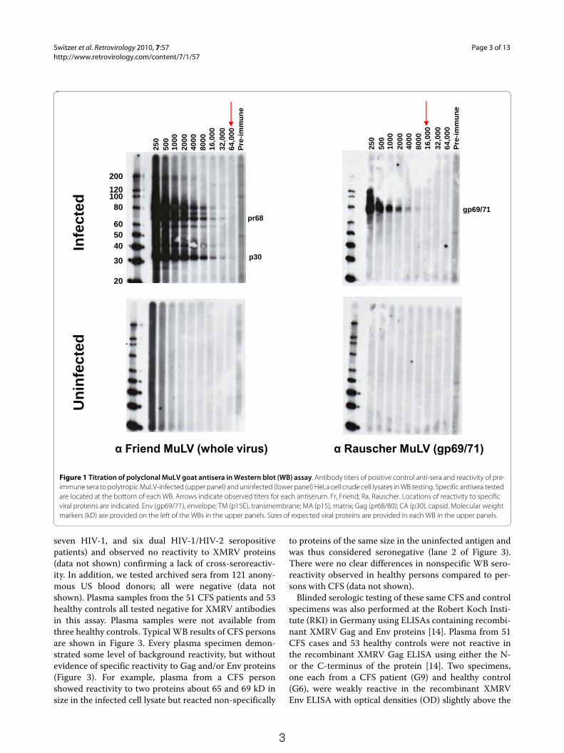
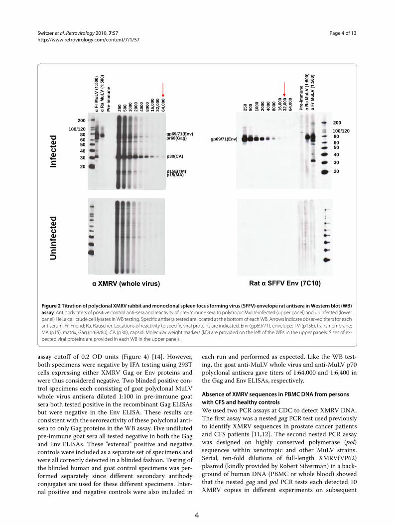
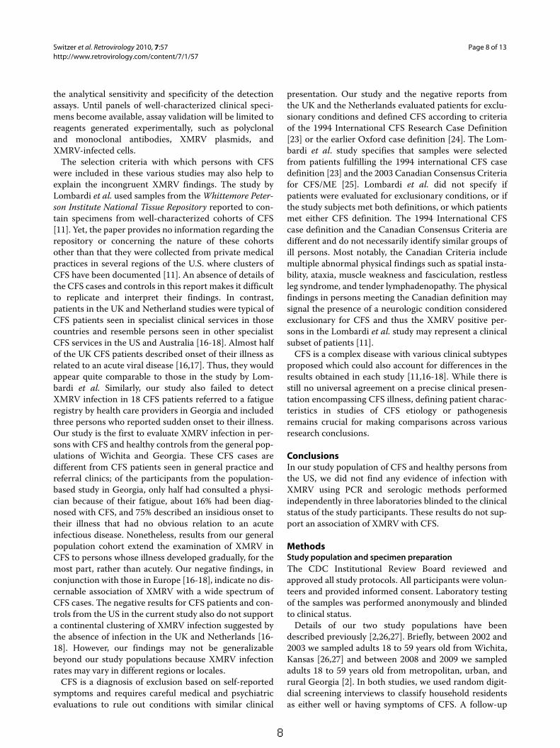
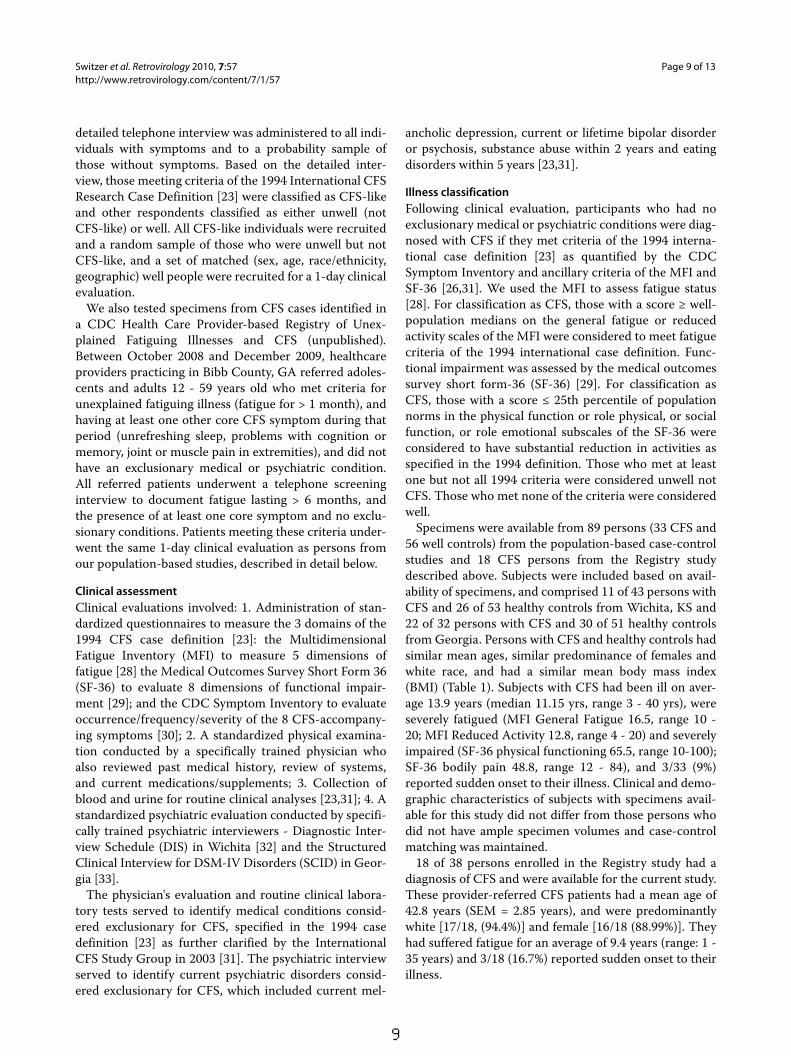
fully for assessing human infection with other zoonoticretroviruses [19,20]. The WB test used lysate from poly-tropic MuLV (PMLV)-infected HeLa cells as antigen.PMLV and XMRV are highly related. They share between87 and 93% nucleotide identity across the genome withXMRV and also have 88 - 97% and 88 - 91% amino acididentity to XMRV Gag and Env proteins, respectively.Partial Gag (123 aa) and Env (55 aa) sequences from ourpolytropic HeLa isolate share 96% and 90% identity toXMRV, respectively. Thus, excellent antigenic cross-reac-tivity between XMRV and our polytropic HeLa isolate isexpected. Specimens were tested for reactivity in parallelagainst control antigens from uninfected HeLa celllysates. Positive seroreactivity was defined as detection ofbands in the infected lysates corresponding to knownviral antigens and a lack of similar reactivity in uninfectedlysates to exclude nonspecific reactivity. Four availableantisera demonstrated good antigenic reactivity to Gagand/or Env proteins (Figures 1 and 2): Goat anti-MuLVpolyclonal antisera to whole virus and to p69/71 Env pro-teins, rabbit anti-XMRV polyclonal antiserum to wholevirus, and rat monoclonal antibody to the Env of spleenfocus forming virus (SFFV), a polytropic MuLV, thatreacts with gp69/71 Env of polytropic and xenotropicMuLV [21]. The anti-XMRV antiserum was used previ-ously to detect XMRV in prostate cancer tissues byimmunohistochemistry [13]. The anti-SFFV antibodywas used by Lombardi et al. in a flow-based antibodycompetition assay to detect antibodies to XMRV Env inCFS patients [11]. All positive control antisera were reac-tive at high titers to various Gag and/or Env proteins (Fig-ures 1 and 2). The anti-MuLV whole virus antiserum andthe anti-XMRV polyclonal antiserum both reacted to thep68/p80 Gag precursor and p30 Gag proteins at titers of1:32,000 and 1:64,000 respectively (Figures 1 and 2). Thepolyclonal anti-gp69/71 Env antiserum and the anti-SFFV monoclonal antibody reacted with the Env gp69/71doublet proteins (Figures. 1 and 2) at a titer of 1:8,000 and1:32,000, respectively (Figures. 1 and 2). The same pat-tern of reactivity was seen using both the anti-MuLVwhole virus and anti-XMRV antisera though a higherlevel of nonspecific reactivity was observed to the HeLalysates with the XMRV antisera (Figures 1 and 2). No spe-cific reactivity was observed for the pre-immune goatsera and to uninfected HeLa lysates (Figures 1 and 2).1:500 dilutions of the whole virus and gp69/71 antiseraand a 1:50 dilution of pre-immune goat sera were thenused as positive and negative controls for testing patientsamples in the WB assay, respectively.
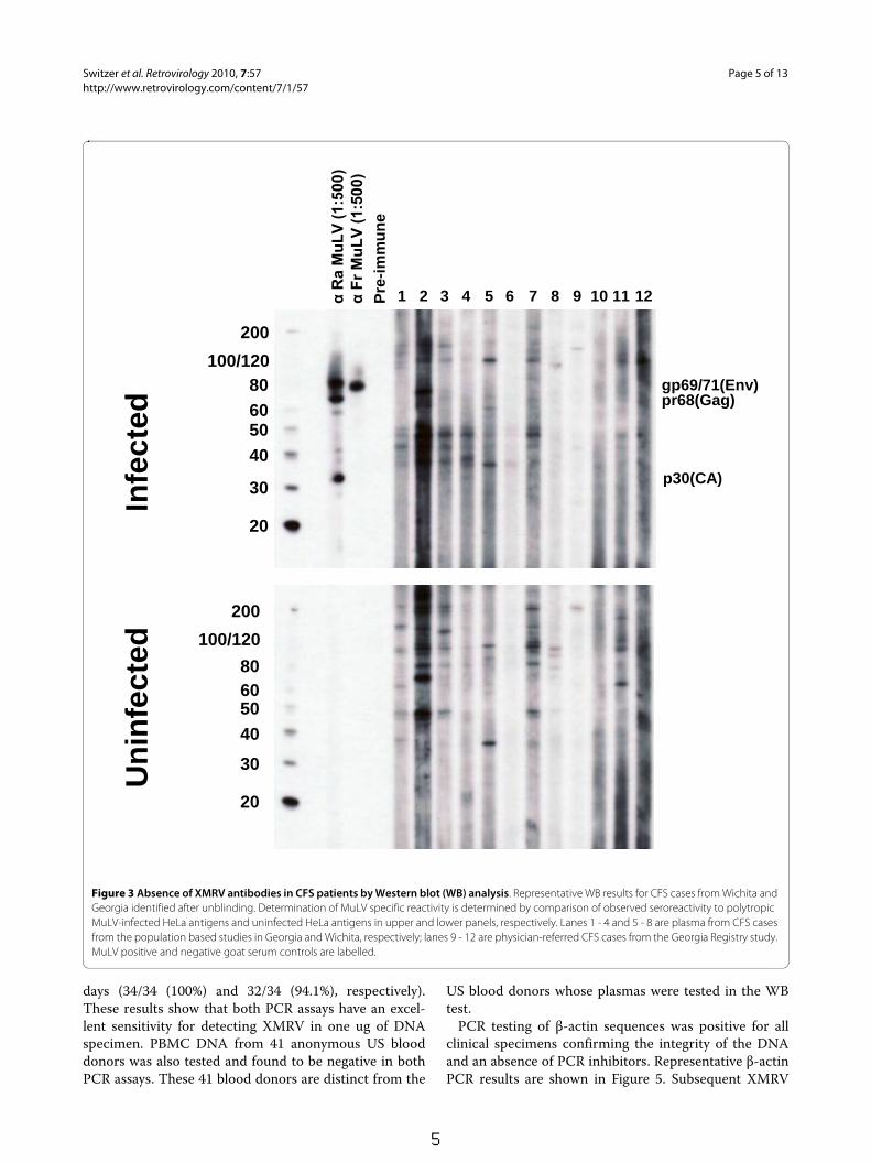
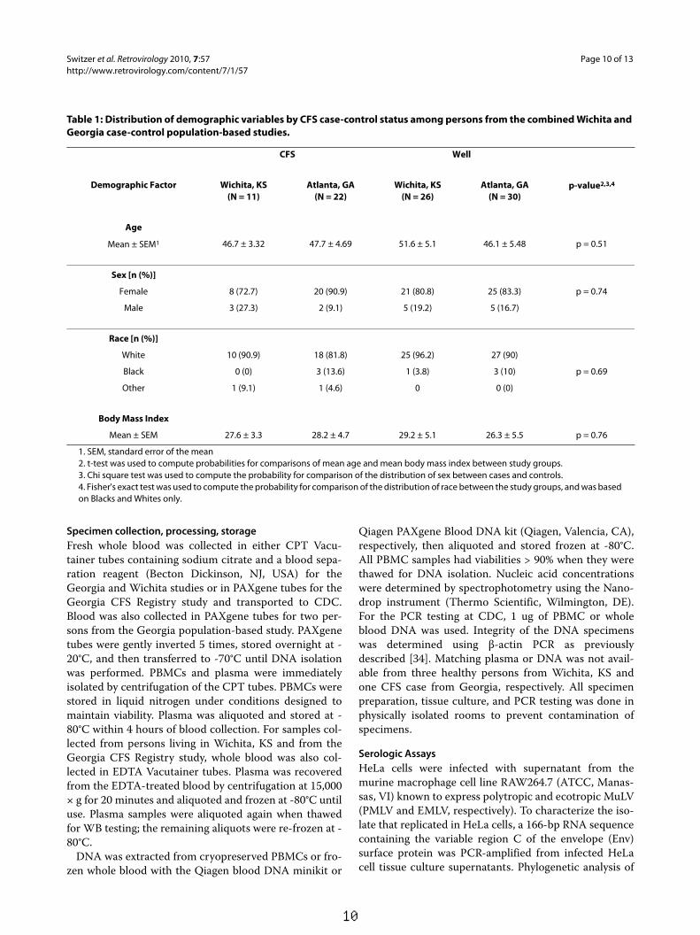
Plasma samples from 51 CFS cases and 53 healthy con-trols were diluted 1:50 and examined for seroreactivity tobands corresponding to Gag (p30 or p68/80) and/or Env(gp69/71 or p15E) proteins present in only the infectedlysate and not the uninfected lysate. We also tested serafrom 26 retrovirus-positive specimens (13 HTLV-1/2,
2
Switzer et al. Retrovirology 2010, 7:57http://www.retrovirology.com/content/7/1/57
Page 3 of 13
seven HIV-1, and six dual HIV-1/HIV-2 seropositivepatients) and observed no reactivity to XMRV proteins(data not shown) confirming a lack of cross-seroreactiv-ity. In addition, we tested archived sera from 121 anony-mous US blood donors; all were negative (data notshown). Plasma samples from the 51 CFS patients and 53healthy controls all tested negative for XMRV antibodiesin this assay. Plasma samples were not available fromthree healthy controls. Typical WB results of CFS personsare shown in Figure 3. Every plasma specimen demon-strated some level of background reactivity, but withoutevidence of specific reactivity to Gag and/or Env proteins(Figure 3). For example, plasma from a CFS personshowed reactivity to two proteins about 65 and 69 kD insize in the infected cell lysate but reacted non-specifically
to proteins of the same size in the uninfected antigen andwas thus considered seronegative (lane 2 of Figure 3).There were no clear differences in nonspecific WB sero-reactivity observed in healthy persons compared to per-sons with CFS (data not shown).
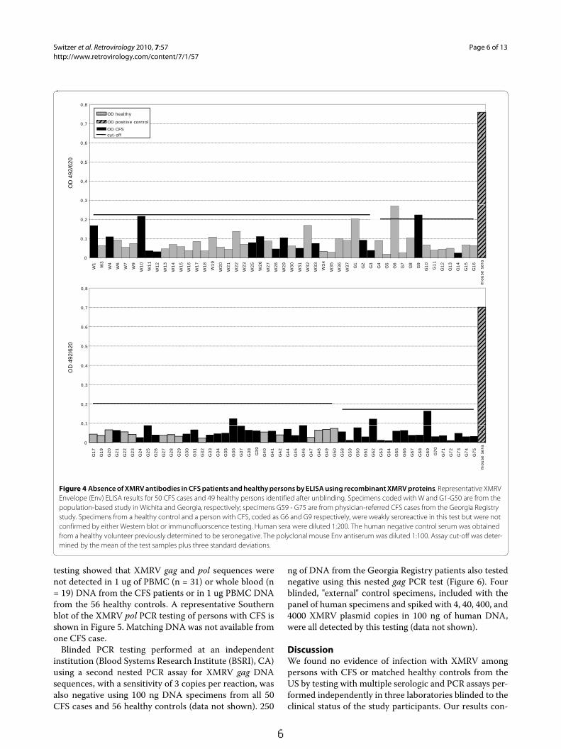
Blinded serologic testing of these same CFS and controlspecimens was also performed at the Robert Koch Insti-tute (RKI) in Germany using ELISAs containing recombi-nant XMRV Gag and Env proteins [14]. Plasma from 51CFS cases and 53 healthy controls were not reactive inthe recombinant XMRV Gag ELISA using either the N-or the C-terminus of the protein [14]. Two specimens,one each from a CFS patient (G9) and healthy control(G6), were weakly reactive in the recombinant XMRVEnv ELISA with optical densities (OD) slightly above the
Figure 1 Titration of polyclonal MuLV goat antisera in Western blot (WB) assay. Antibody titers of positive control anti-sera and reactivity of pre-immune sera to polytropic MuLV-infected (upper panel) and uninfected (lower panel) HeLa cell crude cell lysates in WB testing. Specific antisera tested are located at the bottom of each WB. Arrows indicate observed titers for each antiserum. Fr, Friend; Ra, Rauscher. Locations of reactivity to specific viral proteins are indicated. Env (gp69/71), envelope; TM (p15E), transmembrane; MA (p15), matrix; Gag (pr68/80); CA (p30), capsid. Molecular weight markers (kD) are provided on the left of the WBs in the upper panels. Sizes of expected viral proteins are provided in each WB in the upper panels.
Infe
cted
Un
infe
cted
α Rauscher MuLV (gp69/71)
250
500
1000
4000
2000
8000
16,0
0032
,000
64,0
00P
re-i
mm
un
e
gp69/71
10080
605040
30
20
α Friend MuLV (whole virus)
250
500
1000
4000
2000
8000
16,0
0032
,000
64,0
00P
re-i
mm
un
e
p30
pr68
120
200
3
Switzer et al. Retrovirology 2010, 7:57http://www.retrovirology.com/content/7/1/57
Page 4 of 13
assay cutoff of 0.2 OD units (Figure 4) [14]. However,both specimens were negative by IFA testing using 293Tcells expressing either XMRV Gag or Env proteins andwere thus considered negative. Two blinded positive con-trol specimens each consisting of goat polyclonal MuLVwhole virus antisera diluted 1:100 in pre-immune goatsera both tested positive in the recombinant Gag ELISAsbut were negative in the Env ELISA. These results areconsistent with the seroreactivity of these polyclonal anti-sera to only Gag proteins in the WB assay. Five undilutedpre-immune goat sera all tested negative in both the Gagand Env ELISAs. These "external" positive and negativecontrols were included as a separate set of specimens andwere all correctly detected in a blinded fashion. Testing ofthe blinded human and goat control specimens was per-formed separately since different secondary antibodyconjugates are used for these different specimens. Inter-nal positive and negative controls were also included in
each run and performed as expected. Like the WB test-ing, the goat anti-MuLV whole virus and anti-MuLV p70polyclonal antisera gave titers of 1:64,000 and 1:6,400 inthe Gag and Env ELISAs, respectively.
Absence of XMRV sequences in PBMC DNA from persons with CFS and healthy controlsWe used two PCR assays at CDC to detect XMRV DNA.The first assay was a nested gag PCR test used previouslyto identify XMRV sequences in prostate cancer patientsand CFS patients [11,12]. The second nested PCR assaywas designed on highly conserved polymerase (pol)sequences within xenotropic and other MuLV strains.Serial, ten-fold dilutions of full-length XMRV(VP62)plasmid (kindly provided by Robert Silverman) in a back-ground of human DNA (PBMC or whole blood) showedthat the nested gag and pol PCR tests each detected 10XMRV copies in different experiments on subsequent
Figure 2 Titration of polyclonal XMRV rabbit and monoclonal spleen focus forming virus (SFFV) envelope rat antisera in Western blot (WB) assay. Antibody titers of positive control anti-sera and reactivity of pre-immune sera to polytropic MuLV-infected (upper panel) and uninfected (lower panel) HeLa cell crude cell lysates in WB testing. Specific antisera tested are located at the bottom of each WB. Arrows indicate observed titers for each antiserum. Fr, Friend; Ra, Rauscher. Locations of reactivity to specific viral proteins are indicated. Env (gp69/71), envelope; TM (p15E), transmembrane; MA (p15), matrix; Gag (pr68/80); CA (p30), capsid. Molecular weight markers (kD) are provided on the left of the WBs in the upper panels. Sizes of ex-pected viral proteins are provided in each WB in the upper panels.
Infe
cted
Un
infe
cted
α Fr
MuL
V (1
:500
)
Pre
-im
mu
ne
α R
a M
uLV
(1:5
00)
250
500
1000
4000
2000
8000
16,0
0032
,000
64,0
00Rat α SFFV Env (7C10)
gp69/71(Env)
100/120806050
40
30
20
20025
050
010
00
4000
2000
8000
16,0
0032
,000
α Fr
MuL
V (1
:500
)
64,0
00
Pre
-im
mu
ne
α R
a M
uLV
(1:5
00)
100/1208060504030
20
α XMRV (whole virus)
p30(CA)
p15E(TM)p15(MA)
gp69/71(Env)pr68(Gag)
200
4
Switzer et al. Retrovirology 2010, 7:57http://www.retrovirology.com/content/7/1/57
Page 5 of 13
days (34/34 (100%) and 32/34 (94.1%), respectively).These results show that both PCR assays have an excel-lent sensitivity for detecting XMRV in one ug of DNAspecimen. PBMC DNA from 41 anonymous US blooddonors was also tested and found to be negative in bothPCR assays. These 41 blood donors are distinct from the
US blood donors whose plasmas were tested in the WBtest.
PCR testing of β-actin sequences was positive for allclinical specimens confirming the integrity of the DNAand an absence of PCR inhibitors. Representative β-actinPCR results are shown in Figure 5. Subsequent XMRV
Figure 3 Absence of XMRV antibodies in CFS patients by Western blot (WB) analysis. Representative WB results for CFS cases from Wichita and Georgia identified after unblinding. Determination of MuLV specific reactivity is determined by comparison of observed seroreactivity to polytropic MuLV-infected HeLa antigens and uninfected HeLa antigens in upper and lower panels, respectively. Lanes 1 - 4 and 5 - 8 are plasma from CFS cases from the population based studies in Georgia and Wichita, respectively; lanes 9 - 12 are physician-referred CFS cases from the Georgia Registry study. MuLV positive and negative goat serum controls are labelled.
Pre
-im
mu
ne
α R
a M
uLV
(1:5
00)
α Fr
MuL
V (1
:500
)
1 2 3 4 5 6 7 8 9 10 11 12
100/12080605040
30
200
20
100/120
80605040
30
200
20
p30(CA)
gp69/71(Env)pr68(Gag)
Infe
cted
Un
infe
cted
5
Switzer et al. Retrovirology 2010, 7:57http://www.retrovirology.com/content/7/1/57
Page 6 of 13
testing showed that XMRV gag and pol sequences werenot detected in 1 ug of PBMC (n = 31) or whole blood (n= 19) DNA from the CFS patients or in 1 ug PBMC DNAfrom the 56 healthy controls. A representative Southernblot of the XMRV pol PCR testing of persons with CFS isshown in Figure 5. Matching DNA was not available fromone CFS case.
Blinded PCR testing performed at an independentinstitution (Blood Systems Research Institute (BSRI), CA)using a second nested PCR assay for XMRV gag DNAsequences, with a sensitivity of 3 copies per reaction, wasalso negative using 100 ng DNA specimens from all 50CFS cases and 56 healthy controls (data not shown). 250
ng of DNA from the Georgia Registry patients also testednegative using this nested gag PCR test (Figure 6). Fourblinded, "external" control specimens, included with thepanel of human specimens and spiked with 4, 40, 400, and4000 XMRV plasmid copies in 100 ng of human DNA,were all detected by this testing (data not shown).
DiscussionWe found no evidence of infection with XMRV amongpersons with CFS or matched healthy controls from theUS by testing with multiple serologic and PCR assays per-formed independently in three laboratories blinded to theclinical status of the study participants. Our results con-
Figure 4 Absence of XMRV antibodies in CFS patients and healthy persons by ELISA using recombinant XMRV proteins. Representative XMRV Envelope (Env) ELISA results for 50 CFS cases and 49 healthy persons identified after unblinding. Specimens coded with W and G1-G50 are from the population-based study in Wichita and Georgia, respectively; specimens G59 - G75 are from physician-referred CFS cases from the Georgia Registry study. Specimens from a healthy control and a person with CFS, coded as G6 and G9 respectively, were weakly seroreactive in this test but were not confirmed by either Western blot or immunofluorscence testing. Human sera were diluted 1:200. The human negative control serum was obtained from a healthy volunteer previously determined to be seronegative. The polyclonal mouse Env antiserum was diluted 1:100. Assay cut-off was deter-mined by the mean of the test samples plus three standard deviations.
0,6
0,7
0,8
OD CFScut-off
OD healthy
OD positive control
0,3
0,4
0,5
OD
492
/620
0
0,1
0,2
0,7
0,8
W1
W3
W4
W6
W7
W9
W10
W11
W12
W13
W14
W15
W16
W17
W18
W19
W20
W21
W22
W23
W25
W26
W27
W28
W29
W30
W31
W32
W33
W34
W35
W36
W37
G1
G2
G3
G4
G5
G6
G7
G8
G9
G10
G11
G12
G13
G14
G15
G16
mouse
ser
a
0,4
0,5
0,6
OD
492
/620
0,1
0,2
0,3
O
0
,
G17
G19
G20
G21
G22
G23
G24
G25
G26
G27
G28
G29
G30
G31
G32
G33
G34
G35
G36
G37
G38
G39
G40
G41
G42
G44
G45
G46
G47
G48
G49
G50
G58
G59
G60
G61
G62
G63
G64
G65
G66
G67
G68
G69
G70
G71
G72
G73
G74
G75
mouse
ser
a
6

Switzer et al. Retrovirology 2010, 7:57http://www.retrovirology.com/content/7/1/57
Page 7 of 13
trast with the high rate of XMRV detection reported byLombardi et al. among both CFS patients and controls,but are in agreement with recent data reported in twolarge studies in the UK and a smaller study in the Nether-lands that could not detect XMRV sequences in CFSpatients and one UK study that also failed to detect spe-cific XMRV neutralizing antibody responses in CFS[11,16-18]. Combined, these negative data do not supportXMRV as the etiologic agent of the majority of CFS cases.
Several possibilities could explain these discordantresults, including technical differences in assays used forthe testing in each study. However, the inability of four
independent laboratories to replicate the high XMRVprevalence in CFS cases reported by Lombardi et al. can-not be explained by minor differences in assays used ineach study. In addition, testing at CDC utilized the nestedXMRV gag PCR assay used by Lombardi et al. and Uris-man et al. to identify XMRV infection in CFS and pros-tate cancer patients, respectively [11,12]. Further, toimprove assay sensitivity, we used 1 ug of input DNAwhich is 4-5 times higher than that used by others [11-13,16,17], all while maintaining an assay sensitivity of 10copies. To ensure that our testing would not miss geneti-cally diverse XMRV or MuLV strains, we also used a sen-sitive nested PCR assay with conserved pol gene primersand found that this testing was also negative confirmingthe absence of XMRV/MuLV sequences. While PBMCDNA was used in the majority of specimens, 1 ug wholeblood DNA was also used in testing 19 CFS cases. Thisinput DNA represents about 350 ng of PBMC DNAwhich is similar to the amount used by others [11-13,15,16], thus not affecting the sensitivity of our results.The negative PCR findings were confirmed by an inde-pendent laboratory with a second nested gag PCR assaywhich provided additional evidence for the absence ofXMRV sequences among CFS cases and controls. Theprimary PCR amplification used in this second test is alsothat used by Lombardi et al. which when combined witha nested PCR step has a 3-copy detection threshold.
Antibody responses particularly to Gag and Env pro-teins are hallmarks of immune responses to retroviralinfections including experimental XMRV infection ofmacaques [22]. We used a new WB assay to test for anti-XMRV antibodies and showed by using both monoclonalantibodies and polyclonal antisera that this assaydetected specifically, and with high titers, reactivity toboth XMRV and MuLV Gag and Env proteins. We wereunable to detect antibodies to XMRV Gag and Env in anyof the CFS and controls specimens by using this WBassay. Likewise, negative results were obtained in a sec-ond, independent laboratory by using XMRV-specificELISA-based and IFA assays. Thus, the observed negativeserologic results for all CFS patients reflect an absence ofantibody responses and active XMRV infection. Althoughlimited, the negative WB serology observed in 56 healthycontrols and 121 blood donors also suggests that theXMRV seroprevalence in this population is not high.Screening of larger numbers of US blood donors using ahigh throughput ELISA followed by confirmation in aWB test also showed uncommon seropositivity (~0.1%)[22]. More studies, however, are needed to determine theprevalence of XMRV in healthy populations.
One current limitation of our study, and of others per-forming serologic and PCR testing for XMRV, is theabsence of bona fide positive and negative control speci-mens from infected and uninfected humans to determine
Figure 5 Absence of XMRV polymerase (pol) sequences in CFS pa-tients. A. Representative nested pol PCR results using PBMC DNA spec-imens from persons with CFS identified after unblinding. Lanes 1 - 5, 6 - 10, and 11 - 14 are results for persons with CFS from Wichita, Georgia, and the Georgia registry studies, respectively; lanes 15 and 16, water only controls; lane 17, negative human PBMC DNA control; lanes 18 and 19, assay sensitivity controls consisting of 101 and 103 copies of XMRV VP62 plasmid DNA diluted in a background of 1 ug of human PBMC DNA, respectively. B. Semi-quantitative β-actin PCR results for PBMC DNA specimens above in lanes 1 - 14; lane 15, water control; lanes 16 - 19, 10-fold dilutions of blood donor PBMC DNA starting at 0.1 ug as a positive assay control.
1 2 3 4 5 6 7 8 9 10 11 12 13 14 15 16 17 18 19A.
1° PCR
2° PCR2° PCR
ß-actin
B.
ß actin
Figure 6 Absence of XMRV gag sequences in CFS patients. A. Rep-resentative nested gag PCR results from patients from the Georgia Registry identified after unblinding. Lanes 1 and 20, 100-bp ladder; lanes 2 - 15 are results from CFS patients; lanes 16 - 18 assay sensitivity controls consisting of 10, 3 and 1 copies of XMRV VP62 plasmid DNA diluted in a background of 250 ng of human PBMC DNA; lane 19, water control. B. GAPDH PCR results for same PBMC DNA specimens above.
A. 1 2 3 4 5 6 7 8 9 10 11 12 13 14 15 16 17 18 19 20
2° PCR
B.
GAPDH
7
Switzer et al. Retrovirology 2010, 7:57http://www.retrovirology.com/content/7/1/57
Page 8 of 13
the analytical sensitivity and specificity of the detectionassays. Until panels of well-characterized clinical speci-mens become available, assay validation will be limited toreagents generated experimentally, such as polyclonaland monoclonal antibodies, XMRV plasmids, andXMRV-infected cells.
The selection criteria with which persons with CFSwere included in these various studies may also help toexplain the incongruent XMRV findings. The study byLombardi et al. used samples from the Whittemore Peter-son Institute National Tissue Repository reported to con-tain specimens from well-characterized cohorts of CFS[11]. Yet, the paper provides no information regarding therepository or concerning the nature of these cohortsother than that they were collected from private medicalpractices in several regions of the U.S. where clusters ofCFS have been documented [11]. An absence of details ofthe CFS cases and controls in this report makes it difficultto replicate and interpret their findings. In contrast,patients in the UK and Netherland studies were typical ofCFS patients seen in specialist clinical services in thosecountries and resemble persons seen in other specialistCFS services in the US and Australia [16-18]. Almost halfof the UK CFS patients described onset of their illness asrelated to an acute viral disease [16,17]. Thus, they wouldappear quite comparable to those in the study by Lom-bardi et al. Similarly, our study also failed to detectXMRV infection in 18 CFS patients referred to a fatigueregistry by health care providers in Georgia and includedthree persons who reported sudden onset to their illness.Our study is the first to evaluate XMRV infection in per-sons with CFS and healthy controls from the general pop-ulations of Wichita and Georgia. These CFS cases aredifferent from CFS patients seen in general practice andreferral clinics; of the participants from the population-based study in Georgia, only half had consulted a physi-cian because of their fatigue, about 16% had been diag-nosed with CFS, and 75% described an insidious onset totheir illness that had no obvious relation to an acuteinfectious disease. Nonetheless, results from our generalpopulation cohort extend the examination of XMRV inCFS to persons whose illness developed gradually, for themost part, rather than acutely. Our negative findings, inconjunction with those in Europe [16-18], indicate no dis-cernable association of XMRV with a wide spectrum ofCFS cases. The negative results for CFS patients and con-trols from the US in the current study also do not supporta continental clustering of XMRV infection suggested bythe absence of infection in the UK and Netherlands [16-18]. However, our findings may not be generalizablebeyond our study populations because XMRV infectionrates may vary in different regions or locales.
CFS is a diagnosis of exclusion based on self-reportedsymptoms and requires careful medical and psychiatricevaluations to rule out conditions with similar clinical
presentation. Our study and the negative reports fromthe UK and the Netherlands evaluated patients for exclu-sionary conditions and defined CFS according to criteriaof the 1994 International CFS Research Case Definition[23] or the earlier Oxford case definition [24]. The Lom-bardi et al. study specifies that samples were selectedfrom patients fulfilling the 1994 international CFS casedefinition [23] and the 2003 Canadian Consensus Criteriafor CFS/ME [25]. Lombardi et al. did not specify ifpatients were evaluated for exclusionary conditions, or ifthe study subjects met both definitions, or which patientsmet either CFS definition. The 1994 International CFScase definition and the Canadian Consensus Criteria aredifferent and do not necessarily identify similar groups ofill persons. Most notably, the Canadian Criteria includemultiple abnormal physical findings such as spatial insta-bility, ataxia, muscle weakness and fasciculation, restlessleg syndrome, and tender lymphadenopathy. The physicalfindings in persons meeting the Canadian definition maysignal the presence of a neurologic condition consideredexclusionary for CFS and thus the XMRV positive per-sons in the Lombardi et al. study may represent a clinicalsubset of patients [11].
CFS is a complex disease with various clinical subtypesproposed which could also account for differences in theresults obtained in each study [11,16-18]. While there isstill no universal agreement on a precise clinical presen-tation encompassing CFS illness, defining patient charac-teristics in studies of CFS etiology or pathogenesisremains crucial for making comparisons across variousresearch conclusions.
ConclusionsIn our study population of CFS and healthy persons fromthe US, we did not find any evidence of infection withXMRV using PCR and serologic methods performedindependently in three laboratories blinded to the clinicalstatus of the study participants. These results do not sup-port an association of XMRV with CFS.
MethodsStudy population and specimen preparationThe CDC Institutional Review Board reviewed andapproved all study protocols. All participants were volun-teers and provided informed consent. Laboratory testingof the samples was performed anonymously and blindedto clinical status.
Details of our two study populations have beendescribed previously [2,26,27]. Briefly, between 2002 and2003 we sampled adults 18 to 59 years old from Wichita,Kansas [26,27] and between 2008 and 2009 we sampledadults 18 to 59 years old from metropolitan, urban, andrural Georgia [2]. In both studies, we used random digit-dial screening interviews to classify household residentsas either well or having symptoms of CFS. A follow-up
8
Switzer et al. Retrovirology 2010, 7:57http://www.retrovirology.com/content/7/1/57
Page 9 of 13
detailed telephone interview was administered to all indi-viduals with symptoms and to a probability sample ofthose without symptoms. Based on the detailed inter-view, those meeting criteria of the 1994 International CFSResearch Case Definition [23] were classified as CFS-likeand other respondents classified as either unwell (notCFS-like) or well. All CFS-like individuals were recruitedand a random sample of those who were unwell but notCFS-like, and a set of matched (sex, age, race/ethnicity,geographic) well people were recruited for a 1-day clinicalevaluation.
We also tested specimens from CFS cases identified ina CDC Health Care Provider-based Registry of Unex-plained Fatiguing Illnesses and CFS (unpublished).Between October 2008 and December 2009, healthcareproviders practicing in Bibb County, GA referred adoles-cents and adults 12 - 59 years old who met criteria forunexplained fatiguing illness (fatigue for > 1 month), andhaving at least one other core CFS symptom during thatperiod (unrefreshing sleep, problems with cognition ormemory, joint or muscle pain in extremities), and did nothave an exclusionary medical or psychiatric condition.All referred patients underwent a telephone screeninginterview to document fatigue lasting > 6 months, andthe presence of at least one core symptom and no exclu-sionary conditions. Patients meeting these criteria under-went the same 1-day clinical evaluation as persons fromour population-based studies, described in detail below.
Clinical assessmentClinical evaluations involved: 1. Administration of stan-dardized questionnaires to measure the 3 domains of the1994 CFS case definition [23]: the MultidimensionalFatigue Inventory (MFI) to measure 5 dimensions offatigue [28] the Medical Outcomes Survey Short Form 36(SF-36) to evaluate 8 dimensions of functional impair-ment [29]; and the CDC Symptom Inventory to evaluateoccurrence/frequency/severity of the 8 CFS-accompany-ing symptoms [30]; 2. A standardized physical examina-tion conducted by a specifically trained physician whoalso reviewed past medical history, review of systems,and current medications/supplements; 3. Collection ofblood and urine for routine clinical analyses [23,31]; 4. Astandardized psychiatric evaluation conducted by specifi-cally trained psychiatric interviewers - Diagnostic Inter-view Schedule (DIS) in Wichita [32] and the StructuredClinical Interview for DSM-IV Disorders (SCID) in Geor-gia [33].
The physician's evaluation and routine clinical labora-tory tests served to identify medical conditions consid-ered exclusionary for CFS, specified in the 1994 casedefinition [23] as further clarified by the InternationalCFS Study Group in 2003 [31]. The psychiatric interviewserved to identify current psychiatric disorders consid-ered exclusionary for CFS, which included current mel-
ancholic depression, current or lifetime bipolar disorderor psychosis, substance abuse within 2 years and eatingdisorders within 5 years [23,31].
Illness classificationFollowing clinical evaluation, participants who had noexclusionary medical or psychiatric conditions were diag-nosed with CFS if they met criteria of the 1994 interna-tional case definition [23] as quantified by the CDCSymptom Inventory and ancillary criteria of the MFI andSF-36 [26,31]. We used the MFI to assess fatigue status[28]. For classification as CFS, those with a score ≥ well-population medians on the general fatigue or reducedactivity scales of the MFI were considered to meet fatiguecriteria of the 1994 international case definition. Func-tional impairment was assessed by the medical outcomessurvey short form-36 (SF-36) [29]. For classification asCFS, those with a score ≤ 25th percentile of populationnorms in the physical function or role physical, or socialfunction, or role emotional subscales of the SF-36 wereconsidered to have substantial reduction in activities asspecified in the 1994 definition. Those who met at leastone but not all 1994 criteria were considered unwell notCFS. Those who met none of the criteria were consideredwell.
Specimens were available from 89 persons (33 CFS and56 well controls) from the population-based case-controlstudies and 18 CFS persons from the Registry studydescribed above. Subjects were included based on avail-ability of specimens, and comprised 11 of 43 persons withCFS and 26 of 53 healthy controls from Wichita, KS and22 of 32 persons with CFS and 30 of 51 healthy controlsfrom Georgia. Persons with CFS and healthy controls hadsimilar mean ages, similar predominance of females andwhite race, and had a similar mean body mass index(BMI) (Table 1). Subjects with CFS had been ill on aver-age 13.9 years (median 11.15 yrs, range 3 - 40 yrs), wereseverely fatigued (MFI General Fatigue 16.5, range 10 -20; MFI Reduced Activity 12.8, range 4 - 20) and severelyimpaired (SF-36 physical functioning 65.5, range 10-100);SF-36 bodily pain 48.8, range 12 - 84), and 3/33 (9%)reported sudden onset to their illness. Clinical and demo-graphic characteristics of subjects with specimens avail-able for this study did not differ from those persons whodid not have ample specimen volumes and case-controlmatching was maintained.
18 of 38 persons enrolled in the Registry study had adiagnosis of CFS and were available for the current study.These provider-referred CFS patients had a mean age of42.8 years (SEM = 2.85 years), and were predominantlywhite [17/18, (94.4%)] and female [16/18 (88.99%)]. Theyhad suffered fatigue for an average of 9.4 years (range: 1 -35 years) and 3/18 (16.7%) reported sudden onset to theirillness.
9
Switzer et al. Retrovirology 2010, 7:57http://www.retrovirology.com/content/7/1/57
Page 10 of 13
Specimen collection, processing, storageFresh whole blood was collected in either CPT Vacu-tainer tubes containing sodium citrate and a blood sepa-ration reagent (Becton Dickinson, NJ, USA) for theGeorgia and Wichita studies or in PAXgene tubes for theGeorgia CFS Registry study and transported to CDC.Blood was also collected in PAXgene tubes for two per-sons from the Georgia population-based study. PAXgenetubes were gently inverted 5 times, stored overnight at -20°C, and then transferred to -70°C until DNA isolationwas performed. PBMCs and plasma were immediatelyisolated by centrifugation of the CPT tubes. PBMCs werestored in liquid nitrogen under conditions designed tomaintain viability. Plasma was aliquoted and stored at -80°C within 4 hours of blood collection. For samples col-lected from persons living in Wichita, KS and from theGeorgia CFS Registry study, whole blood was also col-lected in EDTA Vacutainer tubes. Plasma was recoveredfrom the EDTA-treated blood by centrifugation at 15,000× g for 20 minutes and aliquoted and frozen at -80°C untiluse. Plasma samples were aliquoted again when thawedfor WB testing; the remaining aliquots were re-frozen at -80°C.
DNA was extracted from cryopreserved PBMCs or fro-zen whole blood with the Qiagen blood DNA minikit or
Qiagen PAXgene Blood DNA kit (Qiagen, Valencia, CA),respectively, then aliquoted and stored frozen at -80°C.All PBMC samples had viabilities > 90% when they werethawed for DNA isolation. Nucleic acid concentrationswere determined by spectrophotometry using the Nano-drop instrument (Thermo Scientific, Wilmington, DE).For the PCR testing at CDC, 1 ug of PBMC or wholeblood DNA was used. Integrity of the DNA specimenswas determined using β-actin PCR as previouslydescribed [34]. Matching plasma or DNA was not avail-able from three healthy persons from Wichita, KS andone CFS case from Georgia, respectively. All specimenpreparation, tissue culture, and PCR testing was done inphysically isolated rooms to prevent contamination ofspecimens.
Serologic AssaysHeLa cells were infected with supernatant from themurine macrophage cell line RAW264.7 (ATCC, Manas-sas, VI) known to express polytropic and ecotropic MuLV(PMLV and EMLV, respectively). To characterize the iso-late that replicated in HeLa cells, a 166-bp RNA sequencecontaining the variable region C of the envelope (Env)surface protein was PCR-amplified from infected HeLacell tissue culture supernatants. Phylogenetic analysis of
Table 1: Distribution of demographic variables by CFS case-control status among persons from the combined Wichita and Georgia case-control population-based studies.
CFS Well
Demographic Factor Wichita, KS(N = 11)
Atlanta, GA(N = 22)
Wichita, KS(N = 26)
Atlanta, GA(N = 30)
p-value2,3,4
Age
Mean ± SEM1 46.7 ± 3.32 47.7 ± 4.69 51.6 ± 5.1 46.1 ± 5.48 p = 0.51
Sex [n (%)]
Female 8 (72.7) 20 (90.9) 21 (80.8) 25 (83.3) p = 0.74
Male 3 (27.3) 2 (9.1) 5 (19.2) 5 (16.7)
Race [n (%)]
White 10 (90.9) 18 (81.8) 25 (96.2) 27 (90)
Black 0 (0) 3 (13.6) 1 (3.8) 3 (10) p = 0.69
Other 1 (9.1) 1 (4.6) 0 0 (0)
Body Mass Index
Mean ± SEM 27.6 ± 3.3 28.2 ± 4.7 29.2 ± 5.1 26.3 ± 5.5 p = 0.76
1. SEM, standard error of the mean2. t-test was used to compute probabilities for comparisons of mean age and mean body mass index between study groups.3. Chi square test was used to compute the probability for comparison of the distribution of sex between cases and controls.4. Fisher's exact test was used to compute the probability for comparison of the distribution of race between the study groups, and was based on Blacks and Whites only.
10
Switzer et al. Retrovirology 2010, 7:57http://www.retrovirology.com/content/7/1/57
Page 11 of 13
the env sequence showed that the isolate was a PMLV byclustering tightly with other PMLV, and not EMLV (datanot shown). XMRV and PMLV are highly related sharingbetween 87 - 94% nucleotide identity across theirgenomes and 88 - 97% and 88 - 91% amino acid identityto complete Gag and Env proteins, respectively. Indeed,partial Gag (123 aa) and Env (55 aa) sequences from ourpolytropic HeLa isolate share 96% and 90% identity toXMRV, respectively. Thus, the high amino acid related-ness supports the use of this isolate for WB serologic test-ing. Infected and uninfected HeLa crude cell lysates wereprepared for WB testing as previously described [35].Protein concentrations of the lysates were determinedusing the BioRad DC Protein Assay (Hercules, CA).Plasma or serum samples were diluted 1:50 and reactedseparately to 150 ug of infected and uninfected cell lysatesovernight at 4°C after protein separation through 4-12%polyacrylamide gels and transfer to nytran membranes,as previously described [35,36]. Seroreactivity in humanspecimens was detected using peroxidase-conjugatedprotein A/G (Pierce, Rockford, IL) and chemilumines-cence (Amersham, Uppsala, Sweden) [35,36].
Since validated XMRV-positive human sera are notcurrently available, we used experimentally derived poly-clonal antisera and monoclonal antibodies to assess anti-genic reactivity of the WB assay. These reagents includedgoat polyclonal antisera to MuLV (whole virus and gp69/71Env, respectively) available at ATCC (VR-1537 and VR-1521, respectively), and a rabbit anti-XMRV polyclonalantiserum (kindly provided by Ila Singh) and a rat anti-SFFV (7C10) monoclonal antibody (kindly provided bySandra Ruscetti) used previously to detect XMRV proteinexpression and antibodies in prostate cancer and CFSpatients, respectively [11,13,21]. Peroxidase-conjugatedprotein A/G or anti-rat antibody (Sigma, St. Louis, MS)was used to detect bound goat, rabbit, and rat antibodies,respectively. Sensitivity of the assay was estimated usingtwo fold serial dilutions of the MuLV, XMRV, and SFFVpolyclonal and monoclonal antibodies. Cross-reactivityof the WB assay on HIV and HTLV positive plasma wasevaluated on 13 HTLV-1/2 positive, 7 HIV-1-positive,and six HIV-1/HIV-2 dual positive plasma. In addition,sera from 121 HIV and HTLV seronegative anonymousUS blood donors collected in 1998 were tested.
An aliquot of coded plasma from the CFS and healthycontrols was tested at RKI by an ELISA using recombi-nant Gag and Env proteins used recently to investigateXMRV infection in German prostate cancer patients [14].Briefly, recombinant proteins were coated overnight onmicrotiter plates at room temperature in equimolaramounts. The plates were blocked with 2% Marvel milkpowder in phosphate buffered saline (PBS) for 2 h at37°C, washed three times with PBS, 0.05% Tween 20.Patient plasma diluted 1:200 in PBS with 2% milk powder
and 0.05% Tween20 were added into each well and incu-bated for 1 hour at 37°C. Each well was again washedthree times and a 1:1000 dilution of a goat anti-humanIgG-HRP conjugate (Sigma Aldrich, Munich, Germany)in PBS, 2% milk powder, 0.05% Tween 20 (Serva, Heidel-berg, Germany) was added. Following incubation for 1hour at 37°C, each well was again washed three times,and chromogen ortho-phenylendiamin (OPD) in 0.05 Mphosphate-citrate buffer, pH 5.0 containing 4 μl of a 30%solution of the hydrogen peroxide substrate per 10 mlwas added. After 5-10 minutes the color developmentwas stopped by addition of sulphuric acid and the absor-bance at 492 nm/620 nm was measured in a microplatereader. Positive controls included mouse anti-Gag andEnv antisera and pre-immune sera diluted 1:50 in PBSwith 2% milk powder and 0.05% Tween20. In addition, aseparate set of goat sera was also tested in a blinded fash-ion and included external positive and negative controlsconsisting of dilutions of the MuLV whole virus, gp69/71goat polyclonal antisera, or pre-immune goat sera,respectively. Detection of antibody reactivity in the goatsera was done by using rabbit anti-goat HRP conjugate(Dako, Hamburg, Germany).
Samples reactive by ELISA testing were then re-testedusing an immunofluorescence assay (IFA) [14]. Briefly,plasma specimens were diluted 1:200 in blocking bufferand tested against 293T cells expressing codon optimizedsynthetic full-length genes of the XMRV env or gag undercontrol of the CMV promoter and bound to glass slides,as described in detail previously [14]. Following incuba-tion for 60 min at 37°C, the slides were washed exten-sively with PBS and secondary antibodies conjugated tofluorophores were added for 30 min. After thoroughwashing steps with PBS, the cells were mounted in Mow-iol and viewed on a Zeiss (LSM510) confocal laser-scan-ning microscope.
Detection of XMRV sequencesDNA specimens were screened by PCR at the CDC withan XMRV-specific gag and a polymerase (pol) assay thatdetects xenotropic and polytropic MuLV. The XMRVspecific assay uses the primers GAG-O-F and GAG-O-Rand GAG-I-F and GAG-I-R for the primary and nestedPCRs, respectively, and conditions as previouslydescribed [11,12]. This is the same nested PCR test usedby Urisman et al. and Lombardi et al. to detect 413-bpXMRV gag sequences in prostate cancer and CFSpatients, respectively [11,12]. The primers and probes ofthe generic pol PCR assay were designed from an align-ment of complete XMRV and prototypical xenotropic,polytropic, and ecotropic MuLV genomes available atGenBank (accession numbers: xenotropic (XMLV):XMRV VP35 = DQ241301, XMRV VP62 = DQ399707,XMRV VP42 = DQ241302, XMRV WPI-1106 =
11
Switzer et al. Retrovirology 2010, 7:57http://www.retrovirology.com/content/7/1/57
Page 12 of 13
GQ497344, XMRV WPI-1178 = GC497343, MuLV DG-75 = AF221065; MuLV MTCR = NC_001702, mERV Chr9 = AC121813, mERV Chr 4 = AL627077, mERV Chr 1 =AC083892; polytropic (PMLV): mERV Chr 7 =AC167978, mERV Chr 7 = AC127565, mERV Chr 12 =AC153658; ecotropic (EMLV): MuLV AKV = J01998,MuLV BM5eco = AY252102.1, Moloney MuLV = J02255,Rauscher MuLV = NC_001819, Friend MuLV = X02794).The external external XPOLOF (5' CCG TGC CCA ACCCTT ACA ACC TCT 3') and XPOLOR (5' CCG AGGTTC CCT AGG GTT TGT AAT 3') and internal primersXPOLIF (5' TCC ACC CCA CCA GTC AGC CTC TCT3') and XPOLIR (5' AAG TGG CGG CCA GCA GTAAGT CAT 3') were used to generically detect 216-bpXMLV/XMRV pol sequences. All assays were optimizedto achieve the highest sensitivity in detecting XMRVVP62 plasmid DNA in one ug of genomic DNA. One ugof human DNA was used as input for the PCR tests. PCRproducts were visualized by electrophoresis in an ethid-ium bromide-stained 1.8% agarose gel. To furtherincrease the sensitivity and specificity of the PCR assays,amplified gag and pol sequences were confirmed bySouthern blot analysis using the biotinylated oligoprobesXGAGP2 (5' ACC TTG CAG CAC TGG GGA GAT GTC3'), and XPOLP (5' TTG ATG AGG CAC TGC ACAGAG ACC 3') and chemiluminescence detection. Thedetection limit of the assays was evaluated using 10-folddilutions of XMRV VP62 plasmid diluted in a back-ground of one ug of genomic human DNA. Assay speci-ficity was evaluated using PBMC DNA from 41anonymous US blood donors screened negative for HIVand HTLV.
Nested PCR was also performed at BSRI using doubleblinded genomic DNA specimens in order to indepen-dently test for XMRV gag sequences. The first round wasperformed as previously described to detect XMRV inPBMC DNA of CFS patients [11]. Briefly, 100 - 250 ng ofgenomic DNA was amplified using outer gag primers419F (5' ATC AGT TAA CCT ACC CGA GTC GGA C 3')and 1154R (5' GCC GCC TCT TCT TCA TTG TTC TC3') at a final concentration of 0.3 μM, HotStart-IT Fideli-Taq Master Mix (USB Corporation, Cleveland, OH) and 1mM magnesium chloride. PCR was performed using aninitial denaturation step at 94°C for 4 minutes followed by45 cycles of 94°C for 30 seconds, 57°C for 30 seconds and72°C for 1 minute and a final extension step at 72°C for 2minutes. Nested PCR was conducted using 1 μl of thefirst round DNA in the second round reaction. Nestedprimers 488F (5' GGG GAC GAG AGA CAG AGA CA3') and 1107R (5' CAG AGG AGG AAG GTT GTG CT 3')were used at a final concentration of 0.3 μM and amplifi-cation was performed using HotStart-IT FideliTaq. PCR
was performed using an initial denaturation step at 95°Cfor 90 seconds followed by 40 cycles of 95°C for 20 sec-onds, 58°C for 30 seconds and 72°C for 40 seconds and afinal extension step at 72°C for 2 minutes. PCR contami-nation occurring during nested PCR was evaluated byincluding at least one third as many water controls as testsamples in each PCR experiment and were always nega-tive.
Using serial dilutions of a cloned fragment of XMRVgag as a positive control, the nested PCR assay could reli-ably detect at least 3 copies of DNA per reaction, evenwhen spiked into genomic DNA prepared either from293FT cells or donor PBMCs previously validated to benegative for XMRV. Controls of GAPDH (forward - 5'CAT GTT CCA ATA TGA TTC AC 3'; reverse - 5' CCTGGA AGA TGG TGA TG 3'; 75 ng genomic DNA, 3 min-utes at 95°C followed by 45 cycles of 95°C for 20 seconds,55°C for 45 seconds and 72°C for 30 seconds, followed by1 cycle of 72°C for 2 minutes) were performed to ensuresimilar levels of genomic DNA input in each PCR reac-tion.
Competing interestsThe authors declare that they have no competing interests.
Authors' contributionsWMS, WCR, RMH and WH conceived and designed the study. WCR and VRFprovided specimens and data on study population. HJ, HZ, ST, AS, GS, NB, andOH performed specimen testing and data analysis with WMS and WH. WMS,WCR and WH wrote the manuscript. All authors read and approved the finalmanuscript.
AcknowledgementsWe are grateful to Dr. Robert Silverman at the Cleveland Clinic for the VP62 XMRV plasmid, Dr. Ila Singh at the University of Utah for the rabbit anti-XMRV polyclonal sera, and Dr. Sandra Ruscetti at the National Cancer Institute for the rat anti-SFFV (7C10) monoclonal antibody. We also thank Ben Capon and Beth Slikas for development of the gag PCR assay used at the Blood Systems Research Institute. Use of trade names is for identification only and does not imply endorsement by the U.S. Department of Health and Human Services, the Public Health Service, or the Centers for Disease Control and Prevention. The findings and conclusions in this report are those of the authors and do not necessarily represent the views of the Centers for Disease Control and Preven-tion.
Author Details1Laboratory Branch, Division of HIV/AIDS Prevention, National Center for HIV/AIDS, Viral Hepatitis, STD, and TB Prevention, Centers for Disease Control and Prevention, Atlanta, GA 30333, USA, 2Robert Koch-Institute, Centre for Biological Safety 4, Nordufer 20, 13353 Berlin, Germany, 3Blood Systems Research Institute and Department of Laboratory Medicine, UCSF, 270 Masonic Ave., San Francisco, CA 94118, USA and 4Chronic Viral Diseases Branch, Division of Viral and Rickettsial Diseases, National Center for Zoonotic, Vector-Borne and Enteric Diseases, Centers for Disease Control and Prevention, Atlanta, GA 30333, USA
Received: 26 March 2010 Accepted: 1 July 2010 Published: 1 July 2010This article is available from: http://www.retrovirology.com/content/7/1/57© 2010 Switzer et al; licensee BioMed Central Ltd. This is an Open Access article distributed under the terms of the Creative Commons Attribution License (http://creativecommons.org/licenses/by/2.0), which permits unrestricted use, distribution, and reproduction in any medium, provided the original work is properly cited.Retrovirology 2010, 7:57
12
Switzer et al. Retrovirology 2010, 7:57http://www.retrovirology.com/content/7/1/57
Page 13 of 13
References1. Jason LA, Richman JA, Rademaker AW, Jordan KM, Plioplys AV, Taylor RR,
McCready W, Huang CF, Plioplys S: A community-based study of chronic fatigue syndrome. Archives of Internal Medicine 1999, 159:2129-2137.
2. Reeves WC, Jones JF, Maloney E, Heim C, Hoaglin DC, Boneva RS, Morrissey M, Devlin R: Prevalence of chronic fatigue syndrome in metropolitan, urban, and rural Georgia. Popul Health Metr 2007, 8:5.
3. Devanur LD, Kerr JR: Chronic fatigue syndrome. Journal of Clinical Virology 2006, 37:139-150.
4. Jason LA, Benton MC, Valentine L, Johnson A, Torres-Harding S: The economic impact of ME/CFS: individuals and societal level costs. Dynamic Medicine 2008, 7:6.
5. Reynolds KJ, Vernon SD, Bouchery E, Reeves WC: The economic impact of chronic fatigue syndrome. Cost Eff Resour Alloc 2004, 2:4.
6. Solomon L, Nisenbaum R, Reyes M, Papanicolaou DA, Unger ER, Reeves WC: Functional status of persons with chronic fatigue syndrome in the Wichita population. Health Qual Life Outcomes 2003, 1:48.
7. Afari N, Buchwald D: Chronic fatigue syndrome: a review. Am J Psychiatry 2003, 160:221-236.
8. DeFreitas E, Hilliard B, Cheney PR, Bell DS, Kiggundu E, Sankey D, Wroblewska Z, Palladino M, Woodward JP, Koprowski H: Retroviral sequences related to human T-lymphotropic virus type II in patients with chronic fatigue immune dysfunction syndrome. Proc Natl Acad Sci USA 1991, 88:2922-2926.
9. Heneine W, Woods TC, Sinha SD, Khan AS, Chapman LE, Schonberger LB, Folks TM: Lack of evidence for infection with known human and animal retroviruses in patients with chronic fatigue syndrome. Clin Infect Dis 1994, 18(Suppl 1):S121-125.
10. Khan AS, Heneine WM, Chapman LE, Gary HE, Woods TC Jr, Folks TM, Schonberger LB: Assessment of a retrovirus sequence and other possible risk factors for the chronic fatigue syndrome in adults. Ann Intern Med 1993, 118:241-245.
11. Lombardi VC, Ruscetti FW, Das Gupta J, Pfost MA, Hagen KS, Peterson DL, Ruscetti SK, Bagni RK, Petrow-Sadowski C, Gold B, Dean M, Silverman RH, Mikovits JA: Detection of an infectious retrovirus, XMRV, in blood cells of patients with chronic fatigue syndrome. Science 2009, 326:585-589.
12. Urisman A, Molinaro RJ, Fischer N, Plummer SJ, Casey G, Klein EA, Malathi K, Magi-Galluzzi C, Tubbs RR, Ganem D, Silverman RH, DeRisi JL: Identification of a novel Gammaretrovirus in prostate tumors of patients homozygous for R462Q RNASEL variant. PLoS Pathog 2006, 2:e25.
13. Schlaberg R, Choe DJ, Brown KR, Thaker HM, Singh IR: XMRV is present in malignant prostatic epithelium and is associated with prostate cancer, especially high-grade tumors. Proceedings of the National Academy of Sciences of the United States of America 2009, 106:16351-16356.
14. Hohn O, Krause H, Barbarotto P, Niederstadt L, Beimforde N, Denner J, Miller K, Kurth R, Bannert N: Lack of evidence for xenotropic murine leukemia virus-related virus(XMRV) in German prostate cancer patients. Retrovirology 2009, 6:92.
15. Fischer N, Hellwinkel O, Schulz C, Chun FK, Huland H, Aepfelbacher M, Schlomm T: Prevalence of human gammaretrovirus XMRV in sporadic prostate cancer. J Clin Virol 2008, 43:277-283.
16. Erlwein O, Kaye S, McClure MO, Weber J, Wills G, Collier D, Wessely S, Cleare A: Failure to detect the novel retrovirus XMRV in chronic fatigue syndrome. PLoS One 2010, 5:e8519.
17. Groom HC, Boucherit VC, Makinson K, Randal E, Baptista S, Hagan S, Gow JW, Mattes FM, Breuer J, Kerr JR, Stoye JP, Bishop KN: Absence of xenotropic murine leukaemia virus-related virus in UK patients with chronic fatigue syndrome. Retrovirology 2010, 7:10.
18. van Kuppeveld FJ, Jong AS, Lanke KH, Verhaegh GW, Melchers WJ, Swanink CM, Bleijenberg G, Netea MG, Galama JM, van der Meer JW: Prevalence of xenotropic murine leukaemia virus-related virus in patients with chronic fatigue syndrome in the Netherlands: retrospective analysis of samples from an established cohort. BMJ 2010, 340:c1018.
19. Paradis K, Langford G, Long ZF, Heneine W, Sandstrom P, Switzer WM, Chapman LE, Lockey C, Onions D, Otto E, Grp XS: Search for cross-species transmission of porcine endogenous retrovirus in patients treated with living pig tissue. Science 1999, 285:1236-1241.
20. Switzer WM, Bhullar V, Shanmugam V, Cong ME, Parekh B, Lerche NW, Yee JL, Ely JJ, Boneva R, Chapman LE, Folks TM, Heneine W: Frequent simian
foamy virus infection in persons occupationally exposed to nonhuman primates. J Virol 2004, 78:2780-2789.
21. Wolff L, Koller R, Ruscetti S: Monoclonal antibody to spleen focus-forming virus-encoded gp52 provides a probe for the amino-terminal region of retroviral envelope proteins that confers dual tropism and xenotropism. J Virol 1982, 43:472-481.
22. Qiu X, Swanson P, Luk KC, Gupta J, Onlamoon N, Silverman R, Villinger F, Devare S, Schochetman G, Hackett JJ: XMRV: Examination of Viral Kinetics, Tissue Tropism, and Serological Markers of Infection. Conference on Retroviruses and Opportunistic Infections. San Francisco, CA 2010.
23. Fukuda K, Straus SE, Hickie I, Sharpe MC, Dobbins JG, Komaroff A: The chronic fatigue syndrome - a comprehensive approach to its definition and study. Annals of Internal Medicine 1994, 121:953-959.
24. Sharpe MC, Archard LC, Banatvala JE, Borysiewicz LK, Clare AW, David A, Edwards RH, Hawton KE, Lambert HP, Lane RJ, et al.: A report-chronic fatigue syndrome: guidelines for research. J R Soc Med 1991, 84:118-121.
25. Carruthers BM, Jain AK, DeMeirleir DL, Peterson DL, Klimas NG, Lerner AM, Bested AC, Flor-Henry P, Joshi P, Powles ACP, et al.: Myalgic encephalomyelitis/chronic fatigue syndrome: clinical working case definition, diagnostic and treatment protocols. Journal of Chronic Fatigue Syndrome 2003, 11:7-36.
26. Reeves WC, Wagner D, Nisenbaum R, Jones JF, Gurbaxani B, Solomon L, Papanicolaou DA, Unger ER, Vernon SD, Heim C: Chronic fatigue syndrome-a clinically empirical approach to its definition and study. BMC Medicine 2005, 3:19.
27. Reyes M, Nisenbaum R, Hoaglin DC, Unger ER, Emmons C, Randall C, Stewart JA, Abbey S, Jones JF, Gantz N, Minden S, Reeves WC: Prevalence and incidence of chronic fatigue syndrome in Wichita, Kansas. Archives of Internal Medicine 2003, 163:1530-1536.
28. Smets EMA, Garssen B, Bonke B, Dehaes JCJM: The multidimensional fatigue inventory (MFI) psychometric qualities of an instrument to assess fatigue. Journal of Psychosomatic Research 1995, 39:315-325.
29. Ware JE, Sherbourne CD: The Mos 36-item short-form health survey (Sf-36) .1. Conceptual-framework and item selection. Medical Care 1992, 30:473-483.
30. Wagner D, Nisenbaum R, Heim C, Jones JF, Unger ER, Reeves WC: Psychometric properties of a symptom-based questionnaire for the assessment of chronic fatigue syndrome. BMC HlthQuality Life Outcomes 2005, 3:8.
31. Reeves WC, Lloyd A, Vernon SD, Klimas N, Jason LA, Bleijenberg G, Evengard B, White PD, Nisenbaum R, Unger ER, Study ICFS: Identification of ambiguities in the 1994 chronic fatigue syndrome research case definition and recommendations for resolution. Bmc Health Services Research 2003, 3:25.
32. Robbins L, Cottler L, Bucholz K, Compton W: Diagnostic Interview Schedule for DSM-IV (DIS-IV) St. Louis, MO: Washington University; 1995.
33. First MB, Spitzer RL, Gibbon M, Williams JBW: Structured Clinical Interview for DSM-IV-TR Axis I Disorders, Research Version. New York: Biometrics Research, New York State Psychiatric Institute; 2002.
34. Busch MP, Switzer WM, Murphy EL, Thomson R, Heneine W: Absence of evidence of infection with divergent primate T-lymphotropic viruses in United States blood donors who have seroindeterminate HTLV test results. Transfusion 2000, 40:443-449.
35. Hussain AI, Shanmugam V, Bhullar VB, Beer BE, Vallet D, Gautier-Hion A, Wolfe ND, Karesh WB, Kilbourn AM, Tooze Z, Heneine W, Switzer WM: Screening for simian foamy virus infection by using a combined antigen Western blot assay: evidence for a wide distribution among Old World primates and identification of four new divergent viruses. Virology 2003, 309:248-257.
36. Matthews AL, Brown J, Switzer W, Folks TM, Heneine W, Sandstrom PA: Development and validation of a Western immunoblot assay for detection of antibodies to porcine endogenous retrovirus. Transplantation 1999, 67:939-943.
doi: 10.1186/1742-4690-7-57Cite this article as: Switzer et al., Absence of evidence of Xenotropic Murine Leukemia Virus-related virus infection in persons with Chronic Fatigue Syn-drome and healthy controls in the United States Retrovirology 2010, 7:57
13


![[5]Employment Measures - mhlw.go.jp](https://img.pdfslide.us/doc/110x75/622e48b6853e66074b60a095/5employment-measures-mhlwgojp.jpg)