Embed Size (px)
Citation preview

RESEARCH Open Access
Evaluation of immunity against malaria usingluciferase-expressing Plasmodium berghei parasitesIvo Ploemen, Marije Behet, Krystelle Nganou-Makamdop, Geert-Jan van Gemert, Else Bijker, Cornelus Hermsen*
and Robert Sauerwein
Abstract
Background: Measurement of liver stage development is of key interest in malaria biology and vaccine studies.Parasite development in liver cells can be visualized in real-time, both in culture and in live mice, using atransgenic Plasmodium berghei parasite, PbGFP-Luccon, expressing the bioluminescent reporter luciferase. This studyexplores the benefit of using these parasites for the evaluation of immunity against malaria, compared to qRT-PCRtechniques in vivo and in vitro.
Methods: Mice were immunized with either radiation attenuated sporozoites (RAS) or wildtype sporozoites underchloroquine prophylaxis (CPS) and challenged with PbGFP-Luccon. The in vitro transgenic sporozoites neutralizationassay (TSNA) was adapted by replacing PbCS(Pf) parasites for PbGFP-Luccon parasites.
Results: Application of PbGFP-Luccon transgenic parasites provides live quantitative visual information about the relationbetween parasite liver load and protection. Moreover, fast and reproducible results are obtained by using these parasitesin the transgenic sporozoites neutralization assay, measuring functional antibody-mediated immune responses.
Conclusions: PbGFP-Luccon parasites are a straightforward and valuable tool for comprehension of the biologicaland immunological principles underlying protection against malaria.
BackgroundTransgenic organisms that express a bioluminescentreporter are increasingly used due to easy handling andvisualization. Plasmodium berghei parasites, expressingthe bioluminescent reporter luciferase (PbGFP-Luccon)have been used to visualize and quantify parasite devel-opment in vitro in hepatic cells and in vivo in miceusing real-time luminescence imaging [1].Measurement of liver stage development is of key inter-
est in malaria biology and vaccine studies. Protectionagainst the liver stage is one of the targets to abrogatethe infection. Quantification of the number of parasitesin hepatocytes is an important read-out to determineinhibitory activity. This quantification of in vitro [2] andin vivo [3] parasite liver load is usually performed by(qRT)-PCR. This technique, however, is time-consumingand costly, since mice need to be sacrificed at each timepoint for in vitro quantification.
The use of in vivo and in vitro imaging of luciferaseexpressing parasites has some requisites. First, it requiresthat the luciferase expressing parasites are qualitative andquantitative biologically comparable to wildtype in termsof liver and blood infectivity. Second, the in vivo and invitro parasite quantification by measurement of lumines-cence signaling needs to correlate to the established qRT-PCR methods. Previously we showed that the P. bergheiline 676m1cl1 line (PbGFP-Luccon) and wildtype (WT)sporozoites have identical motility, cell traversal and invitro and in vivo hepatocyte infectivity. Moreover, detailedexamination revealed that luciferase expression correlatedtightly with parasite 18S rRNA levels measured by qRT-PCR [1]. Therefore, this transgenic parasite seems suitablefor a quantitative analysis of parasite load.This study aimed to explore the use of PbGFP-Luccon
parasites in both in vivo and in vitro studies evaluatingimmunity against malaria. For the in vivo studies, micewere immunized with either radiation attenuated sporo-zoites (RAS) or wildtype sporozoites under chloroquineprophylaxis (CPS) and subsequently challenged withPbGFP-Luccon. For the in vitro studies, the transgenic
* Correspondence: [email protected] of Medical Microbiology, Radboud University Nijmegen MedicalCenter (RUNMC), Nijmegen, The Netherlands
Ploemen et al. Malaria Journal 2011, 10:350http://www.malariajournal.com/content/10/1/350
© 2011 Ploemen et al; licensee BioMed Central Ltd. This is an Open Access article distributed under the terms of the CreativeCommons Attribution License (http://creativecommons.org/licenses/by/2.0), which permits unrestricted use, distribution, andreproduction in any medium, provided the original work is properly cited.
sporozoites neutralization assay (TSNA) was adaptedby replacing PbCS(Pf) parasites for PbGFP-Lucconparasites [2].
MethodsMiceFemale C57BL6/J mice, eight weeks of age, were pur-chased from Elevage Janvier (France). All studies havebeen performed according to the regulations of the Dutch“Animal On Experimentation act” and the Europeanguidelines 86/609/EEG. Approval was obtained from theRadboud University Experimental Animal Ethical Com-mittee (RUDEC 2009-019).
Mosquito infection and preparation of sporozoitesThe previously described, transgenic P. berghei line676m1cl1 line (PbGFP-Luccon) [1] and its reference cloneof ANKA strain cl15cy1, were used in this study.Anopheles stephensi mosquitoes were infected by feeding
on infected mice using standard methods of mosquitoinfection [4]. On day 21 after infection, the salivary glandsof the mosquitoes were collected by hand-dissection. Sali-vary glands were collected in DMEM (Dulbecco’s Modi-fied Eagle Medium from GIBCO) and homogenized in ahomemade glass grinder. The free sporozoites werecounted in a Bürker-Türk counting chamber using phase-contrast microscopy.
Immunization of mice with radiation attenuatedsporozoites (RAS) or sporozoites under chloroquineprophylaxis (CPS)C57BL/6 mice were immunized with wildtype P. bergheiradiation attenuated sporozoites (RAS) or sporozoitesunder chloroquine prophylaxis (CPS). Immunizationswere performed by i.v injection with three doses of 1 ×10⁴ (RAS and CPS) or 4 × 10³ (CPS) sporozoites, with a7 day interval between the boosts. For CPS immuniza-tion, mice received 800 μg chloroquine base (cq-dipho-sphate Sigma) in PBS i.p, starting from sporozoiteinjection up to two weeks after the last immunization.Absence of blood stage parasites was confirmed byexamination of Giemsa-stained blood smears of tailblood at the end of the chloroquine treatment periodand approximately 1 day before challenge. Mice werechallenged two weeks after ending choroquine treat-ment. Irradiation of sporozoites was performed by expo-sure of infected A. stephensi mosquitoes to 16,000 radof g-radiation (Cesium-137 Gammacel 1000).
Challenge and real time in vivo imaging of liver stagedevelopment in RAS and CPS immunized miceImmunized and control C57BL/6 mice were challengedby the bite of 5-11 infectious mosquitoes or by intrave-nous injection of 1 × 10⁴ PbGFP-Luccon sporozoites in
the tail (200 ul). Control mice consisted of two groups,group 1 received chloroquine similar to the CPS immu-nized mice and group 2 did not receive chloroquine.Giemsa stained bloodsmears were prepared every otherday starting from day 3 to day 21 after challenge, tomonitor for blood stage parasitaemia. Parasite liver loadin animals was visualized through imaging of wholebodies using the in vivo imaging system Lumina (CaliperLife Sciences, USA) as described [1], with some smalladaptations. Briefly, animals were anesthetized using theisofluorane-anesthesia system, their belly was shavedand D-luciferin dissolved in PBS (100 mg/kg; CaliperLife Science, Belgium) was injected subcutaneously (inthe neck). Animals were kept anesthetized during themeasurements, performed within 3-5 minutes after theinjection of D-luciferin. Bioluminescence imaging wasperformed with a 10 cm field of view, medium binningfactor and an exposure time of 300 seconds. Biolumines-cent intensities were expressed in total flux of photons.
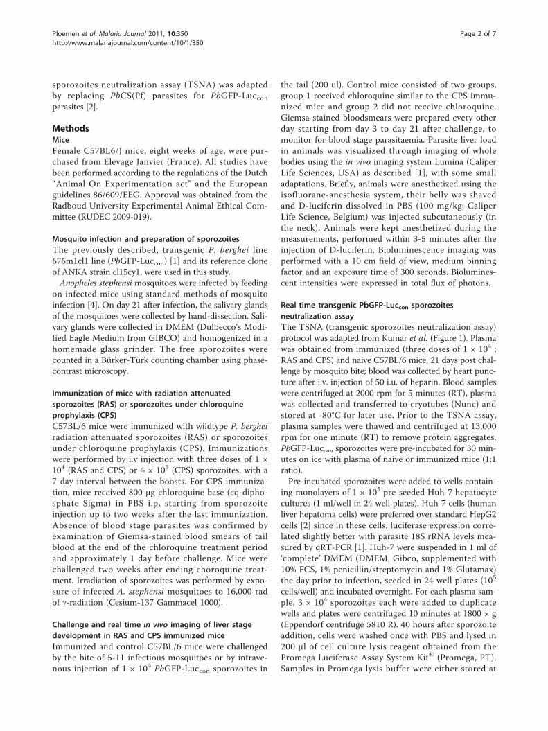
Real time transgenic PbGFP-Luccon sporozoitesneutralization assayThe TSNA (transgenic sporozoites neutralization assay)protocol was adapted from Kumar et al. (Figure 1). Plasmawas obtained from immunized (three doses of 1 × 10⁴ ;RAS and CPS) and naive C57BL/6 mice, 21 days post chal-lenge by mosquito bite; blood was collected by heart punc-ture after i.v. injection of 50 i.u. of heparin. Blood sampleswere centrifuged at 2000 rpm for 5 minutes (RT), plasmawas collected and transferred to cryotubes (Nunc) andstored at -80°C for later use. Prior to the TSNA assay,plasma samples were thawed and centrifuged at 13,000rpm for one minute (RT) to remove protein aggregates.PbGFP-Luccon sporozoites were pre-incubated for 30 min-utes on ice with plasma of naive or immunized mice (1:1ratio).Pre-incubated sporozoites were added to wells contain-
ing monolayers of 1 × 10⁵ pre-seeded Huh-7 hepatocytecultures (1 ml/well in 24 well plates). Huh-7 cells (humanliver hepatoma cells) were preferred over standard HepG2cells [2] since in these cells, luciferase expression corre-lated slightly better with parasite 18S rRNA levels mea-sured by qRT-PCR [1]. Huh-7 were suspended in 1 ml of‘complete’ DMEM (DMEM, Gibco, supplemented with10% FCS, 1% penicillin/streptomycin and 1% Glutamax)the day prior to infection, seeded in 24 well plates (105
cells/well) and incubated overnight. For each plasma sam-ple, 3 × 10⁴ sporozoites each were added to duplicatewells and plates were centrifuged 10 minutes at 1800 × g(Eppendorf centrifuge 5810 R). 40 hours after sporozoiteaddition, cells were washed once with PBS and lysed in200 μl of cell culture lysis reagent obtained from thePromega Luciferase Assay System Kit® (Promega, PT).Samples in Promega lysis buffer were either stored at
Ploemen et al. Malaria Journal 2011, 10:350http://www.malariajournal.com/content/10/1/350
Page 2 of 7

-80°C or processed immediately to measure luminescenceintensity with the Lumina system. The in vivo imaging sys-tem Lumina (Caliper Life Sciences, USA) was used tomeasure luciferase activity of infected Huh-7 cells. Quanti-tative analysis was performed by measuring the lumines-cence signal intensity per well using the ROI settings ofthe Living Image® 3.0 software. ROI measurements areexpressed in total flux of photons. 70 μl of LuciferaseAssay Substrate (Promega Luciferase Assay System Kit®)was added to 20 μl of lysed hepatocyte cultures in a white96-well plate (Dynex Technologies, USA). Biolumines-cence images were acquired with a 7 cm FOV, mediumbinning factor and an exposure time of 10-30 seconds.Percent inhibition was calculated by the following formula;1 - (average bioluminescence in immune plasma cultures/average bioluminescence in naive plasma cultures) ×100%.
ResultsChallenge of immunized mice with PbGFP-LucconsporozoitesMice immunized with CPS or RAS as well as control micewere challenged by PbGFP-Luccon infected mosquitoes andprotection against malaria was evaluated by blood smearreading and real time in vivo imaging. All control
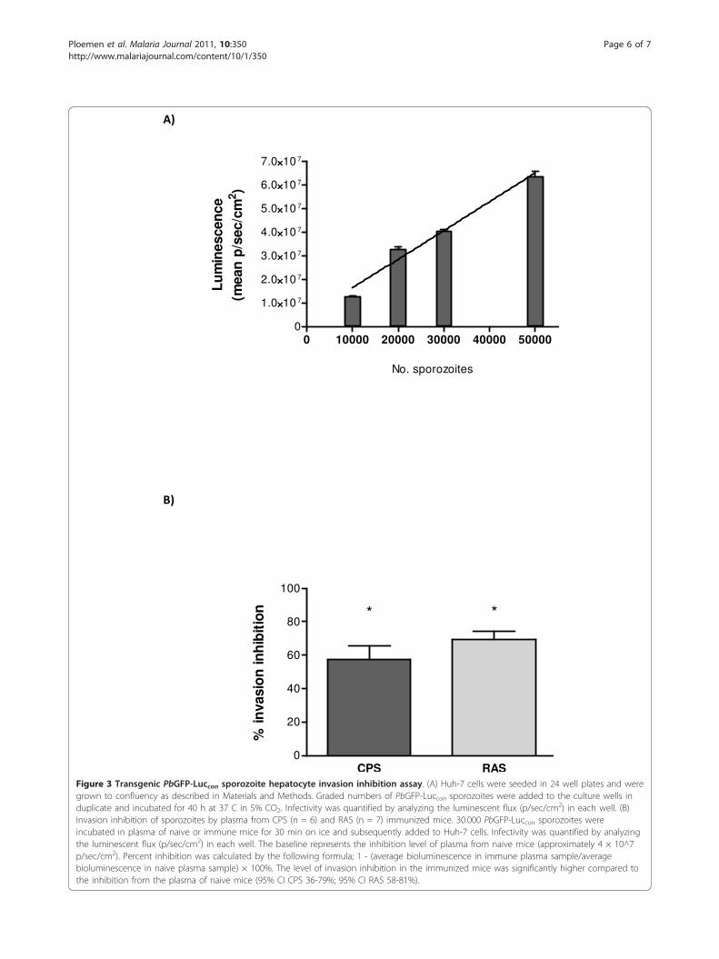
challenged mice (n = 10) developed asexual parasitaemiaand a positive bioluminescent liver signal by real-time invivo imaging at 44 hours post challenge (Figure 2a). Allmice immunized by CPS (n = 10) or RAS (n = 10) with adose regimen of three times 104 sporozoites and challengedby infectious mosquito bites, neither became parasitaemicnor displayed any bioluminescent signal originating fromthe liver (Figure 2a). Next, the robustness of protectiveimmunity was explored by increasing the challenge level inmice that were immunized with CPS by a lower dose regi-men of three times 4 × 10³ sporozoites. Mice were chal-lenged by i.v injection of 1 × 10⁴ PbGFP-Lucconsporozoites and all immunized mice remained negative.These results are in line with the data obtained by in vivoimaging; mice immunized with CPS showed no biolumi-nescent signal, in contrast to control mice, with positiveimages at 30 to 45 hours post challenge (Figure 2b). There-fore, the use of PbGFP-Luccon in a challenge model, com-bined with bioluminescent imaging permits determinationof protective efficacy in the liver post-immunization.
Real time transgenic PbGFP-Luccon sporozoitesneutralization assayTo evaluate the potential benefits of PbGFP-Luccon forassessment of protection in vitro, we adapted the
Real time transgenic sporozoite neutralization assaySchematic representation
HUH 7 hepatocytes (100.000 cells/well)
Incubation for 24hours at 37 C
Lysis of hepatocytes
WtGFP luc sporozoites (30.000 spz/well)Pre incubation of sporozoiteswith plasma(in glass tubes, for 30 minutes on ice)
Addition of pre incubatedsporozoites to hepatocytes
Plasma of naive or immunizedmice
Incubation for 40hours at 37 C
Detection of luciferaseactivity with IVIS
Figure 1 Schematic representation of the adapted transgenic PbGFP-Luccon sporozoite neutralization assay. Neutralization of hepatocyteinvasion by transgenic sporozoites was performed by incubation of naive or immune plasma obtained from (non-) immunized mice with thetransgenic sporozoites. Neutralization was performed for 30 minutes on ice before the antibody/sporozoites mix was added to Huh-7 cellscontaining wells and incubated for 40 h at 37°C. This figure is adapted from figure 1 described by Kumar et al. [2]
Ploemen et al. Malaria Journal 2011, 10:350http://www.malariajournal.com/content/10/1/350
Page 3 of 7

A)
Control RASCPS
44 hr
B)
Control CPS
30 hr
35 hr
45 hr
Figure 2 Real-time in vivo parasite liver load upon challenge in mice immunized with CPS or RAS. (A) Image (2 representative mice foreach group) of the parasite liver load in control (n = 10), CPS (n = 10) and RAS immunized (n = 10) C57BL/6 mice 44 hours post challenge.Mice were immunized i.v with 1 × 10⁴ sporozoites followed by two boosts of 1 × 10⁴ sporozoites. Challenge was performed by infectiousmosquito bites. The rainbow image visible in the naive mice represents the total flux of photons (p/sec/cm2) in that area. (B) Image (2 controlmice and 3 immunized mice) of the parasite liver load in control (n = 3) and CPS immunized (n = 5) C57BL/6 mice 30-45 hours post challenge.Mice were immunized with 4 × 10³ sporozoites by i.v injection followed by two boosts of 4 × 10³ sporozoites. Challenge was performed byinjection of 1 × 10⁴ PbGFP-Luccon sporozoites i.v. The rainbow image visible in the control mice represents the total flux of photons (p/sec/cm2)in that area.
Ploemen et al. Malaria Journal 2011, 10:350http://www.malariajournal.com/content/10/1/350
Page 4 of 7
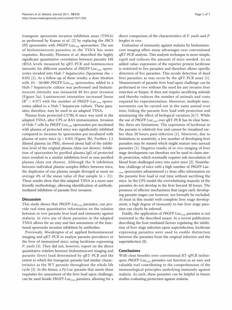
transgenic sporozoite invasion inhibition assay (TSNA)as performed by Kumar et al. [2] by replacing the PbCS(Pf) sporozoites with PbGFP-Luccon sporozoites. The useof bioluminescent parasites in the TSNA has somerequisites. Recently, Ploemen et al. described the highlysignificant quantitative correlation between parasite 18SrRNA levels measured by qRT-PCR and luminescenceintensity for different numbers of PbGFP-Luccon sporo-zoites invaded into Huh-7 hepatocytes (Spearman rho =0.83) [1]. As a follow-up of these results, a dose titrationwith 10 - 50.000 PbGFP-Luccon sporozoites, added to aHuh-7 hepatocyte culture was performed and biolumi-nescent intensity was measured 40 hrs post invasion(Figure 3a). Luminescent intensities increased linear(R2 = 0.97) with the number of PbGFP-Luccon sporo-zoites added to a Huh-7 hepatocyte culture. These para-sites, therefore, may be used in an adapted TSNA.Plasma from protected C57BL/6 mice was used in the
adapted TSNA, after CPS or RAS immunization. Invasionof Huh-7 cells by PbGFP-Luccon sporozoites pre-incubatedwith plasma of protected mice was significantly inhibitedcompared to invasion by sporozoites pre-incubated withplasma of naive mice (p < 0.05) (Figure 3b). Further, 1:1diluted plasma (in PBS), showed about half of the inhibi-tion level of the original plasma (data not shown). Inhibi-tion of sporozoites by purified plasma IgG of protectedmice resulted in a similar inhibition level as non-purifiedplasma (data not shown). Although the % inhibitionbetween individual plasma samples differs between mice,the duplicates of one plasma sample diverged at most onaverage 4% of the mean value of that sample (n = 21).These results show that this adapted TSNA is a more userfriendly methodology, allowing identification of antibody-mediated inhibition of parasite liver invasion.
DiscussionThis study shows that PbGFP-Luccon parasites, can pro-vide real-time quantitative information on the relationbetween in vivo parasite liver load and immunity againstmalaria. In vitro use of these parasites in the adaptedTSNA allows for an easy and fast assessment of the func-tional sporozoite invasion inhibition by antibodies.Previously, Mwakingwe et al. applied bioluminescent
imaging and qRT-PCR to analyse parasite prevalence inthe liver of immunized mice, using luciferase expressingP. yoelii [5]. They did not, however, report on the directquantitative relation between bioluminescent imaging andparasite (liver) load determined by qRT-PCR and theextent to which the transgenic parasite had similar charac-teristics as the WT parasite throughout the whole lifecycle [5]. In the future, a Py-Luc parasite that meets theserequisites for assessment of the liver load upon challenge,can be used beside PbGFP-Luccon parasites, allowing for a
direct comparison of the characteristics of P. yoelii and P.berghei in vivo.Evaluation of immunity against malaria by biolumines-
cent imaging offers many advantages over conventionalqRT-PCR analysis. This analysis technique is more simple,rapid and reduces the amount of mice needed. As anadded value, expression of the reporter protein luciferaseis restricted to live parasites and therefore allows specificdetection of live parasites. This avoids detection of deadliver parasites, as may occur by the qRT-PCR assay [1].Measurement of parasite liver load upon challenge can beperformed in vivo without the need for any invasive liverresection or biopsy. It does not require sacrificing animalsand thereby reduces the number of animals and costsrequired for experimentation. Moreover, multiple mea-surements can be carried out in the same animal overtime, linking the parasite liver load with protection andminimizing the effect of biological variation [6,7]. Whilethe use of PbGFP-Luccon over qRT-PCR has its clear bene-fits, there are limitations. The expression of luciferase inthe parasite is relatively low and cannot be visualized ear-lier than 20 hours post-infection [1]. Moreover, due tolimitations in sensitivity, a low number of developing liverparasites may be missed which might mature into asexualparasites [1]. Negative results of in vivo imaging of liverstage development can therefore not be used to claim ster-ile protection, which eventually requires sub-inoculation ofblood from challenged mice into naive mice [3]. Nonethe-less, challenge of mice with a high number of PbGFP-Luc-con sporozoites administered i.v does offer information onthe parasite liver load in real time without sacrifying themice. In the CPS model the overwhelming majority of theparasites do not develop in the liver beyond 30 hours. Thepresence of effector mechanisms that target early develop-ing parasite stages can however, not formally be excluded.At least in this model with complete liver stage develop-ment, a high degree of immunity to late liver stage para-sites can clearly be inferred.Finally, the application of PbGFP-Luccon parasites is not
restricted to the described assays. In a recent publicationdescribing the host mediated factors regulating the inhibi-tion of liver stage infection upon superinfection, luciferaseexpressing parasites were used to enable distinctionbetween the parasites from the original infection and thesuperinfection [8].
ConclusionsWith clear benefits over conventional RT-qPCR techni-ques, PbGFP-Luccon parasites can function as an easy andvaluable tool contributing to the comprehension of theimmunological principles underlying immunity againstmalaria. As such, these parasites can be helpful in futurestudies evaluating protection against malaria.
Ploemen et al. Malaria Journal 2011, 10:350http://www.malariajournal.com/content/10/1/350
Page 5 of 7
CPS RAS0
20
40
60
80
100
* *
% in
vasi
on
inh
ibit
ion
0 10000 20000 30000 40000 500000
1.0××××10 7
2.0××××10 7
3.0××××10 7
4.0××××10 7
5.0××××10 7
6.0××××10 7
7.0××××10 7
No. sporozoites
Lu
min
esce
nce
(mea
n p
/sec
/cm
2 )
Figure 3 Transgenic PbGFP-Luccon sporozoite hepatocyte invasion inhibition assay. (A) Huh-7 cells were seeded in 24 well plates and weregrown to confluency as described in Materials and Methods. Graded numbers of PbGFP-Luccon sporozoites were added to the culture wells induplicate and incubated for 40 h at 37 C in 5% CO2. Infectivity was quantified by analyzing the luminescent flux (p/sec/cm2) in each well. (B)Invasion inhibition of sporozoites by plasma from CPS (n = 6) and RAS (n = 7) immunized mice. 30.000 PbGFP-Luccon sporozoites wereincubated in plasma of naive or immune mice for 30 min on ice and subsequently added to Huh-7 cells. Infectivity was quantified by analyzingthe luminescent flux (p/sec/cm2) in each well. The baseline represents the inhibition level of plasma from naive mice (approximately 4 × 10^7p/sec/cm2). Percent inhibition was calculated by the following formula; 1 - (average bioluminescence in immune plasma sample/averagebioluminescence in naive plasma sample) × 100%. The level of invasion inhibition in the immunized mice was significantly higher compared tothe inhibition from the plasma of naive mice (95% CI CPS 36-79%; 95% CI RAS 58-81%).
Ploemen et al. Malaria Journal 2011, 10:350http://www.malariajournal.com/content/10/1/350
Page 6 of 7
List of abbreviationsPbGFP-Luccon: Plasmodium berghei that constitutively express fireflyLuciferase and the Green fluorescent protein; PbCS(pf): Plasmodium bergheithat bears the Plasmodium falciparum CS repeats; TSNA: Transgenicsporozoite neutralization assay; RAS: Radiation attenuated sporozoites; CPS:Sporozoites under chloroquine prophylaxis; ROI: Region of interest; q-RTPCR:quantitative real-time polymerase chain reaction; IVIS: in vivo imagingsystem.
AcknowledgementsWe would like to thank Claudia Lagarde for the technical assistance with themouse infections and Anja Scholzen for critical revision of the manuscript.This study was performed within the framework of Top Institute Pharma(Netherlands) project: T4-102. The funders had no role in study design, datacollection and analysis, decision to publish, or preparation of the manuscript.
Authors’ contributionsIP conceived the study, which was largely carried out by IP and MB. KN, GvGand EB, helped design the studies and carry out immunizations andchallenge. IP, MB and CH discussed experiments and results. IP wrote themanuscript which was edited by CH and RS. All authors read and approvedthe final manuscript.
Competing interestsThe authors declare that they have no competing interests.
Received: 25 July 2011 Accepted: 9 December 2011Published: 9 December 2011
References1. Ploemen IH, Prudêncio M, Douradinha BG, Ramesar J, Fonager J, van
Gemert GJ, Luty AJ, Hermsen CC, Sauerwein RW, Baptista FG, Mota MM,Waters AP, Que I, Lowik CW, Khan SM, Janse CJ, Franke-Fayard BM:Visualisation and quantitative analysis of the rodent malaria liver stageby real time imaging. PLoS One 2009, 4:e7881.
2. Kumar KA, Oliveira GA, Edelman R, Nardin E, Nussenzweig V: QuantitativePlasmodium sporozoite neutralization assay (TSNA). J Immunol Methods2004, 292:157-164.
3. Belnoue E, Voza T, Costa FT, Grüner AC, Mauduit M, Rosa DS, Depinay N,Kayibanda M, Vigário AM, Mazier D, Snounou G, Sinnis P, Rénia L:Vaccination with live Plasmodium yoelii blood stage parasites underchloroquine cover induces cross-stage immunity against malaria liverstage. J Immunol 2008, 181:8552-8558.
4. Sinden RE: Infection of mosquitoes with rodent malaria. In Molecularbiology of insect disease vectors: a method manual. Edited by: Crampton JM,Beard CB, Louis C. London, United Kingdom: Chapman and Hall; 1997:67-91.
5. Mwakingwe A, Ting LM, Hochman S, Chen J, Sinnis P, Kim K: Noninvasivereal-time monitoring of liver-stage development of bioluminescentPlasmodium parasites. J Infect Dis 2009, 200:1470-1478.
6. Sadikot RT, Blackwell TS: Bioluminescence imaging. Proc Am Thorac Soc2005, 2:537-2.
7. Welsh DK, Kay SA: Bioluminescence imaging in living organisms. CurrOpin Biotechnol 2005, 16:73-78.
8. Portugal S, Carret C, Recker M, Armitage AE, Gonçalves LA, Epiphanio S,Sullivan D, Roy C, Newbold CI, Drakesmith H, Mota MM: Host-mediatedregulation of superinfection in malaria. Nat Med 2011, 17:732-737.
doi:10.1186/1475-2875-10-350Cite this article as: Ploemen et al.: Evaluation of immunity againstmalaria using luciferase-expressing Plasmodium berghei parasites. MalariaJournal 2011 10:350.
Submit your next manuscript to BioMed Centraland take full advantage of:
• Convenient online submission
• Thorough peer review
• No space constraints or color figure charges
• Immediate publication on acceptance
• Inclusion in PubMed, CAS, Scopus and Google Scholar
• Research which is freely available for redistribution
Submit your manuscript at www.biomedcentral.com/submit
Ploemen et al. Malaria Journal 2011, 10:350http://www.malariajournal.com/content/10/1/350
Page 7 of 7