Embed Size (px)
Citation preview
Yang et al. Journal of Translational Medicine (2014) 12:351 DOI 10.1186/s12967-014-0351-6
RESEARCH Open Access
Electroacupuncture stimulation at sub-specificacupoint and non-acupoint induced distinct brainglucose metabolism change in migraineurs:a PET-CT studyMingxiao Yang1†, Jie Yang1†, Fang Zeng1, Peng Liu2, Zhenhong Lai1, Shufang Deng1, Li Fang3, Wenzhong Song4,Hongjun Xie4* and Fanrong Liang1*
Abstract
Background: Acupuncture has analgesic effect to most pain conditions. Many neuroimaging studies wereconducted to explore acupoint specificity in pain and other condition, but till now there is still discrepancy. Basedon our previous finding, this study investigated the brain metabolism changes of acupuncture analgesia induced bysub-specific acupoint and non-acupoint stimulation.
Methods: 30 migraineurs were included and randomly assigned to 3 groups: Acupuncture Group (AG), ShamAcupuncture Group (SAG) and Migraine Group (MG). In AG, a combination sub-specific points of Shaoyangmeridians, Luxi (TE19), San Yangluo (TE8), and Xi Yangguan(GB33) has been stimulated with electroacupuncture,while non-acupoints for SAG were used and MG received no treatment. Positron emission tomography withcomputed tomography (PET-CT) was used to identify differences in brain glucose metabolism between groups.
Results: In the AG, brain glucose metabolism increase compared with the MG was observed in the middle frontalgyrus, postcentral gyrus, the precuneus, parahippocampus, cerebellum and middle cingulate cortex (MCC), anddecrease were observed in the left hemisphere of Middle Temporal Cortex (MTC).In the SAG, compared with MG,glucose metabolism increased in the poster cingulate cortex (PCC), insula, inferior temporal gyrus, MTC, superiortemporal gyrus, postcentral gyrus, fusiform, inferior parietal lobe, superior parietal lobe, supramarginal gyrus, middleoccipital lobe, angular and precuneus; while, decreased in cerebellum, parahippocampus.
Conclusions: Acupuncture stimulation at both sub-specific acupoint and non-acupoint yields ameliorating effect tomigraine pain, but with evidently differed central mechanism as measured by PET-CT. The pattern of brain glucosemetabolism change in acupoint is pertinent and targeted, while in non-acupoint that was disordered and randomized.These finding may provide new perspectives into the validation of acupoint specificity, optimizing acupunctureanalgesia and revealing central mechanism of acupuncture analgesia by neuroimaging measurement.
Trial registration: This trial was registered in the Chinese Clinical Trial Registry, with registration no. ChiCTR-TRC-11001813.
Keywords: Acupuncture analgesia, PEC-CT, Migraine
* Correspondence: [email protected]; [email protected]†Equal contributors4PET-CT Center, Sichuan Provincial People’s Hospital, Chengdu, Sichuan,China1Acupuncture and Tuina School, Chengdu University of Traditional ChineseMedicine, Chengdu, Sichuan, ChinaFull list of author information is available at the end of the article
© 2014 Yang et al.; licensee BioMed Central. This is an Open Access article distributed under the terms of the CreativeCommons Attribution License (http://creativecommons.org/licenses/by/4.0), which permits unrestricted use, distribution, andreproduction in any medium, provided the original work is properly credited. The Creative Commons Public DomainDedication waiver (http://creativecommons.org/publicdomain/zero/1.0/) applies to the data made available in this article,unless otherwise stated.
Yang et al. Journal of Translational Medicine (2014) 12:351 Page 2 of 9
BackgroundAcupuncture is an essential component of oriental medi-cine that has been persistently and extensively practicedto treat various diseases, pain in particular, in China andmany other Asian countries. Doctors and patients inWest countries usually employ acupuncture as adjunctto conventional drugs for a wide spectrum of pain con-ditions. Though it was dominantly mediated by 5-HTand endogenous opioid system, the mechanism of acu-puncture analgesia remains to be elucidated.In the long history of clinical practice, traditional acu-
puncture formed its therapeutic principles, and the ex-perience of acupuncturists successively enriched thefruitfulness of acupuncture theories. According to trad-itional beliefs, acupuncture point, or termed acupoint,refers to specific point on the body surface that is in-active when the body is in healthy condition, but can befurther activated by certain stimulus, such as needle pene-tration and manipulation, pressing, electrical current, heat.Well-defined acupoints selection principles for practi-tioners to consider in clinical situation were developedand utilized under the umbrella of the existence of acu-point specificity. However, several recent systematic re-views and clinical randomized controlled trials reportedoutcome that there’s minimal, or no superiority in func-tion of acupoint compared with sham acupoint [1-5],which is incompatible with classic acupuncture theory. Sothat it raised questions and doubts to the existence of acu-point specificity [6].In recent years, neuroimaging technology has been
reasonably accepted as a new approach to non-invasivelycharacterize the central interacting mechanism of acu-puncture therapy [7-11]. Integrated response of cerebro-cerebellar and limbic system has been detected by fMRIafter acupuncture stimulation at a specific acupoint [12].By means of that, a growing body of evidence has beenaccumulated to address the existence of acupoint specifi-city. Many interesting articles elicited possible central re-sponse/mechanism of acupuncture specificity werepublished [13-15]. Our former works also indicated thatremarkable modulation on the homeostatic afferent net-work, including the insula, anterior cingulate cortex(ACC), and hypothalamus, might be the specific mech-anism of acupuncture specificity [16]. While, it is neces-sary to point out that the preponderance of previousstudies were undergone within healthy subjects. Sincethe function of acupoint is body-condition dependentaccording to Traditional Chinese Medicine(TCM) per-spectives [17], there’s a paucity of relative studies thatcompares the influence of acupoint on brain activity ofdiseased condition with that of sham acupoint. There-fore, the traits and influence of acupoint specificity foracupuncture analgesia in diseased condition remain un-clear and worth investigation.
Migraine without aura is a disabling disease character-ized by hemispheric headache. Current evidence favors toinclude acupuncture therapy in migraine care for that acu-puncture yielded relatively equivalent effect to positivedrugs, but there’s little side effects [18-20]. The curativefunction of acupuncture for migraine is not limited toinstant cessation for acute pain [21,22], studies alsodemonstrated that acupuncture can reduce frequenciesof migraine attacks as the prophylactic drugs do [23,24].Hence, migraine is considered as a proper indication ofacupuncture analgesia and is frequently chosen as a car-rier to reflect the specificity or mechanism of functionof acupoint. Hence, in this study migraineurs were en-rolled to further investigate acupoint specificity in acu-puncture analgesia.In a previous study, we found that purposeful selection
of specific acupoints on Shaoyang meridians, which werebelieved according to TCM theory to be closely relatedto and also are commonly applied in clinical practice tomigraine treatment, presented preferred instant anal-gesic effect compared to those on Yangming meridians[25]. Meanwhile, results of PET-CT demonstrated thatpain or migraine related brain regions such as MCC,and several parts of the limbic system, have been observedwith more extensive metabolism change after stimulationat specific acupoints on disease dependent meridians com-pared with that of disease independent meridians, whichimplied the specificity of central responses. Secondary tothis, we conducted the present study to explore whetherthere is difference of cerebral response after acupuncturestimulation at sub-specific acupoint on Shaoyang merid-ian, while compared to non-acupoint. The sub-specificacupoint is a concept relative to the specific acupoint,which means that its function is not so potent or focusedas compared to that of the specific one. Specific acupointis defined as points that situated in meridian line with thestrongest and the most concentrated power for certaindisease. The classification of specific acupoint is mostlybased on traditional acupuncture theory. In contrast, non-acupoint is some random point that is absent from merid-ian line, and thus according to its common application itis always referred to as sham point that possesses no spe-cific action [26,27]. Therefore, this study as a continuumwas performed on the basis of previous successful applica-tion of PET-CT paradigm in migraine to indicate centralresponse, and this is also a deepened exploration to theprevious findings.Migraine patients were recruited as voluntary subjects,
which is identical to former study to observe subject indiseased condition. Also, fluorodeoxyglucose positronemission tomography combined with computed tomog-raphy (FDG-PET/CT) was main tool to visualize glucosemetabolism of diverse brain regions in migraine patients.By trial designed like this, we expected to observe the
Yang et al. Journal of Translational Medicine (2014) 12:351 Page 3 of 9
difference of cerebral response in relevant groups inorder to further investigate mechanism of acupunctureanalgesia and acupoint specificity.
Materials and methodsEthical approvalThe whole research protocol had been systematicallyreviewed and further approved by the ethics committeeof the Teaching Hospital of Chengdu University of Tra-ditional Chinese Medicine. The approval identifier is2007KL-001. Before the commencement of the trial,each subject was clearly explained about the trial proce-dures and participants that were included in this trialprovided with written informed consent.
Subjects and experimental paradigmDuring the period of April 2008 to December 2009, wehave included thirty right-handed patients with acutemigraine without aura. The migraine patients (12 malesand 18 Females; mean age 33.28 ± 8.03 years) were ran-domized into three groups in a 1:1:1 ratio. Patient assignedto Acupuncture at sub-specific acupoint Group (AG) re-ceived stimulation at sub-specific acupoints on Shaoyangmeridians; Sham Acupuncture at non-acupoint Group(SAG) will conduct stimulation at non-acupoints whichwere located neither at acupoint site nor in meridian lines;and Migraineur Wait-List control Group (MG) as a blankcontrol will deliver no treatment to participants. After thecompletion of PET-CT scan, patients in SAG and MGwere given traffic compensation fee for their contribu-tions, and also free acupuncture treatments were providedas required. Subjects were matched by gender, age, hand-edness, and education.
Inclusion criteriaEligible participant met all the following criterion wereincluded: (1) diagnosed as acute migraine without auraaccording to the classification criteria of the Inter-national Headache Society for the diagnosis of migrainewithout aura; (2) unilateral headache in the left hemi-sphere; (3) Frequency of migraine attacks no less thatonce per month during the last three months before in-clusion; (4) 2 ≤migraine pain at recruitment and beforescanning ≤ 8 as measured by Visual Analogue Scale(VAS) with terminal descriptors of “no pain” and “worstpain possible” in a range between 0 and 10; (5) durationof migraine attack no more than 24 hours to the begin-ning of the scan; (6) aged 20–45 years; (7) no otherneurological disorders assessed by normal skull CT orMRI; (8) rescue medication should be avoided within24 hours since the onset of the acute attack; (9)Fullyagreed with all trial procedures and returned written in-formed consent.
Exclusion criteriaPatients were excluded if any one of the following exclu-sion criterion was matched: (1) headaches that were sec-ondary to organic disorders other than migraine, such ascerebropathy, vascular malformation, hypertension, or ar-teriosclerosis; (2) other conditions like psychosis, bleedingdisorders, or allergies; (3) other pain conditions that maycontaminate the nociceptive sensation of migraine; (4) in-cluded in other study; (5) pregnancy or lactating, or partic-ipants considering to conceive in the following 6 months;(6) medication with vasoactive agent in the last two weeks;(7) with emotional disorders such as major anxiety dis-order and/or depression; and (8) presence of any contrain-dications to PET-CT or electroacupuncture.
InterventionsIncluded participants were averagely assigned to either oneof the three groups by means of simple randomization. Theacupoint combination for the AG included three sub-specific acupoints on Shaoyang meridians, Luxi (TE19),San Yangluo (TE8), and Xi Yangguan (GB33). Non-acupoints applied in the SAG were predefined and uti-lized in previous studies of our team [21]. The locationsof these non-acupoints were described as follows: (1)Sham-point 1:The medial side of the arm at the anteriorborder of the insertion of the deltoid muscle at the junc-tion of the deltoid and biceps muscles; (2) Sham-point2:The edge of the tibia ,1–2 cm lateral and horizontal tothe Zusanli, ST36; (3) Sham-point 3: On the ulnar sideof the arm, half way between the epicondylus medialisof the humerus and the ulnar side of the wrist. Thesethree points were sited distant to the traditionally rec-ognized acupoints or meridians lines. Illustrations ofthese points were showed in Figure 1. Only points onthe left side of the body were used in the experiment.After sterilization in local dermal area of sub-specific
acupoint/non-acupoint, sterile disposable acupunctureneedles (25–40 mm in length and 0.30 mm in diameter;Hwato acupuncture needles manufactured by SuzhouMedical Supplies Co., Ltd., Suzhou, China) were insertedtransversely or obliquely into to the points to a depth of15–30 mm. After needle insertion, in AG needles wereevenly twirled and rotated to induce the DeQi sensation(soreness, numbness, distention, heaviness, dull pain, etc.)[28,29]. After the DeQi phenomenon was achieved and re-ported by patient, auxiliary needles were perpendicularlypunctured 2 mm lateral to the points, to 2 mm in depth,without manual manipulation. Electrodes of Han’s acu-point nerve stimulator (HANS; model LH 200A; TENS,Nanjing, China) were then connected to the needles byacupuncturist. While, in SAG HANS was connected toneedles after insertion and required no DeQi sensationelicited. The frequency of stimulation was 100 Hz, and theintensity of the electricity stimulus varied from 0.1 to
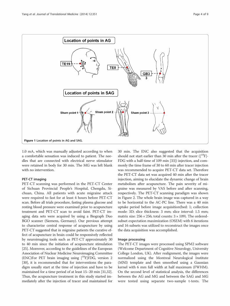
Figure 1 Location of points in AG and SAG.
Yang et al. Journal of Translational Medicine (2014) 12:351 Page 4 of 9
1.0 mA, which was manually adjusted according to whena comfortable sensation was induced to patient. The nee-dles that are connected with electrical nerve stimulatorwere retained in body for 30 min. The MG was left blankwith no intervention.
PET-CT imagingPET-CT scanning was performed in the PET-CT Centerof Sichuan Provincial People’s Hospital, Chengdu, Si-chuan, China. All patients with acute migraine attackwere required to fast for at least 4 hours before PET-CTscan. Before all trials procedure, fasting plasma glucose andresting blood pressure were examined prior to acupuncturetreatment and PET-CT scan to avoid faint. PET-CT im-aging data sets were acquired by using a Biograph DuoBGO scanner (Siemens, Germany). Our previous attemptto characterize central response of acupuncture by usingPET-CT suggested that in migraine patients the curative ef-fect of acupuncture in brain could be responsively reflectedby neuroimaging tools such as PET-CT approximately 30to 40 min since the initiation of acupuncture stimulation[25]. Moreover, according to the guidelines of the EuropeanAssociation of Nuclear Medicine Neuroimaging Committee(ENC)For PET brain imaging using [18F]FDG, version 2[30], it is recommended that for interventions the para-digm usually start at the time of injection and have to bemaintained for a time period of at least 15–20 min [31,32].Thus, the acupuncture treatment in this study started im-mediately after the injection of tracer and maintained for
30 min. The ENC also suggested that the acquisitionshould not start earlier than 30 min after the tracer ([18F]-FDG with a half-time of 109 min [33]) injection, and com-monly the time frame of 30 to 60 min after tracer injectionwas recommended to acquire PET-CT data set. Thereforethe PET-CT data set was acquired 40 min after the tracerinjection, aiming to elucidate the dynamic change of brainmetabolism after acupuncture. The pain severity of mi-graine was measured by VAS before and after scanning,respectively. The PET-CT scanning paradigm was shownin Figure 2. The whole brain image was captured in a wayto be horizontal to the AC-PC line. There was a 40 minuptake period before image acquisition(bed: 1; collectionmode: 3D; slice thickness: 3 mm; slice interval: 1.5 mm;matrix size: 256 × 256; total counts: 3 × 10⁹). The ordered-subset expectation maximization (OSEM) with 6 iterationsand 16 subsets was utilized to reconstruct the images oncethe data acquisition was accomplished.
Image processingThe PET-CT images were processed using SPM2 software(Welcome Department of Cognitive Neurology, UniversityCollege London, UK). After realignment, the images werenormalized using the Montreal Neurological Institute(MNI) template and then smoothed using a Gaussiankernel with 6 mm full width at half maximum (FWHM).On the second level of statistical analysis, the differencesbetween the AG and MG and between the SAG and MGwere tested using separate two-sample t-tests. The
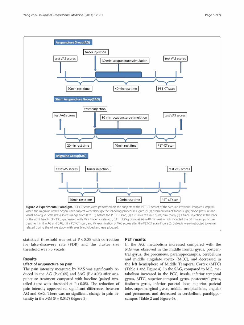
Figure 2 Experimental Paradigm. PET-CT scans were performed on the subjects at the PET-CT center of the Sichuan Provincial People’s Hospital.When the migraine attack began, each subject went through the following procedure(Figure 2): (1) examinations of blood sugar, blood pressure andVisual Analogue Scale (VAS) scores (range from 0 to 10) before the PET-CT scan; (2) a 20 min rest in a quiet, dim room; (3) a tracer injection at the backof the right hand (18F-FDG; synthesized with Mini Tracer accelerator; 0.11 mCi/kg dosage); (4) a 40 min rest, which included the 30 min acupuncturetreatment in the AG and SAG; (5) a PET-CT scan; and (6) examination of VAS scores after the PET-CT scan (Figure 2). Subjects were instructed to remainrelaxed during the whole study, with eyes blindfolded and ears plugged.
Yang et al. Journal of Translational Medicine (2014) 12:351 Page 5 of 9
statistical threshold was set at P < 0.05 with correctionfor false-discovery rate (FDR) and the cluster sizethreshold was >5 voxels.
ResultsEffect of acupuncture on painThe pain intensity measured by VAS was significantly re-duced in the AG (P < 0.05) and SAG (P < 0.05) after acu-puncture treatment compared with baseline (paired two-tailed t-test with threshold at P < 0.05). The reduction ofpain intensity appeared no significant differences betweenAG and SAG. There was no significant change in pain in-tensity in the MG (P = 0.047) (Figure 3).
PET resultsIn the AG, metabolism increased compared with theMG was observed in the middle frontal gyrus, postcen-tral gyrus, the precuneus, parahippocampus, cerebellumand middle cingulate cortex (MCC), and decreased inthe left hemisphere of Middle Temporal Cortex (MTC)(Table 1 and Figure 4). In the SAG, compared to MG, me-tabolism increased in the PCC, insula, inferior temporalgyrus, MTC, superior temporal gyrus, postcentral gyrus,fusiform gyrus, inferior parietal lobe, superior parietallobe, supramarginal gyrus, middle occipital lobe, angularand precuneus, and decreased in cerebellum, parahippo-campus (Table 2 and Figure 4).
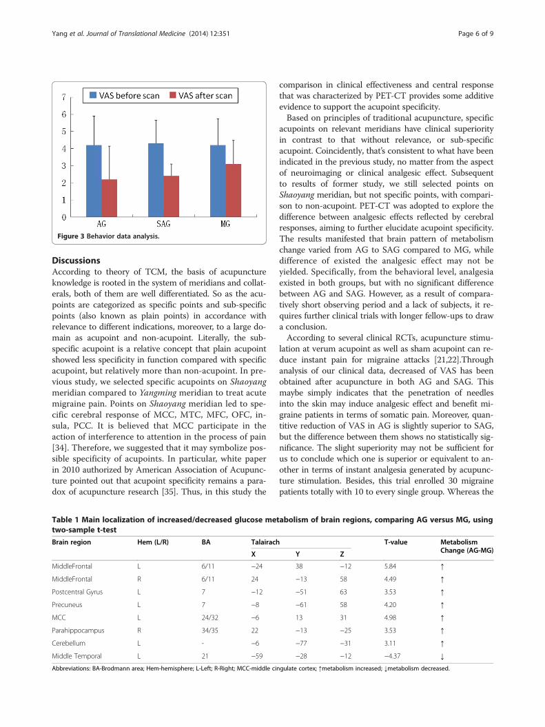
Figure 3 Behavior data analysis.
Yang et al. Journal of Translational Medicine (2014) 12:351 Page 6 of 9
DiscussionsAccording to theory of TCM, the basis of acupunctureknowledge is rooted in the system of meridians and collat-erals, both of them are well differentiated. So as the acu-points are categorized as specific points and sub-specificpoints (also known as plain points) in accordance withrelevance to different indications, moreover, to a large do-main as acupoint and non-acupoint. Literally, the sub-specific acupoint is a relative concept that plain acupointshowed less specificity in function compared with specificacupoint, but relatively more than non-acupoint. In pre-vious study, we selected specific acupoints on Shaoyangmeridian compared to Yangming meridian to treat acutemigraine pain. Points on Shaoyang meridian led to spe-cific cerebral response of MCC, MTC, MFC, OFC, in-sula, PCC. It is believed that MCC participate in theaction of interference to attention in the process of pain[34]. Therefore, we suggested that it may symbolize pos-sible specificity of acupoints. In particular, white paperin 2010 authorized by American Association of Acupunc-ture pointed out that acupoint specificity remains a para-dox of acupuncture research [35]. Thus, in this study the
Table 1 Main localization of increased/decreased glucose mettwo-sample t-test
Brain region Hem (L/R) BA Talairach
X
MiddleFrontal L 6/11 −24
MiddleFrontal R 6/11 24
Postcentral Gyrus L 7 −12
Precuneus L 7 −8
MCC L 24/32 −6
Parahippocampus R 34/35 22
Cerebellum L - −6
Middle Temporal L 21 −59
Abbreviations: BA-Brodmann area; Hem-hemisphere; L-Left; R-Right; MCC-middle cin
comparison in clinical effectiveness and central responsethat was characterized by PET-CT provides some additiveevidence to support the acupoint specificity.Based on principles of traditional acupuncture, specific
acupoints on relevant meridians have clinical superiorityin contrast to that without relevance, or sub-specificacupoint. Coincidently, that’s consistent to what have beenindicated in the previous study, no matter from the aspectof neuroimaging or clinical analgesic effect. Subsequentto results of former study, we still selected points onShaoyang meridian, but not specific points, with compari-son to non-acupoint. PET-CT was adopted to explore thedifference between analgesic effects reflected by cerebralresponses, aiming to further elucidate acupoint specificity.The results manifested that brain pattern of metabolismchange varied from AG to SAG compared to MG, whiledifference of existed the analgesic effect may not beyielded. Specifically, from the behavioral level, analgesiaexisted in both groups, but with no significant differencebetween AG and SAG. However, as a result of compara-tively short observing period and a lack of subjects, it re-quires further clinical trials with longer fellow-ups to drawa conclusion.According to several clinical RCTs, acupuncture stimu-
lation at verum acupoint as well as sham acupoint can re-duce instant pain for migraine attacks [21,22].Throughanalysis of our clinical data, decreased of VAS has beenobtained after acupuncture in both AG and SAG. Thismaybe simply indicates that the penetration of needlesinto the skin may induce analgesic effect and benefit mi-graine patients in terms of somatic pain. Moreover, quan-titive reduction of VAS in AG is slightly superior to SAG,but the difference between them shows no statistically sig-nificance. The slight superiority may not be sufficient forus to conclude which one is superior or equivalent to an-other in terms of instant analgesia generated by acupunc-ture stimulation. Besides, this trial enrolled 30 migrainepatients totally with 10 to every single group. Whereas the
abolism of brain regions, comparing AG versus MG, using
T-value MetabolismChange (AG-MG)Y Z
38 −12 5.84 ↑
−13 58 4.49 ↑
−51 63 3.53 ↑
−61 58 4.20 ↑
13 31 4.98 ↑
−13 −25 3.53 ↑
−77 −31 3.11 ↑
−28 −12 −4.37 ↓
gulate cortex; ↑metabolism increased; ↓metabolism decreased.
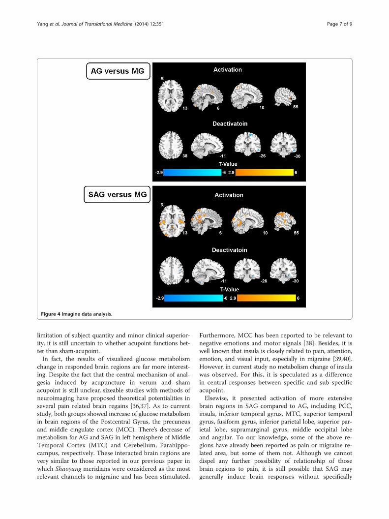
Figure 4 Imagine data analysis.
Yang et al. Journal of Translational Medicine (2014) 12:351 Page 7 of 9
limitation of subject quantity and minor clinical superior-ity, it is still uncertain to whether acupoint functions bet-ter than sham-acupoint.In fact, the results of visualized glucose metabolism
change in responded brain regions are far more interest-ing. Despite the fact that the central mechanism of anal-gesia induced by acupuncture in verum and shamacupoint is still unclear, sizeable studies with methods ofneuroimaging have proposed theoretical potentialities inseveral pain related brain regains [36,37]. As to currentstudy, both groups showed increase of glucose metabolismin brain regions of the Postcentral Gyrus, the precuneusand middle cingulate cortex (MCC). There’s decrease ofmetabolism for AG and SAG in left hemisphere of MiddleTemporal Cortex (MTC) and Cerebellum, Parahippo-campus, respectively. These interacted brain regions arevery similar to those reported in our previous paper inwhich Shaoyang meridians were considered as the mostrelevant channels to migraine and has been stimulated.
Furthermore, MCC has been reported to be relevant tonegative emotions and motor signals [38]. Besides, it iswell known that insula is closely related to pain, attention,emotion, and visual input, especially in migraine [39,40].However, in current study no metabolism change of insulawas observed. For this, it is speculated as a differencein central responses between specific and sub-specificacupoint.Elsewise, it presented activation of more extensive
brain regions in SAG compared to AG, including PCC,insula, inferior temporal gyrus, MTC, superior temporalgyrus, fusiform gyrus, inferior parietal lobe, superior par-ietal lobe, supramarginal gyrus, middle occipital lobeand angular. To our knowledge, some of the above re-gions have already been reported as pain or migraine re-lated area, but some of them not. Although we cannotdispel any further possibility of relationship of thosebrain regions to pain, it is still possible that SAG maygenerally induce brain responses without specifically
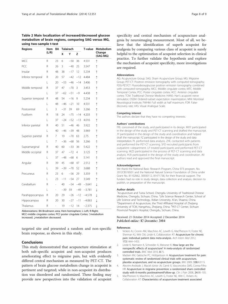
Table 2 Main localization of increased/decreased glucosemetabolism of brain regions, comparing SAG versus MG,using two-sample t-test
Regions Hem(L/R)
BA Talairach T-value MetabolismChange(SAG-MG)
X Y Z
MCC R 23 6 −50 36 4.531 ↑
PCC R 26 3 −40 25 3.547 ↑
Insular R 48 38 −17 12 5.234 ↑
Inferior temporal R 20 57 −42 −12 4.484 ↑
L 20 −53 −46 −14 3.406 ↑
Middle temporal R 37 47 −70 3 3.453 ↑
L 37 −62 −51 −9 4.438 ↑
Superior temporal R 48 54 −16 9 5.234 ↑
L 48 −46 −21 10 4.531 ↑
Postcentral L 3 −31 39 69 3.266 ↑
Fusiform R 18 24 −75 −14 4.203 ↑
L 37 −24 −52 −13 4.016 ↑
Inferior parietal R 40 51 −46 46 3.922 ↑
L 40 −46 −39 48 3.969 ↑
Superior parietal R 7 19 −70 63 2.75 ↑
L 7 −36 −68 56 3.266 ↑
Supramarginal R 40 60 −33 36 5.422 ↑
Middle occipital R 37 47 −72 4 3.125 ↑
L 37 −48 −68 6 5.141 ↑
Angular R 39 45 −68 47 2.512 ↑
L 39 −39 −70 52 3.688 ↑
Precuneus R 23 6 −56 29 3.359 ↑
L 23 −11 −54 27 3.549 ↑
Cerebellum R - 40 −54 −49 −3.641 ↓
L - −39 59 −49 −3.781 ↓
Parahippocampus R 20 29 −26 −12 −4.578 ↓
Hippocampus R 20 30 −27 −11 −4.063 ↓
Thalamus R - 19 −12 14 −2.375 ↓
Abbreviations: BA-Brodmann area; Hem-hemisphere; L-Left; R-Right;MCC-middle cingulate cortex; PCC-poster cingulate Cortex; ↑metabolismincreased; ↓metabolism decreased.
Yang et al. Journal of Translational Medicine (2014) 12:351 Page 8 of 9
targeted site and presented a random and non-specificbrain response, as shown in this study.
ConclusionsThis study demonstrated that acupuncture stimulation atboth sub-specific acupoint and non-acupoint producesameliorating effect to migraine pain, but with evidentlydiffered central mechanism as measured by PET-CT. Thepattern of brain glucose metabolism change in acupoint ispertinent and targeted; while in non-acupoint its distribu-tion was disordered and randomized. These finding mayprovide new perspectives into the validation of acupoint
specificity and central mechanism of acupuncture anal-gesia by neuroimaging measurement. Most of all, we be-lieve that the identification of superb acupoint foranalgesia by comparing various class of acupoint is surelyhelpful to the optimization of acupoint selection in clinicalpractice. To further validate the hypothesis and explorethe mechanism of acupoint specificity, more investigationsare required.
AbbreviationsAG: Acupuncture Group; SAG: Sham Acupuncture Group; MG: MigraineGroup; PET-CT: Positron emission tomography with computed tomography;FDG-PET/CT: Fluorodeoxyglucose positron emission tomography combinedwith computed tomography; MCC: Middle cingulate cortex; MTC: MiddleTemporal Cortex; PCC: Poster cingulate cortex; ACC: Anterior cingulatecortex; TCM: Traditional Chinese Medicine; HANS: Han’s acupoint nervestimulator; OSEM: Ordered-subset expectation maximization; MNI: MontrealNeurological Institute; FWHM: Full width at half maximum; FDR: False-discovery rate; VAS: Visual Analogue Scale.
Competing interestThe authors declare that they have no competing interests.
Authors’ contributionsFRL conceived of the study, and participated in its design. MXY participatedin the design of the study and PET-CT scanning and drafted the manuscript.JY participated in the design of the study and coordination and helpeddraft the manuscript. FZ participated in the design of the study and datainterpretation. PL performed data analysis. ZHL contacted with patientsand performed the PET-CT scanning. SFD recruited participants fromoutpatient >department. LF treated participants and performed PET-CTscanning. WZS participated in the process of PET-CT scanning and dataanalysis. HJX participated in the design of the study and coordination. Allauthors read and approved the final manuscript.
AcknowledgementWe thank the National Basic Research Program, China 973 program, No.2012CB518501 and the National Natural Science Foundation of China underGrant No. 81102662, 30930112, 81471738, for their financial support. Thefunders had no role in study design, data collection and analysis, decision topublish, or preparation of the manuscript.
Author details1Acupuncture and Tuina School, Chengdu University of Traditional ChineseMedicine, Chengdu, Sichuan, China. 2Life Science Research Center, School ofLife Science and Technology, Xidian University, Xi’an, Shaanxi, China.3Department of Acupuncture, the Third Affiliated Hospital of ZhejiangUniversity of TCM, Hangzhou, Zhejiang, China. 4PET-CT Center, SichuanProvincial People’s Hospital, Chengdu, Sichuan, China.
Received: 21 October 2014 Accepted: 2 December 2014
References1. Vickers AJ, Cronin AM, Maschino AC, Lewith G, MacPherson H, Foster NE,
Sherman KJ, Witt CM, Linde K, Collaboration AT: Acupuncture for chronicpain: individual patient data meta-analysis. Arch Intern Med 2012,172:1444–1453.
2. Linde K, Niemann K, Schneider A, Meissner K: How large are thenonspecific effects of acupuncture? A meta-analysis of randomizedcontrolled trials. BMC Med 2010, 8:75.
3. Madsen MV, Gøtzsche PC, Hróbjartsson A: Acupuncture treatment for pain:systematic review of randomised clinical trials with acupuncture,placebo acupuncture, and no acupuncture groups. BMJ 2009, 338:A3115.
4. Alecrim-Andrade J, Maciel-Júnior JA, Carnè X, Vasconcelos GMS, Correa-FilhoHR: Acupuncture in migraine prevention: a randomized sham controlledstudy with 6-months posttreatmentFollow-up. Clin J Pain 2008, 24:98–105.
5. MacPherson H, Maschino AC, Lewith G, Foster NE, Witt C, Vickers AJ,Collaboration AT: Characteristics of acupuncture treatment associated
Yang et al. Journal of Translational Medicine (2014) 12:351 Page 9 of 9
with outcome: an individual patient meta-analysis of 17,922 patientswith chronic pain in randomised controlled trials. PLoS One 2013,8:e77438.
6. Zhao L, Chen J, Liu C-Z, Li Y, Cai D-J, Tang Y, Yang J, Liang F-R: A reviewof acupoint specificity research in China: status quo and prospects.Evid Based Complement Altern Med 2012, 2012:1–16.
7. Dhond RP, Yeh C, Park K, Kettner N, Napadow V: Acupuncture modulatesresting state connectivity in default and sensorimotor brain networks.Pain 2008, 136:407–418.
8. Dhond RP, Kettner N, Napadow V: Neuroimaging acupuncture effects inthe human brain. J Altern Complement Med 2007, 13:603–616.
9. Napadow V, Kettner N, Liu J, Li M, Kwong K, Vangel M, Makris N, Audette J,Hui K: Hypothalamus and amygdala response to acupuncture stimuli incarpal tunnel syndrome. Pain 2007, 130:254–266.
10. Bai L, Qin W, Tian J, Dong M, Pan X, Chen P, Dai J, Yang W, Liu Y:Acupuncture modulates spontaneous activities in the anticorrelatedresting brain networks. Brain Res 2009, 1279:37–49.
11. Huang W, Pach D, Napadow V, Park K, Long X, Neumann J, Maeda Y,Nierhaus T, Liang F, Witt CM: Characterizing acupuncture stimuli usingbrain imaging with fMRI-a systematic review and meta-analysis of theliterature. PLoS One 2012, 7:e32960.
12. Hui KK, Liu J, Marina O, Napadow V, Haselgrove C, Kwong KK, Kennedy DN,Makris N: The integrated response of the human cerebro-cerebellar andlimbic systems to acupuncture stimulation at ST 36 as evidenced byfMRI. Neuroimage 2005, 27:479–496.
13. Zhang W-T, Jin Z, Luo F, Zhang L, Zeng Y-W, Han J-S: Evidence from brainimaging with fMRI supporting functional specificity of acupoints inhumans. Neurosci Lett 2004, 354:50–53.
14. Campbell A: Point specificity of acupuncture in the light of recent clinicaland imaging studies. Acupunct Med 2006, 24:118–122.
15. Huang Y, Tang C, Wang S, Lu Y, Shen W, Yang J, Chen J, Lin R, Cui S, XiaoH: Acupuncture regulates the glucose metabolism in cerebral functionalregions in chronic stage ischemic stroke patients–-a PET-CT cerebralfunctional imaging study. BMC Neurosci 2012, 13:75.
16. Zeng F, Qin W, Ma T, Sun J, Tang Y, Yuan K, Li Y, Liu J, Liu X, Song W, Lan L,Liu M, Yu S, Gao X, Tian J, Liang F: Influence of acupuncture treatment oncerebral activity in functional dyspepsia patients and its relationshipwith efficacy. Am J Gastroenterol 2012, 107:1236–1247.
17. Hogeboom C, Sherman K, Cherkin D: Variation in diagnosis and treatmentof chronic low back pain by traditional Chinese medicine acupuncturists.Complement Ther Med 2001, 9:154–166.
18. Vickers AJ, Rees RW, Zollman CE, McCarney R, Smith CM, Ellis N, Fisher P,Van Haselen R: Acupuncture for chronic headache in primary care: large,pragmatic, randomised trial. BMJ 2004, 328:744.
19. Melchart D, Thormaehlen J, Hager S, Liao J, Linde K, Weidenhammer W:Acupuncture versus placebo versus sumatriptan for early treatment ofmigraine attacks: a randomized controlled trial. J Intern Med 2003,253:181–188.
20. Linde K, Allais G, Brinkhaus B, Manheimer E, Vickers A, White AR:Acupuncture for migraine prophylaxis. Cochrane Database Syst Rev 2009,1:CD001218.
21. Li Y, Liang F, Yang X, Tian X, Yan J, Sun G, Chang X, Tang Y, Ma T, Zhou L:Acupuncture for treating acute attacks of migraine: a randomizedcontrolled trial. Headache 2009, 49:805–816.
22. Wang LP, Zhang XZ, Guo J, Liu HL, Zhang Y, Liu CZ, Yi JH, Wang LP, ZhaoJP, Li SS: Efficacy of acupuncture for acute migraine attack: a multicentersingle blinded, randomized controlled Trial. Pain Med 2012, 13:623–630.
23. Diener H-C, Kronfeld K, Boewing G, Lungenhausen M, Maier C, MolsbergerA, Tegenthoff M, Trampisch H-J, Zenz M, Meinert R: Efficacy of acupuncturefor the prophylaxis of migraine: a multicentre randomised controlledclinical trial. Lancet Neurol 2006, 5:310–316.
24. Wang LP, Zhang XZ, Guo J, Liu HL, Zhang Y, Liu CZ, Yi JH, Zhao JP, Li SS:Efficacy of acupuncture for migraine prophylaxis: a single-blinded,double-dummy, randomized controlled trial. Pain 2011, 152:1864–1871.
25. Yang J, Zeng F, Feng Y, Fang L, Qin W, Liu X, Song W, Xie H, Chen J, LiangF: A PET-CT study on the specificity of acupoints through acupuncturetreatment in migraine patients. BMC Complement Altern Med 2012,12:1472–6882.
26. Li Y, Zheng H, Witt CM, Roll S, Yu S-G, Yan J, Sun G-J, Zhao L, Huang W-J,Chang X-R: Acupuncture for migraine prophylaxis: a randomizedcontrolled trial. Can Med Assoc J 2012, 184:401–10.
27. Linde K, Streng A, Jürgens S, Hoppe A, Brinkhaus B, Witt C, Wagenpfeil S,Pfaffenrath V, Hammes MG, Weidenhammer W: Acupuncture for patientswith migraine: a randomized controlled trial. JAMA 2005, 293:2118–2125.
28. Kong J, Gollub R, Huang T, Polich G, Napadow V, Hui K, Vangel M, Rosen B,Kaptchuk TJ: Acupuncture de qi, from qualitative history to quantitativemeasurement. J Altern Complement Med 2007, 13:1059–1070.
29. Yang J, Yang M-X, Zeng F, Wu X, Chen J, Liu Y-Q, Feng Y, Liang F-R: Visualizedcharacterization for cerebral response of acupuncture deqi: paradoxunderway. Evid Based Complement Altern Med 2013, 2013:894750.
30. Varrone A, Asenbaum S, Vander Borght T, Booij J, Nobili F, Någren K,Darcourt J, Kapucu ÖL, Tatsch K, Bartenstein P: EANM procedure guidelinesfor PET brain imaging using [18F]FDG, version 2. Eur J Nucl Med MolImaging 2009, 36:2103–2110.
31. Schreckenberger M, Spetzger U, Sabri O, Meyer PT, Zeggel T, Zimny M,Gilsbach J, Buell U: Localisation of motor areas in brain tumour patients:a comparison of preoperative [18F]FDG-PET and intraoperative corticalelectrostimulation. Eur J Nucl Med 2001, 28:1394–1403.
32. Herholz K, Pietrzyk U, Karbe H, Würker M, Wienhard K, Heiss W-D: Individualmetabolic anatomy of repeating words demonstrated by MRI-guidedpositron emission tomography. Neurosci Lett 1994, 182:47–50.
33. Nuutila P, Raitakari M, Laine H, Kirvelä O, Takala T, Utriainen T, Mäkimattila S,Pitkänen O-P, Ruotsalainen U, Iida H: Role of blood flow in regulatinginsulin-stimulated glucose uptake in humans. Studies using bradykinin,[15O] water, and [18F]Fluoro-deoxy-glucose and positron emissiontomography. J Clin Investig 1996, 97:1741.
34. Brown CA, Jones AK: A role for midcingulate cortex in the interruptiveeffects of pain anticipation on attention. Clin Neurophysiol 2008,119:2370–2379.
35. Langevin HM, Wayne PM, MacPherson H, Schnyer R, Milley RM, Napadow V,Lao L, Park J, Harris RE, Cohen M: Paradoxes in acupuncture research:strategies for moving forward. Evid Based Complement Altern Med 2010,2011:1–11.
36. Li X, Liu X, Song W, Tang Y, Zeng F, Liang F: Effect of acupuncture atacupoints of the Shaoyang Meridian on cerebral glucose metabolism inthe patient of chronic migraine. Zhongguo Zhen Jiu 2008, 28:854–859.
37. Iannetti G, Zambreanu L, Wise R, Buchanan T, Huggins J, Smart T, VennartW, Tracey I: Pharmacological modulation of pain-related brain activityduring normal and central sensitization states in humans. Proc Natl AcadSci U S A 2005, 102:18195–18200.
38. Pereira MG, de Oliveira L, Erthal FS, Joffily M, Mocaiber IF, Volchan E, PessoaL: Emotion affects action: midcingulate cortex as a pivotal node ofinteraction between negative emotion and motor signals. Cogn AffectBehav Neurosci 2010, 10:94–106.
39. Nagai M, Kishi K, Kato S: Insular cortex and neuropsychiatric disorders:a review of recent literature. Eur Psychiatry 2007, 22:387–394.
40. Afridi SK, Giffin NJ, Kaube H, Friston KJ, Ward NS, Frackowiak RS, Goadsby PJ:A positron emission tomographic study in spontaneous migraine. ArchNeurol 2005, 62:1270–1275.
Submit your next manuscript to BioMed Centraland take full advantage of:
• Convenient online submission
• Thorough peer review
• No space constraints or color figure charges
• Immediate publication on acceptance
• Inclusion in PubMed, CAS, Scopus and Google Scholar
• Research which is freely available for redistribution
Submit your manuscript at www.biomedcentral.com/submit