Embed Size (px)
Citation preview
RESEARCH Open Access
EFEMP1 suppresses malignant glioma growth andexerts its action within the tumor extracellularcompartmentYuanjie Hu1, Peter Dion Pioli1, Eric Siegel2, Qinghua Zhang3, Jodi Nelson4, Abhishek Chaturbedi4,Marlon S Mathews4, Daniel I Ro4, Selma Alkafeef1, Nelson Hsu4, Mark Hamamura5, Liping Yu6, Kenneth R Hess7,Bruce J Tromberg8, Mark E Linskey4 and Yi-Hong Zhou1,4*
Abstract
Purpose: There are conflicting reports regarding the function of EFEMP1 in different cancer types. In this study, wesought to evaluate the role of EFEMP1 in malignant glioma biology.
Experimental Design: Real-time qRT-PCR was used to quantify EFEMP1 expression in 95 glioblastoma multiforme(GBM). Human high-grade glioma cell lines and primary cultures were engineered to express ectopic EFEMP1, asmall hairpin RNA of EFEMP1, or treated with exogenous recombinant EFEMP1 protein. Following treatment,growth was assayed both in vitro and in vivo (subcutaneous (s.c.) and intracranial (i.c.) xenograft model systems).
Results: Cox regression revealed that EFEMP1 is a favorable prognostic marker for patients with GBM. Over-expression of EFEMP1 eliminated tumor development and suppressed angiogenesis, cell proliferation, and VEGFAexpression, while the converse was true with knock-down of endogenous EFEMP1 expression. The EFEMP1suppression of tumor onset time was nearly restored by ectopic VEGFA expression; however, overall tumor growthrate remained suppressed. This suggested that inhibition of angiogenesis was only partly responsible for EFEMP1’simpact on glioma development. In glioma cells that were treated by exogenous EFEMP1 protein or over-expressedendogenous EFEMP1, the EGFR level was reduced and AKT signaling activity attenuated. Mixing of EFEMP1 proteinwith cells prior to s.c. and i.c. implantations or injection of the protein around the established s.c. xenografts, bothsignificantly suppressed tumorigenicity.
Conclusions: Overall, our data reveals that EEFEMP1 suppresses glioma growth in vivo, both by modulating thetumor extracellular microenvironment and by altering critical intracellular oncogenic signaling pathways.
BackgroundFibulins are a seven-member family of secreted glyco-proteins, which are characterized by repeated epidermalgrowth-factor-like domains and a unique C-terminalstructure [1]. Recent studies exploring the role of fibu-lins in cancer biology have yielded conflicting results.Different members of the fibulin family have beenshown to demonstrate either tumor-suppressive oroncogenic activity [2]. Paradoxically, an individual fibu-lin can also demonstrate either tumor-suppressive or
oncogenic behavior tied to tissue-specific expression. Anexample of this phenomenon is fibulin 3, officiallynamed EGF-containing fibulin-like extracellular matrixprotein 1 (EFEMP1).In support of a possible tumor-suppression role,
EFEMP1 was discovered to have an anti-angiogenic func-tion via suppression of endothelial cell sprouting [3].There are additional reports showing that: (A) tumorigeni-city of fibrosarcoma cells was inhibited by EFEMP1 over-expression, (B) reduced EFEMP1 expression and/orEFEMP1 promoter methylation occurs in lung, liver,breast, prostate, and nasopharyngeal carcinomas [4-9], and(C) a decrease in EFEMP1 expression in hepatocellularand nasopharyngeal carcinoma is correlated with a worse
* Correspondence: [email protected] of Biological Chemistry, University of California Irvine, 5171California Ave., Suite 150, Irvine, CA (92697), USAFull list of author information is available at the end of the article
Hu et al. Molecular Cancer 2011, 10:123http://www.molecular-cancer.com/content/10/1/123
© 2011 Hu et al; licensee BioMed Central Ltd. This is an Open Access article distributed under the terms of the Creative CommonsAttribution License (http://creativecommons.org/licenses/by/2.0), which permits unrestricted use, distribution, and reproduction inany medium, provided the original work is properly cited.
prognosis [5,9]. In contrast, a potential cancer-promotingfunction of EFEMP1 was implied in two clinical studies; inone study, the level of EFEMP1 expression was correlatedto poor prognosis for cervical cancer [10], while the otherstudy demonstrated EFEMP1 over-expression in breastcarcinoma [11]. In addition, pancreatic adenocarcinomacells, EFEMP1 over-expression was shown to promotexenograft formation [12]. The potentially variable tissue-specific effects of EFEMP1 on cancer patient prognosis arereflected in the corresponding tissue-derived cancer invitro assays, revealing the ability of EFEMP1 to either acti-vate [13] or suppress [9] AKT signaling activity in pan-creatic or nasopharyngeal carcinoma cell lines,respectively.In glioma cells, EFEMP1 was shown to enhance in
vitro substrate-specific cell adhesion and promote cellmotility and dispersion [14]. However, to date, there hasbeen no in vivo study of EFEMP1 effects on humanglioma biology. Results from microarray analysesrevealed that EFEMP1 is up-regulated by transcriptionfactor PAX6 - a tumor suppressor in malignant gliomas[15-19]. As a protein functioning in the extracellularmilieu, given its potential tumor-suppressive role, thereis an interest to develop EFEMP1 into a new therapeuticagent for patients with malignant glioma. We thus car-ried out an in-depth study of EFEMP1 expression as aprognostic marker in the most malignant grade ofglioma, glioblastoma multiforme (GBM). We utilizedvarious human malignant glioma cell lines and primarycultures to examine the mechanisms of EFEMP1 tumorsuppression. Most importantly we demonstrated an invivo tumor suppression effect of EFEMP1 in both sub-cutaneous and intracranial xenograft models.
Materials and methodsGBM cDNA samples, patient follow-up, and geneexpression quantificationWe included 95 glioblastoma multiforme (GBM) cDNAsamples and patients’ overall survival data from our pre-viously described glioma prognosis project [20]. cDNAsamples of human glioma cell cultures and subcuta-neous (s.c.) xenografts were made from 2-3 μg totalRNA using superscript reverse transcriptase II (Invitro-gen). Real-time qRT-PCR were carried out in a StepOnereal-time PCR instrument (Applied Biosystems, FosterCity) usingAqRT-PCR Standard-1020 (for EFEMP1, VEGFA) and
Standard-1057 (for KDR) and primer sets for the mar-ker/target gene and reference gene (ACTB), provided byZiren Research LLC (Irvine, CA).
Glioma cell lines and primary culturesHigh-grade glioma cell lines U251HF, SNB19 andLN229 were gifts from A. Yung’s lab at M.D. Anderson
Cancer Center [17]. Glioma primary cultures werederived from patient glioma specimens requested andcultured according to approved IRB and IBC protocols.Single-cell suspensions were prepared by digestingtumor pieces with 0.05% Trypsin-EDTA for 30-45 minfollowed by mechanical dissociation (passing through aglass pipette until smooth) in cold tissue-dissociationbuffer (DMEM/F12 containing 0.10 mg/ml DNase and10% fetal bovine serum). Dissociated cells were culturedin DMEM/F12 medium supplemented with 5% bovineserum in collagen-coated (2 μg/cm2) culture plates. Allcultures were grown at 37°C in a tissue culture incuba-tor with 5% CO2.
Plasmid and lentiviral vectors, transfection and infection,and EFEMP1 proteinFull-length EFEMP1 cDNA of protein-coding region byall three transcription variants (NM_001039348,NM_001039349, NM_004105) was PCR-amplified usinga 5’ primer that contains a Hind III site and a 3’ primerwith or without an octapeptide FLAG in front of thestop codon. The PCR fragment was then cloned intopcDNA3.1+ and verified by sequencing. The stableEFEMP1-transfected clones of U251HF were establishedvia transfection of plasmid DNA of EFEMP1/pcDNA3.1+ or EFEMP1-CF (C-terminal FLAG tag)/pcDNA3.1+.Positive clones were verified by real-time qRT-PCR andimmunoblotting with FLAG antibody (Figure 1).Plasmid constructs of three VEGFA isoforms, VEGF-
121, VEGF-165, VEGF-189, and control-vector LacZwere kindly provided by Dr. Shi-Yuan Cheng [21]. Eachplasmid construct was transfected into the EFEMP1-transfected clone (EFEMP1c6) of U251HF, and subjectedto dual selection with neomycin (400 mg/ml) and hygro-mycin B (75 mg/ml) for a duration of 2-3 weeks. Lenti-viral vectors pGIPZ-shEFEMP1 (expressing a smallhairpin RNA of EFEMP1) and pGIPZ-Empty were pro-cured from Open Biosystems (Huntsville, AL). Infectiouslentivirus was produced by co-transfection of the lenti-viral vector construct with packaging plasmid psPAX2and envelope plasmid pCMV-VSVG in HEK-293T cells,following the manufacturer’s protocol. The infectedglioma primary cultures were selected for 1-2 weeks inculture medium containing 1.25 μg/ml puromycin priorto analysis.Human recombinant EFEMP1 protein was from
Abnova (Walnut, CA). Protein was dissolved in vehicle(50 mM Tris-HCl, 10 mM reduced Glutathione, pH =8.0).
In vitro cell and in vivo tumor growth assaysWe used an MTT assay to measure glioma cell prolif-eration in vitro previously described, which providesresults consistent with viable-cell counting [18]. For
Hu et al. Molecular Cancer 2011, 10:123http://www.molecular-cancer.com/content/10/1/123
Page 2 of 12
formation of intracranial (i.c.) xenografts, cells (1 × 10⁵
/3 μl DMEM/F12) were injected into the frontal lobe of4-6 week old female nude mice (stain NCrNu-M, Taco-nic, Hudson, NY), using a Harvard Apparatus Model 11Plus Syringe Pump and a mouse stereotactic frame.Mice were observed daily until moribund signs (hunch-back and motionless) appeared, and were terminated thefollowing day (which was recorded as the survival date).For formation of subcutaneous (s.c.) xenografts, cells(2.5 or 1 × 10⁶ cells/50 μl DMEM/F12) were subcuta-neously injected into nude mice anterior to their rightand left thighs on both sides. Tumor measurementswere taken every 3-4 days after implantation, and tumorvolume was calculated using the formula V = (L*W2)/2(L, length; W, width).
Modulated imaging (MI) and magnetic resonance imaging(MRI)Mice with s.c. xenografts were subjected to modulatedimaging as described previously [19], which provides
quantitative measures of the in vivo concentrations ofoxyhemoglobin (OHb), deoxyhemoglobin (RHb) andtotal hemoglobin (THb). THb is an index of angiogen-esis, while oxygen saturation (StO2), the ratio of OHb toTHb, reflects tumor-cell metabolic activity.Mice with i.c. xenografts were anesthetized using 50
mg/kg sodium pentobarbital, and transported insidesterile cylindrical mouse-isolation containers that havesmall exhaust holes that allow the administration of gasanesthesia during the imaging studies. Isoflurane wasused for anesthesia during imaging. MR images wereacquired using a 7T small animal imaging system. T2-weighted images were acquired using a 2D spin-echopulse sequence with the following parameters: TR = 3.5s, TE = 50 ms, matrix size = 256 × 256, FOV = 30 mm,slice thickness = 0.8 mm, NEX = 2. T1-weighted imageswere acquired using a 2D spin-echo pulse sequencewith the following parameters: TR = 350 ms, TE = 10ms, matrix size = 256 × 256, FOV = 30 mm, slice thick-ness = 1 mm, NEX = 6. 0.2 mmol/kg of gadodiamide
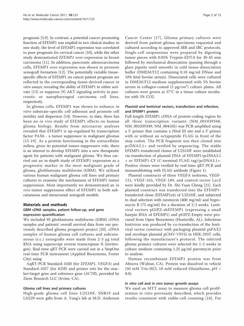
Figure 1 EFEMP1 over-expression suppressed U251HF cell growth in vivo not in vitro. A, FLAG immunoblot of conditioned-medium (CM)proteins from cell cultures of U251HF (p) and stable transfectants of FLAG-tagged EFEMP1. B & C, real-time qRT-PCR quantification of EFEMP1mRNA levels in U251HF transfected with FLAG-tagged or untagged EFEMP1 constructs, normalized to GAPDH, and compared with untransfectedcell arbitrarily set to unity. D, MTT detection of cell in vitro growth speed. E, Kaplan-Meier survival curves for mice after i.c. implantation withU251HF and its EFEMP1 transfectants.
Hu et al. Molecular Cancer 2011, 10:123http://www.molecular-cancer.com/content/10/1/123
Page 3 of 12
contrast agent were injected i.v. into the animal prior toMR imaging. SMIS NMR software was used to calculatethe dimensions of the brain tumor in the i.c. xenograftmice. Tumor volume measurement (Volume = Area ×Depth × 1/2 in mm3) involved calculating the area ofthe image with largest tumor size by the “free hand”ROI option in the software, which highlights the peri-meter of the tumor in that slice. Depth was calculatedby multiplying the number of slices where visible tumorwas seen by 0.8 (thickness of each slice).
Immunofluorescence assayImmunofluorescence of s.c. xenograft cryosections (10μm) was carried out using PECAM-1 [CD31] (1:300,Millipore Corp, Temecula, CA) and Ki67 (1:1000,Abcam Inc, Cambridge, MA) primary antibodies. Fluros-cein anti-rat IgG (H+L) and rhodamine anti-rabbit IgG(H+L) were used as secondary antibodies. The fluores-cence signals were detected using IMAGEJ 1.42 (NIH-IMAGE) which provide counting (blood vessel number)and total area (size of total blood vessels in a micro-scope image). The blood vessel density (BVD) was com-puted based on the vessel number per image of 1.02mm2 for 4-6 areas from three s.c. xenografts. For the invivo tumor-cell proliferation index (PI), information waspresented as the percentage of positive Ki67-stainingcells to the total number of DAPI-staining nuclei.
Western blotting, VEGFA enzyme immunometric assaysConditioned medium (CM) was harvested from 48-hourcell cultures and spun through Vivaspin 20 columns toincrease protein concentration. The TnT T7 QuickTranscription/Translation System (Promega, Madison,WI) was used to synthesize EFEMP1-FLAG protein,which was used as a positive control in immunoblottingfor detection of EFEMP1-FLAG using a FLAG M2monoclonal antibody (Sigma, St. Louis, MO, 1:1000dilution). Proteins of cell culture and s.c. xenograftswere extracted in radioimmunoprecipitation assay(RIPA) buffer containing 1X protease inhibitor cocktail(Roche) with a glass homogenizer (for tumors), and sub-jected to western blotting. Antibodies used include Fibu-lin-3 (mab3-5) from Santa Cruz Biotechnology (SantaCruz, California), Actin from EMD Bioscience (SanDiego, CA), EGFR, AKT, and pAKT(Ser 473) from CellSignaling Technology (Danvers, MA). A human VEGFAenzyme immunometric assay (ELISA) kit (AssayDesigns, Ann Arbor, MI) was used for quantification ofVEGFA isoform VEGF165 in tumor protein extractaccording to the manufacturer’s instructions.
Antibody arrayProtein microarray analysis were carried out using aphospho-specific antibody microarray (Full Moon
Biosystems, Inc) according to the manufacturer’s proto-col. Briefly, 100 μg of cell lysate in 50 μL of reactionmixture were labeled with 1.43 μL biotin in 10 μg/μL N,N-dimethyformamide. The resulting biotin-labeled pro-teins were diluted 1:20 in coupling solution beforeapplying to the array for conjugation. The antibodymicroarray was blocked with blocking solution for 30min at room temperature, rinsed with Milli-Q-gradewater for 3 min, dried with compressed nitrogen, andincubated with the biotin-labeled cell lysates at 4°Covernight. The slide was washed twice with 60 mL of 1×wash solution for 10 min. The conjugated-labeled pro-tein was detected using Cy3-streptavidin. The imagesand data were acquired on Axon GenePix scanner.Phospho-specific antibody activities were compared bycorresponding total protein.
Statistical analysisData was examined prior to analysis for adherence todistributional assumptions, and transformed if necessaryvia logarithmic transformation to stabilize variance.One-way ANOVAs with standard post hoc comparisonswere used to identify effects of EFEMP1 on cell MTTvalue, tumor weight, blood-vessel density, proliferationindex, and gene expression between multiple stabletransfectants and the untransfected cells. Mixed-modelsANOVAs were used to analyze longitudinally measuredtumor volumes for effects due to knock-down ofEFEMP1 using shRNA and rescue by exogenousEFEMP1 protein. Group average tumor volumes ± stan-dard errors (SEs) were calculated as the grand mean ofaverage volumes across all time points. Doubling timesand SEs were calculated from log2-scale growth ratesand their SEs by inversion and delta method, respec-tively. Overall survivals in mice implanted i.c. with dif-ferent EFEMP1 transfectants were estimated via Kaplan-Meier curves and compared for differences via logranktest. Overall survivals in humans with GBM were corre-lated with EFEMP1 expression in their tumor cDNAsamples by means of Cox regression, with the univariatepredictor being the log10-scaled ratio of EFEMP1 vs.ACTB mRNA as measured by absolute real-time qRT-PCR. SAS versions 9.1.3 and 9.2 (The SAS Institute,Cary, NC) were used for all analyses.
ResultsEFEMP1 expression in GBM is related to a longer overallsurvival of patientsWe investigated the prognostic value of EFEMP1 basedon its expression in 95 GBM specimens. The mRNAlevels of EFEMP1 and reference gene ACTB were quan-tified using absolute real-time qRT-PCR [22]. Thelog10-transformed ratio of EFEMP1 to ACTB was usedas the univariate predictor in a Cox regression with
Hu et al. Molecular Cancer 2011, 10:123http://www.molecular-cancer.com/content/10/1/123
Page 4 of 12
overall survival time as outcome variable. The parameterestimate for EFEMP1 expression in GBM was -0.182[likelihood-ratio P = 0.097], equivalent to a 17%decrease in mortality with each 10-fold increase in theEFEMP1/ACTB ratio, which suggests that the overallsurvival time increases as EFEMP1 expression levelsincrease. This finding suggested that EFEMP1 may beimportant to study for its tumor suppressor function inGBM.
EFEMP1 suppresses tumorigenicity of human high gradeglioma cell line U251HFWe started an investigation of EFEMP1 using thehuman malignant glioma cell line U251HF, which ishighly tumorigenic and forms GBM-like infiltratingnecrotic tumors in i.c. xenograft model systems [17].Since EFEMP1 is a secreted protein within the extracel-lular matrix compartment, we initially determined theability of transfected cells to secret a FLAG-taggedEFEMP1 in culture medium. As shown in Figure 1A,western blotting of proteins secreted by cells showedFLAG-tagged EFEMP1. Secreted protein levels corre-lated with EFEMP1 mRNA expression quantified byreal-time qRT-PCR (Figure 1B).We focused the functional assays on the U251HF cell
line transfected with un-tagged EFEMP1 (EFEMP1c2:9-fold increase of EFEMP1 mRNA, EFEMP1c6: 15-foldincrease of EFEMP1 mRNA, Figure 1C). As shown Fig-ure 1D, there was no obvious effect of EFEMP1 expres-sion on in vitro cell growth, nor were there anymorphological changes (data not shown). However,EFEMP1 over-expression dramatically suppressedtumorigenicity (Figure 1E). The survival of mice after i.c. implantation of the EFEMP1-transfected cells was sig-nificantly prolonged (EFEMP1c6) and in some instances,animals were completely free of tumor (EFEMP1c2). Asexpected, animals implanted with the un-transfectedparental cells (U251HF) all died of their tumor between24-46 days post-implantation. Two of the five miceimplanted with EFEMP1c6 cells died between 57-60days, one died at 90 days, while the remaining two sur-vived beyond 95 days after implantation. A repeat of thei.c. implantation experiment confirmed EFEMP1-mediated suppression of U251HF tumorigenicity in thein vivo i.c. xenograft model system.
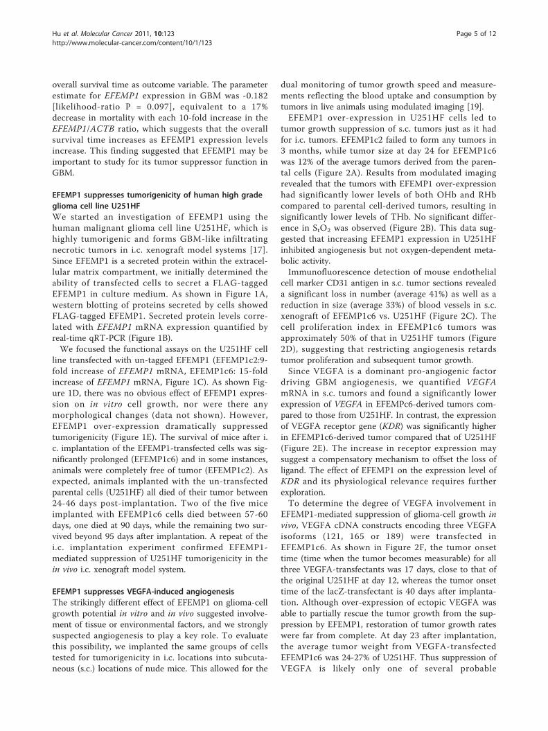
EFEMP1 suppresses VEGFA-induced angiogenesisThe strikingly different effect of EFEMP1 on glioma-cellgrowth potential in vitro and in vivo suggested involve-ment of tissue or environmental factors, and we stronglysuspected angiogenesis to play a key role. To evaluatethis possibility, we implanted the same groups of cellstested for tumorigenicity in i.c. locations into subcuta-neous (s.c.) locations of nude mice. This allowed for the
dual monitoring of tumor growth speed and measure-ments reflecting the blood uptake and consumption bytumors in live animals using modulated imaging [19].EFEMP1 over-expression in U251HF cells led to
tumor growth suppression of s.c. tumors just as it hadfor i.c. tumors. EFEMP1c2 failed to form any tumors in3 months, while tumor size at day 24 for EFEMP1c6was 12% of the average tumors derived from the paren-tal cells (Figure 2A). Results from modulated imagingrevealed that the tumors with EFEMP1 over-expressionhad significantly lower levels of both OHb and RHbcompared to parental cell-derived tumors, resulting insignificantly lower levels of THb. No significant differ-ence in StO2 was observed (Figure 2B). This data sug-gested that increasing EFEMP1 expression in U251HFinhibited angiogenesis but not oxygen-dependent meta-bolic activity.Immunofluorescence detection of mouse endothelial
cell marker CD31 antigen in s.c. tumor sections revealeda significant loss in number (average 41%) as well as areduction in size (average 33%) of blood vessels in s.c.xenograft of EFEMP1c6 vs. U251HF (Figure 2C). Thecell proliferation index in EFEMP1c6 tumors wasapproximately 50% of that in U251HF tumors (Figure2D), suggesting that restricting angiogenesis retardstumor proliferation and subsequent tumor growth.Since VEGFA is a dominant pro-angiogenic factor
driving GBM angiogenesis, we quantified VEGFAmRNA in s.c. tumors and found a significantly lowerexpression of VEGFA in EFEMPc6-derived tumors com-pared to those from U251HF. In contrast, the expressionof VEGFA receptor gene (KDR) was significantly higherin EFEMP1c6-derived tumor compared that of U251HF(Figure 2E). The increase in receptor expression maysuggest a compensatory mechanism to offset the loss ofligand. The effect of EFEMP1 on the expression level ofKDR and its physiological relevance requires furtherexploration.To determine the degree of VEGFA involvement in
EFEMP1-mediated suppression of glioma-cell growth invivo, VEGFA cDNA constructs encoding three VEGFAisoforms (121, 165 or 189) were transfected inEFEMP1c6. As shown in Figure 2F, the tumor onsettime (time when the tumor becomes measurable) for allthree VEGFA-transfectants was 17 days, close to that ofthe original U251HF at day 12, whereas the tumor onsettime of the lacZ-transfectant is 40 days after implanta-tion. Although over-expression of ectopic VEGFA wasable to partially rescue the tumor growth from the sup-pression by EFEMP1, restoration of tumor growth rateswere far from complete. At day 23 after implantation,the average tumor weight from VEGFA-transfectedEFEMP1c6 was 24-27% of U251HF. Thus suppression ofVEGFA is likely only one of several probable
Hu et al. Molecular Cancer 2011, 10:123http://www.molecular-cancer.com/content/10/1/123
Page 5 of 12
Figure 2 EFEMP1 suppression of tumorigenicity, in vivo cell proliferation, and VEGFA-induced angiogenesis. A, weights of s.c. xenograftsdissected 24 days after implantation with 5 × 10⁶ cells of U251HF and its EFEMP1 transfectants. B, total (THb) hemoglobin concentration andtissue oxygen saturation (StO2) of s.c. xenografts in live mice by modulated imaging before tumor dissections. C-D, immunofluorescence oftumor frozen-sections, with CD31 antibody detecting blood vessel density and Ki67 antibody detecting proliferation index after DAPI-counterstaining of the nuclei. E, real-time qRT-PCR quantification of gene expressions in s.c. tumors above, with mean and SD for 6-9 tumors,normalized to ACTB. F, tumor growth curve based on tumor volume measurement after s.c. implantation.
Hu et al. Molecular Cancer 2011, 10:123http://www.molecular-cancer.com/content/10/1/123
Page 6 of 12
mechanisms underlying EFEMP1 suppression of gliomagrowth and EFEMP1 exhibits some VEGF-independentanti-tumor effects.We then further investigated the effect of reducing
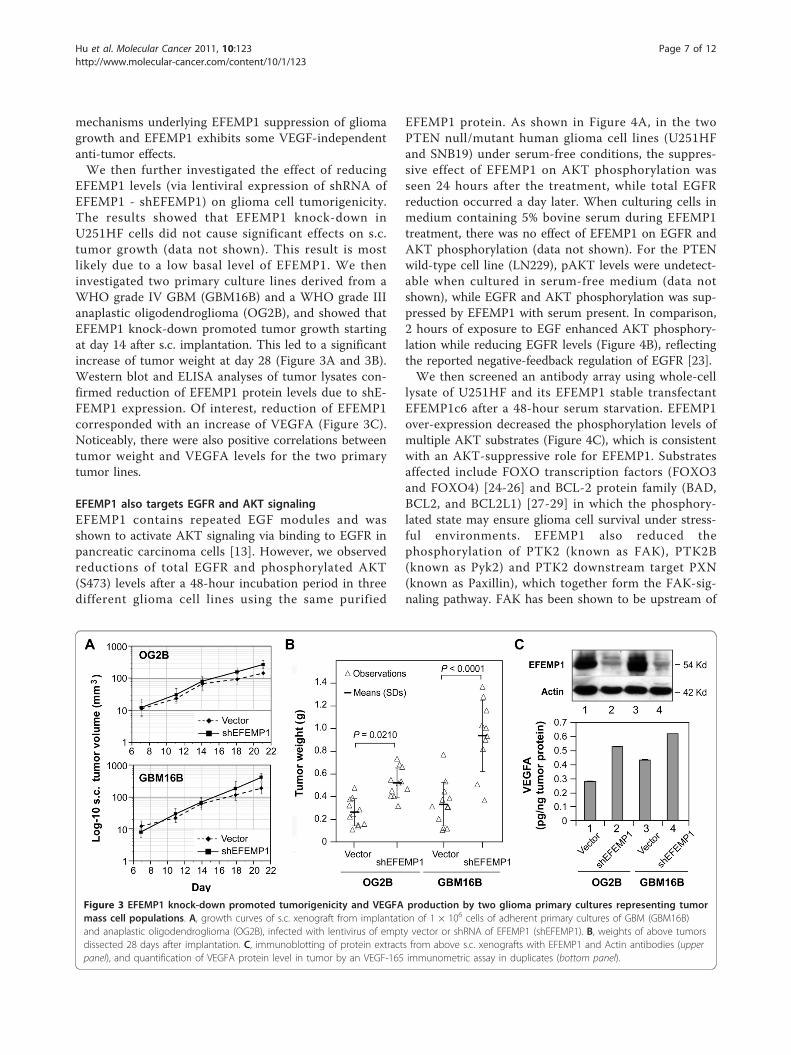
EFEMP1 levels (via lentiviral expression of shRNA ofEFEMP1 - shEFEMP1) on glioma cell tumorigenicity.The results showed that EFEMP1 knock-down inU251HF cells did not cause significant effects on s.c.tumor growth (data not shown). This result is mostlikely due to a low basal level of EFEMP1. We theninvestigated two primary culture lines derived from aWHO grade IV GBM (GBM16B) and a WHO grade IIIanaplastic oligodendroglioma (OG2B), and showed thatEFEMP1 knock-down promoted tumor growth startingat day 14 after s.c. implantation. This led to a significantincrease of tumor weight at day 28 (Figure 3A and 3B).Western blot and ELISA analyses of tumor lysates con-firmed reduction of EFEMP1 protein levels due to shE-FEMP1 expression. Of interest, reduction of EFEMP1corresponded with an increase of VEGFA (Figure 3C).Noticeably, there were also positive correlations betweentumor weight and VEGFA levels for the two primarytumor lines.
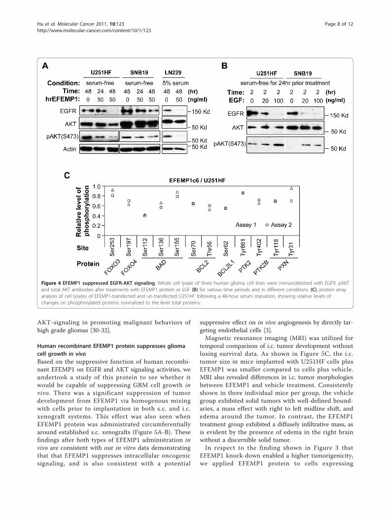
EFEMP1 also targets EGFR and AKT signalingEFEMP1 contains repeated EGF modules and wasshown to activate AKT signaling via binding to EGFR inpancreatic carcinoma cells [13]. However, we observedreductions of total EGFR and phosphorylated AKT(S473) levels after a 48-hour incubation period in threedifferent glioma cell lines using the same purified
EFEMP1 protein. As shown in Figure 4A, in the twoPTEN null/mutant human glioma cell lines (U251HFand SNB19) under serum-free conditions, the suppres-sive effect of EFEMP1 on AKT phosphorylation wasseen 24 hours after the treatment, while total EGFRreduction occurred a day later. When culturing cells inmedium containing 5% bovine serum during EFEMP1treatment, there was no effect of EFEMP1 on EGFR andAKT phosphorylation (data not shown). For the PTENwild-type cell line (LN229), pAKT levels were undetect-able when cultured in serum-free medium (data notshown), while EGFR and AKT phosphorylation was sup-pressed by EFEMP1 with serum present. In comparison,2 hours of exposure to EGF enhanced AKT phosphory-lation while reducing EGFR levels (Figure 4B), reflectingthe reported negative-feedback regulation of EGFR [23].We then screened an antibody array using whole-cell
lysate of U251HF and its EFEMP1 stable transfectantEFEMP1c6 after a 48-hour serum starvation. EFEMP1over-expression decreased the phosphorylation levels ofmultiple AKT substrates (Figure 4C), which is consistentwith an AKT-suppressive role for EFEMP1. Substratesaffected include FOXO transcription factors (FOXO3and FOXO4) [24-26] and BCL-2 protein family (BAD,BCL2, and BCL2L1) [27-29] in which the phosphory-lated state may ensure glioma cell survival under stress-ful environments. EFEMP1 also reduced thephosphorylation of PTK2 (known as FAK), PTK2B(known as Pyk2) and PTK2 downstream target PXN(known as Paxillin), which together form the FAK-sig-naling pathway. FAK has been shown to be upstream of
Figure 3 EFEMP1 knock-down promoted tumorigenicity and VEGFA production by two glioma primary cultures representing tumormass cell populations. A, growth curves of s.c. xenograft from implantation of 1 × 10⁶ cells of adherent primary cultures of GBM (GBM16B)and anaplastic oligodendroglioma (OG2B), infected with lentivirus of empty vector or shRNA of EFEMP1 (shEFEMP1). B, weights of above tumorsdissected 28 days after implantation. C, immunoblotting of protein extracts from above s.c. xenografts with EFEMP1 and Actin antibodies (upperpanel), and quantification of VEGFA protein level in tumor by an VEGF-165 immunometric assay in duplicates (bottom panel).
Hu et al. Molecular Cancer 2011, 10:123http://www.molecular-cancer.com/content/10/1/123
Page 7 of 12
AKT-signaling in promoting malignant behaviors ofhigh grade gliomas [30-32].
Human recombinant EFEMP1 protein suppresses gliomacell growth in vivoBased on the suppressive function of human recombi-nant EFEMP1 on EGFR and AKT signaling activities, weundertook a study of this protein to see whether itwould be capable of suppressing GBM cell growth invivo. There was a significant suppression of tumordevelopment from EFEMP1 via homogenous mixingwith cells prior to implantation in both s.c. and i.c.xenograft systems. This effect was also seen whenEFEMP1 protein was administrated circumferentiallyaround established s.c. xenografts (Figure 5A-B). Thesefindings after both types of EFEMP1 administration invivo are consistent with our in vitro data demonstratingthat that EFEMP1 suppresses intracellular oncogenicsignaling, and is also consistent with a potential
suppressive effect on in vivo angiogenesis by directly tar-geting endothelial cells [3].Magnetic resonance imaging (MRI) was utilized for
temporal comparison of i.c. tumor development withoutlosing survival data. As shown in Figure 5C, the i.c.tumor size in mice implanted with U251HF cells plusEFEMP1 was smaller compared to cells plus vehicle.MRI also revealed differences in i.c. tumor morphologiesbetween EFEMP1 and vehicle treatment. Consistentlyshown in three individual mice per group, the vehiclegroup exhibited solid tumors with well-defined bound-aries, a mass effect with right to left midline shift, andedema around the tumor. In contrast, the EFEMP1treatment group exhibited a diffusely infiltrative mass, asis evident by the presence of edema in the right brainwithout a discernible solid tumor.In respect to the finding shown in Figure 3 that
EFEMP1 knock-down enabled a higher tumorigenicity,we applied EFEMP1 protein to cells expressing
Figure 4 EFEMP1 suppressed EGFR-AKT signaling. Whole cell lysate of three human glioma cell lines were immunoblotted with EGFR, pAKTand total AKT antibodies after treatments with EFEMP1 protein or EGF (B) for various time periods and in different conditions. (C), protein arrayanalysis of cell lysates of EFEMP1-transfected and un-transfected U251HF following a 48-hour serum starvation, showing relative levels ofchanges on phosphorylated proteins normalized to the level total proteins.
Hu et al. Molecular Cancer 2011, 10:123http://www.molecular-cancer.com/content/10/1/123
Page 8 of 12
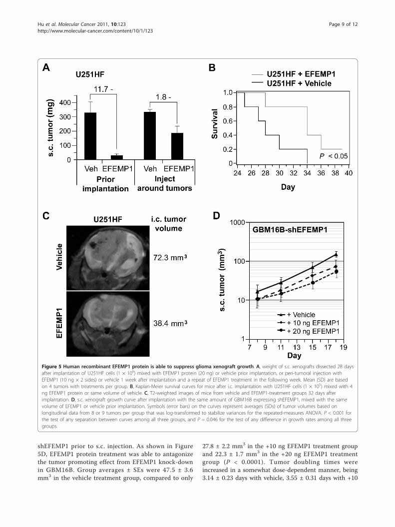
shEFEMP1 prior to s.c. injection. As shown in Figure5D, EFEMP1 protein treatment was able to antagonizethe tumor promoting effect from EFEMP1 knock-downin GBM16B. Group averages ± SEs were 47.5 ± 3.6mm3 in the vehicle treatment group, compared to only
27.8 ± 2.2 mm3 in the +10 ng EFEMP1 treatment groupand 22.3 ± 1.7 mm3 in the +20 ng EFEMP1 treatmentgroup (P < 0.0001). Tumor doubling times wereincreased in a somewhat dose-dependent manner, being3.14 ± 0.23 days with vehicle, 3.55 ± 0.31 days with +10
Figure 5 Human recombinant EFEMP1 protein is able to suppress glioma xenograft growth. A, weight of s.c. xenografts dissected 28 daysafter implantation of U251HF cells (1 × 10⁶) mixed with EFEMP1 protein (20 ng) or vehicle prior implantation, or peri-tumoral injection withEFEMP1 (10 ng × 2 sides) or vehicle 1 week after implantation and a repeat of EFEMP1 treatment in the following week. Mean (SD) are basedon 4 tumors with treatments per group. B, Kaplan-Meier survival curves for mice after i.c. implantation with U251HF cells (1 × 10⁵) mixed with 4ng EFEMP1 protein or same volume of vehicle. C, T2-weighted images of mice from vehicle and EFEMP1-treatment groups 32 days afterimplantation. D, s.c. xenograft growth curve after implantation with the same amount of GBM16B expressing shEFEMP1, mixed with the samevolume of EFEMP1 or vehicle prior implantation. Symbols (error bars) on the curves represent averages (SDs) of tumor volumes based onlongitudinal data from 8 or 9 tumors per group that was log-transformed to stabilize variances for the repeated-measures ANOVA. P < 0.001 forthe test of any separation between curves among all three groups, and P = 0.046 for the test of any difference in growth rates among all threegroups.
Hu et al. Molecular Cancer 2011, 10:123http://www.molecular-cancer.com/content/10/1/123
Page 9 of 12
ng EFEMP1, and 4.24 ± 0.42 days in the +20 ngEFEMP1 (P = 0.046).
DiscussionThe overall results of this study revealed a role forEFEMP1 in the suppression of glioma growth, via inde-pendent blockade of EGFR and AKT signaling pathwaysand the repression of VEGFA-induced angiogenesis.Activation of these molecular pathways is a well-knownmolecular-pathological feature of GBM. The finding of afavorable clinical prognosis effect from EFEMP1 expres-sion in GBM patients is consistent with these laboratoryfindings. EFEMP1 anti-angiogenic effects appear to takeplace through both VEGFA-dependent and VEGFA-independent mechanisms. EFEMP1-mediated suppres-sion of glioma-cell expression of VEGFA would result inthe suppression of VEGFA stimulation of angiogenesis,which is in addition to EFEMP1’s direct inhibition ofendothelial cells sprouting [3].In addition to the overall improvement of mouse sur-
vival resulting from EFEMP1 over-expression, MRI dataof i.c. glioma morphologies revealed EFEMP1’s functionin potentiating glioma-cell infiltration, as shown by invitro studies [14]. This is the first demonstration ofEFEMP1’s role in the regulation of glioma cell invasionin an i.c. xenograft system. In our GBM cell line/pri-mary culture in vivo systems, the tumor-suppressiveeffect of EFEMP1 overwhelmed its pro-invasive activity.Further investigation is needed to understand the mole-cular context of EFEMP1 in control of different malig-nant behaviors of cancer cells.This study showed that EFEMP1 repressed AKT activ-
ity and phosphorylation of multiple AKT substrates inhuman malignant glioma cells. These results differedfrom observations in pancreatic adenocarcinoma cells,where EFEMP1 enhanced AKT phosphorylation [13],but were in accordance with findings in nasopharyngealcarcinomas cells, where EFEMP1 suppressed AKT phos-phorylation. Such discrepancies may be related to differ-ent membrane receptors and downstream signalingpathways given the plethora of molecular and cellularabnormalities in different cancer cell types.EGFR amplification occurs in 40 to 70% of primary
GBMs, but is not observed in lower-grade astrocytomas[33]. Activation of EGFR and PI3K/AKT signaling path-ways are clearly the causes of malignant behavior ofGBM and emerges as common pathways regulating cel-lular proliferation, survival, migration and invasion [34].Our data revealed the negative regulation of EGFR byEFEMP1, via down-regulation of total EGFR levels. It iswell-documented that receptor activation by EGF leadsto the internalization and degradation of EGFR [35].The EFEMP1-initiated reduction of total EGFR,although not as quick as EGF-initiated negative-feedback
regulation of EGFR, could involve a similar protein-degradation mechanism, since EGFR mRNA levels werenot reduced upon EFEMP1 treatment.We have shown that EFEMP1-mediated down-regula-
tion of AKT signaling occurs in PTEN null/mutantglioma cells under serum-starvation conditions but notin serum-containing medium. This is consistent withthe lack of an EFEMP1-mediated effect on cell prolifera-tion in serum-containing culture medium, in contrast tothe dramatic suppression of tumor formation in both s.c. and i.c. xenograft systems. Our data revealed thatEFEMP1 suppresses tumor growth via multiple mechan-isms. In addition to the aforementioned effect onattenuating EGFR/AKT signaling activities, we demon-strate EFEMP1 involvement in regulation of the tumormicroenvironment through down-regulation of glioma-cell VEGFA production. The mechanism of EFEMP1 inthe down-regulation of VEGFA expression is not clari-fied by this study, though it could be an indirect effectfrom inhibition of AKT signaling [36].Current anti-angiogenic agents such as bevacizumab, a
humanized VEGFA antibody, have met with limited suc-cess in glioma patients due to its promoting cell inva-sion [37, 38]. Either EGFR-targeting monoclonalantibody or inhibition of tyrosine kinases activity inglioma patients produced minimal tumor response andno improvement in overall survival [39, 40]. The identi-fication for EFEMP1 of a VEGFA-independent mechan-ism in addition to its VEGFA-dependent mechanism insuppressing angiogenesis and EFGR- and AKT-signalingactivities suggests that it may be possible to developadditional anti-angiogenic agents that are complemen-tary to bevacizamab, or potentially still effective in theface of bevacizumab resistance. While more studies areneeded to further define the role of EFEMP1 (i.e. anti-angiogenic vs. pro-invasive), results from this study pro-vide a mechanistic rationale on developing EFEMP1, orEFEMP1-derived signaling peptide, as a new potenttherapy for patients with high-grade gliomas.In reviewing published studies on EFEMP1 in cancer
and incorporating results from this study, it should benoted that EFEMP1’s function, whether promoting orinhibiting cancer growth, may be dependent on a mole-cular context that differs by cancer cell type and malig-nancy stage. The identification of proteins that arecooperative with EFEMP1 in the regulation of intracellu-lar signaling pathways and the tumor microenvironmentwould distinguish the differential effect of EFEMP1 incancer. The tumor-suppressive effect of EFEMP1 toglioma cells may also be applicable to other cancertypes that have hyperactivation of EGFR and AKT sig-naling pathways. Some of these are lung, liver, breast,and prostate cancers, where EFEMP1 promoter methyla-tion and/or expression were down-regulated [4-8].
Hu et al. Molecular Cancer 2011, 10:123http://www.molecular-cancer.com/content/10/1/123
Page 10 of 12
ConclusionOur data revealed that EFEMP1 is a favorable prognos-tic factor for GBM, has a tumor-suppressive effect inmalignant glioma cells, and acts in the extracellularcompartment via independent blocking of EGFR andAKT signaling pathways while also repressing VEGFA-induced angiogenesis. Results of these findings justifytherapeutic development of EFEMP1-derived agents forGBM. In addition, the discovery of the function ofEFEMP1 as a tumor suppressor that modulates EGFRsuggests a need to further investigate EFEMP1 as apotential predictive marker for anti-EGFR therapies ofcancer, including glioblastoma, lung, breast, prostate,and liver cancers.
Author’s informationCorresponding author YHZ, assistant professor in UCIrvine since 2006, has started her research focus sinceher discovery of PAX6 suppressing GBM cell growth invivo and as a favourable prognosis factor in high-gradegliomas and, with mechanisms involved in suppressionof glioma invasion, survival under oxidative stress, andangiogenesis published in Clinical Cancer Research in2003, Cancer Research in 2006, and Journal of Neuro-Oncology in 2005 and 2010. This study is a continua-tion of her study of PAX6 in glioma, focusing on identi-fication of a therapeutic target/agent for GBMtreatment. From the initial discovery of EFEMP1 as apotential target of PAX6 in 2004 while YHZ wasresearch assistant professor in UAMS, her lab focusedon experiments designed to explore the role of EFEMP1as an effector of PAX6 suppression of in vivo growth bysuppression of angiogenesis, and the clinical prognosiseffect of EFEMP1 for the translational meaning ofresults from this study. Overall 16 researchers partici-pated in this study from a period of 6 years.First author YH, PhD candidate, UC Irvine, has parti-
cipated in PAX6 and EFEMP1 studies for 5 years, co-authored in CR and JNO papers from Zhou lab. KRH,professor, MD Anderson Cancer Center, has been colla-borating with YHZ since 2003 on glioma prognosisstudy based on real-time PCR quantified gene expres-sion data.
List of abbreviationsBVD: blood vessel density; ELISA: immunometric assay; MI: Modulatedimaging; MRI: magnetic resonance imaging; GBM: Glioblastoma multiforme; i.c.: intracranial; s.c.: subcutaneous; Ohb: oxyhemoglobin; RHb:deoxyhemoglobin; THb: total hemoglobin; StO2: oxygen saturation; PI:proliferation index.
AcknowledgementsThis work was supported in part by the University of California, Irvine, Schoolof Medicine and Committee on Research Award, Chao FamilyComprehensive Cancer Center Seed Grant, a generous Stern Family gift
administered through the UC Irvine Foundation, and the Arkansas CancerResearch Center at the University of Arkansas for Medical Sciences.
Author details1Department of Biological Chemistry, University of California Irvine, 5171California Ave., Suite 150, Irvine, CA (92697), USA. 2Department ofBiostatistics, University of Arkansas for Medical Sciences, 4301 W. MarkhamSt., Little Rock, AR (72205), USA. 3National Engineering Center for Biochip atShanghai & Shanghai Biochip Co., Lt, 151 Libing Rd, Shanghai (201203),China. 4Neurological Surgery, University of California Irvine, 5171 CaliforniaAve., Suite 150, Irvine, CA (92697), USA. 5Tu & Yuen Center for FunctionalOnco-Imaging, University of California Irvine, 5171 California Ave., Suite 150,Irvine, CA (92697), USA. 6Ziren Research LLC, 9841 Irvine Center Drive, Irvine,CA (92618), USA. 7Department of Biostatistics, University of Texas M. D.Anderson Cancer Center, 1515 Holcombe Blvd, Houston, TX (77030), USA.8Beckman Laser Institute, University of California Irvine, 5171 California Ave.,Suite 150, Irvine, CA (92697), USA.
Authors’ contributionsYH, PDP, QZ, JN, AC, MSM, DR, SA, NH, MH, and LY have participated inprocessing the experiments of with data presented in this manuscript and/or verified the conclusions of this study. KRH performed the Cox analysisand data interpretation, ES performed all statistical analyses, BT contributedto the MI data interpretation, and ML participated in data discussion. YHZconceived this study, made the constructs, participated in its design andcoordination. The manuscript was written by YHZ, edited by ES, ML, andPDP. All authors read and approved the final manuscript.
Competing interestsThe authors declare that they have no competing interests.
Received: 26 May 2011 Accepted: 28 September 2011Published: 28 September 2011
References1. de Vega S, Iwamoto T, Yamada Y: Fibulins: multiple roles in matrix
structures and tissue functions. Cell Mol Life Sci 2009, 66:1890-1902.2. Gallagher WM, Currid CA, Whelan LC: Fibulins and cancer: friend or foe?
Trends Mol Med 2005, 11:336-340.3. Albig AR, Neil JR, Schiemann WP: Fibulins 3 and 5 antagonize tumor
angiogenesis in vivo. Cancer Res 2006, 66:2621-2629.4. Yue W, Dacic S, Sun Q, Landreneau R, Guo M, Zhou W, Siegfried JM, Yu J,
Zhang L: Frequent inactivation of RAMP2, EFEMP1 and Dutt1 in lungcancer by promoter hypermethylation. Clin Cancer Res 2007, 13:4336-4344.
5. Nomoto S, Kanda M, Okamura Y, Nishikawa Y, Qiyong L, Fujii T,Sugimoto H, Takeda S, Nakao A: Epidermal growth factor-containingfibulin-like extracellular matrix protein 1, EFEMP1, a novel tumor-suppressor gene detected in hepatocellular carcinoma using doublecombination array analysis. Ann Surg Oncol 2010, 17:923-932.
6. Kim YJ, Yoon HY, Kim SK, Kim YW, Kim EJ, Kim IY, Kim WJ: EFEMP1 as aNovel DNA Methylation Marker for Prostate Cancer: Array-Based DNAMethylation and Expression Profiling. Clin Cancer Res 2011, 17:4523-4530.
7. Zhang Y, Wang R, Song H, Huang G, Yi J, Zheng Y, Wang J, Chen L:Methylation of multiple genes as a candidate biomarker in non-smallcell lung cancer. Cancer Lett 2011, 303:21-28.
8. Sadr-Nabavi A, Ramser J, Volkmann J, Naehrig J, Wiesmann F, Betz B,Hellebrand H, Engert S, Seitz S, Kreutzfeld R, et al: Decreased expression ofangiogenesis antagonist EFEMP1 in sporadic breast cancer is caused byaberrant promoter methylation and points to an impact of EFEMP1 asmolecular biomarker. Int J Cancer 2009, 124:1727-1735.
9. Hwang CF, Chien CY, Huang SC, Yin YF, Huang CC, Fang FM, Tsai HT, Su LJ,Chen CH: Fibulin-3 is associated with tumour progression and a poorprognosis in nasopharyngeal carcinomas and inhibits cell migration andinvasion via suppressed AKT activity. J Pathol 2010, 222:367-379.
10. En-lin S, Sheng-guo C, Hua-qiao W: The expression of EFEMP1 in cervicalcarcinoma and its relationship with prognosis. Gynecol Oncol 2010,117:417-422.
11. Davidson B, Stavnes HT, Holth A, Chen X, Yang Y, Shih Ie M, Wang TL:Gene expression signatures differentiate ovarian/peritoneal serouscarcinoma from breast carcinoma in effusions. J Cell Mol Med 2011,15:535-544.
Hu et al. Molecular Cancer 2011, 10:123http://www.molecular-cancer.com/content/10/1/123
Page 11 of 12
12. Seeliger H, Camaj P, Ischenko I, Kleespies A, De Toni EN, Thieme SE, Blum H,Assmann G, Jauch KW, Bruns CJ: EFEMP1 expression promotes in vivotumor growth in human pancreatic adenocarcinoma. Mol Cancer Res2009, 7:189-198.
13. Camaj P, Seeliger H, Ischenko I, Krebs S, Blum H, De Toni EN, Faktorova D,Jauch KW, Bruns CJ: EFEMP1 binds the EGF receptor and activates MAPKand Akt pathways in pancreatic carcinoma cells. Biol Chem 2009,390:1293-1302.
14. Hu B, Thirtamara-Rajamani KK, Sim H, Viapiano MS: Fibulin-3 is uniquelyupregulated in malignant gliomas and promotes tumor cell motility andinvasion. Mol Cancer Res 2009, 7:1756-1770.
15. Zhou YH, Tan F, Hess KR, Yung WK: The expression of PAX6, PTEN,vascular endothelial growth factor, and epidermal growth factorreceptor in gliomas: relationship to tumor grade and survival. Clin CancerRes 2003, 9:3369-3375.
16. Zhou YH, Wu X, Tan F, Shi YX, Glass T, Liu TJ, Wathen K, Hess KR, Gumin J,Lang F, Yung WK: PAX6 suppresses growth of human glioblastoma cells.J Neurooncol 2005, 71:223-229.
17. Mayes DA, Hu Y, Teng Y, Siegel E, Wu X, Panda K, Tan F, Yung WK,Zhou YH: PAX6 suppresses the invasiveness of glioblastoma cells andthe expression of the matrix metalloproteinase-2 gene. Cancer Res 2006,66:9809-9817.
18. Chang JY, Hu Y, Siegel E, Stanley L, Zhou YH: PAX6 increases glioma cellsusceptibility to detachment and oxidative stress. J Neurooncol 2007,84:9-19.
19. Zhou YH, Hu Y, Mayes D, Siegel E, Kim JG, Mathews MS, Hsu N, Eskander D,Yu O, Tromberg BJ, Linskey ME: PAX6 suppression of glioma angiogenesisand the expression of vascular endothelial growth factor A. J Neurooncol2010, 96:191-200.
20. Zhou YH, Hess KR, Raj VR, Yu L, Liu L, Yung AWK, Linskey ME:Establishment of prognostic models for astrocytic and oligodendroglialbrain tumors with standardized quantification of marker geneexpression and clinical variables. Biomarker Insights 2010, 2010:153-168.
21. Guo P, Xu L, Pan S, Brekken RA, Yang ST, Whitaker GB, Nagane M,Thorpe PE, Rosenbaum JS, Su Huang HJ, et al: Vascular endothelial growthfactor isoforms display distinct activities in promoting tumorangiogenesis at different anatomic sites. Cancer Res 2001, 61:8569-8577.
22. Zhou YH, Raj VR, Siegel E, Yu L: Standardization of gene expressionquantification by absolute real-time qRT-PCR system using a singlestandard for marker and reference genes. Biomarker Insights 2010,2010:79-85.
23. Huang PH, Xu AM, White FM: Oncogenic EGFR signaling networks inglioma. Science signaling 2009, 2:re6.
24. Dansen TB, Burgering BM: Unravelling the tumor-suppressive functions ofFOXO proteins. Trends in cell biology 2008, 18:421-429.
25. Brunet A, Bonni A, Zigmond MJ, Lin MZ, Juo P, Hu LS, Anderson MJ,Arden KC, Blenis J, Greenberg ME: Akt promotes cell survival byphosphorylating and inhibiting a Forkhead transcription factor. Cell 1999,96:857-868.
26. Kops GJ, de Ruiter ND, De Vries-Smits AM, Powell DR, Bos JL, Burgering BM:Direct control of the Forkhead transcription factor AFX by protein kinaseB. Nature 1999, 398:630-634.
27. Datta SR, Dudek H, Tao X, Masters S, Fu H, Gotoh Y, Greenberg ME: Aktphosphorylation of BAD couples survival signals to the cell-intrinsicdeath machinery. Cell 1997, 91:231-241.
28. Datta SR, Ranger AM, Lin MZ, Sturgill JF, Ma YC, Cowan CW, Dikkes P,Korsmeyer SJ, Greenberg ME: Survival factor-mediated BADphosphorylation raises the mitochondrial threshold for apoptosis.Developmental cell 2002, 3:631-643.
29. Willimott S, Wagner SD: Post-transcriptional and post-translationalregulation of Bcl2. Biochemical Society transactions 2010, 38:1571-1575.
30. Natarajan M, Hecker TP, Gladson CL: FAK signaling in anaplasticastrocytoma and glioblastoma tumors. Cancer journal 2003, 9:126-133.
31. Gutenberg A, Bruck W, Buchfelder M, Ludwig HC: Expression of tyrosinekinases FAK and Pyk2 in 331 human astrocytomas. Acta Neuropathologica2004, 108:224-230.
32. Bellis SL, Miller JT, Turner CE: Characterization of tyrosine phosphorylationof paxillin in vitro by focal adhesion kinase. The Journal of biologicalchemistry 1995, 270:17437-17441.
33. Ekstrand AJ, Sugawa N, James CD, Collins VP: Amplified and rearrangedepidermal growth factor receptor genes in human glioblastomas reveal
deletions of sequences encoding portions of the N- and/or C-terminaltails. Proc Natl Acad Sci USA 1992, 89:4309-4313.
34. Louis DN: Molecular pathology of malignant gliomas. Annual review ofpathology 2006, 1:97-117.
35. Soubeyran P, Kowanetz K, Szymkiewicz I, Langdon WY, Dikic I: Cbl-CIN85-endophilin complex mediates ligand-induced downregulation of EGFreceptors. Nature 2002, 416:183-187.
36. Gomez-Manzano C, Fueyo J, Jiang H, Glass TL, Lee HY, Hu M, Liu JL,Jasti SL, Liu TJ, Conrad CA, Yung WK: Mechanisms underlying PTENregulation of vascular endothelial growth factor and angiogenesis.Annals of Neurology 2003, 53:109-117.
doi:10.1186/1476-4598-10-123Cite this article as: Hu et al.: EFEMP1 suppresses malignant gliomagrowth and exerts its action within the tumor extracellularcompartment. Molecular Cancer 2011 10:123.
Submit your next manuscript to BioMed Centraland take full advantage of:
• Convenient online submission
• Thorough peer review
• No space constraints or color figure charges
• Immediate publication on acceptance
• Inclusion in PubMed, CAS, Scopus and Google Scholar
• Research which is freely available for redistribution
Submit your manuscript at www.biomedcentral.com/submit
Hu et al. Molecular Cancer 2011, 10:123http://www.molecular-cancer.com/content/10/1/123
Page 12 of 12


![cells inhibits angiogenesis in glioblastoma · cells * glioma Downregulation INTRODUCTION Angiogenesis is a key event in the progression of malignant gliomas [1,2]. It is a highly](https://img.pdfslide.us/doc/110x75/5ecd7b084c46b638be2fbb49/cells-inhibits-angiogenesis-in-glioblastoma-cells-glioma-downregulation-introduction.jpg)