Embed Size (px)
Citation preview
RESEARCH Open Access
A whole genome screen for HIV restriction factorsLi Liu1, Nidia MM Oliveira1†, Kelly M Cheney1†, Corinna Pade1, Hanna Dreja1, Ann-Marie H Bergin2, Viola Borgdorff2
, David H Beach2, Cleo L Bishop2, Matthias T Dittmar1 and Áine McKnight1*
Abstract
Background: Upon cellular entry retroviruses must avoid innate restriction factors produced by the host cell. Forhuman immunodeficiency virus (HIV) human restriction factors, APOBEC3 (apolipoprotein-B-mRNA-editing-enzyme),p21 and tetherin are well characterised.
Results: To identify intrinsic resistance factors to HIV-1 replication we screened 19,121 human genes and identified114 factors with significant inhibition of infection. Those with a known function are involved in a broad spectrumof cellular processes including receptor signalling, vesicle trafficking, transcription, apoptosis, cross-nuclearmembrane transport, meiosis, DNA damage repair, ubiquitination and RNA processing. We focused on the PAF1complex which has been previously implicated in gene transcription, cell cycle control and mRNA surveillance.Knockdown of all members of the PAF1 family of proteins enhanced HIV-1 reverse transcription and integration ofprovirus. Over-expression of PAF1 in host cells renders them refractory to HIV-1. Simian Immunodeficiency Virusesand HIV-2 are also restricted in PAF1 expressing cells. PAF1 is expressed in primary monocytes, macrophages andT-lymphocytes and we demonstrate strong activity in MonoMac1, a monocyte cell line.
Conclusions: We propose that the PAF1c establishes an anti-viral state to prevent infection by incomingretroviruses. This previously unrecognised mechanism of restriction could have implications for invasion of cells byany pathogen.
BackgroundViruses usurp normal cellular processes to completetheir life cycle. Once inside the cell cytoplasm viralRNA is reverse transcribed into single stranded cDNAfollowed by double stranded (ds)DNA. The dsDNA incells forms a pre integration complex (PIC) whichincludes viral proteins and interacts with numerous cellcomponents. Eventually the PIC is transported into thenucleus for host DNA integration.The use of small-interfering RNA (siRNA) screens has
greatly extended our knowledge of the cellular processeshijacked by viruses for infection and the componentsneeded by HIV to facilitate these early steps in replica-tion [1-4]. For example TNPO3, was identified by twoscreens to be a required for a replication step in theHIV life cycle [1,2]. TNPO3 was later shown to facilitatenuclear import of the PIC [5].
Host cells, however, have evolved intrinsic resistancefactors to mitigate viral replication. Several host restric-tion factors have been identified that prevent the pro-gression of HIV replication during the early phase of thelife cycle. The best characterised of these are encoded bythe TRIM5a and the APOBEC gene families [6,7]. APO-BECs interact with the nascent DNA during reversetranscription [6]. TRIM5a interacts with incoming viralcapsids (CA) resulting in premature disassembly [7].TRIM28/KAP1 has recently been shown to restrict inte-gration of HIV-1 [8]. p21(Waf1/Cip1/Sdi1) (p21) wasidentified to act during or after reverse transcription[9,10]. SAMHD1 acts prior to integration, possibly bydegrading or preventing the accumulation of HIV DNA[11]. Another restriction factor Tetherin (BST-2/CD317)acts post integration to prevent viruses from leaving thecell during the budding stage of the life cycle [12].To detect intrinsic anti-viral restriction factors acting at
the early, post fusion stages of HIV-1 replication, HeLa-CD4 cells were transfected with an siRNA library targeting19,121 human genes and then challenged with an HIV-189.6R pseudovirus carrying a GFP reporter gene (HIV-1
* Correspondence: [email protected]† Contributed equally1Centre for Immunology and Infectious Disease, Blizard Institute, Barts andThe London School of Medicine and Dentistry, Queen Mary University ofLondon, 4 Newark Street, London E1 2AT, UKFull list of author information is available at the end of the article
Liu et al. Retrovirology 2011, 8:94http://www.retrovirology.com/content/8/1/94
© 2011 Liu et al; licensee BioMed Central Ltd. This is an Open Access article distributed under the terms of the Creative CommonsAttribution License (http://creativecommons.org/licenses/by/2.0), which permits unrestricted use, distribution, and reproduction inany medium, provided the original work is properly cited.
gag/pol/tat and rev, HIV-2 MCR Env). The negative fac-tors identified perform a diverse range of cellular activities.Those with known function are involved in receptor sig-nalling, vesicle trafficking, transcription, apoptosis, cross-nuclear membrane transport, meiosis, DNA damagerepair, ubiquitination and RNA processing. Our screen foranti-HIV factors can serve as a platform to understandingthe host’s adaptation viral infection.
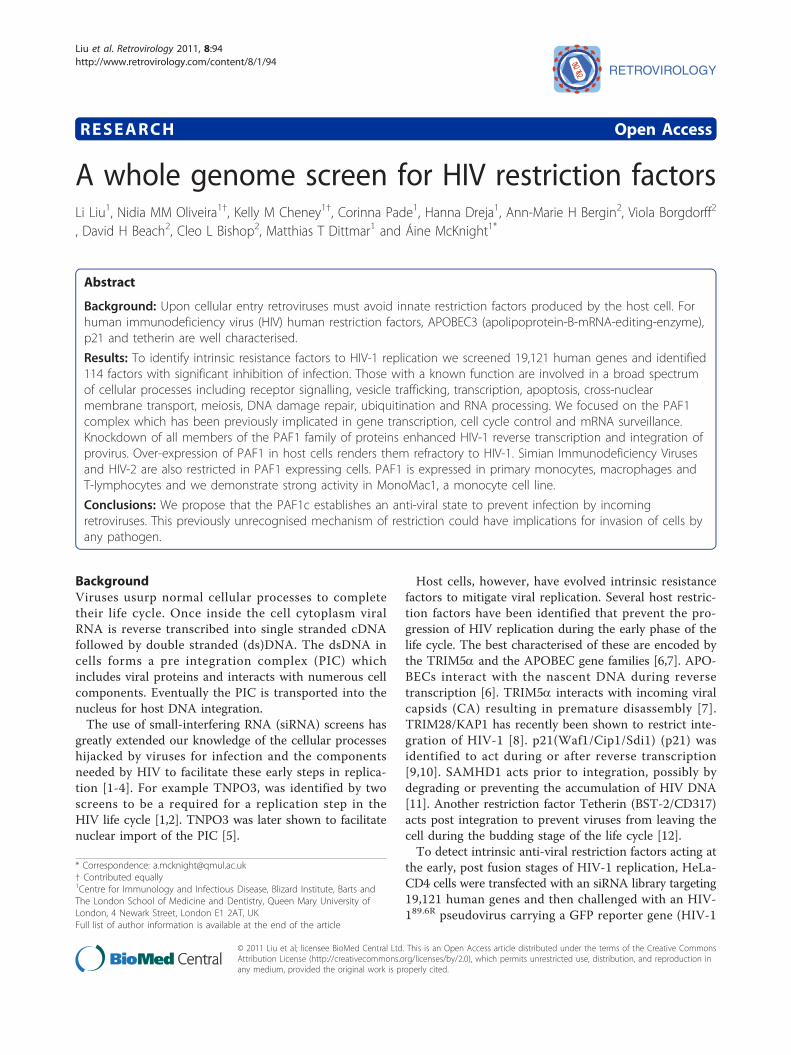
ResultsSystem SetupTo detect human cellular restriction factors that operateat the early stages of HIV-1 replication, we developed asingle round infectious HIV pseudotype assay to siRNAscreen HeLa-CD4 cells. The HIV pseudotype HIV89.6R,has an HIV-2 Env MCR (derived from the primary
isolate prCBL-23). HeLa-CD4 cells contain ectopicallyexpressed CD4 but naturally express the co-receptorCXCR4. Both receptors are used by HIV89.6R to entercells. HIV89.6R was evaluated for tropism in HeLa-CD4cells. Although HIV89.6R replicates efficiently on NP2-CD4-CXCR4 cells it is restricted on HeLa-CD4 cells(Figure 1B) while HIV8.2N grows equally well on bothcell types and was used as a positive control for viralreplication and to monitor the GFP expression andsiRNA effects (Figure 1C). The viral pseudotypesHIV89.6R and HIV8.2N are only capable of a single roundof infection so the number of GFP expressing cells isequivalent to virus infectious units (or focus formingunits, FFU). An increase in infectious units after siRNAgene knockdown followed by virus challenge after 72hours indicated rescue of viral replication.
A B
C
D
Figure 1 siRNA screen setup. 1A Screen strategy and results. 1B Infectious units/μl of HIV8.2N and HIV89.6R virus stocks following challenge onHeLa-CD4 and NP2-CD4-CXCR4 cells. Results are mean ± SD of a representative experiment performed in triplicate. 1C GFP+ foci following viruschallenge of HeLa-CD4 cells of HIV89.6R and HIV8.2N. Green, virus; red, cells. 1D siRNA knockdown of AP2M1 and PAF1 rescues infection of HeLa-CD4 cells by HIV89.6R compared with a negative control siRNA (CB). HIV8.2N is the non-restricted positive control. % infection: AP2M1 3.57%, PAF10.65%, CB 0.0004%. Green, virus; red, cells.
Liu et al. Retrovirology 2011, 8:94http://www.retrovirology.com/content/8/1/94
Page 2 of 15
To optimise the screen we used negative control siR-NAs targeting cyclophilin B (CB; siGLO), PLK1 andGFP. The reverse transfection protocol was almost 100%efficient, CB protein expression was reduced by 60%, thePLK1 siRNA (cell killer control) reduced cell number bymore than 99% and GFP siRNA reduced GFP intensityby 67.8% (data not shown). siRNAs against the CD4receptor and the nuclear importin TNPO3 were used totest the effects of the siRNAs on the inhibition of non-restricted HIV8.2N infectivity. The results show that theinfection was reduced by 95.8% and 93.0%, respectively(Additional file 1, A1).
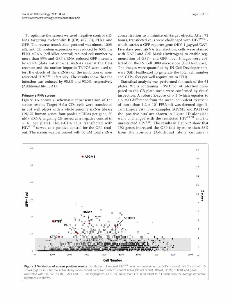
Primary siRNA screenFigure 1A shows a schematic representation of thescreen results. Target HeLa-CD4 cells were transfectedin 384 well plates with a whole genome siRNA library(19,121 human genes, four pooled siRNAs per gene, 30nM). siRNA targeting CB served as a negative control (n= 16 per plate). HeLa-CD4 cells transfected withHIV8.2N served as a positive control for the GFP read-out. The screen was performed with 30 nM total siRNA
concentration to minimise off-target effects. After 72hours, transfected cells were challenged with HIV89.6R -which carries a GFP reporter gene (HIV-1 gag/pol/GFP).Five days post siRNA transfection, cells were stainedwith DAPI and Cell Mask (Invitrogen) to enable seg-mentation of GFP+ and GFP- foci. Images were col-lected on the IN Cell 1000 microscope (GE Healthcare).The images were quantified by IN Cell Developer soft-ware (GE Healthcare) to generate the total cell numberand GFP+ foci per well (equivalent to FFU).Statistical analysis was performed for each of the 61
plates. Wells containing > 3SD foci of infection com-pared to the CB plate mean were confirmed by visualinspection. A robust Z-score of > 3 (which equates toa > 3SD difference from the mean; equivalent to rescueof more than 1.2 × 10³ FFU/ml) was deemed signifi-cant (Figure 2A). Two examples (AP2M1 and PAF1) ofthe ‘positive hits’ are shown in Figure 1D alongsidewells challenged with the restricted HIV89.6R and theunrestricted HIV8.2N. The results in Figure 2 show that192 genes increased the GFP foci by more than 3SDfrom the controls (Additional file 2 contains a
AP2M1
RTF1 PAF1
CTR9
SETDB1
DNM2
Figure 2 Validation of screen positive results. Distribution of rescued HIV89.6R infection determined by GFP+ foci/well (left Y axis) with Z-scores (right Y axis) for the siRNA library (open circles) compared with CB control siRNA (closed circles). AP2M1, DNM2, SETDB1 and genesassociated with the PAF1c CTR9, PAF1 and RTF1 are highlighted. GFP+ foci more than 3 SD (equivalent to 7.43 foci) from the average of controlinfections are shown.
Liu et al. Retrovirology 2011, 8:94http://www.retrovirology.com/content/8/1/94
Page 3 of 15
complete list of genes and their corresponding Z-scores, A2).
Secondary screenNext, we performed a secondary validation screen for183 of the 192 candidates from the primary screen.Transfection of the original pooled siRNAs was per-formed in triplicate and the same Z-score thresholdapplied. This yielded 114 genes, equivalent to a 62.3%validation rate (Additional file 3). Ingenuity PathwayAnalysis (IPA, http://www.ingenuity.com) was used toanalyse the 114 confirmed genes. Network, functionaland pathway analysis was carried out with the IPA soft-ware. The identified genes are involved in a wide varietyof networks, such as cell signalling, molecular transport,nucleic acid metabolism, cell cycle, DNA replication,and recombination and repair. Additional file 4 sum-marises the analysis (see also additional file 5, Tableincluding gene ontology (GO) terms, A5). Functionalanalysis shows the participation of skeletal development,cell signalling, molecular transport, nucleic acid metabo-lism, cell cycle, cell-to-cell signalling and interaction,lipid metabolism, renal and urological disease, reproduc-tive system development, cell-mediated immuneresponse, DNA replication, RNA post-transcriptionalmodification, recombination and repair, antigen presen-tation and the humoral immune response. Pathway ana-lysis reveals possible pathways in which these genes maybe involved and include the CXCR4 signalling pathway,virus entry via the endocytic pathway, clathrin-mediatedendocytosis and dendritic cell maturation.
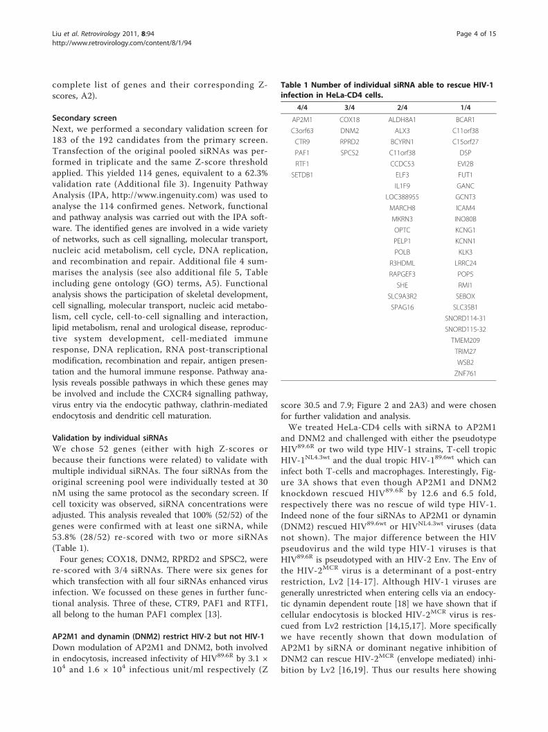
Validation by individual siRNAsWe chose 52 genes (either with high Z-scores orbecause their functions were related) to validate withmultiple individual siRNAs. The four siRNAs from theoriginal screening pool were individually tested at 30nM using the same protocol as the secondary screen. Ifcell toxicity was observed, siRNA concentrations wereadjusted. This analysis revealed that 100% (52/52) of thegenes were confirmed with at least one siRNA, while53.8% (28/52) re-scored with two or more siRNAs(Table 1).Four genes; COX18, DNM2, RPRD2 and SPSC2, were
re-scored with 3/4 siRNAs. There were six genes forwhich transfection with all four siRNAs enhanced virusinfection. We focussed on these genes in further func-tional analysis. Three of these, CTR9, PAF1 and RTF1,all belong to the human PAF1 complex [13].
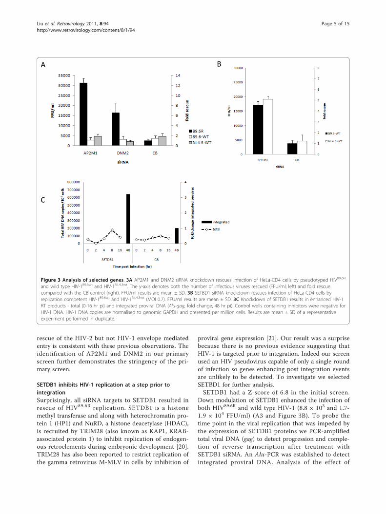
AP2M1 and dynamin (DNM2) restrict HIV-2 but not HIV-1Down modulation of AP2M1 and DNM2, both involvedin endocytosis, increased infectivity of HIV89.6R by 3.1 ×104 and 1.6 × 10⁴ infectious unit/ml respectively (Z
score 30.5 and 7.9; Figure 2 and 2A3) and were chosenfor further validation and analysis.We treated HeLa-CD4 cells with siRNA to AP2M1
and DNM2 and challenged with either the pseudotypeHIV89.6R or two wild type HIV-1 strains, T-cell tropicHIV-1NL4.3wt and the dual tropic HIV-189.6wt which caninfect both T-cells and macrophages. Interestingly, Fig-ure 3A shows that even though AP2M1 and DNM2knockdown rescued HIV89.6R by 12.6 and 6.5 fold,respectively there was no rescue of wild type HIV-1.Indeed none of the four siRNAs to AP2M1 or dynamin(DNM2) rescued HIV89.6wt or HIVNL4.3wt viruses (datanot shown). The major difference between the HIVpseudovirus and the wild type HIV-1 viruses is thatHIV89.6R is pseudotyped with an HIV-2 Env. The Env ofthe HIV-2MCR virus is a determinant of a post-entryrestriction, Lv2 [14-17]. Although HIV-1 viruses aregenerally unrestricted when entering cells via an endocy-tic dynamin dependent route [18] we have shown that ifcellular endocytosis is blocked HIV-2MCR virus is res-cued from Lv2 restriction [14,15,17]. More specificallywe have recently shown that down modulation ofAP2M1 by siRNA or dominant negative inhibition ofDNM2 can rescue HIV-2MCR (envelope mediated) inhi-bition by Lv2 [16,19]. Thus our results here showing
Table 1 Number of individual siRNA able to rescue HIV-1infection in HeLa-CD4 cells.
4/4 3/4 2/4 1/4
AP2M1 COX18 ALDH8A1 BCAR1
C3orf63 DNM2 ALX3 C11orf38
CTR9 RPRD2 BCYRN1 C15orf27
PAF1 SPCS2 C11orf38 DSP
RTF1 CCDC53 EVI2B
SETDB1 ELF3 FUT1
IL1F9 GANC
LOC388955 GCNT3
MARCH8 ICAM4
MKRN3 INO80B
OPTC KCNG1
PELP1 KCNN1
POLB KLK3
R3HDML LRRC24
RAPGEF3 POP5
SHE RMI1
SLC9A3R2 SEBOX
SPAG16 SLC35B1
SNORD114-31
SNORD115-32
TMEM209
TRIM27
WSB2
ZNF761
Liu et al. Retrovirology 2011, 8:94http://www.retrovirology.com/content/8/1/94
Page 4 of 15
rescue of the HIV-2 but not HIV-1 envelope mediatedentry is consistent with these previous observations. Theidentification of AP2M1 and DNM2 in our primaryscreen further demonstrates the stringency of the pri-mary screen.
SETDB1 inhibits HIV-1 replication at a step prior tointegrationSurprisingly, all siRNA targets to SETDB1 resulted inrescue of HIV89.6R replication. SETDB1 is a histonemethyl transferase and along with heterochromatin pro-tein 1 (HP1) and NuRD, a histone deacetylase (HDAC),is recruited by TRIM28 (also known as KAP1, KRAB-associated protein 1) to inhibit replication of endogen-ous retroelements during embryonic development [20].TRIM28 has also been reported to restrict replication ofthe gamma retrovirus M-MLV in cells by inhibition of
proviral gene expression [21]. Our result was a surprisebecause there is no previous evidence suggesting thatHIV-1 is targeted prior to integration. Indeed our screenused an HIV pseudovirus capable of only a single roundof infection so genes enhancing post integration eventsare unlikely to be detected. To investigate we selectedSETBD1 for further analysis.SETDB1 had a Z-score of 6.8 in the initial screen.
Down modulation of SETDB1 enhanced the infection ofboth HIV89.6R and wild type HIV-1 (8.8 × 10³ and 1.7-1.9 × 10⁴ FFU/ml) (A3 and Figure 3B). To probe thetime point in the viral replication that was impeded bythe expression of SETDB1 proteins we PCR-amplifiedtotal viral DNA (gag) to detect progression and comple-tion of reverse transcription after treatment withSETDB1 siRNA. An Alu-PCR was established to detectintegrated proviral DNA. Analysis of the effect of
A B
C
Figure 3 Analysis of selected genes. 3A AP2M1 and DNM2 siRNA knockdown rescues infection of HeLa-CD4 cells by pseudotyped HIV89.6R
and wild type HIV-189.6wt and HIV-1NL4.3wt. The y-axis denotes both the number of infectious viruses rescued (FFU/ml; left) and fold rescuecompared with the CB control (right). FFU/ml results are mean ± SD. 3B SETBD1 siRNA knockdown rescues infection of HeLa-CD4 cells byreplication competent HIV-189.6wt and HIV-1NL4.3wt (MOI 0.7). FFU/ml results are mean ± SD. 3C Knockdown of SETDB1 results in enhanced HIV-1RT products - total (0-16 hr pi) and integrated proviral DNA (Alu-gag, fold change, 48 hr pi). Control wells containing inhibitors were negative forHIV-1 DNA. HIV-1 DNA copies are normalised to genomic GAPDH and presented per million cells. Results are mean ± SD of a representativeexperiment performed in duplicate.
Liu et al. Retrovirology 2011, 8:94http://www.retrovirology.com/content/8/1/94
Page 5 of 15
SETDB1 knockdown on the levels of total HIV-1 DNAshowed no apparent difference while there was a 3.2fold increase in integrated proviruses (Figure 3C).Thus our results strongly suggested that SETDB1 is
also involved in inhibition of HIV-1 replication. Consis-tent with this hypothesis Allouch et al. report thatTRIM28 indeed restricts replication of HIV-1 integra-tion through recruitment of HDAC [8]. The moleculardetails will be worth pursuing.
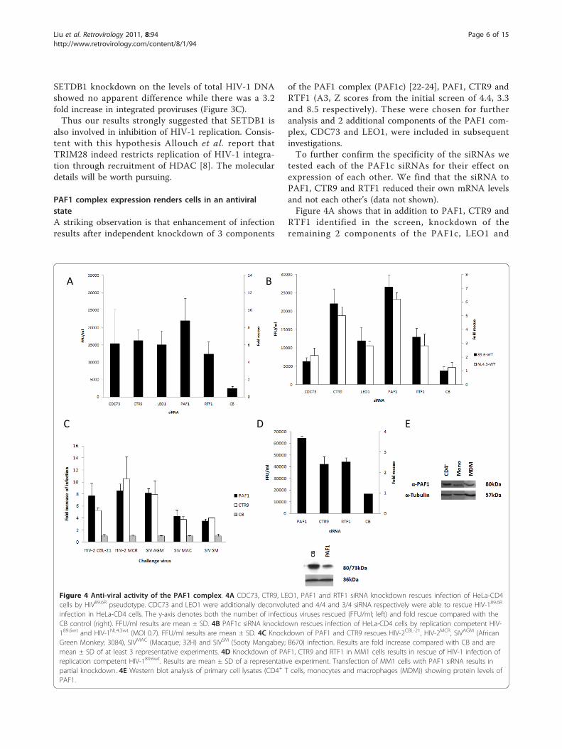
PAF1 complex expression renders cells in an antiviralstateA striking observation is that enhancement of infectionresults after independent knockdown of 3 components
of the PAF1 complex (PAF1c) [22-24], PAF1, CTR9 andRTF1 (A3, Z scores from the initial screen of 4.4, 3.3and 8.5 respectively). These were chosen for furtheranalysis and 2 additional components of the PAF1 com-plex, CDC73 and LEO1, were included in subsequentinvestigations.To further confirm the specificity of the siRNAs we
tested each of the PAF1c siRNAs for their effect onexpression of each other. We find that the siRNA toPAF1, CTR9 and RTF1 reduced their own mRNA levelsand not each other’s (data not shown).Figure 4A shows that in addition to PAF1, CTR9 and
RTF1 identified in the screen, knockdown of theremaining 2 components of the PAF1c, LEO1 and
A
C
B
D E
Figure 4 Anti-viral activity of the PAF1 complex. 4A CDC73, CTR9, LEO1, PAF1 and RTF1 siRNA knockdown rescues infection of HeLa-CD4cells by HIV89.6R pseudotype. CDC73 and LEO1 were additionally deconvoluted and 4/4 and 3/4 siRNA respectively were able to rescue HIV-189.6R
infection in HeLa-CD4 cells. The y-axis denotes both the number of infectious viruses rescued (FFU/ml; left) and fold rescue compared with theCB control (right). FFU/ml results are mean ± SD. 4B PAF1c siRNA knockdown rescues infection of HeLa-CD4 cells by replication competent HIV-189.6wt and HIV-1NL4.3wt (MOI 0.7). FFU/ml results are mean ± SD. 4C Knockdown of PAF1 and CTR9 rescues HIV-2CBL-21, HIV-2MCR, SIVAGM (AfricanGreen Monkey; 3084), SIVMAC (Macaque; 32H) and SIVSM (Sooty Mangabey; B670) infection. Results are fold increase compared with CB and aremean ± SD of at least 3 representative experiments. 4D Knockdown of PAF1, CTR9 and RTF1 in MM1 cells results in rescue of HIV-1 infection ofreplication competent HIV-189.6wt. Results are mean ± SD of a representative experiment. Transfection of MM1 cells with PAF1 siRNA results inpartial knockdown. 4E Western blot analysis of primary cell lysates (CD4+ T cells, monocytes and macrophages (MDM)) showing protein levels ofPAF1.
Liu et al. Retrovirology 2011, 8:94http://www.retrovirology.com/content/8/1/94
Page 6 of 15
CDC73 also rescued infection of HIV89.6R. LEO1 andCDC73 were false negatives in the primary screen. Nextwe tested the effect of inhibition of expression of thesefactors against the wild type viruses HIV-1NL4.3wt andHIV-189.6wt. Figure 4B shows that knockdown of anyone of all five components of the PAF1c rescued infec-tion of wild type viruses by 1.7-7.2 fold (equivalent to6.2 × 103-2.7 × 10⁴ FFU/ml).To determine whether the anti-viral effect observed
was restricted to HIV-1 we tested for activity againstHIV-2 and SIV isolates. We used siRNA to two PAF1ccomponents, PAF1 and CTR9. Figure 4C shows thatinhibiting expression of either of these proteins resultedin rescue of HIV-2 (CBL-21 and MCR) and SIV (AfricanGreen Monkey, Macaque and Sooty Mangabey) by 4-10fold.We wanted to test if there was an effect of PAF1c in
cells of more physiological relevance to HIV-1 infection.Figure 4D shows that treatment of the differentiatedmonocytoid cell line MonoMac1 (MM1) with siRNAtargeting PAF1, CTR9 and RTF1 resulted in rescue ofHIV-189.6wt (2.5-4.7 × 10⁴ FFU/ml, 2.5-4 fold). In Figure4E Western blot analysis reveals that PAF1 is expressedin primary CD4+ T-cells, monocytes and monocytederived macrophages. Attempts however to knock downexpression in these cell types resulted in cytotoxicity.There are a number of recognised steps in the early
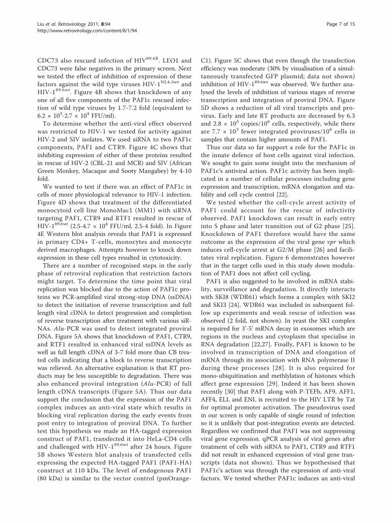
phase of retroviral replication that restriction factorsmight target. To determine the time point that viralreplication was blocked due to the action of PAF1c pro-teins we PCR-amplified viral strong-stop DNA (ssDNA)to detect the initiation of reverse transcription and fulllength viral cDNA to detect progression and completionof reverse transcription after treatment with various siR-NAs. Alu-PCR was used to detect integrated proviralDNA. Figure 5A shows that knockdown of PAF1, CTR9,and RTF1 resulted in enhanced viral ssDNA levels aswell as full length cDNA of 3-7 fold more than CB trea-ted cells indicating that a block to reverse transcriptionwas relieved. An alternative explanation is that RT pro-ducts may be less susceptible to degradation. There wasalso enhanced proviral integration (Alu-PCR) of fulllength cDNA transcripts (Figure 5A). Thus our datasupport the conclusion that the expression of the PAF1complex induces an anti-viral state which results inblocking viral replication during the early events frompost entry to integration of proviral DNA. To furthertest this hypothesis we made an HA-tagged expressionconstruct of PAF1, transfected it into HeLa-CD4 cellsand challenged with HIV-189.6wt after 24 hours. Figure5B shows Western blot analysis of transfected cellsexpressing the expected HA-tagged PAF1 (PAF1-HA)construct at 110 kDa. The level of endogenous PAF1(80 kDa) is similar to the vector control (pmOrange-
C1). Figure 5C shows that even though the transfectionefficiency was moderate (30% by visualisation of a simul-taneously transfected GFP plasmid; data not shown)inhibition of HIV-189.6wt was observed. We further ana-lysed the levels of inhibition of various stages of reversetranscription and integration of proviral DNA. Figure5D shows a reduction of all viral transcripts and pro-virus. Early and late RT products are decreased by 6.3and 2.8 × 10⁵ copies/106 cells, respectively, while thereare 7.7 × 10³ fewer integrated proviruses/106 cells insamples that contain higher amounts of PAF1.Thus our data so far support a role for the PAF1c in
the innate defence of host cells against viral infection.We sought to gain some insight into the mechanism ofPAF1c’s antiviral action. PAF1c activity has been impli-cated in a number of cellular processes including geneexpression and transcription, mRNA elongation and sta-bility and cell cycle control [22].We tested whether the cell-cycle arrest activity of
PAF1 could account for the rescue of infectivityobserved. PAF1 knockdown can result in early entryinto S phase and later transition out of G2 phase [25].Knockdown of PAF1 therefore would have the sameoutcome as the expression of the viral gene vpr whichinduces cell-cycle arrest at G2/M phase [26] and facili-tates viral replication. Figure 6 demonstrates howeverthat in the target cells used in this study down modula-tion of PAF1 does not affect cell cycling.PAF1 is also suggested to be involved in mRNA stabi-
lity, surveillance and degradation. It directly interactswith SKI8 (WDR61) which forms a complex with SKI2and SKI3 [24]. WDR61 was included in subsequent fol-low up experiments and weak rescue of infection wasobserved (2 fold, not shown). In yeast the SKI complexis required for 3’-5’ mRNA decay in exosomes which areregions in the nucleus and cytoplasm that specialise inRNA degradation [22,27]. Finally, PAF1 is known to beinvolved in transcription of DNA and elongation ofmRNA through its association with RNA polymerase IIduring these processes [28]. It is also required formono-ubiquitination and methlylation of histones whichaffect gene expression [29]. Indeed it has been shownrecently [30] that PAF1 along with P-TEFb, AF9, AFF1,AFF4, ELL and ENL is recruited to the HIV LTR by Tatfor optimal promoter activation. The pseudovirus usedin our screen is only capable of single round of infectionso it is unlikely that post-integration events are detected.Regardless we confirmed that PAF1 was not suppressingviral gene expression. qPCR analysis of viral genes aftertreatment of cells with siRNA to PAF1, CTR9 and RTF1did not result in enhanced expression of viral gene tran-scripts (data not shown). Thus we hypothesised thatPAF1c’s action was through the expression of anti-viralfactors. We tested whether PAF1c induces an anti-viral
Liu et al. Retrovirology 2011, 8:94http://www.retrovirology.com/content/8/1/94
Page 7 of 15
state through activation of transcription of known anti-HIV factors. Following treatment of HeLa-CD4 cellswith PAF1 siRNA, the mRNA levels of p21, APO-BEC3G, Tetherin, TRIM5a, SAMHD1 and TREX1 weredetermined by qPCR. No difference was seen in theexpression of these restriction factors when comparedwith untreated cells (data not shown) suggesting thatPAF1 does not exert its anti-viral activity through theaction of these known restrictive genes.We conclude that PAF1c induces an anti-viral state in
cells and inhibits infection of HIV-1, 2 and SIV byblocking progression of the early events of infectionduring reverse transcription and up to integration. Ourdata suggest that the anti-viral effect of PAF1 is notmediated through control of the cell cycle, enhancementof expression of viral transcripts, enhancement of viralgenome stability or enhancement of transcription ofknown anti-viral restriction factors such as p21.
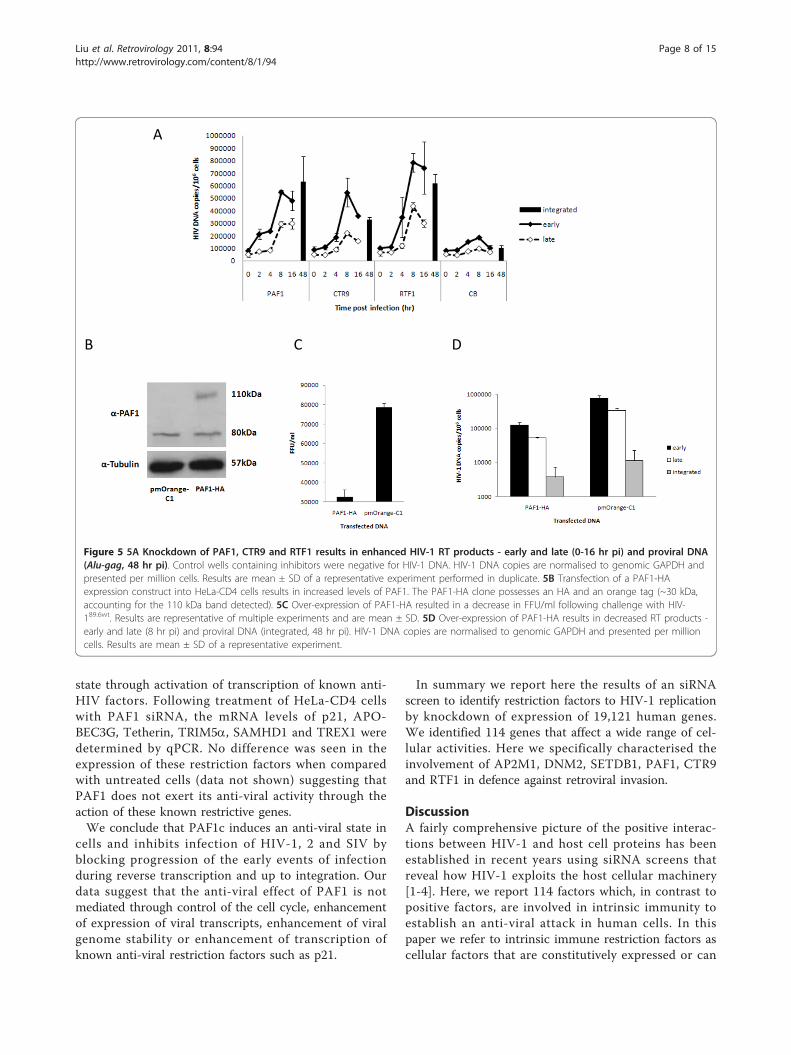
In summary we report here the results of an siRNAscreen to identify restriction factors to HIV-1 replicationby knockdown of expression of 19,121 human genes.We identified 114 genes that affect a wide range of cel-lular activities. Here we specifically characterised theinvolvement of AP2M1, DNM2, SETDB1, PAF1, CTR9and RTF1 in defence against retroviral invasion.
DiscussionA fairly comprehensive picture of the positive interac-tions between HIV-1 and host cell proteins has beenestablished in recent years using siRNA screens thatreveal how HIV-1 exploits the host cellular machinery[1-4]. Here, we report 114 factors which, in contrast topositive factors, are involved in intrinsic immunity toestablish an anti-viral attack in human cells. In thispaper we refer to intrinsic immune restriction factors ascellular factors that are constitutively expressed or can
A
CB D
Figure 5 5A Knockdown of PAF1, CTR9 and RTF1 results in enhanced HIV-1 RT products - early and late (0-16 hr pi) and proviral DNA(Alu-gag, 48 hr pi). Control wells containing inhibitors were negative for HIV-1 DNA. HIV-1 DNA copies are normalised to genomic GAPDH andpresented per million cells. Results are mean ± SD of a representative experiment performed in duplicate. 5B Transfection of a PAF1-HAexpression construct into HeLa-CD4 cells results in increased levels of PAF1. The PAF1-HA clone possesses an HA and an orange tag (~30 kDa,accounting for the 110 kDa band detected). 5C Over-expression of PAF1-HA resulted in a decrease in FFU/ml following challenge with HIV-189.6wt. Results are representative of multiple experiments and are mean ± SD. 5D Over-expression of PAF1-HA results in decreased RT products -early and late (8 hr pi) and proviral DNA (integrated, 48 hr pi). HIV-1 DNA copies are normalised to genomic GAPDH and presented per millioncells. Results are mean ± SD of a representative experiment.
Liu et al. Retrovirology 2011, 8:94http://www.retrovirology.com/content/8/1/94
Page 8 of 15
be generated in cells in the absence of help from otherarms of the immune system. Such factors however areunlikely to act in isolation of the immune system andmay indeed be the initial trigger for innate and adaptiveimmune responses in the host. The proteins identifiedwith known functions are associated with a vast array ofcellular processes such as receptor signalling, vesicletrafficking, transcription, mRNA processing, DNA/RNAsurveillance, cross-nuclear membrane transport andubiquitination.In our screen individual silencing of 114 genes rescued
infectivity of HIV-1 by more than 103 FFU/ml and forsome genes as many as 2 × 10⁵ infectious units/ml (1.6-12.6 fold). The 52 most potent siRNAs ‘hits’ werefurther validated using 4 unpooled siRNAs. Rescue ofviral infection by 2 or more of the 4 siRNAs wasobserved for 28 genes. Of these, AP2M1, DNM2, CTR9,PAF1, RTF1 and SETDB1 were further characterised forinhibition of HIV infection.At the time of starting this screen only APOBEC3G/F
and rhesus macaque TRIM5a proteins were known asfactors that restrict HIV-1 replication. APOBECs arepackaged into virus particles in the producer cells butact on the next round of infection in the new target cell[6]. The HEK 293T cells used to produce the pseudo-virus in our screen do not express APOBECs [6] soidentification of these proteins was not expected.Neither did we identify TRIM5a because it is a speciesspecific restriction factor and the human protein does
not significantly affect HIV-1 [7,31]. Since the screenwas completed two other factors have been reported;p21 acts during reverse transcription and is active instem cells, macrophages and CD4+ lymphocytes[9,10,32] while SAMHD1 expression is confined to den-dritic and myeloid lineage cells [11]. Tetherin, whichacts at the late phase of the viral life cycle prevents viralbudding [12], was not identified in this screen becausewe probed the siRNA library with an HIV pseudotypethat is infectious for only a single round to the integra-tion of provirus and GFP expression.Using a simple model of viral restriction, intrinsic
restriction factors can be loosely divided into; detectorsof invasion, messengers, mediators and effectors thateither prevent infection of the virus or, in the case ofinterferon (IFN) signalling, alert neighbouring cells. IFNactivation of the JAK-STAT pathway ultimately resultsin the expression of anti-viral genes. For example theAPOBEC and TRIM gene families are induced by type IIFNs [33].After entry into the cell cytoplasm HIV-1 uncoating
involves the disassembly of the viral matrix (MA) andcapsid (CA) and release of the genomic RNA and theassociated proteins. Thus these viral components couldpotentially be seen by the host cell as ‘non-self’ mole-cules. Pattern Recognition Receptors (PRRs) or viral sen-sors recognise distinct pathogen associated molecularpatterns (PAMPs). For example, recently TRIM5a wasshown to be a PRR that recognises the viral CA even-tually resulting in immune signalling and AP-1 and NF-�B activation [34]. Of similar interest, silencing ofCOX18 (a cytochrome c oxidase assembly protein)resulted in rescue (3.3 fold). COX18 is essential for theproduction of COX2 (PTGS2) [35] whose expression isattenuated by TRIM5a knockdown [34]. It seems unli-kely that COX18 itself is a direct PRR. It is more plausi-ble that COX18 is an essential downstream componentthat through its interaction with COX2 may commissionor interact with one or more ‘effectors’ of restriction.During and after CA disassembly the viral nucleic
acids and those produced during reverse transcription,RNA fragments, DNA and DNA:RNA hybrids maybecome exposed to cytoplasmic PRRs. Proteins thatinteract with nucleic acids might also play a role in pat-tern recognition. Our screen did not identify any of theclassical RNA-recognising PRRs such as toll like recep-tors TLR3, 7 or 8 or RIG-1 like receptors RIG-1 orMDA-5 [36]. The best characterised DNA receptor DAI(DNA dependent activator of IFN regulatory factors)responds to adenoviral DNA [37]. In this screen silen-cing of POLB polymerase (DNA directed) rescued viralinfection (3.5 fold). POLB rescue was validated with 2individual siRNAs. POLB is involved in DNA damagerepair and performs base excision repair required for
Figure 6 Silencing of PAF1 does not affect cell cycleprogression. HeLa-CD4 cells transfected with PAF1 or CB siRNAwere compared to nocodazole (NOC) treated controls. Cell cycleprofiles were determined by flow cytometric analysis of PI-labelledcultures. Western blot confirms PAF1 silencing compared to CBcontrol.
Liu et al. Retrovirology 2011, 8:94http://www.retrovirology.com/content/8/1/94
Page 9 of 15
DNA maintenance, replication, recombination [38].Interestingly, POLB is specifically up regulated afterinfection of cells with human herpes virus 16.Once PAMPS are engaged multiple signalling cascades
are activated and establish an anti-viral state [39]. Onepotent hit IL1F9, an interleukin cytokine, is switched onby IFNg. Interestingly expression of this gene is alsoinduced after herpes simplex virus infection-1 [40]. Sig-nal induced antiviral responses will also use de novotranscription and translation to establish an anti-viralenvironment. Three transcription factors ALX3, ELF3and PELP1 (3, 4 and 3.4 fold, validated 2/4 siRNAs)identified in the screen could be involved in thisprocess.The screen identified a number of genes that are
involved in ubiquitination of proteins. Conjugation ofthe ubiquitin monomer to proteins is mediated bythree families of enzymes; E1, E2 or E3 substrate speci-fic ubiquitin ligases [41]. E3 enzymes are critical andinteract with both E2 and substrate. Generally, ubiqui-tination leads to the degradation of proteins by theproteasome. In viral restriction ubiquitination could beinvolved in the degradation process of either viral pro-teins or recycling of restriction proteins. The processof ubiquitination is involved in TRIM5a mediatedrestriction [7]. TRIM5a has E3 ligase activity. When E2binds to the RING domain E3 ligase activity ofTRIM5a transfers the ligase from E2 to TRIM5a [42].HIV-1 Vif appears to use ubiquitination as a means ofpreventing the action of APOBEC proteins, by target-ing the Cul5-elongin B elongin C-Rbx ubiquitin ligase.This results in the polyubiquitination of APOBEC3Gwhich is then degraded by proteosomes [43-47]. Astrong ‘hit’ in our screen was the E3 ubiguitin ligaseMARCH8 (RNF178, MIR) [48] whose knockdownresulted in rescue (4.6 fold and 2/4 unpooled siRNA).A second strong hit was MKRN3 (markorin 3, ring fin-ger protein, 3) where 2 individual siRNAs rescuedinfection (4.7 fold). Silencing of RNF19A, TRIM25 andTRIM27, which all belong (or are related) to E3 ligasesalso rescued infectivity (3-3.5 fold between 1 × 10³ and3 × 103 FFU/ml). Interestingly TRIM25 is essential forRIG-1 mediated IFNb production and antiviral activity[49]. However of the latter three genes only TRIM27was validated with individual siRNA with only 1 out of4 siRNAs confirming the restriction phenotype. Addi-tionally knockdown of WSB2, a bridge-protein whichconnects substrate-binding domains and E3 ubiquitinprotein ligases had a modest rescue (3.2 fold, 1 of 4individual siRNA).
Nuclear importOnce production of the viral cDNA is complete it mustbe transported to and enter the nucleus through
specialised nuclear pores using host cell transportmechanisms [50]. CA appears to remain associated withthe PIC for some time following its production [51,52]but disassociates from the reverse transcription complexprior to nuclear entry [53]. Nuclear trafficking is likelyto be another “check point” for the host cell. A trun-cated protein CPSF6 (cleavage and polyadenylation fac-tor) a member of the S/R family protein impairs nuclearentry of PIC [54]. This restriction can be overcome byCA mutants. In our study invalidation of NPIP (nuclearpore complex interacting protein) resulted in enhancedinfection. The function of NPIP is not clear, though itco localises with NUP62, a protein needed for HIV PICtransport from cytoplasm to nucleus [2].
RNA speciesWe might expect that some retroviral elements wouldbe involved in anti-viral defence. Friend-virus-suscept-ibility factor (Fv-1) a gag region of an endogenous retro-virus [55], restricts murine leukaemia virus (MLV) at astage after cellular entry and before integration [56].MLV CA is the target for Fv-1 [57-59]. We did notidentify endogenous retroviral genes in this screen.However BCYRN1 which is believed to be retroposition-ally generated [60] was identified (5.8 fold, validated 2/4). BCYRN1 belongs to a family of interspersed repeti-tive DNA sequences and encodes a neural smalluntranslated non-messenger RNA. The specific neuronalexpression of BCYRN1 is intriguing. Immune privilegedsites such as the brain may rely more heavily on innateimmune factors to prevent viral infection. The tissuespecificity of BCYRN1 implies that it is unlikely to playa role in HIV-1 pathogenesis.
Genes with no obvious roleA number of genes that currently have no tentativefunctional role in retroviral defence were identified inthe primary screen and validated with at least 2 indivi-dual siRNAs: SPSC2 (removes signal peptides from pro-teins as they translocate into the endoplasmic reticulumlumen), R3HDML (a serine protease inhibitor), SPAG16(a component of microtubules) and RAPGEF3 (Rap gua-nine nucleotide exchange factor 3). OPTC is a proteinthat is associated with the extracellular matrix in theeye. ALDH8A1 is an aldehyde dehydrogenase andLOC388955 (PREL1 domain-containing protein 1) is amitochondrial pseudogene. A functional role cannot beprecluded as genes are frequently found to play a practi-cal role in cellular processes far removed from thosewith which they were originally associated. Severalgenes, C3orf63, RPRD2, C11orf38, SLC9A3R2 and SHEhad no identified function. Further studies on thesegenes may shed light on currently poorly understoodbiological processes.
Liu et al. Retrovirology 2011, 8:94http://www.retrovirology.com/content/8/1/94
Page 10 of 15
Endocytosis genes AP2M1 and DNM2Some factors identified in our screen might have beenpredicted: AP2M1 and DNM2, two components of theendocytic pathway were identified in the primary screento restrict infection of cells by the pseudovirus but notby wild type HIV-1 viruses. This observation can beexplained - we previously showed HIV-1 and 2 virusescan be blocked post-entry by a restriction factor Lv2 butonly if the virus was delivered to the cytoplasm using aspecific endocytic route mediated by an HIV-2 virusMCR envelope [14,16,17]. The entry route is specific tothe HIV-2 envelope because pseudoviruses with VSV-Gare rescued [15]. We recently showed that blockingentry of HIV-2 MCR virus by an AP2M1/DNM2 depen-dent route rescued Lv2 restriction. Unlike the wild typeHIV-1 viruses, the HIV-1 pseudovirus used in ourscreen was generated with an Env derived from HIV-2MCR. So inhibition of the pseudovirus (but not the wildtype HIV-1) would be expected to be rescued by siRNAsto AP2M1 and DNM2.Other factors involved in endocytosis will be interest-
ing to probe with respect to the Lv2 restriction.CCDC53 (coiled-coil domain containing protein), acomponent of the WASH complex, is present on thesurface of endosomes and recruits the Arp2/3 complexfor actin polymerisation [61]. Two GTPases (or asso-ciated proteins) were identified: RAPGEF3 (Rap guaninenucleotide exchange factor 3) and RAB37 (small GTPasethat regulates vesicle trafficking) and knockdown ofeither resulted in a 3.7 or 2.8 fold rescue. It will beinteresting to further evaluate their role in Lv2 restric-tion. However, apart from vesicle trafficking, GTPasesregulate a myriad of cellular functions including signaltransduction and cytoskeletal organisation.
SETDB1We did not predict that silencing of SETDB1 (SETdomain, bifurcated 1) would rescue replication ofHIV. SETDB1 is involved in TRIM family proteinTRIM28 (also known as KAP1) restriction of bothexogenous MLV and endogenous retrovirus expressionpost integration in murine cells [21]. SETDB1, a his-tone methlytransferase, the NuRD histone deacetlyasecomplex (HDAC) and heterochromatin associated pro-tein HP-1 are targeted to proviral DNA by TRIM28 torepress transcription. Furthermore, when transfectedinto human cells, SETDB1 increases HIV-LTR trans-activation in conditions where Tat levels were subopti-mal [62] but no function in the viral life cycle prior tointegration had been previously described. Our screenwith single round pseudotype virus should only iden-tify genes involved in the early phase in HIV replica-tion and not those post integration. Surprisingly,SETDB1 silencing rescued wild type HIV-1 replication
by almost 2 × 10⁴ FFU/ml (4.6 fold). Indeed all 4 siR-NAs individually silencing SETDB1 resulted in viralrescue and was a gene among our most significant‘hits’. Upon further investigation we found that therewas a small increase in reverse transcripts and thatthere was a relatively strong increase in integratedproviral DNA. Recently, Allouch et al. described asimilar phenotype for restriction by TRIM28 throughrecruitment of HDAC [8]. It will be interesting toprobe the molecular details of this interaction ingreater depth.
PAF1 complex expression renders cells restrictive tolentiviral infectionOur validation of the PAF1c showed that all of itsknown components are important for the observedrestriction. PAF1c expression restricts HIV-1, 2 and SIV.It restricts infection of a monocytic cell line and isexpressed in primary CD4+ T cells, monocytes andmacrophages. The restriction results in fewer viral tran-scripts both in the early and late stage of infection. Italso results in less integrated proviral DNA. We are notsure however if this phenotype is due to PAF1’s directaction on replicating virus or if its action is throughactivation of expression of one or more unknownrestriction factors.A role for PAF1 in the cell cycle has been recently
demonstrated [25] and studies using an in vitro disas-sembly model have suggested that uncoating requirescell cycle dependent host cell factors [63]. However thecell cycle was unperturbed by inhibition of PAF1 expres-sion in the Hela-CD4 cells we used in the screen sug-gesting that PAF1’s cell cycle activity is not involved inthis restriction.There are two possibilities for the action of PAF1c in
viral restriction: First, PAF1 interacts with RNA poly-merase II and is proposed to be involved in transcrip-tion, elongation and stability of mRNA, surveillance anddegradation [24]. Indeed a large quantity of PAF1c islocated in the nucleus so it is also possible that its activ-ity in gene transcription may enhance expression ofanti-viral factors, some of which are already ‘hits’ in ourprimary screen. Additionally we have excluded a role forAPOBEC3G, TRIM5a, p21, tetherin and the recentlydescribed SAMHD1.Second, it has also been demonstrated that SKI8
(WDR61) interacts with the PAF1c [24]. SKI8 is alsopart of the SKI complex which is involved in mRNAdecay [24,27]. Given the ability of PAF1/SKI8 to interactwith DNA and RNA and the localisation of both thesecomponents in the cytoplasm it is possible that PAF1c/SKI8 could act as a PRR. DNA:RNA hybrids or degrad-ing RNA genomes are possible targets. To distinguishbetween the two possibilities of PAF1’s action it may be
Liu et al. Retrovirology 2011, 8:94http://www.retrovirology.com/content/8/1/94
Page 11 of 15
of use to determine which genes are enhanced in thepresence or absence of PAF1 using gene array analysis.
ConclusionsIn summary we describe the identification of 114 genesthat are involved in restriction of HIV replication duringthe early stages of the viral life cycle. Preliminary char-acterisation of 6 genes AP2M1, DNM2, CTR9, PAF1,RTF1 and SETDB1 confirms their biological role in ret-roviral restriction. This preliminary characterisationattests to the robustness of the primary screen. It isimprobable that the genes identified evolved in resis-tance to HIV-1 infection. It is more plausible they areactive against other viruses or pathogens that invadehost cells and will be a platform for a general under-standing of innate immune or intrinsic defence againstinvasion of known and emerging pathogens. Variation inthe expression patterns and polymorphisms in thesegenes may lead to an understanding of the reasons whysome individuals are more or less susceptible to specificinfectious diseases.
MethodsCellsCulture of HEK 293T, HeLa-CD4, NP2-CD4-CXCR4,MonoMac 1 (MM1) and C8166, and their optimal cul-ture conditions, have been described previously[17,64,65]. Peripheral blood mononuclear cells (PBMC)were prepared from seronegative donors by density-gra-dient centrifugation (Lymphoprep, Axis-Shield). CD4+ Tlymphocytes were isolated using CD4 Microbeadsaccording to manufacturer’s instructions (Miltenyi Bio-tech). Monocyte-derived macrophages (MDM) were pre-pared by adherence as described previously [66], exceptthat cells were harvested (for monocyte preparations)and replated at 2 × 10⁶ cells/ml following the initialovernight incubation, and left to differentiate for 7-14days in RPMI 1640 supplemented with 20% autologoushuman serum and 20 ng/ml macrophage colony stimu-lating factor (M-CSF; R&D Systems).
Plasmids and virus productionThe expression plasmid for PAF1 was generated by PCRamplifying the open reading frame from HeLa-CD4cDNA which was subcloned into the pmOrange-C1 vec-tor (Clontech). Primers are available upon request.Infectious molecular clones for wild type (wt) HIV-189.6wt and HIV-1NL4.3wt were obtained from the Centrefor AIDS Research (NIBSC, UK). Restricted pseudotypedvirions (HIV89.6R) were generated by combining thetransfer vector pCSGW with the restrictive HIV-2 envel-ope MCR [16,17] and the core construct p8.91-89.6gag.For the non-restricted viral particles (HIV8.2N), we usedMCN Env and p8.2-89.6 core, where the gag fragment
from p8.91-89.6gag was transferred into pCMVΔ8.2according to the method described [67]. Virus stockswere prepared from infectious full-length and chimericHIV clones by polyethylenimine (Polysciences) transfec-tion of HEK 293T cells. SIV and HIV-2CBL-21 stockswere grown in C8166 cells.
siRNA screenThe screen was performed using the siRNA library(QIAGEN, the Human Whole Genome siRNA Set V4.0)which enables gene silencing studies of 19,121 genesfrom the RefSeq database (NCBI handbook). HeLa-CD4plated at 600 cells/well in a 384-well plate were trans-fected with 30 nM siRNA from the library or controlsiRNA PPIB (cyclophilin B, referred to as CB) usingHiPerfect (QIAGEN) and the CyBio Vario (CyBio, Ger-many) liquid handling system. The system was opti-mised using siRNAs to CB and GFP. The siRNAsagainst receptor CD4 and the nuclear importin TNPO3reduced infectivity of HIV8.2N by 95.8% and 93.0%,respectively.Plates were incubated for 72 hr before challenge with
HIV89.6R (MOI 0.7 on NP2-CD4-CXCR4). 48 hr postvirus challenge, GFP expressing cells were recorded bythe IN Cell Analyzer 1000 automated imaging systemand analysed by IN Cell Developer software (GE Health-care). Wells containing a GFP+ foci Z-score > 3 relativeto the CB control (n = 16 per plate) were confirmed byvisual inspection. 183/192 hits from the original screenwere retested in triplicate. The top 52 hits were thenfurther validated with four individual siRNAs per target.Targeted proteins rescuing infection by more than twoout of four deconvoluted siRNAs were considered vali-dated hits.
siRNA transfection and infection with replicationcompetent virusHeLa-CD4 cells were seeded at 6.3 × 10⁴ cells/well in 6-well plates. 72 hr after siRNA transfection (15 nMPAF1, CTR9, RTF1, LEO1/7.5 nM CDC73/30 nM allother siRNA), cells were challenged with HIV-189.6wt
and HIV-1NL4.3wt (MOI 0.2) for up to 5 hr. 1 hr prior toviral challenge, inhibitors (Raltegravir (1 μM), AZT (100μM), SCH-D (1 μM) and AMD3100 (250 nM)) wereadded to control wells. Infection was assessed after 24-48 hr by intracellular p24 staining, real-time quantitativePCR (qPCR) or mRNA analysis.
Western blotSDS-PAGE separated cellular proteins, immobilised onHybond-P PVDF membrane (GE Healthcare) weredetected with the primary rabbit polyclonal antibodyagainst (RbpAb-) PAF1, RbpAb-GAPDH and rat pAb-tubulin (Abcam) and a secondary horseradish peroxidise
Liu et al. Retrovirology 2011, 8:94http://www.retrovirology.com/content/8/1/94
Page 12 of 15
conjugated goat anti-rabbit/rat antibody (Promega). Pro-teins were visualised using a chemiluminescence kit(ECL, GE Healthcare).
In situ immunostaining for p24 antigenInfected cells were fixed with cold (-20°C) methanol:acetone (1:1), washed with PBS then immunostained forp24 using mouse anti-HIV-1 p24 monoclonal antibodiesEVA365 and 366 (NIBSC) (1:50) or anti-HIV-2 patienthuman plasma (1:1000) (to detect HIV-2 and SIVinfected cells), as previously described [68]. Infectedcells were blue (regarded as foci of infection (FFU/ml))and quantitated by light microscopy.
First round Alu-gag PCRDNA was extracted at various time points after infectionwith the QIAamp DNA Blood Mini Kit (QIAGEN).Integrated HIV-1 DNA was measured by nested PCR, aspreviously described [69].
qPCR for HIV-1 DNAThe isolated DNA was subjected to qPCR to determinethe number of early (negative strand strong stop) andlate (gag) transcripts, normalised for cell number bygenomic GAPDH as previously described.
cDNA synthesis and mRNA analysisTotal HeLa-CD4 RNA was extracted an RNeasy PlantMini Kit (QIAGEN) and cDNA was synthesised withSuperscript III First Strand Synthesis System (Invitrogen),according to manufacturer’s instructions. The cDNAproduced was subjected to qPCR as described [69].
Transfection of suspension cell linessiRNA transfection of MM1 cells was performed usingthe AMAXA Nucleofector Kit V (Lonza Cologne AG)according to manufacturer’s instructions. 48 hr follow-ing viral challenge, 20 ng/ml of phorbol 12-myristate13-acetate (PMA) was added to induce cellular adher-ence prior to intracellular p24 staining.
Cell cycle analysisHeLa-CD4 cells were transfected with PAF1 or CBsiRNA for 48 hr, or treated with nocodazole (40 ng/ml;Sigma) for 18 hr before cells were harvested. Cells werefixed with ice-cold 70% ethanol, washed and pelleted.Cells were RNaseA-treated (100 μg/ml; Sigma), stainedwith propidium iodide (50 μg/ml; Sigma) and analysedby flow cytometry (LSRII, BD Biosciences).
Additional material
Additional file 1: Efficiency of siRNA knockdown on HIV infection.siRNA knockdown of the HIV receptor CD4 and nuclear importin TNPO3
inhibits infection of HIV-18.2N by 95.8% and 93% respectively comparedwith CB control siRNA. Green, virus; red, cells.
Additonal file 2: Results of primary screen with Z-scores. Positiveresults from primary screen are listed. Z-scores are given for all hitsgreater than 3SD from CB control siRNA.
Additional file 3: siRNA knockdown of 114 genes rescues HIVinfection. Infection of HIV89.6R pseudotyped virus was rescued in HeLa-CD4 cells following siRNA knockdown of 114 genes. The y-axis denotesboth the number of infectious viruses rescued (focus forming units/ml,FFU/ml; left) and fold rescue compared with the CB control (right). FFU/ml results are mean ± SD.
Additional file 4: Pathway analysis of most potent screen hits.Ingenuity Pathway Analysis (IPA http://www.ingenuity.com) wasperformed on the validated screen hits showing the functions, if known,associated with the most potent.
Additional file 5: Network, functional and pathway analysis. A list of114 confirmed factors including heat map and GO terms.
AcknowledgementsThis work was supported by an MRC Senior Non-Clinical Fellowship awardedto AM (G117/547) and a Wellcome Trust VIP award (LL). The monoclonalantibodies to p24 (EVA365 and 366) were provided by the EU ProgrammeEVA Centre for AIDS Reagents, NIBSC, UK (AVIP Contract Number LSHP-CT-2004-503487).
Author details1Centre for Immunology and Infectious Disease, Blizard Institute, Barts andThe London School of Medicine and Dentistry, Queen Mary University ofLondon, 4 Newark Street, London E1 2AT, UK. 2Centre for CutaneousResearch, Blizard Institute, Barts and The London School of Medicine andDentistry, Queen Mary University of London, 4 Newark Street, London E12AT, UK.
Authors’ contributionsLL developed, executed and validated the siRNA screen with help andadvice from NMMO, ÁMcK, MTD, A-MB, VB and CLB (Figures 1, A1, 2, A3, A4).KMC and NMMO performed the biological and virological assays withassistance from HD and CP (Figures 3c, 4c-e, 5, 6) and LL (Figure 3a-b, 4a-b).ÁMcK conceived and guided the project with input from DHB and MTD. Allauthors contributed to writing the paper.
Competing interestsThe authors declare that they have no competing interests.
Received: 8 August 2011 Accepted: 14 November 2011Published: 14 November 2011
References1. Brass AL, Dykxhoorn DM, Benita Y, Yan N, Engelman A, Xavier RJ,
Lieberman J, Elledge SJ: Identification of host proteins required for HIVinfection through a functional genomic screen. Science 2008, 319:921-926.
2. Konig R, Zhou Y, Elleder D, Diamond TL, Bonamy GM, Irelan JT, Chiang CY,Tu BP, De Jesus PD, Lilley CE, Seidel S, Opaluch AM, Caldwell JS,Weitzman MD, Kuhen KL, Bandyopadhyay S, Ideker T, Orth AP, Miraglia LJ,Bushman FD, Young JA, Chanda SK: Global analysis of host-pathogeninteractions that regulate early-stage HIV-1 replication. Cell 2008,135:49-60.
3. Zhou H, Xu M, Huang Q, Gates AT, Zhang XD, Castle JC, Stec E, Ferrer M,Strulovici B, Hazuda DJ, Espeseth AS: Genome-scale RNAi screen for hostfactors required for HIV replication. Cell Host Microbe 2008, 4:495-504.
4. Yeung ML, Houzet L, Yedavalli VS, Jeang KT: A genome-wide short hairpinRNA screening of jurkat T-cells for human proteins contributing toproductive HIV-1 replication. J Biol Chem 2009, 284:19463-19473.
5. Christ F, Thys W, De Rijck J, Gijsbers R, Albanese A, Arosio D, Emiliani S,Rain JC, Benarous R, Cereseto A, Debyser Z: Transportin-SR2 imports HIVinto the nucleus. Curr Biol 2008, 18:1192-1202.
Liu et al. Retrovirology 2011, 8:94http://www.retrovirology.com/content/8/1/94
Page 13 of 15
6. Sheehy AM, Gaddis NC, Choi JD, Malim MH: Isolation of a human genethat inhibits HIV-1 infection and is suppressed by the viral Vif protein.Nature 2002, 418:646-650.
7. Stremlau M, Owens CM, Perron MJ, Kiessling M, Autissier P, Sodroski J: Thecytoplasmic body component TRIM5alpha restricts HIV-1 infection in OldWorld monkeys. Nature 2004, 427:848-853.
8. Allouch A, Di Primio C, Alpi E, Lusic M, Arosio D, Giacca M, Cereseto A: TheTRIM Family Protein KAP1 Inhibits HIV-1 Integration. Cell Host Microbe2011, 9:484-495.
9. Chen H, Li C, Huang J, Cung T, Seiss K, Beamon J, Carrington MF, Porter LC,Burke PS, Yang Y, Ryan BJ, Liu R, Weiss RH, Pereyra F, Cress WD, Brass AL,Rosenberg ES, Walker BD, Yu XG, Lichterfeld M: CD4+ T cells from elitecontrollers resist HIV-1 infection by selective upregulation of p21. J ClinInvest 2011, 121:1549-1560.
10. Zhang J, Scadden DT, Crumpacker CS: Primitive hematopoietic cells resistHIV-1 infection via p21. J Clin Invest 2007, 117:473-481.
11. Laguette N, Sobhian B, Casartelli N, Ringeard M, Chable-Bessia C, Segeral E,Yatim A, Emiliani S, Schwartz O, Benkirane M: SAMHD1 is the dendritic-and myeloid-cell-specific HIV-1 restriction factor counteracted by Vpx.Nature 2011, 474(7353):654-7.
12. Neil SJ, Zang T, Bieniasz PD: Tetherin inhibits retrovirus release and isantagonized by HIV-1 Vpu. Nature 2008, 451:425-430.
13. Mueller CL, Jaehning JA: Ctr9, Rtf1, and Leo1 are components of thePaf1/RNA polymerase II complex. Mol Cell Biol 2002, 22:1971-1980.
14. Marchant D, Neil SJ, Aubin K, Schmitz C, McKnight A: An envelope-determined, pH-independent endocytic route of viral entry determinesthe susceptibility of human immunodeficiency virus type 1 (HIV-1) andHIV-2 to Lv2 restriction. J Virol 2005, 79:9410-9418.
15. McKnight A, Griffiths DJ, Dittmar M, Clapham P, Thomas E: Characterizationof a late entry event in the replication cycle of humanimmunodeficiency virus type 2. J Virol 2001, 75:6914-6922.
16. Reuter S, Kaumanns P, Buschhorn SB, Dittmar MT: Role of HIV-2 envelopein Lv2-mediated restriction. Virology 2005, 332:347-358.
17. Schmitz C, Marchant D, Neil SJ, Aubin K, Reuter S, Dittmar MT, McKnight A:Lv2, a novel postentry restriction, is mediated by both capsid andenvelope. J Virol 2004, 78:2006-2016.
18. Miyauchi K, Kim Y, Latinovic O, Morozov V, Melikyan GB: HIV enters cellsvia endocytosis and dynamin-dependent fusion with endosomes. Cell2009, 137:433-444.
19. Harrison IP, McKnight A: Cellular entry via an actin and clathrin-dependent route is required for Lv2 restriction of HIV-2. Virology 2011,415:47-55.
20. Rowe HM, Jakobsson J, Mesnard D, Rougemont J, Reynard S, Aktas T,Maillard PV, Layard-Liesching H, Verp S, Marquis J, Spitz F, Constam DB,Trono D: KAP1 controls endogenous retroviruses in embryonic stemcells. Nature 2010, 463:237-240.
21. Wolf D, Goff SP: TRIM28 mediates primer binding site-targeted silencingof murine leukemia virus in embryonic cells. Cell 2007, 131:46-57.
22. Mueller CL, Porter SE, Hoffman MG, Jaehning JA: The Paf1 complex hasfunctions independent of actively transcribing RNA polymerase II. MolCell 2004, 14:447-456.
23. Rozenblatt-Rosen O, Hughes CM, Nannepaga SJ, Shanmugam KS,Copeland TD, Guszczynski T, Resau JH, Meyerson M: The parafibromintumor suppressor protein is part of a human Paf1 complex. Mol Cell Biol2005, 25:612-620.
24. Zhu B, Mandal SS, Pham AD, Zheng Y, Erdjument-Bromage H, Batra SK,Tempst P, Reinberg D: The human PAF complex coordinates transcriptionwith events downstream of RNA synthesis. Genes Dev 2005, 19:1668-1673.
25. Moniaux N, Nemos C, Deb S, Zhu B, Dornreiter I, Hollingsworth MA,Batra SK: The human RNA polymerase II-associated factor 1 (hPaf1): anew regulator of cell-cycle progression. PLoS One 2009, 4:e7077.
26. Emerman M: HIV-1, Vpr and the cell cycle. Curr Biol 1996, 6:1096-1103.27. Ridley SP, Sommer SS, Wickner RB: Superkiller mutations in
Saccharomyces cerevisiae suppress exclusion of M2 double-strandedRNA by L-A-HN and confer cold sensitivity in the presence of M and L-A-HN. Mol Cell Biol 1984, 4:761-770.
28. Pokholok DK, Hannett NM, Young RA: Exchange of RNA polymerase IIinitiation and elongation factors during gene expression in vivo. Mol Cell2002, 9:799-809.
29. Hampsey M, Reinberg D: Tails of intrigue: phosphorylation of RNApolymerase II mediates histone methylation. Cell 2003, 113:429-432.
30. Sobhian B, Laguette N, Yatim A, Nakamura M, Levy Y, Kiernan R,Benkirane M: HIV-1 Tat assembles a multifunctional transcriptionelongation complex and stably associates with the 7SK snRNP. Mol Cell2011, 38:439-451.
31. Kaumanns P, Hagmann I, Dittmar MT: Human TRIM5alpha mediatedrestriction of different HIV-1 subtypes and Lv2 sensitive and insensitiveHIV-2 variants. Retrovirology 2006, 3:79.
32. Bergamaschi A, David A, Le Rouzic E, Nisole S, Barre-Sinoussi F, Pancino G:The CDK inhibitor p21Cip1/WAF1 is induced by FcgammaR activationand restricts the replication of human immunodeficiency virus type 1and related primate lentiviruses in human macrophages. J Virol 2009,83:12253-12265.
33. Cullen BR: Role and mechanism of action of the APOBEC3 family ofantiretroviral resistance factors. J Virol 2006, 80:1067-1076.
34. Pertel T, Hausmann S, Morger D, Zuger S, Guerra J, Lascano J, Reinhard C,Santoni FA, Uchil PD, Chatel L, Bisiaux A, Albert ML, Strambio-De-Castillia C,Mothes W, Pizzato M, Grutter MG, Luban J: TRIM5 is an innate immunesensor for the retrovirus capsid lattice. Nature 2011, 472:361-365.
35. Souza RL, Green-Willms NS, Fox TD, Tzagoloff A, Nobrega FG: Cloning andcharacterization of COX18, a Saccharomyces cerevisiae PET generequired for the assembly of cytochrome oxidase. J Biol Chem 2000,275:14898-14902.
36. Viswanathan K, Fruh K, DeFilippis V: Viral hijacking of the host ubiquitinsystem to evade interferon responses. Curr Opin Microbiol 2010,13:517-523.
37. Takaoka A, Wang Z, Choi MK, Yanai H, Negishi H, Ban T, Lu Y, Miyagishi M,Kodama T, Honda K, Ohba Y, Taniguchi T: DAI (DLM-1/ZBP1) is a cytosolicDNA sensor and an activator of innate immune response. Nature 2007,448:501-505.
38. Braithwaite EK, Kedar PS, Stumpo DJ, Bertocci B, Freedman JH, Samson LD,Wilson SH: DNA polymerases beta and lambda mediate overlapping andindependent roles in base excision repair in mouse embryonicfibroblasts. PLoS One 2010, 5:e12229.
39. Douville RN, Hiscott J: The interface between the innate interferonresponse and expression of host retroviral restriction factors. Cytokine2011, 52:108-115.
40. Kumar S, McDonnell PC, Lehr R, Tierney L, Tzimas MN, Griswold DE,Capper EA, Tal-Singer R, Wells GI, Doyle ML, Young PR: Identification andinitial characterization of four novel members of the interleukin-1 family.J Biol Chem 2000, 275:10308-10314.
41. Pickart CM: Mechanisms underlying ubiquitination. Annu Rev Biochem2001, 70:503-533.
42. Lienlaf M, Hayashi F, Di Nunzio F, Tochio N, Kigawa T, Yokoyama S, Diaz-Griffero F: Contribution of E3-ubiquitin ligase activity to HIV-1 restrictionby TRIM5{alpha}rh: Structure of the RING domain of TRIM5{alpha}. J Virol2011, 85(17):8725-37, Epub 2011 Jul 6.
43. Conticello SG, Harris RS, Neuberger MS: The Vif protein of HIV triggersdegradation of the human antiretroviral DNA deaminase APOBEC3G.Curr Biol 2003, 13:2009-2013.
44. Marin M, Rose KM, Kozak SL, Kabat D: HIV-1 Vif protein binds the editingenzyme APOBEC3G and induces its degradation. Nat Med 2003,9:1398-1403.
45. Mehle A, Strack B, Ancuta P, Zhang C, McPike M, Gabuzda D: Vifovercomes the innate antiviral activity of APOBEC3G by promoting itsdegradation in the ubiquitin-proteasome pathway. J Biol Chem 2004,279:7792-7798.
46. Sheehy AM, Gaddis NC, Malim MH: The antiretroviral enzyme APOBEC3Gis degraded by the proteasome in response to HIV-1 Vif. Nat Med 2003,9:1404-1407.
47. Yu X, Yu Y, Liu B, Luo K, Kong W, Mao P, Yu XF: Induction of APOBEC3Gubiquitination and degradation by an HIV-1 Vif-Cul5-SCF complex.Science 2003, 302:1056-1060.
48. Goto E, Ishido S, Sato Y, Ohgimoto S, Ohgimoto K, Nagano-Fujii M, Hotta H:c-MIR, a human E3 ubiquitin ligase, is a functional homolog ofherpesvirus proteins MIR1 and MIR2 and has similar activity. J Biol Chem2003, 278:14657-14668.
49. Gack MU, Shin YC, Joo CH, Urano T, Liang C, Sun L, Takeuchi O, Akira S,Chen Z, Inoue S, Jung JU: TRIM25 RING-finger E3 ubiquitin ligase isessential for RIG-I-mediated antiviral activity. Nature 2007, 446:916-920.
50. Fassati A: HIV infection of non-dividing cells: a divisive problem.Retrovirology 2006, 3:74.
Liu et al. Retrovirology 2011, 8:94http://www.retrovirology.com/content/8/1/94
Page 14 of 15
51. Arhel NJ, Souquere-Besse S, Munier S, Souque P, Guadagnini S,Rutherford S, Prevost MC, Allen TD, Charneau P: HIV-1 DNA Flap formationpromotes uncoating of the pre-integration complex at the nuclear pore.EMBO J 2007, 26:3025-3037.
52. Dismuke DJ, Aiken C: Evidence for a functional link between uncoatingof the human immunodeficiency virus type 1 core and nuclear importof the viral preintegration complex. J Virol 2006, 80:3712-3720.
53. Fassati A, Goff SP: Characterization of intracellular reverse transcriptioncomplexes of human immunodeficiency virus type 1. J Virol 2001,75:3626-3635.
54. Lee K, Ambrose Z, Martin TD, Oztop I, Mulky A, Julias JG, Vandegraaff N,Baumann JG, Wang R, Yuen W, Takemura T, Shelton K, Taniuchi I, Li Y,Sodroski J, Littman DR, Coffin JM, Hughes SH, Unutmaz D, Engelman A,KewalRamani VN: Flexible use of nuclear import pathways by HIV-1. CellHost Microbe 2010, 7:221-233.
55. Best S, Le Tissier P, Towers G, Stoye JP: Positional cloning of the mouseretrovirus restriction gene Fv1. Nature 1996, 382:826-829.
56. Hartley JW, Rowe WP, Huebner RJ: Host-range restrictions of murineleukemia viruses in mouse embryo cell cultures. J Virol 1970, 5:221-225.
57. Boone LR, Innes CL, Glover PL, Linney E: Development andcharacterization of an Fv-1-sensitive retrovirus-packaging system: single-hit titration kinetics observed in restrictive cells. J Virol 1989,63:2592-2597.
58. Bowerman B, Brown PO, Bishop JM, Varmus HE: A nucleoprotein complexmediates the integration of retroviral DNA. Genes Dev 1989, 3:469-478.
59. Rommelaere J, Donis-Keller H, Hopkins N: RNA sequencing providesevidence for allelism of determinants of the N-, B- or NB-tropism ofmurine leukemia viruses. Cell 1979, 16:43-50.
60. Brosius J: Gene duplication and other evolutionary strategies: from theRNA world to the future. J Struct Funct Genomics 2003, 3:1-17.
61. Derivery E, Sousa C, Gautier JJ, Lombard B, Loew D, Gautreau A: The Arp2/3 activator WASH controls the fission of endosomes through a largemultiprotein complex. Dev Cell 2009, 17:712-723.
62. Van Duyne R, Easley R, Wu W, Berro R, Pedati C, Klase Z, Kehn-Hall K,Flynn EK, Symer DE, Kashanchi F: Lysine methylation of HIV-1 Tatregulates transcriptional activity of the viral LTR. Retrovirology 2008, 5:40.
63. Auewarakul P, Wacharapornin P, Srichatrapimuk S, Chutipongtanate S,Puthavathana P: Uncoating of HIV-1 requires cellular activation. Virology2005, 337:93-101.
64. Genois N, Robichaud GA, Tremblay MJ: Mono Mac 1: a new in vitro modelsystem to study HIV-1 infection in human cells of the mononuclearphagocyte series. J Leukoc Biol 2000, 68:854-864.
65. Salahuddin SZ, Markham PD, Wong-Staal F, Franchini G, Kalyanaraman VS,Gallo RC: Restricted expression of human T-cell leukemia–lymphomavirus (HTLV) in transformed human umbilical cord blood lymphocytes.Virology 1983, 129:51-64.
66. Marchant D, Neil SJ, McKnight A: Human immunodeficiency virus types 1and 2 have different replication kinetics in human primary macrophageculture. J Gen Virol 2006, 87:411-418.
67. Bock M, Bishop KN, Towers G, Stoye JP: Use of a transient assay forstudying the genetic determinants of Fv1 restriction. J Virol 2000,74:7422-7430.
68. McKnight A, Clapham PR, Weiss RA: HIV-2 and SIV infection of nonprimatecell lines expressing human CD4: restrictions to replication at distinctstages. Virology 1994, 201:8-18.
69. Cheney KM, McKnight A: Interferon-alpha mediates restriction of humanimmunodeficiency virus type-1 replication in primary humanmacrophages at an early stage of replication. PLoS One 2010, 5:e13521.
doi:10.1186/1742-4690-8-94Cite this article as: Liu et al.: A whole genome screen for HIV restrictionfactors. Retrovirology 2011 8:94.
Submit your next manuscript to BioMed Centraland take full advantage of:
• Convenient online submission
• Thorough peer review
• No space constraints or color figure charges
• Immediate publication on acceptance
• Inclusion in PubMed, CAS, Scopus and Google Scholar
• Research which is freely available for redistribution
Submit your manuscript at www.biomedcentral.com/submit
Liu et al. Retrovirology 2011, 8:94http://www.retrovirology.com/content/8/1/94
Page 15 of 15


![ImmuLisa Enhanced dsDNA Antibody ELISA Anti-dsDNA.pdf · 2013-06-13 · EN 1 ImmuLisa™ Enhanced dsDNA Antibody ELISA Double Stranded DNA Antibody ELISA [IVD] For in vitro diagnostic](https://img.pdfslide.us/doc/110x75/5e2b5f357941f82acb3441b8/immulisa-enhanced-dsdna-antibody-anti-dsdnapdf-2013-06-13-en-1-immulisaa.jpg)