Embed Size (px)
Citation preview

Schröder et al. Behavioral and Brain Functions 2013, 9:7http://www.behavioralandbrainfunctions.com/content/9/1/7
RESEARCH Open Access
A boy with homozygous microdeletion ofNEUROG1 presents with a congenital cranialdysinnervation disorder [Moebius syndromevariant]Julia C Schröder1*, Anne K Läßig2, Danuta Galetzka1, Angelika Peters1, John C Castle3, Stefan Diederich1,Ulrich Zechner1, Wibke Müller-Forell4, Annerose Keilmann2 and Oliver Bartsch1
Abstract
Background: We report on a 6-year-old Turkish boy with profound sensorineural deafness, balance disorder, severedisorder of oral motor function, and mild developmental delay. Further findings included scaphocephaly,plagiocephaly, long palpebral fissures, high narrow palate, low-set posteriorly rotated ears, torticollis, hypoplasticgenitalia and faulty foot posture. Parents were consanguineous.
Methods and results: Computed tomography and magnetic resonance imaging showed bilateral single widenedcochlear turn, narrowing of the internal auditory canal, and bilateral truncation of the vestibulo-cochlear nerve.Microarray analysis and next generation sequencing showed a homozygous deletion of chromosome 5q31.1spanning 115.3 kb and including three genes: NEUROG1 (encoding neurogenin 1), DCNP1 (dendritic cell nuclearprotein 1, C5ORF20) and TIFAB (TIFA-related protein). The inability to chew and swallow, deafness and balancedisorder represented congenital palsies of cranial nerves V (trigeminal nerve) and VIII (vestibulo-cochlear nerve) andthus a congenital cranial dysinnervation disorder.
Conclusions: Based on reported phenotypes of neurog1 null mutant mice and other vertebrates, we stronglypropose NEUROG1 as the causative gene in this boy. The human NEUROG1 resides within the DFNB60 locus fornon-syndromic autosomal recessive deafness on chromosome 5q22-q31, but linkage data have excluded it frombeing causative in the DFNB60 patients. Given its large size (35 Mb, >100 genes), the 5q22-q31 area could harbormore than one deafness gene. We propose NEUROG1 as a new gene for syndromic autosomal recessive hearingloss and congenital cranial dysinnervation disorder including cranial nerves V and VIII.
Keywords: Congenital cranial dysinnervation disorder, Moebius syndrome variant, Mondini dysplasia, Sensorineuraldeafness, Disorder of oral motor function, Aplasia/hypoplasia of cranial sensory ganglia, Aplasia/hypoplasia of cranialnerves V and VIII, NEUROG1, Homozygous deletion, Autosomal recessive
* Correspondence: [email protected] of Human Genetics, University Medical Centre of the JohannesGutenberg University Mainz, Langenbeckstrasse 1, D-55101, Mainz, GermanyFull list of author information is available at the end of the article
© 2013 Schröeder et al.; licensee BioMed Central Ltd. This is an Open Access article distributed under the terms of theCreative Commons Attribution License (http://creativecommons.org/licenses/by/2.0), which permits unrestricted use,distribution, and reproduction in any medium, provided the original work is properly cited.

Schröder et al. Behavioral and Brain Functions 2013, 9:7 Page 2 of 7http://www.behavioralandbrainfunctions.com/content/9/1/7
BackgroundThe term congenital cranial dysinnervation disorder (CCDD)describes a heterogeneous group of neurodevelopmentaldiseases affecting the cranial nerves and its nuclei [1]. Atpresent, a total of 10 phenotypes fall under the CCDD um-brella, including congenital ptosis, Duane syndrome, hori-zontal gaze palsy, and congenital facial palsy [2]. Someauthors also include Moebius syndrome, which is defined byaffection of cranial nerves VI and VII, and the Moebius syn-drome variants (predominant affection of cranial nervesother than VI and VII) in the entity of CCDD [1,2]. InCCDD, seven disease genes have been identified to date, buta number of additional loci and phenotypes still await geneelucidation [2]. Current evidence supports the concept thatthe CCDDs are primarily due to neurogenic disturbances ofbrainstem or cranial nerve development [2].The cranial sensory ganglia can be separated into two
groups according to the origin of their constituentneurons. The proximal ganglia originate from thetrigeminal and otic placodes and the neural crest,whereas the distal ganglia are derived from the otherepibranchial placodes (facial, glossopharyngeal and vagusplacode) [3]. Hence, the proximal ganglia include theganglia for cranial nerves V, VIII and XI (trigeminal,vestibulo-cochlear and accessory nerves). The distal gan-glia include the geniculate ganglion for cranial nerve VII,and cranial nerves IX and X are formed by both prox-imal (IXth and Xth, superior-jugular) and distal (IXth,petrosal and Xth, nodose) ganglia [4]. Cranial nerve V(trigeminal nerve) includes motoric nerve fibers inaddition to sensory ones; these are derived from the nu-cleus motorius nervi trigemini in the rhombencephalon.Cranial nerve VI (abducens nerve) does not contain sen-sory nerve fibers. Neuronal development of all cranialsensory ganglia requires different basic helix-loop-helixtranscription factors [5], including neurogenin1 (neurog1)and neurogenin2 (neurog2) [6]. Altered expression ofneuronal differentiation helix-loop-helix transcription fac-tor genes deranges neuronal differentiation in the centraland the peripheral nervous system [5]. Studies in null mu-tant mice for neurog1 and neurog2 revealed a developmen-tal halt of neurogenesis in cranial sensory ganglia atearliest stages [7]. Neurog1 was essential for the formationof the proximal cranial sensory neurons and neurog2 wasfound to be more involved in control of the distal cranialsensory neurons [8-10].Clinical findings in CCDDs can include hearing loss
and anatomic deviations of the cochlea and the vestibu-lar apparatus [1]. Disturbances in embryogenesis of theotic placode can lead to sensorineural hearing loss andcochlear malformation. A common malformation of thecochlea is Mondini dysplasia, which is characterized by acochlea of only 1.5 turns instead of the normal 2.5 turns.The interscalar septum between the middle and the
apical coil is absent, leading to a common apical cham-ber with cystic dilatation. The vestibular system isaffected in some cases [11]. Sensorineural hearing losscan also be caused by developmental disorders of thecranial sensory ganglia and the vestibulo-cochlear nerve.Here we report on a 6-year-old boy with consanguin-
eous parents displaying numerous clinical features, in-cluding profound sensorineural hearing loss, bilateralaplasia of cranial nerve VIII, cochlear hypoplasia (incom-plete Mondini dysplasia), a severe disorder of oral motorfunction and torticollis. Genomic analysis identified ahomozygous 5q31.1 deletion spanning 115 kb and in-cluding the Neurogenin1 gene (NEUROG1). The con-genital palsy of cranial nerves V (causing the severe gulpand mouth motor disorder) and VIII (causing thedeafness) represents a congenital cranial dysinnervationdisorder or Moebius syndrome variant and we suggestNEUROG1 as the likely causative gene in this boy.
MethodsThis research was performed with the approval of theEthics Board of the University Medical Center Mainzand in compliance with the Helsinki Declaration.
Clinical reportThe 6-year-old proband was the first child of healthyconsanguineous Turkish parents. His parents were sec-ond cousins. The parents reported that their commongrandfather developed hearing impairment before age50 years and that a great-granddaughter of their grand-father´s sister had received cochlea implants due to con-genital deafness. Further data were not available. Theproband was born by vacuum extraction after an un-eventful pregnancy of 40 weeks gestation. The birthweight was 3,750 g (50th to 75th percentile), length52 cm (50th percentile), head circumference 35 cm(50th percentile) and Apgar scores were 8 and 9 at 1and 5 minutes. After birth, he was ventilated for one dayand treated with antibiotics due to suspected neonatalsepsis. At age 4 months, he was hospitalized because ofepisodes of screaming followed by sudden tonus loss. Globaldevelopmental delay, muscular hypotonia and torticollis werenoticed. A metabolic disease was suspected but no diagnosiswas made. A brainstem evoked response audiometry (BERA)test at age 8 months indicated profound bilateral deafness.Cranial computed tomography imaging showed bilateralstenosis of the internal auditory canal with a maximumdiameter of 1.1 mm and a hypoplastic cochlea with only asingle widened cochlear turn (Figure 1). Magnetic resonanceimaging confirmed the abnormalities and Mondini malfor-mation was diagnosed. The vestibulocochlear nerve wasassumed to be aplastic because no nerval structure reachingthe cochlea could be identified. At age 8 months the boy wasfitted with hearing aids and at age 12 months with a cochlear
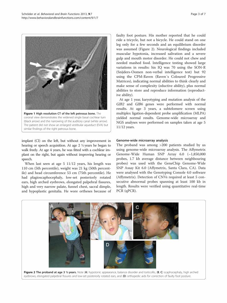
Figure 1 High resolution CT of the left petrosus bone. Thecoronal view demonstrates the widened single basal cochlear turn(black arrow) and the narrowing of the auditory canal (white arrow).The patient did not show an enlarged vestibular aqueduct (EVA) butsimilar findings of the right petrosus bone.
Schröder et al. Behavioral and Brain Functions 2013, 9:7 Page 3 of 7http://www.behavioralandbrainfunctions.com/content/9/1/7
implant (CI) on the left, but without any improvement inhearing or speech acquisition. At age 2 ½ years he began towalk freely. At age 4 years, he was fitted with a cochlear im-plant on the right, but again without improving hearing orspeech.When last seen at age 5 11/12 years, his length was
110 cm (5th percentile), weight was 21 kg (50th percent-ile) and head circumference 53 cm (75th percentile). Hehad plagioscaphocephaly, low-set posteriorly rotatedears, high arched eyebrows, elongated palpebral fissures,high and very narrow palate, funnel chest, sacral dimple,and hypoplastic genitalia. He wore ortheses because of
Figure 2 The proband at age 3 ½ years. Note (A) hypotonic appearanceeyebrows, elongated palpebral fissures and low-set posteriorly rotated ears
faulty foot posture. His mother reported that he couldride a tricycle, but not a bicycle. He could stand on oneleg only for a few seconds and an equilibrium disorderwas assumed (Figure 2). Neurological findings includedmuscular hypotonia, increased salivation and a severegulp and mouth motor disorder. He could not chew andneeded mashed food. Intelligence testing showed largevariations in results: his IQ was 70 using the SON-R(Snijders-Oomen non-verbal intelligence test) but 92using the CPM-Raven (Raven´s Coloured ProgressiveMatrices), indicating normal abilities to think clearly andmake sense of complexity (eductive ability), plus normalabilities to store and reproduce information (reproduct-ive ability).At age 1 year, karyotyping and mutation analysis of the
GJB2 and GJB6 genes were performed with normalresults. At age 3 years, a subtelomere screen usingmultiplex ligation-dependent probe amplification (MLPA)yielded normal results. Genome-wide microarray andNGS analyses were performed on samples taken at age 511/12 years.
Genome-wide microarray analysisThe proband was among >200 patients studied by ususing genome-wide microarray analysis. The AffymetrixGenome-Wide Human SNP Array 6.0 (~1,850,000probes, 1.7 kb average distance between neighbouringprobes) was used with the GeneChip Genome-WideSNP Assay Kit 6.0 (Affymetrix, Santa Clara, CA). Datawere analysed with the Genotyping Console 4.0 software(Affymetrix). Detection of CNVs required at least 5 con-secutive abnormal probes spanning at least 100 kb inlength. Results were verified using quantitative real-timePCR (qPCR).
, balance disorder and torticollis, (B, C) scaphocephaly, high arched, and (D) orthopedic aids for correction of faulty foot posture.

Schröder et al. Behavioral and Brain Functions 2013, 9:7 Page 4 of 7http://www.behavioralandbrainfunctions.com/content/9/1/7
Mapping of the precise molecular breakpoints using nextgeneration sequencingUsing one flow cell lane of an Illumina HiSeq 2000, wegenerated 115,420,519 50 nt single end reads, with101,701,184 reads passing standard quality filters. Readswere aligned to the human reference genome (GRCh37/hg19) using the Bowtie algorithm (version 0.12.7) [12].7,417,427 (7%) of the reads failed to align and 17,125,015(17%) aligned to multiple locations and were removed,leaving 77,455,596 (76%) unambiguously aligned reads.After sorting reads based on genomic position usingthe samtools software package [13], breakpoints weredetermined using a statistical approach implemented inthe R programming language. Based on the existing gen-etic and microarray data, we expected a homozygous dele-tion and thus expected no sequence reads to align to thedeleted region. Away from the suspected deletion and out-side of centromeres, we determined the distribution of agenomic position to the closest sequence read. Using thisdistribution, we determined the likelihood that a givenposition was in a homozygously deleted locus: a genomicposition far from the closest sequencing read is morelikely to be in a deleted locus. For a given position, theassigned p-value represents the fraction of all genomicpositions that are an equal or greater distance from theclosest read. The region identified by the Affymetrix SNP6.0 GeneChip and the FREEC algorithm was examined indetail, including the aligned reads and the resultant p-values (Figure 3A, B) and identified a region where noreads aligned.
Figure 3 Definition of the homozygous deletion by NGS and breakpo(black bars) and RefSeq genes (blue). (B) A plot showing a p-value represendeletion. (C) The sequence at the start and stop of the homozygous deleti
The precise deletion sequence locus was determinedusing PCR with primers designed based on theNGS results, GCTTTTTCTCCTAAATTCTCTGG (for-ward) and TCAGCCATCTTTGTTGTTTCC (reverse)(Figure 3C). PCR was performed according to standardprotocols. The reaction mixture consisted of 2.5 μl 10xPCR buffer, 2.5 μl 50 mM MgCl2, 2.5 μl 10 mM dNTPmix, 1.0 μl (100 ng) of each forward and reverse primer,0.5 μl (2.5 U) Taq polymerase, 14 μl PCR-grade waterund 100 ng template DNA. PCR amplification wascarried out with an initial denaturation step at 94�C for3 min, 35 cycles of 94�C for 30 sec, primer-specificannealing temperature of 55�C for 30 sec, 72�C for60 sec and a final extension step at 72�C for 10 min.Following Exo/SAP digestion, PCR products weresequenced using the CEQ DTCS Quick Start Kit(Beckman Coulter, Krefeld, Germany) and separated ona Beckman Coulter CEQ 8000 Genetic Analysis System.Sequencing data were analysed using the BLAST pro-gram (http://www.ensembl.org/Homo_sapiens/blastviewand http://www.ncbi.nlm.nih.gov/BLAST).
ResultsResults of microarray analysisMicroarray analysis showed a homozygous deletion arr5q31.1(134,764,990–134,875,777)x0 (GRCh37/hg19, ISCN2009) in the patient and identical but heterozygousdeletions in the parents (Figure 4). The deletions wereconfirmed using qPCR. The proximal breakpoint onchromosome 5q31.1 was identified between 134,764,553
int-spanning PCR analysis. (A) The locus, showing aligned readsting the likelihood that a genomic position is not in a homozygouson; the black bars represent the location of primer sequences.
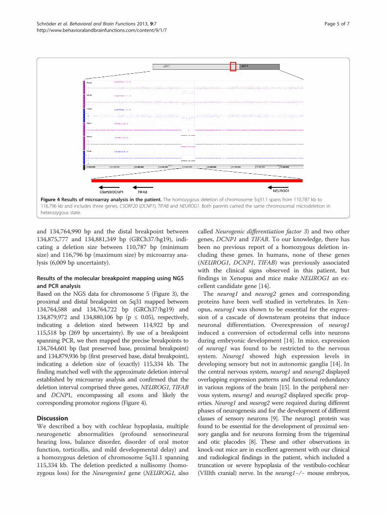
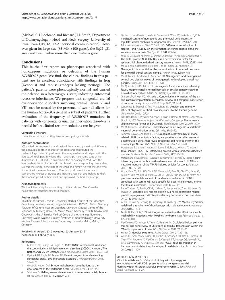
Figure 4 Results of microarray analysis in the patient. The homozygous deletion of chromosome 5q31.1 spans from 110.787 kb to116.796 kb and includes three genes, C5ORF20 (DCNP1), TIFAB and NEUROG1. Both parents carried the same chromosomal microdeletion inheterozygous state.
Schröder et al. Behavioral and Brain Functions 2013, 9:7 Page 5 of 7http://www.behavioralandbrainfunctions.com/content/9/1/7
and 134,764,990 bp and the distal breakpoint between134,875,777 and 134,881,349 bp (GRCh37/hg19), indi-cating a deletion size between 110,787 bp (minimumsize) and 116,796 bp (maximum size) by microarray ana-lysis (6,009 bp uncertainty).
Results of the molecular breakpoint mapping using NGSand PCR analysisBased on the NGS data for chromosome 5 (Figure 3), theproximal and distal breakpoint on 5q31 mapped between134,764,588 and 134,764,722 bp (GRCh37/hg19) and134,879,972 and 134,880,106 bp (p ≤ 0.05), respectively,indicating a deletion sized between 114,922 bp and115,518 bp (269 bp uncertainty). By use of a breakpointspanning PCR, we then mapped the precise breakpoints to134,764,601 bp (last preserved base, proximal breakpoint)and 134,879,936 bp (first preserved base, distal breakpoint),indicating a deletion size of (exactly) 115,334 kb. Thefinding matched well with the approximate deletion intervalestablished by microarray analysis and confirmed that thedeletion interval comprised three genes, NEUROG1, TIFABand DCNP1, encompassing all exons and likely thecorresponding promotor regions (Figure 4).
DiscussionWe described a boy with cochlear hypoplasia, multipleneurogenetic abnormalities (profound sensorineuralhearing loss, balance disorder, disorder of oral motorfunction, torticollis, and mild developmental delay) anda homozygous deletion of chromosome 5q31.1 spanning115,334 kb. The deletion predicted a nullisomy (homo-zygous loss) for the Neurogenin1 gene (NEUROG1, also
called Neurogenic differentiation factor 3) and two othergenes, DCNP1 and TIFAB. To our knowledge, there hasbeen no previous report of a homozygous deletion in-cluding these genes. In humans, none of these genes(NEUROG1, DCNP1, TIFAB) was previously associatedwith the clinical signs observed in this patient, butfindings in Xenopus and mice make NEUROG1 an ex-cellent candidate gene [14].The neurog1 and neurog2 genes and corresponding
proteins have been well studied in vertebrates. In Xen-opus, neurog1 was shown to be essential for the expres-sion of a cascade of downstream proteins that induceneuronal differentiation. Overexpression of neurog1induced a conversion of ectodermal cells into neuronsduring embryonic development [14]. In mice, expressionof neurog1 was found to be restricted to the nervoussystem. Neurog1 showed high expression levels indeveloping sensory but not in autonomic ganglia [14]. Inthe central nervous system, neurog1 and neurog2 displayedoverlapping expression patterns and functional redundancyin various regions of the brain [15]. In the peripheral ner-vous system, neurog1 and neurog2 displayed specific prop-erties. Neurog1 and neurog2 were required during differentphases of neurogenesis and for the development of differentclasses of sensory neurons [9]. The neurog1 protein wasfound to be essential for the development of proximal sen-sory ganglia and for neurons forming from the trigeminaland otic placodes [8]. These and other observations inknock-out mice are in excellent agreement with our clinicaland radiological findings in the patient, which included atruncation or severe hypoplasia of the vestibulo-cochlear(VIIIth cranial) nerve. In the neurog1−/− mouse embryos,

Schröder et al. Behavioral and Brain Functions 2013, 9:7 Page 6 of 7http://www.behavioralandbrainfunctions.com/content/9/1/7
similar malformations of peripheral neural structures withabsence of the vestibular-cochlear ganglion and of all affer-ent, efferent, and autonomic nerve fibers of the VIIIth cra-nial nerve were reported [10]. The efferent and autonomicfibers were thought to be lost as a secondary effect due tothe absence of the afferents [10].Furthermore, the anatomical deviations in the inner
ear of the patient correspond to those in the knockoutmice. The boy’s internal auditory canal was narrowedand the cochlea was hypoplastic with only one singlewidened cochlear turn. In the neurog1−/− mutant mice,the inner ear showed an overall reduction in size andthe cochlea only had 1.25 turns, as opposed to 1.75turns in the control littermates [10]. Moreover, the pa-tient presented with a balance disorder, and thevestibulo-cochlear system of the neurog1−/− mutantmice showed a distinct missing utriculosaccular ductwith only a small saccular recess [10].This patient and the neurog1−/− knockout mice also
shared the severe disorder of oral motor function. Theboy was unable to swallow and to chew food and showedincreased salivation and speech difficulties, while all new-born neurog1−/− mice were unable to suckle, lacked milkin their stomachs and died within 12 hours after birth[10]. Ma et al. suggested that the absence of the trigeminalganglia and associated defects of the Vth nerve resulted ina lack of sensory innervation and neonatal lethality byinterfering with suckling. Correspondingly, we assume amalfunction of the Vth cranial nerve in the boy that couldbe caused by lack of sensory innervation or a missingmotor innervation due to defective development of therhombencephalon, which is the origin of the motoricfibers of the Vth cranial nerve. Taken together, the ana-tomic and functional abnormalities in mice perfectlymatch the symptoms in this patient. Therefore, westrongly infer that Neurog1 is a neuronal determinationgene for the cranial sensory neurons that give rise to cra-nial nerves V and VIII in numerous vertebrates includingXenopus, mouse and humans.In contrast, we consider TIFAB and DCNP1 unlikely can-
didate genes for the abnormalities in this patient. TIFAB ishighly expressed in spleen and inhibits TIFA-mediated cel-lular functions by inducing a conformational change in theTIFA protein [16]. TIFAB impedes activation of NF-kappaBand acts as a negative regulator of TRAF6-induced cellularfunctions such as B cell proliferation and maturation ofmacrophages [17]. DCNP1 is a genetic factor that has beenreported to be involved in asthma susceptibility. A case–control study among subjects with asthma revealed anassociation of variants of DCNP1 with serum immuno-globulin E (IgE) levels [18]. DCNP1 may also play a role inthe pathogenesis of depressive disorders because it wasshown to enhance corticotropin-releasing hormone expres-sion in the hypothalamic paraventricular nucleus. Hence,
DNCP1 interferes with hypothalamic stress response andmay contribute to the pathogenesis of major depression [19].The clinical findings in our proband perfectly match
with those in patients with CCDD, especially of theHOXA1 spectrum: among other symptoms, these patientsshow severe bilateral sensory-neural hearing loss due toabsence of the cochlear and vestibular apparatus, often ac-companied by absence of the eighth cranial nerve [1].Every causative gene characterized in context of CCDDs isassociated with neuronal development at the nuclear,brainstem, or peripheral nerve level [1,2]. For example,the responsible gene in Duane Retraction Syndrome(CHN1) is involved in ocular motor axon path finding ofthe sixth nerve. Homozygous mutations in HOXA1 causean early and profound brainstem patterning defect andheterozygous mutations in KIF21A, which are causativefor congenital fibrosis of the extraocular muscles type 1,lead to disturbance of anterograde organelle transport inneuronal cells [2].Taken together, the clinical and molecular genetic findings
in our proband most closely match the term congenital cra-nial dysinnervation disorder and there are also analogies toMoebius syndrome and its variants (MIM 157900). In alarge study of Moebius syndrome including 37 Dutchpatients, the following clinical observations are consistentwith those observed in our proband: feeding problems atbirth due to insufficient suckling or swallowing, nasal dys-arthria and delayed language development, congenitaldeafness, motor disabilities, and malformations of the ex-tremities of variable severity. A lack of sensation of the lips,cheek, forehead and cornea in some patients indicated a de-fect of the sensory root of the trigeminal nerve [20]. Otherauthors have described external ear deformities, deafnessand pharyngeal involvement in patients with Moebius syn-drome variants [21-23].Moreover, the clinical observations made in our pro-
band partially correspond with those described in patientsharboring the homozygous HOXB1 c.619C > T mutation[24]: the phenotype included bilateral facial palsy, hearingloss, and strabismus. Two affected brothers from consan-guineous parents showed a “masked facies” without anyfacial movement, sensorineural hearing loss, feeding diffi-culties and speech delay. MR imaging of the older brotherrevealed bilateral absence of the facial nerve and bilateralcochlear malformation with abnormal tapering of thebasal turn. In contrast to our case, the vestibulocochlearnerve was preserved on both sides. An affected brotherand sister from another family not known to be consan-guineous also presented with bilateral facial weakness andsensorineural hearing loss [24].The human NEUROG1 maps within the interval of the
DFNB60 locus for non-syndromic autosomal recessivehearing loss on 5q22-q31, but linkage data have excludedNEUROG1 from being causative in the DFNB60 patients
Schröder et al. Behavioral and Brain Functions 2013, 9:7 Page 7 of 7http://www.behavioralandbrainfunctions.com/content/9/1/7
(Michael S. Hildebrand and Richard J.H. Smith, Departmentof Otolaryngology - Head and Neck Surgery, University ofIowa, Iowa City, IA, USA, personal communication). How-ever, given its large size (35 Mb, >100 genes), the 5q22-q31area could well harbor more than one deafness gene.
ConclusionsThis is the first report on phenotypes associated withhomozygous mutations or deletions of the humanNEUROG1 gene. We find, the clinical findings in this pa-tient are in excellent coincidence with findings in frog(Xenopus) and mouse embryos lacking neurog1. Thepatient`s parents were phenotypically normal and carriedthe deletion in a heterozygous state, indicating autosomalrecessive inheritance. We propose that congenital cranialdysinnervation disorders involving cranial nerves V andVIII may be caused by the presence of two null alleles forthe human NEUROG1 gene in a subset of patients. Furtherevaluation of the frequency of NEUROG1 mutations inpatients with congenital cranial dysinnervation disorders isneeded before clinical recommendations can be given.
Competing interestsThe authors declare that they have no competing interests.
Authors’ contributionsJCS carried out sequencing and drafted the manuscript. AKL and AK werethe pedaudiologists in charge of the child and contributed thepedaudiologic data. DG performed the array analysis and helped with thefigures. AP took part in writing the manuscript, it contains parts of herdissertation. JC, SD and UZ carried out the NGS analysis. WMF was theneuroradiologist in charge and contributed the corresponding clinical data.OB was the clinical geneticist of the child and family, he was the one whofirst noticed the resemblance to Moebius syndrome, initiated andcoordinated molecular studies and literature research and helped to draftthe manuscript. All authors read and approved the final manuscript.
AcknowledgementsWe thank the family for consenting to this study and Mrs. CorneliaPoarangan for excellent technical support.
Author details1Institute of Human Genetics, University Medical Centre of the JohannesGutenberg University Mainz, Langenbeckstrasse 1, D-55101, Mainz, Germany.2Division of Communication Disorders, University Medical Centre of theJohannes Gutenberg University Mainz, Mainz, Germany. 3TRON-TranslationalOncology at the University Medical Centre of the Johannes GutenbergUniversity Mainz, Mainz, Germany. 4Institute of Neuroradiology, UniversityMedical Centre of the Johannes Gutenberg University Mainz, Mainz,Germany.
Received: 31 August 2012 Accepted: 23 January 2013Published: 18 February 2013
References1. Gutowski NJ, Bosley TM, Engle EC: 110th ENMC International Workshop:
the congenital cranial dysinnervation disorders (CCDDs). Naarden, TheNetherlands, 25–27 October, 2002. Neuromuscul Disord 2003, 13:573–578.
2. Oystreck DT, Engle EC, Bosley TM: Recent progress in understandingcongenital cranial dysinnervation disorders. J Neuroophthalmol 2011,31:69–77.
3. Webb JF, Noden DM: Ectodermal placodes: contributions to thedevelopment of the vertebrate head. Am Zool 1993, 33:434–447.
4. Schlosser G: Making senses development of vertebrate cranial placodes.Int Rev Cell Mol Biol 2010, 283:129–234.
5. Fischer T, Faus-Kessler T, Welzl G, Simeone A, Wurst W, Prakash N: Fgf15-mediated control of neurogenic and proneural gene expressionregulates dorsal midbrain neurogenesis. Dev Biol 2011, 350:496–510.
6. Takano-Maruyama M, Chen Y, Gaufo GO: Differential contribution ofNeurog1 and Neurog2 on the formation of cranial ganglia along theanterior-posterior axis. Dev Dyn 2012, 241:229–241.
7. Fode C, Gradwohl G, Morin X, Dierich A, LeMeur M, Goridis C, Guillemot F:The bHLH protein NEUROGENIN 2 is a determination factor forepibranchial placode-derived sensory neurons. Neuron 1998, 20:483–494.
8. Ma Q, Chen Z, del Barco Barrantes I, de la Pompa JL, Anderson DJ:Neurogenin1 is essential for the determination of neuronal precursorsfor proximal cranial sensory ganglia. Neuron 1998, 20:469–482.
9. Ma Q, Fode C, Guillemot F, Anderson DJ: Neurogenin1 and neurogenin2control two distinct waves of neurogenesis in developing dorsal rootganglia. Genes Dev 1999, 13:1717–1728.
10. Ma Q, Anderson DJ, Fritzsch B: Neurogenin 1 null mutant ears developfewer, morphologically normal hair cells in smaller sensory epitheliadevoid of innervation. J Assoc Res Otolaryngol 2000, 1:129–143.
11. Graham JM, Phelps PD, Michaels L: Congenital malformations of the earand cochlear implantation in children: Review and temporal bone reportof common cavity. J Laryngol Otol Suppl 2000, 25:1–14.
12. Langmead B, Trapnell C, Pop M, Salzberg SL: Ultrafast and memory-efficient alignment of short DNA sequences to the human genome.Genome Biol 2009, 10:R25.
13. Li H, Handsaker B, Wysoker A, Fennell T, Ruan J, Homer N, Marth G, Abecasis G,Durbin R, 1000 Genome Project Data Processing Subgroup: The sequencealignment/map format and SAM tools. Bioinformatics 2009, 25:2078–2079.
14. Ma Q, Kintner C, Anderson DJ: Identification of neurogenin, a vertebrateneuronal determination gene. Cell 1996, 87:43–52.
15. Sommer L, Ma Q, Anderson DJ: Neurogenins, a novel family of atonal-related bHLH transcription factors, are putative mammalian neuronaldetermination genes that reveal progenitor cell heterogeneity in thedeveloping CNS and PNS. Mol Cell Neurosci 1996, 8:221–241.
16. Matsumura T, Semba K, Azuma S, Ikawa S, Gohda J, Akiyama T, Inoue J:TIFAB inhibits TIFA, TRAF-interacting protein with a forkhead-associateddomain. Biochem Biophys Res Commun 2004, 317:230–234.
17. Matsumura T, Kawamura-Tsuzuku J, Yamamoto T, Semba K, Inoue J: TRAF-interacting protein with a forkhead-associated domain B (TIFAB) is anegative regulator of the TRAF6-induced cellular functions. J Biochem2009, 146:375–381.
18. Kim Y, Park CS, Shin HD, Choi JW, Cheong HS, Park BL, Choi YH, Jang AS,Park SW, Lee YM, Lee EJ, Park SG, Lee JY, Lee JK, Han BG, Oh B, Kimm K: Apromoter nucleotide variant of the dendritic cell-specific DCNP1associates with serum IgE levels specific for dust mite allergens amongthe Korean asthmatics. Genes Immun 2007, 8:369–378.
19. Zhou T, Wang S, Ren H, Qi XR, Luchetti S, Kamphuis W, Zhou JN, Wang G,Swaab DF: Dendritic cell nuclear protein-1, a novel depression-relatedprotein, upregulates corticotropin-releasing hormone expression. Brain2010, 133:3069–3079.
20. Verzijl HT, van der Zwaag B, Cruysberg JR, Padberg GW: Moebius syndromeredefined: a syndrome of rhombencephalic maldevelopment. Neurology2003, 61:327–333.
21. Terzis JK, Karypidis D: Direct tongue neurotization: the effect on speechintelligibility in patients with Moebius syndrome. Plast Reconstr Surg 2010,125:150–160.
22. MacDermot KD, Winter R, Taylor D, Baraitser M: Oculofacialbulbar palsy inmother and son: review of 26 reports of familial transmission within the“Moebius spectrum of defects”. J Med Genet 1991, 28:18–26.
23. Kumar D: Moebius syndrome. J Med Genet 1990, 27:122–126.24. Webb BD, Shaaban S, Gaspar H, Cunha LF, Schubert CR, Hao K, Robson CD,
Chan WM, Andrews C, MacKinnon S, Oystreck DT, Hunter DG, Iacovelli AJ,Ye X, Camminady A, Engle EC, Jabs EW: HOXB1 founder mutation inhumans recapitulates the phenotype of Hoxb1−/− mice. Am J Hum Genet2012, 91:171–179.
doi:10.1186/1744-9081-9-7Cite this article as: Schröder et al.: A boy with homozygousmicrodeletion of NEUROG1 presents with a congenital cranialdysinnervation disorder [Moebius syndrome variant]. Behavioral andBrain Functions 2013 9:7.