Embed Size (px)
Citation preview

R E S EA RCH L E T T E R
Nonmucoid conversion of mucoid Pseudomonas aeruginosainduced by sulfate-stimulated growth
Kyung Bae Min1, Kang-Mu Lee1, Young Taek Oh1 & Sang Sun Yoon1,2
1Department of Microbiology and Immunology, Brain Korea PLUS Project for Medical Science, Seoul, Korea; and 2Institute for Immunology and
Immunological Diseases, Yonsei University College of Medicine, Seoul, Korea
Correspondence: Sang Sun Yoon,
Department of Microbiology and
Immunology, Yonsei University College of
Medicine, 250 Seongsanno, Seodaemun-gu,
Seoul 120-752, Korea.
Tel.: +82 2 2228 1824;
fax: +82 2 392 7088;
e-mail: [email protected]
Received 14 July 2014; accepted 11
September 2014. Final version published
online 31 October 2014.
DOI: 10.1111/1574-6968.12600
Editor: Mark Schembri
Keywords
Pseudomonas aeruginosa; mucoid; sulfate;
nonmucoid reversion; alginate.
Abstract
Alginate-overproducing mucoid Pseudomonas aeruginosa, responsible for
chronic airway infections in cystic fibrosis (CF) patients, is resistant to antibi-
otic treatments and host immune clearance. In this study, we performed a phe-
notype microarray screen and identified sulfate ion as a molecule that can
suppress alginate production. When a mucoid P. aeruginosa strain CM21 and
additional mucoid isolates were grown with 5% sodium sulfate, significantly
decreased levels of alginate were produced. Suppression of alginate production
was also induced by other sulfate salts. Expression of a reporter gene fused to
the algD promoter was considerably decreased when grown with sulfate. Fur-
thermore, bacterial cell shape was abnormally altered in CM21, but not in
PAO1, a prototype nonmucoid strain, suggesting that sulfate-stimulated cell
shape change is associated with transcriptional suppression of the alginate
operon. Finally, a CM21 lpxC mutant defective in lipid A biosynthesis contin-
ued to produce alginate and maintained the correct cell shape when grown
with sulfate. These results suggest a potential involvement of lipoploysaccharide
biosynthesis in the sulfate-induced reversion to nonmucoid phenotype. This
study proposes a novel strategy that can be potentially applied to treat persis-
tent infection by recalcitrant mucoid P. aeruginosa.
Introduction
The Gram-negative bacterium Pseudomonas aeruginosa is
a major opportunistic pathogen in human cystic fibrosis
(CF). In chronic CF airways, P. aeruginosa strains that
have acquired selected phenotypes are recovered and
such phenotypic changes are induced for survival in the
harsh environment of the human lung (Burns et al.,
2001; Hogardt et al., 2007; Mena et al., 2008). One of
these changes is an overproduction of a capsule-like
polysaccharide called alginate (Evans & Linker, 1973).
Mucoid P. aeruginosa strains isolated from sputum of
CF patients have a mutation in the mucA gene, encod-
ing MucA, a negative regulator of AlgT that activates
transcription of the alginate biosynthesis operon (Martin
et al., 1993). A truncated version of MucA, encoded by
a mutated mucA gene, no longer binds to AlgT (Xie
et al., 1996), and expression of the alginate biosynthesis
operon is continuously activated (Govan & Deretic,
1996; Xie et al., 1996; Boucher et al., 1997; Bragonzi
et al., 2009). Besides this major pathway to mucoid con-
version, a stringent starvation response or sigma factor
competition were also reported to regulate alginate pro-
duction in P. aeruginosa (May et al., 1991; Yin et al.,
2013).
In mucoid P. aeruginosa, expression of virulence factors
is more down-regulated than acute infection, and this is
in response to the need to thrive in infected tissue or
organs (Hogardt & Heesemann, 2010). However, mucoid
P. aeruginosa exhibit several unique virulence-associated
characteristics compared with nonmucoid counterparts.
First, mucoid variants are more resistant to antibiotic
treatment (Govan & Fyfe, 1978) and phagocytic immune
activity (Cabral et al., 1987). Such a resistant nature is
due to the presence of an alginate capsule around the cell.
Secondly, mucoid strains are less potent in activating the
type III secretion system, an important virulence determi-
nant (Wu et al., 2004). These phenotypes are considered
to contribute to the persistent survival in the patient air-
way (Yu et al., 1998). For efficient treatment, it would
FEMS Microbiol Lett 360 (2014) 157–166 ª 2014 Federation of European Microbiological Societies.Published by John Wiley & Sons Ltd. All rights reserved
MIC
ROBI
OLO
GY
LET
TER
S

therefore be beneficial to switch mucoid P. aeruginosa
back to a nonmucoid form.
When a mucoid P. aeruginosa strain was grown stati-
cally with reduced oxygen potential, nonmucoid rever-
tants were recovered that acquired spontaneous
mutations in the algT gene (Wyckoff et al., 2002). In
addition, overexpression of the rpoD gene resulted in
suppressed production of alginate AlgT/U (Yin et al.,
2013). These previous studies suggest that reversion to a
nonmucoid form can be achieved by genetic alterations.
To our knowledge, however, conversion to a nonmu-
coid form by treatment with a defined chemical com-
pound has not previously been elucidated. Here, we
performed a phenotype microarray analysis with a clini-
cally isolated mucoid P. aeruginosa strain and revealed
that alginate production was suppressed by sulfate ions.
This highlights a previously undescribed means to
weaken mucoid P. aeruginosa, a major threat to human
healthcare.
Materials and methods
Phenotype microarray, bacterial strains and
growth conditions
Bacterial strains and plasmids used in this study are
shown in Table 1. Phenotype microarray (Biolog Inc.,
Hayward, CA) experiments were performed as described
elsewhere (Johnson et al., 2008). The mucoid CM21
strain was grown in each well of phenotype microarray
plates for 24 h. Bacterial cells were then inoculated into
150 9 20-mm Luria–Bertani (LB) agar plates using a
96-well pin replicator. After 24 h growth at 37 °C, mor-
phology of the CM21 colonies was examined and growth
conditions that induced nonmucoid reversion were iden-
tified. CF strain P. aeruginosa PAO1 and Escherichia coli
were grown at 37 °C on LB plates or in LB broth. Genta-
micin (Sigma-Aldrich, St. Louis, MO) at 50 lg mL�1 was
added for selecting transformed bacteria. Sodium sulfate,
ammonium sulfate, magnesium sulfate and sodium
chloride were all purchased from Sigma-Aldridge. The
reporter plasmid, pMEXRalgD, was a kind gift from
Dr Toyofuku (University of Tsukuba, Japan) and it has
been modified from pMEX9 (Toyofuku et al., 2007) by
replacing the xylE reporter gene with a gene encoding
Ds-Red.
Alginate assay
Pseudomonas aeruginosa strains were grown at 37 °C in
LB broth and LB broth containing sodium sulfate
(LB+SS), ammonium sulfate (LB+AS), magnesium sulfate
(LB+MS) and sodium chloride (LB+SC) for 24 h with
vigorous shaking. Bacterial cells were grown in 10 mL LB
overnight and a 1-mL aliquot of each bacterial culture
was pelleted by centrifugation at 16000 9 g for 5 min.
Twenty microliters of culture supernatant was mixed with
80 lL distilled water and assayed for quantification of
alginate as described previously (Damron et al., 2009).
OD600 values of bacterial suspensions were used for nor-
malization. The amount of uronic acid was analysed with
a standard curve constructed with the use of alginic acid
from brown algae (Sigma-Aldrich).
Transposon random mutagenesis
For random transposon insertion mutagenesis, E. coli
SM10/kpir harboring pBTK30 (Kim et al., 2012) and
CM21 were used as donor and recipient strains, respec-
tively. Gentamicin-resistant trans-conjugants were grown
in LB agar plates containing 50 lg mL�1 gentamicin,
50 lg mL�1 Irgasan (Sigma) and 5% sodium sulfate.
Mucoid mutants were selected and the transposon inser-
tion site was determined by arbitrary PCR as described by
Kim et al. (2012).
Complementation of lpxC mutant
A low copy plasmid pJN105 was used for complementa-
tion of the lpxC mutant. The gentamicin resistance gene
of pJN105 was replaced with a carbenicillin resistance
marker and named pJN105c (Table 1). The lpxC gene of
the parental strain CM21 was amplified and cloned into
pJN105c. Primers to amplify the lpxC ORF and its
endogenous promoter region were lpxC_EcoRI_F: 50-CCATTGAATTCTGGAGAAACCGGTGAAGGTCG-30 and lpxC_
EcoRI_R: 50-ATGTGGAATTCGAAAACCTTCGCGAATCCGCC-30 (EcoRI restriction recognition sequence
underlined in each primer sequence). The CM21 lpxC::Tn
mutant was then transformed with pJN105c::lpxC
or pJN105c by electroporation. Carbenicillin at 50 or
75 lg mL�1 was used for selective growth in broth or on
agar plates, respectively.
Growth curve measurement
Pseudomonas aeruginosa strains PAO1 and CM21 were
grown in LB or in LB supplemented with 5% sodium sul-
fate for 20 h at 37 °C with shaking at 200 r.p.m. Initial
culture volume was 10 mL and bacterial growth was
monitored by measuring OD600 values.
DsRed fluorescence mesurement
CM21 transformed with pMEXRalgD plasmid was incu-
bated in 352 mM sodium sulfate, ammonium sulfate,
FEMS Microbiol Lett 360 (2014) 157–166ª 2014 Federation of European Microbiological Societies.Published by John Wiley & Sons Ltd. All rights reserved
158 K.B. Min et al.
magnesium sulfate and sodium chloride, and 50 lg mL�1
gentamicin added to LB broth, consisting of 1% tryptone
(w/v) and 0.5% yeast extract (w/v), with vigorous shaking
for 24 h at 37 °C. Each 200-lL aliquot of bacterial cul-
ture was transferred to a 96-well plate. Fluorescence was
measured using a fluorometer. Excitation and emission
wavelength were 555 and 600 nm, respectively. For nor-
malization based on OD600 value, each 1-mL aliquots of
bacterial culture was pelleted by centrifugation at 16000
9 g for 5 min and resuspended in phosphate-buffered
saline.
Confocal microscopy
Bacterial shape and red fluorescent signal were observed
using FV-1000 confocal microscope (Olympus Optical,
Tokyo, Japan) following procedures described previously
(Yoon et al., 2011; Lee et al., 2012). Excitation and emis-
sion wavelength were 555 and 600 nm, respectively, for
observation of DsRed fluorescence.
Statistical analysis
Data are expressed as means � SD. An unpaired Student’s
t-test was used to analyse the average difference between
the nonsulfate and sulfate-treated groups, and a P value of
< 0.05 was considered statistically significant. All experi-
ments were repeated to verify the reproducibility.
Results
Alginate production was decreased during
growth with 5% sodium sulfate
To determine the growth condition that suppresses algi-
nate production, a mucoid CF isolate strain, CM21, was
grown in plates of the phenotype microarray (PM). After
overnight growth, strain CM21 was reinoculated into LB
agar plates using a 96-well pin replicator and we screened
for CM21 colonies that showed a nonmucoid phenotype.
Among 1919 wells, reduced alginate production was only
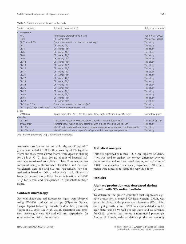
Table 1. Strains and plasmids used in the study
Strain or plasmid Relevant characteristic(s) Reference or source
P. aeruginosa
PAO1 Nonmucoid prototype strain; Alg� Yoon et al. (2002)
FRD1 CF isolate; Alg+ Yoon et al. (2006)
PAO1 mucA::Tn Transposon insertion mutant of mucA; Alg+ This study
CM2 CF isolate; Alg� This study
CM4 CF isolate; Alg+ This study
CM6 CF isolate; Alg� This study
CM8 CF isolate; Alg+ This study
CM9 CF isolate; Alg+ This study
CM12 CF isolate; Alg+ This study
CM13 CF isolate; Alg+ This study
CM15 CF isolate; Alg+ This study
CM19 CF isolate; Alg+ This study
CM21 CF isolate; Alg+ This study
CM22 CF isolate; Alg� This study
CM23 CF isolate; Alg+ This study
CM24 CF isolate; Alg� This study
CM25 CF isolate; Alg� This study
CM28 CF isolate; Alg� This study
CM46 CF isolate; Alg+ This study
CM51 CF isolate; Alg+ This study
CM52 CF isolate; Alg+ This study
CM21 lpxC::Tn Transposon insertion mutant of lpxC This study
CM21 lpxC::Tn/pJN105c::lpxC lpxC::Tn complementation strain This study
E. coli
SM10/kpir Donor strain, Kmr, thi-1, thr, leu, tonA, lacY, supE, recA::RP4-2-Tc::Mu, kpir Laboratory strain
Plasmids
pBTK30 Transposon vector for construction of a random mutant library, Gmr Kim et al. (2012)
pMEXRalgD Transcriptional fusion of algD promoter with a gene encoding DsRed, Gmr This study
pJN105c pJN105 with carbenicillin resistance marker in replace of gentamicin resistance marker This study
pJN105c::lpxC pJN105c with wild-type copy of lpxC gene with its endogenous promoter This study
Alg+, mucoid phenotype; Alg�; nonmucoid phenotype.
FEMS Microbiol Lett 360 (2014) 157–166 ª 2014 Federation of European Microbiological Societies.Published by John Wiley & Sons Ltd. All rights reserved
Sulfate-induced suppression of alginate production 159

observed in D08 well of the Microplate No. 9 that con-
tained 5% sodium sulfate. Quantitative alginate assay fur-
ther confirmed suppressed alginate production by sodium
sulfate. As shown in Fig. 1a, the level of alginate pro-
duced by CM21 during growth with sodium sulfate was
significantly decreased when compared with that of
LB-grown CM21. The suppressed level of alginate pro-
duced by CM21 was similar to that produced by PAO1, a
nonmucoid prototype P. aeruginosa strain (Fig. 1b), sug-
gesting that alginate production was almost completely
inhibited by sodium sulfate. A well-known mucoid strain
FRD1 (Lee et al., 2013) also responded to the presence of
sodium sulfate and decreased production of alginate was
observed (Fig. 1a). Moreover, PAO1-derived mucA
mutants, which were determined to be mucoid (Xie et al.,
1996), also responded to the presence of sodium sulfate
and exhibited nonmucoid phenotype (Supporting Infor-
mation, Fig. S1). We then examined whether alginate
production is affected by sodium sulfate in a dose-depen-
dent manner. A gradual decrease in alginate production
was observed in response to increasing concentrations of
sodium sulfate (Fig. 1b). Together, our results demon-
strated that sodium sulfate can suppress alginate produc-
tion in mucoid P. aeruginosa strains.
Environmental factors that influence bacterial growth,
such as temperature (Leitao et al., 1992) and oxygen
potential (Krieg et al., 1986), were reported to play roles
in alginate production. We therefore explored whether
suppressed alginate production is associated with altered
bacterial growth by sodium sulfate. Based on our growth
curve experiments shown in Fig. 1c and d, bacterial
growth was only marginally affected by sodium sulfate.
Although a 2-h delay was observed during the exponen-
tial phase of growth, final OD600 values were comparable
between growth in LB and LB supplemented with 5%
sodium sulfate (Fig. 1c and d). Of note is that such a
delay was invariably observed, irrespective of whether the
tested strain was nonmucoid or mucoid. These results
suggest that sulfate-stimulated suppression of alginate
production is probably not due to the growth inhibition
of the CM21 mucoid strain by sodium sulfate.
Sulfate-stimulated suppression of alginate
production in other mucoid P. aeruginosa CF
isolates
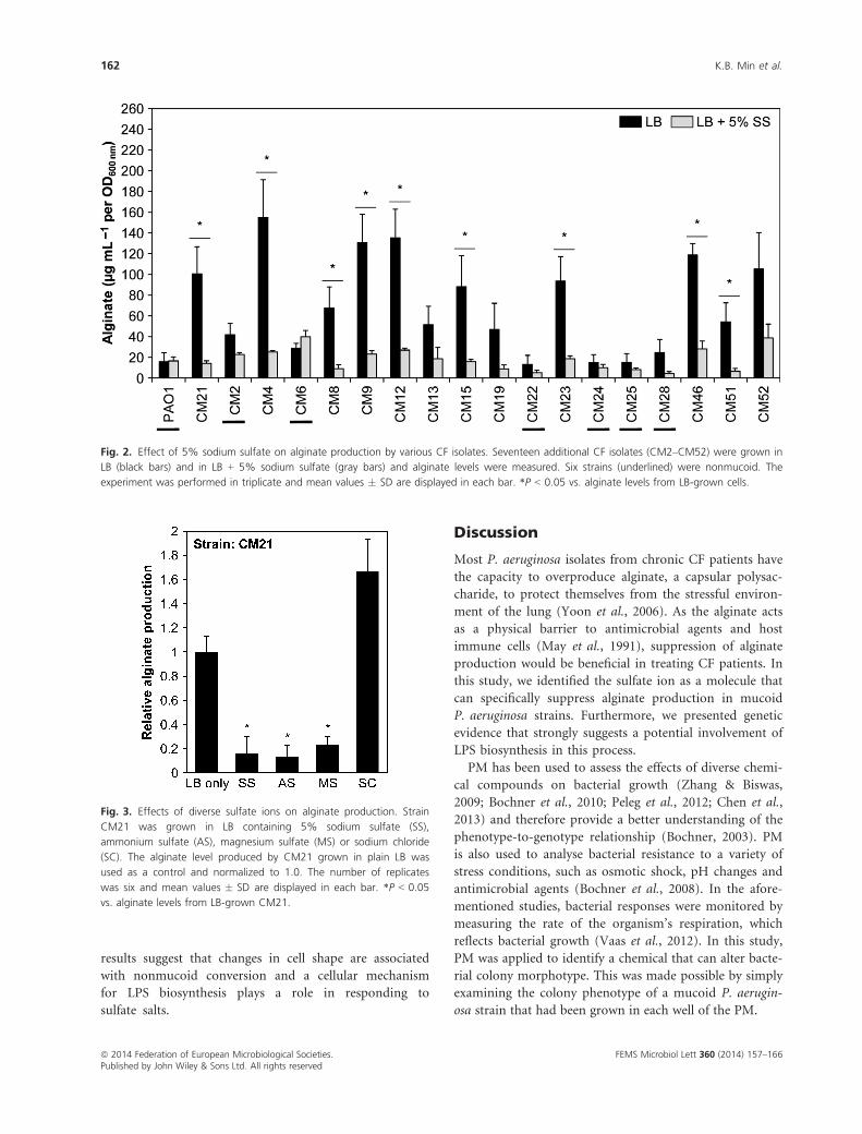
Next, we sought to elucidate the effect of sodium sulfate
on bacterial ability to produce alginate in 17 other P. aeru-
ginosa CF isolates. Among these strains, CM02, CM06,
CM22, CM24, CM25 and CM28 were nonmucoid, while
the other 11 strains were mucoid. To verify sulfate-induced
conversion to a nonmucoid phenotype in a quantitative
manner, we used an alginate assay with bacterial culture
supernatants. As shown in Fig. 2, all the mucoid strains
showed decreased levels of alginate when grown with 5%
sodium sulfate, although bacterial response to sodium sul-
fate varied between strains. CM4, CM12 and CM51 exhib-
ited the most dramatic decrease in alginate production in
response to the treatment, while strain CM52 showed only
minimal decrease in the assay (Fig. 2). Again, nonmucoid
strains grown in LB media produced low levels of alginate,
further verifying the specificity of our alginate assay.
Other sulfate salts also suppressed alginate
production in the mucoid CM21 strain
Our results demonstrated that sodium sulfate can sup-
press alginate production in all the tested mucoid strains.
Next, we examined whether the inhibitory alginate pro-
duction can also be achieved by other sulfate salts. When
strain CM21 was grown in LB containing equal concen-
trations (352 mM) of ammonium sulfate or magnesium
sulfate, we observed a similar degree of suppression of
alginate production (Fig. 3). To rule out the possibility
that the suppressed alginate production is caused by
osmotic stress due to the presence of high concentrations
of sulfate salts, we tested the effect of NaCl on alginate
production. Of note, CM21 strain produced higher levels
of alginate during growth in LB supplemented with NaCl.
Together, these results suggest that suppressed production
of alginate by sodium sulfate is induced by the sulfate
ion, and not by sodium ions or by any secondary effect
due to the presence of high osmotic stress.
Sulfate-induced suppression of alginate
production is regulated at the transcriptional
level
Next, we sought to assess the effect of sulfate on expression
of the algD gene, the first gene of the alginate biosynthesis
operon that encodes GDP-mannose 6-dehydrogenase. To
address this, we used a transcriptional fusion construct,
pMEXRalgD, in which the DsRed-coding gene was cloned
downstream of the algD promoter (Toyofuku et al., 2007,
2014). PAO1 transformed with pMEXRalgD failed to
exhibit a red fluorescent signal, while a robust red fluo-
rescent signal was detected in CM21 harboring the same
plasmid (Fig. 4a and f). This result further confirmed that
the alginate biosynthesis operon is actively transcribed in
the mucoid CM21 strain. Note that the red fluorescent
signal was significantly decreased in the CM21/pMEX-
RalgD strain grown in LB amended with sodium sulfate,
ammonium sulfate or magnesium sulfate (Fig. 4g–i).Consistent with this finding, alginate production was sup-
pressed under these growth conditions (Fig. 3). When
grown with NaCl, a culture condition that permitted
FEMS Microbiol Lett 360 (2014) 157–166ª 2014 Federation of European Microbiological Societies.Published by John Wiley & Sons Ltd. All rights reserved
160 K.B. Min et al.

robust alginate production, the CM21/pMEXRalgD strain
exhibited a comparable level of red fluorescent signal
(Fig. 4j). Consistent with the image analysis, red fluores-
cent intensities of CM21/pMEXRalgD cells were signifi-
cantly decreased when grown with sodium sulfate,
ammonium sulfate and magnesium sulfate (Fig. S2).
Intriguingly, we observed significant cell shape changes of
the mucoid CM21 strain in response to growth with
sodium sulfate, ammonium sulfate or magnesium sulfate.
A round cell shape was observed when grown with
sodium sulfate (Fig. 4g) or ammonium sulfate (Fig. 4h),
whereas CM21 strain became highly elongated upon
growth with magnesium sulfate (Fig. 4i). Cell shape
changes were not detected in the nonmucoid PAO1 strain
under these growth conditions (Fig. 4b–d). Together, ourresults suggest that sulfate-induced suppression of alginate
production is probably accompanied by bacterial cell
shape change.
Isolation of CM21-derived mutants that
remained mucoid during growth with sodium
sulfate
To provide insight into the nonmucoid conversion in
response to sodium sulfate, we screened a library of
CM21 transposon insertion mutants looking for a
mutant that remained mucoid during sulfate-stimulated
growth. Among c. 32 000 mutants tested, three inde-
pendent mutants were found to be mucoid in LB agar
plates containing 5% sodium sulfate. Importantly, all of
the three mutants were determined to have transposon
insertion in the lpxC (PA4406) gene. Transposon inser-
tion occurred at different sites, all of which were
located near the 30 end of the gene (data not
shown).The lpxC gene encodes LpxC enzyme, UDP-3-
N-acetylglucosamine deacetylase, a key enzyme involved
in lipid A biosynthesis, the first committed step for
lipopolysaccharide (LPS) production (Hyland et al.,
1997). The lpxC::Tn mutant maintained a mucoid phe-
notype in the presence of 5% sodium sulfate, while its
parental CM21 strain again showed nonmucoid colonies
in the same growth media, and a quantitative alginate
assay further verified that the lpxC::Tn mutant continu-
ously produced alginate in LB media containing sodium
sulfate (Fig. 5a and b). When the mutant was comple-
mented with its wild-type copy of the lpxC gene, sul-
fate responsiveness was recovered and thus alginate
production was markedly decreased (Fig. 5c and d).
Importantly, the normal rod-shape morphology was not
changed after growth with sulfate salts (Fig. S3). These
(a) (b)
(c) (d)
Fig. 1. Identification of sodium sulfate as a
molecule that can suppress alginate
production. (a) Levels of alginate produced by
PAO1 (nonmucoid), CF isolate FRD1 (mucoid)
and CM21 (mucoid) strains in LB (black bars)
and in LB containing 5% sodium sulfate (SS,
gray bars). The number of replicates was three
and mean values � SD are displayed in each
bar. *P < 0.05 vs. alginate levels from LB-
grown FRD1, **P < 0.005 vs. alginate levels
from LB-grown CM21. (b) Dose-dependent
effect of sodium sulfate on alginate
production. Mucoid CM21 strain was grown
in LB with increasing concentrations of sodium
sulfate. *P < 0.05 vs. alginate levels from
CM21 grown in LB, **P < 0.005 vs. alginate
levels from LB-grown CM21. The number of
replicates was six and mean values � SD are
displayed in each bar. (c, d) Effect of 5%
sodium sulfate on bacterial growth. (c)
Nonmucoid wild-type PAO1 and (d) mucoid
CF isolate CM21 were grown in LB (solid line
with black triangles) and in LB containing 5%
sulfate (dotted line with black squares) for
20 h. Bacterial growth was monitored by
measuring the optical density (OD) at 600 nm
every 2 h.
FEMS Microbiol Lett 360 (2014) 157–166 ª 2014 Federation of European Microbiological Societies.Published by John Wiley & Sons Ltd. All rights reserved
Sulfate-induced suppression of alginate production 161
results suggest that changes in cell shape are associated
with nonmucoid conversion and a cellular mechanism
for LPS biosynthesis plays a role in responding to
sulfate salts.
Discussion
Most P. aeruginosa isolates from chronic CF patients have
the capacity to overproduce alginate, a capsular polysac-
charide, to protect themselves from the stressful environ-
ment of the lung (Yoon et al., 2006). As the alginate acts
as a physical barrier to antimicrobial agents and host
immune cells (May et al., 1991), suppression of alginate
production would be beneficial in treating CF patients. In
this study, we identified the sulfate ion as a molecule that
can specifically suppress alginate production in mucoid
P. aeruginosa strains. Furthermore, we presented genetic
evidence that strongly suggests a potential involvement of
LPS biosynthesis in this process.
PM has been used to assess the effects of diverse chemi-
cal compounds on bacterial growth (Zhang & Biswas,
2009; Bochner et al., 2010; Peleg et al., 2012; Chen et al.,
2013) and therefore provide a better understanding of the
phenotype-to-genotype relationship (Bochner, 2003). PM
is also used to analyse bacterial resistance to a variety of
stress conditions, such as osmotic shock, pH changes and
antimicrobial agents (Bochner et al., 2008). In the afore-
mentioned studies, bacterial responses were monitored by
measuring the rate of the organism’s respiration, which
reflects bacterial growth (Vaas et al., 2012). In this study,
PM was applied to identify a chemical that can alter bacte-
rial colony morphotype. This was made possible by simply
examining the colony phenotype of a mucoid P. aerugin-
osa strain that had been grown in each well of the PM.
Fig. 2. Effect of 5% sodium sulfate on alginate production by various CF isolates. Seventeen additional CF isolates (CM2–CM52) were grown in
LB (black bars) and in LB + 5% sodium sulfate (gray bars) and alginate levels were measured. Six strains (underlined) were nonmucoid. The
experiment was performed in triplicate and mean values � SD are displayed in each bar. *P < 0.05 vs. alginate levels from LB-grown cells.
Fig. 3. Effects of diverse sulfate ions on alginate production. Strain
CM21 was grown in LB containing 5% sodium sulfate (SS),
ammonium sulfate (AS), magnesium sulfate (MS) or sodium chloride
(SC). The alginate level produced by CM21 grown in plain LB was
used as a control and normalized to 1.0. The number of replicates
was six and mean values � SD are displayed in each bar. *P < 0.05
vs. alginate levels from LB-grown CM21.
FEMS Microbiol Lett 360 (2014) 157–166ª 2014 Federation of European Microbiological Societies.Published by John Wiley & Sons Ltd. All rights reserved
162 K.B. Min et al.

Our results clearly suggested that sulfate-induced sup-
pression of alginate production occurred in association
with cell shape change. Because such a morphological
change was not observed in nonmucoid PAO1, the sensi-
tivity of mucoid strains to sulfate ions is probably due to
the distinct differences in cell surface properties between
(a) (b) (c) (d) (e)
(f) (g) (h) (i) (j)
Fig. 4. Confocal micrographs of algD promoter reporter strains. Nonmucoid wild-type strain PAO1 and mucoid CF isolate strain CM21 were
transformed with the plasmid pMEXRalgD, in which the algD promoter was fused with the coding sequence of DsRed red fluorescent protein.
Reporter strains were grown in LB (a and f), LB+SS (b and g), LB+AS (c and h), LB+MS (d and i) or LB+SC (e and j) and analyzed for red
fluorescence. All images were acquired at 91000 magnification. Scale bars = 5 lm.
(a) (b)
(c) (d)
Fig. 5. Effect of the lpxC gene mutation on
the ability of CM21 to produce alginate. (a)
Colony morphotype of CM21 and CM21
lpxC::Tn mutant in an LB agar plate containing
5% sodium sulfate. (b) Levels of alginate
produced by CM21 (black bars) and its lpxC
mutant (gray bars). The alginate level
produced by CM21 grown in plain LB was
used as a control and normalized to 1.0. The
experiment was performed in triplicate and
mean values � SD are displayed in each bar.
*P < 0.05 vs. alginate levels from LB-grown
CM21. (c) Colony morphotype of the control
strain, CM21 lpxC::Tn/pJN105c, and
complemented strain, CM21 lpxC::Tn/
pJN105c::lpxC. (d) Levels of alginate produced
by the lpxC complemented strain, CM21 lpxC::
Tn/pJN105c::lpxC (black bars), and the control
strain, CM21 lpxC::Tn/pJN105c (gray bars).
The alginate level produced by the
complemented strain grown in plain LB was
used as a control and normalized to 1.0. The
experiment was performed in triplicate and
mean values � SD are displayed in each bar.
*P < 0.05 vs. alginate levels from LB-grown
CM21 lpxC::Tn/pJN105c::lpxC. The experiment
was performed in triplicate and means � SD
are displayed in each bar. *P < 0.05 vs.
alginate levels from LB-grown lpxC::Tn
mutant.
FEMS Microbiol Lett 360 (2014) 157–166 ª 2014 Federation of European Microbiological Societies.Published by John Wiley & Sons Ltd. All rights reserved
Sulfate-induced suppression of alginate production 163

mucoid and nonmucoid cells. To produce and secrete
alginate, a polymeric substance with high molecular
weight, mucoid P. aeruginosa requires a multiprotein
complex spanning the outer membrane, the periplasm
and the inner membrane (Remminghorst & Rehm, 2006).
Our results also showed that the transcriptional activity
of the algD promoter was down-regulated in the presence
of the sulfate ion. It will be important to investigate how
sulfate-induced changes in cell surface properties lead to
the signal that eventually suppressed the transcription of
the alginate operon.
Changes in bacterial cell morphology are triggered by
various stimuli. During anaerobic NO�3 respiration, cell
elongation was observed in nonmucoid P. aeruginosa
(Yoon et al., 2011). Of note is that such a unique cell
shape change was not detected in mucoid P. aeruginosa.
Together, these results suggest that bacterial response to a
given stimulation may differ in nonmucoid vs. mucoid
P. aeruginosa. Recently, it was reported that nonmucoid
strains of P. aeruginosa rapidly converted to a spherical
shape in response to treatment with a class of b-lactamantibiotics (Monahan et al., 2014). Again, it will be of
interest to examine if the similar change can be induced
in mucoid strains.
Transposon mutants disrupted in the lpxC gene were
not responsive to sulfate ions and remained mucoid in
the presence of 5% sulfate. The recovery of lpxC mutants
was counterintuitive, because the lpxC gene was consid-
ered to be an essential gene (Barb & Zhou, 2008). For the
regulation of lipid A level, E. coli utilizes FtsH protease
that recognizes a conserved degradation motif located in
the C-terminal region of LpxC (Ogura et al., 1999). As
the level of lipid A increases, FtsH protease degrades LpxC
to maintain balanced lipid A levels for membrane stability
and viability (Katz & Ron, 2008). Unlike E. coli LpxC,
P. aeruginosa LpxC was reported to be highly stable under
conditions in which FtsH is active (Langklotz et al.,
2011). Moreover, chemical inhibitors for E. coli LpxC
failed to inhibit P. aeruginosa LpxC (Mdluli et al., 2006).
These findings suggest that regulation of LPS content in
P. aeruginosa may not be mediated by a conventional
process that involves proteolytic degradation of LpxC. In
the current study, three independent lpxC mutants were
recovered with robust ability to produce alginate in
sulfate media. Importantly, the mutant cells maintained
their normal morphology (Fig. S3). Although more
experiments are necessary to precisely determine the
mechanism involved, uninterrupted production of lipid A
is required for the sulfate-induced conversion to a non-
mucoid phenotype in mucoid P. aeruginosa strains. Of
note is that lipid A extracted from mucoid P. aeruginosa
isolates was found to be modified with palmitate or
aminoarabinose and these modifications were not detected
in environmental isolates (Ernst et al., 2007). Therefore, it
is likely that an altered lipid A moiety is responsible for the
“mucoid-specific” response to excess sulfate.
In conclusion, we have identified a novel mechanism
by which the mucoid phenotype of clinical P. aeruginosa
isolates can be shut off. Development of an effective strat-
egy to treat mucoid P. aeruginosa infection is necessary
and will be facilitated by establishing a means of reverting
mucoid isolates to the nonmucoid phenotype. We antici-
pate that our current results will stimulate further investi-
gations, with the ultimate goal of eradicating this
clinically important opportunistic pathogen.
Acknowledgements
This work was supported by grants from the National
Research Foundation (NRF) of Korea, funded by the Kor-
ean Government (MSIP), No. 2014R1A2A2A01002861
and No. 2014R1A4A1008625. The authors have declared
no conflicts of interest.
References
Barb AW & Zhou P (2008) Mechanism and inhibition of
LpxC: an essential zinc-dependent deacetylase of bacterial
lipid A synthesis. Curr Pharm Biotechnol 9: 9–15.Bochner BR (2003) New technologies to assess genotype–
phenotype relationships. Nat Rev Genet 4: 309–314.Bochner BR, Giovannetti L & Viti C (2008) Important
discoveries from analysing bacterial phenotypes. Mol
Microbiol 70: 274–280.Bochner B, Gomez V, Ziman M, Yang S & Brown SD (2010)
Phenotype microarray profiling of Zymomonas mobilis ZM4.
Appl Biochem Biotechnol 161: 116–123.Boucher JC, Yu H, Mudd MH & Deretic V (1997) Mucoid
Pseudomonas aeruginosa in cystic fibrosis: characterization of
muc mutations in clinical isolates and analysis of clearance
in a mouse model of respiratory infection. Infect Immun 65:
3838–3846.Bragonzi A, Paroni M, Nonis A, Cramer N, Montanari S,
Rejman J, Di Serio C, Doring G & Tummler B (2009)
Pseudomonas aeruginosa microevolution during cystic
fibrosis lung infection establishes clones with adapted
virulence. Am J Respir Crit Care Med 180: 138–145.Burns JL, Gibson RL, McNamara S et al. (2001) Longitudinal
assessment of Pseudomonas aeruginosa in young children
with cystic fibrosis. J Infect Dis 183: 444–452.Cabral DA, Loh BA & Speert DP (1987) Mucoid Pseudomonas
aeruginosa resists nonopsonic phagocytosis by human
neutrophils and macrophages. Pediatr Res 22: 429–431.Chen B, Liang W, Wu R, Liang P & Kan B (2013) Phenotype
microarray screening of carbon sources used by Vibrio
cholerae identifies genes regulated by the cAMP receptor
protein. Can J Microbiol 59: 472–478.
FEMS Microbiol Lett 360 (2014) 157–166ª 2014 Federation of European Microbiological Societies.Published by John Wiley & Sons Ltd. All rights reserved
164 K.B. Min et al.

Damron FH, Qiu D & Yu HD (2009) The Pseudomonas
aeruginosa sensor kinase KinB negatively controls alginate
production through AlgW-dependent MucA proteolysis. J
Bacteriol 191: 2285–2295.Ernst RK, Moskowitz SM, Emerson JC, Kraig GM, Adams KN,
Harvey MD, Ramsey B, Speert DP, Burns JL & Miller SI
(2007) Unique lipid a modifications in Pseudomonas
aeruginosa isolated from the airways of patients with cystic
fibrosis. J Infect Dis 196: 1088–1092.Evans LR & Linker A (1973) Production and characterization
of the slime polysaccharide of Pseudomonas aeruginosa. J
Bacteriol 116: 915–924.Govan JR & Deretic V (1996) Microbial pathogenesis in cystic
fibrosis: mucoid Pseudomonas aeruginosa and Burkholderia
cepacia. Microbiol Rev 60: 539–574.Govan JR & Fyfe JA (1978) Mucoid Pseudomonas aeruginosa
and cystic fibrosis: resistance of the mucoid form to
carbenicillin, flucloxacillin and tobramycin and the isolation
of mucoid variants in vitro. J Antimicrob Chemother 4:
233–240.Hogardt M & Heesemann J (2010) Adaptation of Pseudomonas
aeruginosa during persistence in the cystic fibrosis lung. Int J
Med Microbiol 300: 557–562.Hogardt M, Hoboth C, Schmoldt S, Henke C, Bader L &
Heesemann J (2007) Stage-specific adaptation of
hypermutable Pseudomonas aeruginosa isolates during
chronic pulmonary infection in patients with cystic fibrosis.
J Infect Dis 195: 70–80.Hyland SA, Eveland SS & Anderson MS (1997) Cloning,
expression, and purification of UDP-3-O-acyl-GlcNAc
deacetylase from Pseudomonas aeruginosa: a metalloamidase
of the lipid A biosynthesis pathway. J Bacteriol 179:
2029–2037.Johnson DA, Tetu SG, Phillippy K, Chen J, Ren Q & Paulsen
IT (2008) High-throughput phenotypic characterization of
Pseudomonas aeruginosa membrane transport genes. PLoS
Genet 4: e1000211.
Katz C & Ron EZ (2008) Dual role of FtsH in regulating
lipopolysaccharide biosynthesis in Escherichia coli. J Bacteriol
190: 7117–7122.Kim S, Rahman M, Seol SY, Yoon SS & Kim J (2012)
Pseudomonas aeruginosa bacteriophage PA1O requires type
IV pili for infection and shows broad bactericidal and
biofilm removal activities. Appl Environ Microbiol 78:
6380–6385.Krieg DP, Bass JA & Mattingly SJ (1986) Aeration selects for
mucoid phenotype of Pseudomonas aeruginosa. J Clin
Microbiol 24: 986–990.Langklotz S, Schakermann M & Narberhaus F (2011) Control
of lipopolysaccharide biosynthesis by FtsH-mediated
proteolysis of LpxC is conserved in enterobacteria but not
in all gram-negative bacteria. J Bacteriol 193: 1090–1097.Lee KM, Go J, Yoon MY, Park Y, Kim SC, Yong DE & Yoon
SS (2012) Vitamin B12-mediated restoration of defective
anaerobic growth leads to reduced biofilm formation in
Pseudomonas aeruginosa. Infect Immun 80: 1639–1649.
Lee CR, Lee JH, Jeong BC & Lee SH (2013) Lipid a
biosynthesis of multidrug-resistant pathogens – a novel drug
target. Curr Pharm Des 19: 6534–6550.Leitao JH, Fialho AM & Sa-Correia I (1992) Effects of growth
temperature on alginate synthesis and enzymes in
Pseudomonas aeruginosa variants. J Gen Microbiol 138:
605–610.Martin DW, Holloway BW & Deretic V (1993)
Characterization of a locus determining the mucoid status
of Pseudomonas aeruginosa: AlgU shows sequence
similarities with a Bacillus sigma factor. J Bacteriol 175:
1153–1164.May TB, Shinabarger D, Maharaj R et al. (1991) Alginate
synthesis by Pseudomonas aeruginosa: a key pathogenic
factor in chronic pulmonary infections of cystic fibrosis
patients. Clin Microbiol Rev 4: 191–206.Mdluli KE, Witte PR, Kline T et al. (2006) Molecular
validation of LpxC as an antibacterial drug target in
Pseudomonas aeruginosa. Antimicrob Agents Chemother 50:
2178–2184.Mena A, Smith EE, Burns JL, Speert DP, Moskowitz SM,
Perez JL & Oliver A (2008) Genetic adaptation of
Pseudomonas aeruginosa to the airways of cystic fibrosis
patients is catalyzed by hypermutation. J Bacteriol 190:
7910–7917.Monahan LG, Turnbull L, Osvath SR, Birch D, Charles IG &
Whitchurch CB (2014) Rapid conversion of Pseudomonas
aeruginosa to a spherical cell morphotype facilitates
tolerance to carbapenems and penicillins but increases
susceptibility to antimicrobial peptides. Antimicrob Agents
Chemother 58: 1956–1962.Ogura T, Inoue K, Tatsuta T et al. (1999) Balanced
biosynthesis of major membrane components through
regulated degradation of the committed enzyme of lipid A
biosynthesis by the AAA protease FtsH (HflB) in Escherichia
coli. Mol Microbiol 31: 833–844.Peleg AY, de Breij A, Adams MD et al. (2012) The success of
acinetobacter species; genetic, metabolic and virulence
attributes. PLoS ONE 7: e46984.
Remminghorst U & Rehm BH (2006) Bacterial alginates: from
biosynthesis to applications. Biotechnol Lett 28: 1701–1712.Toyofuku M, Nomura N, Fujii T, Takaya N, Maseda H,
Sawada I, Nakajima T & Uchiyama H (2007) Quorum
sensing regulates denitrification in Pseudomonas aeruginosa
PAO1. J Bacteriol 189: 4969–4972.Toyofuku M, Zhou S, Sawada I, Takaya N, Uchiyama H &
Nomura N (2014) Membrane vesicle formation is associated
with pyocin production under denitrifying conditions in
Pseudomonas aeruginosa PAO1. Environ Microbiol 16:
2927–2938.Vaas LA, Sikorski J, Michael V, Goker M & Klenk HP (2012)
Visualization and curve-parameter estimation strategies for
efficient exploration of phenotype microarray kinetics. PLoS
ONE 7: e34846.
Wu W, Badrane H, Arora S, Baker HV & Jin S (2004)
MucA-mediated coordination of type III secretion and
FEMS Microbiol Lett 360 (2014) 157–166 ª 2014 Federation of European Microbiological Societies.Published by John Wiley & Sons Ltd. All rights reserved
Sulfate-induced suppression of alginate production 165

alginate synthesis in Pseudomonas aeruginosa. J Bacteriol
186: 7575–7585.Wyckoff TJ, Thomas B, Hassett DJ & Wozniak DJ (2002)
Static growth of mucoid Pseudomonas aeruginosa selects for
non-mucoid variants that have acquired
flagellum-dependent motility. Microbiology 148: 3423–3430.Xie ZD, Hershberger CD, Shankar S, Ye RW & Chakrabarty
AM (1996) Sigma factor-anti-sigma factor interaction in
alginate synthesis: inhibition of AlgT by MucA. J Bacteriol
178: 4990–4996.Yin Y, Withers TR, Wang X & Yu HD (2013) Evidence for
sigma factor competition in the regulation of alginate
production by Pseudomonas aeruginosa. PLoS ONE 8:
e72329.
Yoon SS, Hennigan RF, Hilliard GM et al. (2002)
Pseudomonas aeruginosa anaerobic respiration in biofilms:
relationships to cystic fibrosis pathogenesis. Dev Cell 3: 593–603.
Yoon SS, Coakley R, Lau GW et al. (2006) Anaerobic killing
of mucoid Pseudomonas aeruginosa by acidified nitrite
derivatives under cystic fibrosis airway conditions. J Clin
Invest 116: 436–446.Yoon MY, Lee KM, Park Y & Yoon SS (2011) Contribution of
cell elongation to the biofilm formation of Pseudomonas
aeruginosa during anaerobic respiration. PLoS ONE 6:
e16105.
Yu H, Hanes M, Chrisp CE, Boucher JC & Deretic V (1998)
Microbial pathogenesis in cystic fibrosis: pulmonary
clearance of mucoid Pseudomonas aeruginosa and
inflammation in a mouse model of repeated respiratory
challenge. Infect Immun 66: 280–288.Zhang J & Biswas I (2009) A phenotypic microarray analysis
of a Streptococcus mutans liaS mutant. Microbiology 155: 61–68.
Supporting Information
Additional Supporting Information may be found in the
online version of this article:
Fig. S1. Colony morphology of PAO1 mucA::Tn strain
grown in LB agar plate containing either 0% or 5%
sodium sulfate.
Fig. S2. Relative fluorescence level of CM21 transformed
with pMEXRalgD plasmid.
Fig. S3. Confocal microscope images of CM21 lpxC::Tn
strain grown under 5% sodium sulfate (SS), ammonium
sulfate (AS), magnesium sulfate (MS) and sodium chlo-
ride (SC).
FEMS Microbiol Lett 360 (2014) 157–166ª 2014 Federation of European Microbiological Societies.Published by John Wiley & Sons Ltd. All rights reserved
166 K.B. Min et al.