Embed Size (px)
Citation preview
Research ArticleZuogui Jiangtang Jieyu Formulation Prevents Hyperglycaemiaand Depressive-Like Behaviour in Rats by Reducing theGlucocorticoid Level in Plasma and Hippocampus
YuHong Wang,1 Hui Yang,2 Wei Li,2 Pan Meng,1 YuanShan Han,1
Xiuli Zhang,1 DeLiang Cao,1 and Yuansheng Tan1,2
1Hunan University of Chinese Medicine, No. 300, Bachelor Road, Changsha, Hunan 410208, China2First Hospital of Hunan University of Chinese Medicine, Hunan, China
Correspondence should be addressed to YuHong Wang; wyh [email protected] and Yuansheng Tan; [email protected]
Received 30 September 2014; Revised 8 January 2015; Accepted 9 January 2015
Academic Editor: Kazuo Toda
Copyright © 2015 YuHong Wang et al. This is an open access article distributed under the Creative Commons Attribution License,which permits unrestricted use, distribution, and reproduction in any medium, provided the original work is properly cited.
Aim. To determine whether Zuogui Jiangtang Jieyu prescription (ZGJTJY) has hypoglycemic and antidepressant effects which aremediated by corticosterone through adjustment of 11𝛽-hydroxysteroid dehydrogenase type 1 (11𝛽-HSD1) and glucocorticoid (GR)levels. Materials and Methods. The diabetes-related depression rats were randomly divided into four groups: the model group,metformin (1.8mg/kg) combined with fluoxetine (10.8mg/kg) group, and ZGJTJY high and low dose groups. Four weeks aftermodeling, blood glucose, behavior, and cognitive function of depression were detected. The expressions of 11𝛽-HSD1 and GR inhippocampus were measured by western blotting and immunohistochemical experiments. Results. We found that (1) the treatmentwith ZGJTJY (10.26 g/kg) increases themotor activities and improves cognition ability. (2) ZGJTJY (10.26 g/kg) significantly relievesthe disorder in blood and the relative indexes. (3) ZGJTJY (10.26 g/kg) can reduce hippocampal corticosterone expression levelsand further improve hippocampus pathological changes. (4) ZGJTJY increased the expression of GR accompanied with decreasing11𝛽-HSD1 in hippocampus. Conclusions. ZGJTJY inhibits the expression of 11𝛽-HSD1 and increases GR in hippocampus andsubsequentlymodulates blood glucose levels, and therefore it is potential property that ZGJTJY could be of benefit for the treatmentof behavior and cognitive function of diabetes-related depression.
1. Introduction
Diabetes, a global chronic disease, affects almost 382 millionpeople [1]. Although a stable blood glucose can reduce therisk of complications occurring and reduce the impact onpatients’ normal life [2], it is unfortunately that a well-controlled blood glucose is not the key point that helps to stopthe central erosion which is caused by diabetes mellitus. Thiserosion not only worsens the physical condition and qualityof life [3], but also is regarded as themajormortality predictorin patients [4, 5].
It is regretful to mention that the research of type 2diabetes-related depression is usually assessed in terms ofclinical aspect. In the past decade, the experimental researchis barely related to the treatment of diabetes with depres-sion, except a study presented in 2014 that evaluated the
therapeutic effects of Zuogui Jiangtang Jieyu prescription onthe aspects of glucose and behavioral activity [6]. Althoughthis paper is important from an experimental standpoint,a further investigation seems to be necessary at present.Hippocampus is recognized as not only the first tissue thatis affected by diabetes mellitus, but also the major partthat related to depression [7, 8]. We hypothesized that theprotective effect of Zuogui Jiangtang Jieyu prescription inhippocampus is an important contributor to its hypoglycemicand antidepression function.
It is common to improve the classical Chinese prescrip-tion through the combination with characters of moderndisease to adapt the illness in China. Zuogui Jiangtang JieyuFang, a prescription based on the characteristics of diabetes-related depression, is a Chinese herbal prescription on thestrength of Zuogui Wan which has a long history in reducing
Hindawi Publishing CorporationEvidence-Based Complementary and Alternative MedicineVolume 2015, Article ID 158361, 10 pageshttp://dx.doi.org/10.1155/2015/158361

2 Evidence-Based Complementary and Alternative Medicine
blood glucose and it was listed in the Chinese authority“Pharmacopoeia” and developed by Jingyue Zhang duringMing dynasty. Zuogui Jiangtang Jieyu prescription includ-ing Astragalus, Hypericum perforatum, cooked Rehmannia,cornel, medlar, dodder, Eucommia ulmoides, Salvia milti-orrhiza, cortex moutan, and radix achyranthis bidentataeand added to turmeric and hyperforin perforatum whichhave hypoglycemic and antidepressant effects significantly.Various researches indicated that some Chinese medicineherbs including Astragalus polysaccharide, corni fructus,and Achyranthes aspera are efficiently improving the glucosehomeostasis through a variety of pathways [9–11]. Zhang etal. [12] demonstrated that curcumin, a natural polyphenoliccompound of Curcuma longa, had an antidepression effect byincreasing brain-derived neurotrophic factor (BDNF) whichis one of the underlying mechanisms in treatment. Tian etal. [13] have studied adhyperforin, a novel constituent ofHypericum perforatum L. The effects of inhibited uptake ofserotonin, norepinephrine, dopamine, and displayed robustbinding affinities for the serotonin and norepinephrine trans-porters provide the first evidence for Hypericum perforatumL.’s antidepressant-like activity.
Recent researches suggest that the biological bases ofdiabetes and depression are related to HPA axis disorder andthen the disorder causes an abnormal increase of cortisol(in human) and corticosterone (CORT in rodents) [14–17],which are easily penetrated into the blood-brain barrierand damage the nerves. Previous studies found that thereare two essential proteins, 11𝛽-HSD1 and GR, expressed inhippocampus and closely related to CORT. The functions ofthese two proteins include CORT activation and stimulationof HPA axis negative feedback activity, and they are activatedby binding to CORT, respectively [14, 18, 19]. Thus, a partialincrease of CORT and damaged hippocampal neuron usuallyattribute to the disordered expression of 11𝛽-HSD1 and GR[19, 20]. In addition, hippocampus is a key target organ thatregulates emotion and cognitive function, which is essentialfor occurrence of depression [8]. Therefore, we believe thathyperactivity of HPA axis in diabetes contributes to anabnormal high level of CORT and it has been overtransferredto hippocampus. Moreover, a deregulated 11𝛽-HSD1 leads tothe CORT hyperactivity. Meanwhile, decreased expression ofGR leads to a situation that CORT fails to cause negativefeedback and regulate the HPA axis disorders. And it causesCORT accumulation in hippocampus, which injures neuronsand induces diabetes-related depression.
In order to test the above hypothesis, we intend to verifyZGJTJY on the treatment of diabetes-related depressionfrom the aspects of ethology and hematology. And thenthe expression of 11𝛽-HSD1 and GR in the hippocampuswas detected through immunohistochemistry and westernblotting, while determining the content of CORT in thehippocampus by ELISA. Finally, hippocampal morphologywas observed through HE stain. In addition, the purposeof this study is to determine the reasons of the abnormalincrease of CORT in hippocampus in rats with diabetes-related depression, as well as neuronal damage, and verify thetherapeutic effect of ZGJTJY.
2. Materials and Methods
2.1. Drugs and Reagent. The raw material of ZGJTJY waspurchased and concentrated to oral liquid (1.14 g/mL) in theFirst Hospital of Hunan University of Chinese Medicine.The prescription consists of 18 g Astragalus, 3 g Hypericumperforatum, 9 g turmeric, 15 g cooked Rehmannia, 12 g cornel,12 g medlar, 9 g dodder, 9 g Eucommia ulmoides, 12 g Salviamiltiorrhiza, 6 g cortex moutan, and 9 g radix achyranthisbidentatae. Metformin hydrochloride tablets (0.25 g) andfluoxetine hydrochloride capsules (20mg) were purchasedfrom Hunan Xiangya Pharmaceutical and Patheon, France,respectively. High fat diet consists of 10% cholesterol, 0.2%propylthiouracil, 20% lard oil, 20% Tween 80, and 20%propylene glycol and then is added to still water to 100mL.
2.2. Animal Materials. 75 male SD rats weighing 180–200 g were obtained from Hunan Province Slack Scene ofLaboratory Animal Company and kept in SPF LaboratoryAnimal Center in Hunan Chinese Medicine University. Theexperiment has been ethically acceptable and where relevantconforms to the national guidelines for animal usage in theresearch.
2.3. Rats Molding. 10mL/kg high fat diet was given byintragastric injection for 14 days except control group. Afterthat, abrosia with water supply was performed for 24 h before38mg/kg streptozotocin (Sigma-Aldrich Co., USA) injectionon the tail. Control groupwas infused with 2mL/kg 0.1mol/Lcitrate buffer instead. After 72 h, fast blood glucose wasdetected to screen the diabetes rats. And then, the screenedrats were exposed to 28 days of unpredictable chronic mildstress (UCMS) including (I) 4∘C ice water bath (5min), (II)45∘C hot stimulus (5min), (III) pour cage 45∘C (24 h), (IV)noise (8 h), (V) day and night upside down (24 h), (VI) dampbedding (200mL/cage, 24 h), and (VII) clip tail (1min). Thestress was performed once per day randomly.
After molding, diabetes-related depression rats weredivided into five groups consisting of vehicle, DMGB/F(treated with 1.8mg/kg DMGB and 10.8mg/kg fluoxetine),ZGJTJY/H, and ZGJTJY/L (treated with gradient concentra-tion of Zuogui Jiangtang Jieyu by 2.28 g/m and 0.57 g/mL,resp.). In addition, control group was given equal volume ofnormal saline instead.
2.4. Open Field Test. Open field test was carried out in an80 cm × 80 cm × 40 cm open field chamber. The floor ofthe chamber was divided into 25 equilateral squares. Thehorizontal movement (four feet within a square counted asone score) and vertical movement (two front paws to vacatecounted as one score) were counted within 3min after 1minadaptation. The test was taken once every week.
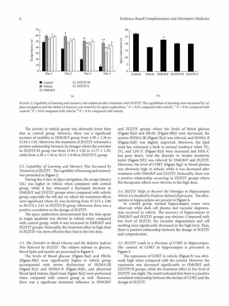
2.5. Morris Water Maze Test. Spatial learning and memorywere tested byMorris watermaze.TheMorris watermazewasfilled with water and divided into four quadrants. There wasan underwater platform placed in one of the quadrants. Afterthat, in the place navigation test, the time for rats to locate
Evidence-Based Complementary and Alternative Medicine 3
the underwater platformwas regarded as evasive latency (EL)and it lasted for four days. And space exploration was carriedout on the 5th day, the platform was removed, and time forrats to locate the platform quadrant was recorded as spaceexploration time (SET).
2.6. Surgery. Rats were anesthetized with 4mL/kg 10% chlo-ral hydrate and mounted on the operating desk after abrosiafor 24 h. The heart was exposed and then 150–200mL 0.01MPBS was implanted quickly from aorta on the apex cordis,while scissoring the auricula dextra and implanting 150–200mL 4% paraformaldehyde through aorta slowly untilclear fluid came out from auricula dextra. After that, theywere beheaded in brain and the hippocampuswas isolated onthe cryostage, and then it was fixed in 4% paraformaldehydefor 6–8 h and kept in ice saline (2.5mL/g) after infusion andlimbs stiffness.
2.7. Blood Test. The blood in serum tubes was centrifugedand the levels of fasting plasma glucose (FPG), fasting insulin(FINS), glycated hemoglobin (HbAlc), total cholesterol(TC), triglyceride (TG), high-density lipoprotein-cholesterol(HDL-C), and low-density lipoprotein-cholesterol (LDL-C)were tested, respectively, as previous study described [10].The homeostasis model assessment of insulin resistance wascalculated as follows:
(HOMA-IR) = FPG × FINS22.5
(ISI) = ln [ 1
FPG × FINS]
(HOMA-B) = 20 × FINSFPG − 3.5
.
(1)
2.8. ELISA Experiments. One side of hippocampus wassampled for enzyme-linked immunosorbent assay.The tissuewas ground in ice saline by refiner before it was centrifugedfor 10000 r/min 10min. Supernatant was collected in −80∘Cuntil the content of CORT was measured by ELISA kit(R&D Systems, USA) following the manufacturer’s protocol.Samples were added to Microelisa Stripplate and incubatedin 37∘C for 30min before 50𝜇L HRP-conjugate reagent wasadded, which followed by 30min incubation at 37∘C. Andthen, chromogen solution and stop solution were addedbefore the optical density value was measured at 450 nm.In the meantime, the standard curve was made. Moreover,the level of CORT in blood plasma was measured by CORTELISA kit (R&D Systems, USA).
2.9. Histopathology Experiment. The brain was removedsurgically and fixed in 4% formalin before the tissue wasdehydrated, paraffin-embedded, and sliced into section. Afterthat, HE stain was performed and then observed under lightmicroscope.
2.10. Western Blotting Experiments. The other side of hip-pocampus was applied for western blotting (Western Blotting
05
1015202530354045
Control Vehicle DMGB/F ZGJTJY/H ZGJTJY/L
Auto
nom
ic ac
tivity
(sco
re)
##
#
∗∗
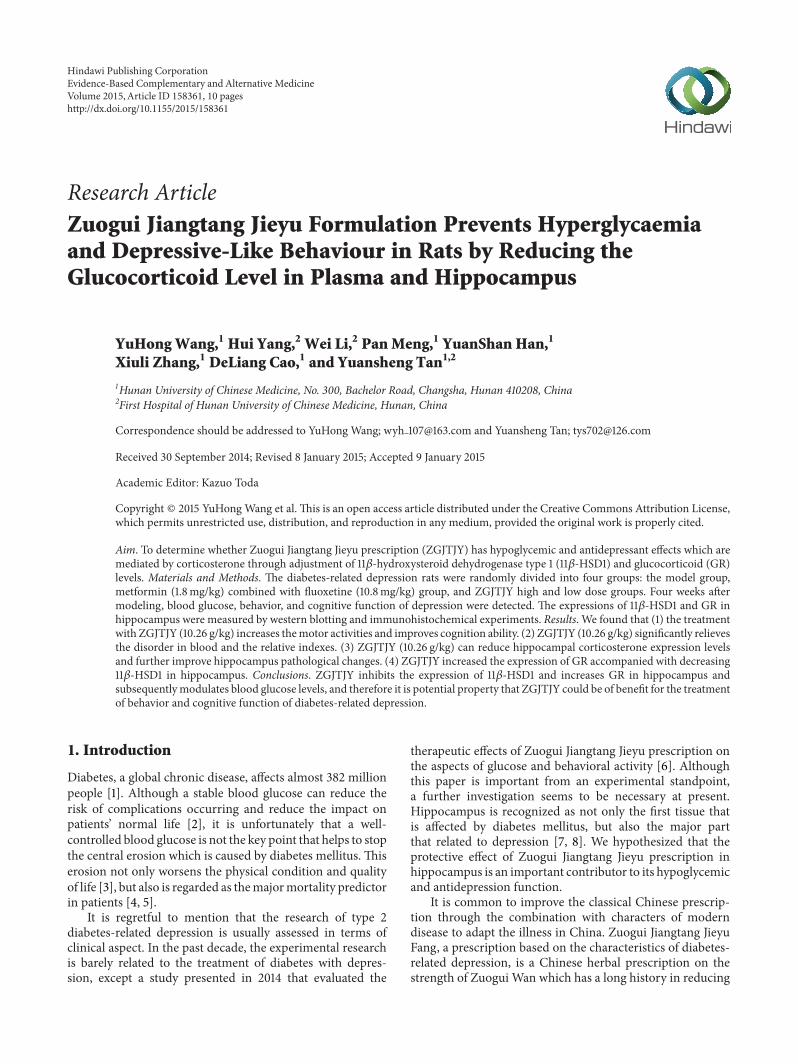
Figure 1: ZGJTJY increases the activity of rats with diabetes-related depression.The autonomic activitywas recorded by countinghorizontalmovement and vertical movement in open field test. ∗𝑃 <0.05 compared with control, ∗∗𝑃 < 0.01 compared with control;#𝑃 < 0.05 compared with vehicle; ##𝑃 < 0.01 compared with vehicle.
Kit, Beyotime Institute of Biotechnology, China). The tissuewas homogenized in cell lysis buffer (NP-40 lysis buffer)with protease inhibitor and it was ground by refiner beforesupernatant was kept in −20∘C after centrifuge. The proteinwas electrophoretically resolved on 10% SDS-polyacrylamidegels and then transferred to nitrocellulose membranes at100mA for 2.5 h. After that, the membranes were blockedin skimmed milk for 1 h at room temperature and overnightat 4∘C in anti-11𝛽-HSD1 (1 : 1000; Cell Signaling Technology,USA), anti-GR (1 : 1000; Cell Signaling Technology, USA),and anti-𝛽-actin (1 : 800; Cell Signaling Technology, USA),receptively. Then the membrane was incubated with HRPantibody (Boster Co., Wuhan, China) at dilution of 1 : 1000.Finally, themembranes were observed by the use of enhancedchemiluminescence kit (ECL, Amersham).
2.11. Immunohistochemical Experiments. The brain was per-formed for immunohistochemical experiment. After the tis-sues were dehydrated, embedded in paraffin, and sliced intosections, the slides were incubated in 3% hydrogen peroxidefor 10min and heated in 0.01M citrate buffer. After that,they were incubated with the goat serum sealing fluid for20min and covered with 50 𝜇L anti-rabbit, which followed by4∘C overnight. Then, the sections were rewarmed at 37∘C for45min and incubated with SABC for 30min, which followedby DAB coloration under the microscope and the sectionswere redyed with hematoxylin (2min). Finally, dehydration,hyalinization, andmountingwere performed and the sectionswere observed under high magnification (×400) with picturetaken.
2.12. Statistics. All the data were based on SPSS16.0 andanalyzed by one-way analysis of variance (ANOVA), 𝑡-testwith two-side test. A level of 𝑃 < 0.05 was set as statisticallysignificant.
3. Results
3.1. ZGJTJY Helps to Increase the State of Behavioral Activity.Themobility of different groups is presented in Figure 1.
4 Evidence-Based Complementary and Alternative Medicine
0
10
20
30
40
50
60
70
Day 1 Day 2 Day 3 Day 4
Esca
pe la
tenc
y (s
)
####
##
∗∗ ∗∗
∗
ControlVehicleDMGB/F
ZGJTJY/HZGJTJY/L
(a)
0
5
10
15
20
25
30
Control Vehicle DMGB/F ZGJTJY/H ZGJTJY/L
Spac
e exp
lora
tion
(s) #
(b)
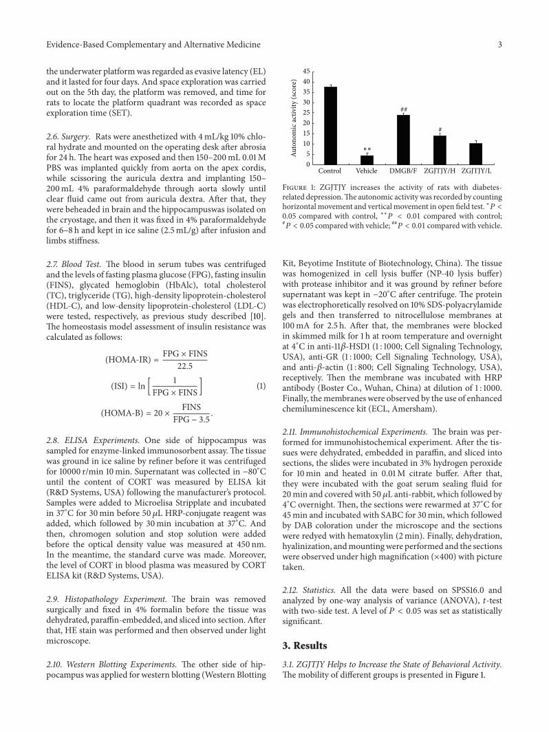
Figure 2: Capability of learning and memory was enhanced after treatment with ZGJTJY. The capabilities of learning were measured by (a)place navigation and the ability of memory was tested by (b) space exploration. ∗𝑃 < 0.05 compared with control, ∗∗𝑃 < 0.01 compared withcontrol; #𝑃 < 0.05 compared with vehicle; ##𝑃 < 0.01 compared with vehicle.
The activity in vehicle group was obviously lower thanthat in control group. However, there was a significantincrease of mobility in DMGB/F group from 4.38 ± 1.36 to23.94 ± 1.02. Moreover, the treatment of ZGJTJY witnessed apositive relationship between its dosages where the activitiesin ZGJTJY/H group rise from 23.94 ± 1.02 to 13.77 ± 1.29,while from 4.38 ± 1.36 to 10.21 ± 0.98 in ZGJTJY/L group.
3.2. Capability of Learning and Memory Was Increased byTreatment of ZGJTJY. Thecapability of learning andmemorywas presented in Figure 2.
During the 4 days of place navigation, the escape latency(EL) was higher in vehicle when compared with controlgroup, while it has witnessed a fluctuated decrease inDMGB/F and ZGJTJY groups when compared with vehiclegroup, especially the 4th day on which the treatment effectswere significant where EL was declining from 47.22 S ± 3.86to 38.32 S ± 2.81 in ZGJTJY/H group. Moreover, there was apositive correlation in the dosage of ZGJTJY.
The space exploration demonstrated that the time spentin target quadrant was shorter in vehicle when comparedwith control group, while it was increased in DMGB/F andZGJTJY groups. Noticeably, the treatment effect in high doseof ZGJTJY was more efficient than that in the low dose.
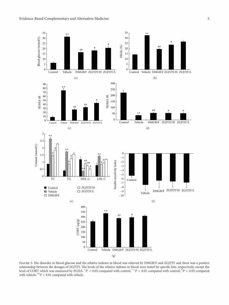
3.3. The Disorder in Blood Glucose and the Relative IndexesWas Relieved by ZGJTJY. The relative indexes in glucose,blood lipid, and insulin are presented in Figure 3.
The levels of blood glucose (Figure 3(a)) and HbAlc(Figure 3(b)) were significantly higher in vehicle groupaccompanied with severe dysfunction of HOMA-IR(Figure 3(c)) and HOMA-B (Figure 3(d)), and abnormalblood lipid indexes (lipid triad, Figure 3(e)) were performedwhen compared with control group as well. However,there was a significant treatment influence in DMGB/F
and ZGJTJY groups where the levels of blood glucose(Figure 3(a)) and HbAlc (Figure 3(b)) were decreased, theserious HOMA-IR (Figure 3(c)) was relieved, and HOMA-B(Figure 3(d)) was slightly improved. Moreover, the lipidtriad has witnessed a back to normal tendency when TC,TG, and LDL-C (Figure 3(e)) were increased and HDL-Chas gone down. And the disorder in insulin sensitivityindex (Figure 3(f)) was relieved by DMGB/F and ZGJTJY.Moreover, the level of CORT (Figure 3(g)) in blood plasmawas obviously high in vehicle, while it was decreased aftertreatment with DMGB/F and ZGJTJY. Noticeably, there wasa positive relationship occurring in ZGJTJY groups wherethe therapeutic effects were obvious in the high dose.
3.4. ZGJTJY Helps to Recover the Damages in HippocampusWhichAre Insulted byDiabetes-RelatedDepression. Thealter-nations in hippocampus are present in Figure 4.
In control group, normal hippocampus cones wereobserved, while dark cell plasma and vacuolar degenera-tion occurred in vehicle. The recovery of hippocampus inDMGB/F and ZGJTJY groups was obvious. Compared withlow level of ZGJTJY, the vacuolar degeneration and cellswelling were significantly decreased in the high level. Thus,there is positive relationship between the dosage of ZGJTJYand cytoprotection.
3.5. ZGJTJY Leads to a Decrease of CORT in Hippocampus.The content of CORT in hippocampus is presented inFigure 5.
The expression of CORT in vehicle (Figure 5) was obvi-ously high when compared with the control. However, theexpression was decreased significantly in DMGB/F andZGJTJY/H groups, while the treatment effect in low level ofZGJTJYwas slight.The result indicated that there is a positivecorrelated relationship between the decline of CORT and thedosage of ZGJTJY.
Evidence-Based Complementary and Alternative Medicine 5
0
5
10
15
20
25
30
35
Control Vehicle DMGB/F ZGJTJY/H ZGJTJY/L
Bloo
d gl
ucos
e (m
mol
/L)
## ##
∗∗
(a)
###
0
5
10
15
20
25
30
35
Control Vehicle DMGB/F ZGJTJY/H ZGJTJY/L
HbA
lc (%
)
∗∗
(b)
0102030405060708090
Control Vehicle DMGB/F ZGJTJY/H ZGJTJY/L
HO
MA-
IR
####
#
∗∗
(c)
HO
MA-
IR
0
50
100
150
200
250
300
Control Vehicle DMGB/F ZGJTJY/H ZGJTJY/L
## # #∗∗
(d)
0
0.5
1
1.5
2
2.5
3
TC TG HDL-C LDL-C
Con
tent
(mm
ol/L
)
##
#
#####
##
#
∗∗
∗∗
∗∗
∗∗
ControlVehicleDMGB/F
ZGJTJY/HZGJTJY/L
(e)
Control
Vehicle DMGB/F ZGJTJY/H ZGJTJY/L
0
Insu
lin se
nsiti
vity
inde
x
−1
−2
−3
−4
−5
−6
−7
−8
−9
−10
(f)
050
100150200250300350400
Control Vehicle DMGB/F ZGJTJY/H ZGJTJY/L
## #∗∗
CORT
(𝜇g/
g)
(g)
Figure 3: The disorder in blood glucose and the relative indexes in blood was relieved by DMGB/F and ZGJTJY and there was a positiverelationship between the dosages of ZGJTJY. The levels of the relative indexes in blood were tested by specific kits, respectively, except thelevel of CORT which was measured by ELISA. ∗𝑃 < 0.05 compared with control, ∗∗𝑃 < 0.01 compared with control; #𝑃 < 0.05 comparedwith vehicle; ##𝑃 < 0.01 compared with vehicle.

6 Evidence-Based Complementary and Alternative Medicine
(a) (b) (c)
Normal hippocampusVacuolar degeneration
(d)
Normal hippocampusVacuolar degeneration
(e)
Figure 4: Cytoprotection was obvious in ZGJTJY group where the normal hippocampus appeared to increase. The morphology changes inhippocampus were observed by HE stain. (a) Control; (b) vehicle; (c) DMGB/F; (d) ZGJTJY/H; (e) ZGJTJY/L.
0
4
8
12
16
20
Control Vehicle DMGB/F ZGJTJY/H ZGJTJY/L
## #
Cor
ticos
tero
ne (𝜇
g/g)
∗∗
Figure 5: The expression of CORT was inhibited by treatmentof ZGJTJY. The CORT expression in hippocampus was measuredby ELISA kit following the manufacturer’s protocol. ∗𝑃 < 0.05compared with control; ∗∗𝑃 < 0.01 compared with control; #𝑃 <0.05 compared with vehicle, ##𝑃 < 0.01 compared with vehicle;𝑛 = 8, 𝑥 ± 𝑠.
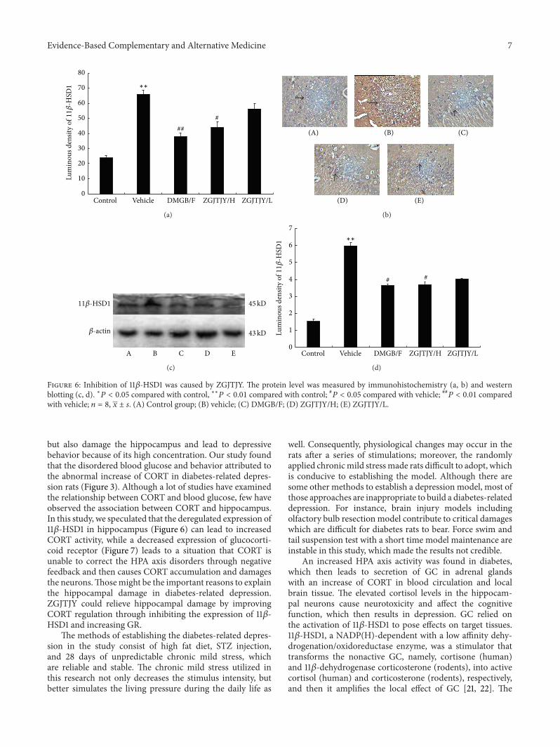
3.6. Downregulation of 11𝛽-HSD1 Was Caused by ZGJTJY inHippocampus. The protein level of 11𝛽-HSD1 was measuredby immunohistochemistry (Figures 6(a) and 6(b)) and west-ern blotting (Figures 6(c) and 6(d)). The result (Figure 6(b))shows that 11𝛽-HSD1-like positive immunoreactivity neuronhas become stronger in vehicle group when compared withcontrol. Thus the luminous density of 11𝛽-HSD1 positivecell was significantly higher in group vehicle (Figure 6(a)).However, there is an obvious decline of 11𝛽-HSD1 inDMGB/F(𝑃 < 0.05, Figure 6(a)) and ZGJTJY groups. Furthermore, a
positive correlation was found between the decrease of 11𝛽-HSD1 and the dose of ZGJTJYwhere the high dose of ZGJTJYwitnessed a dramatic decrease of 11𝛽-HSD1 (Figure 6(a)). Asshown in Figures 6(c) and 6(d), the western blotting testindicates that the changes between different groups nearlyshared the same tendency with immunohistochemistry.
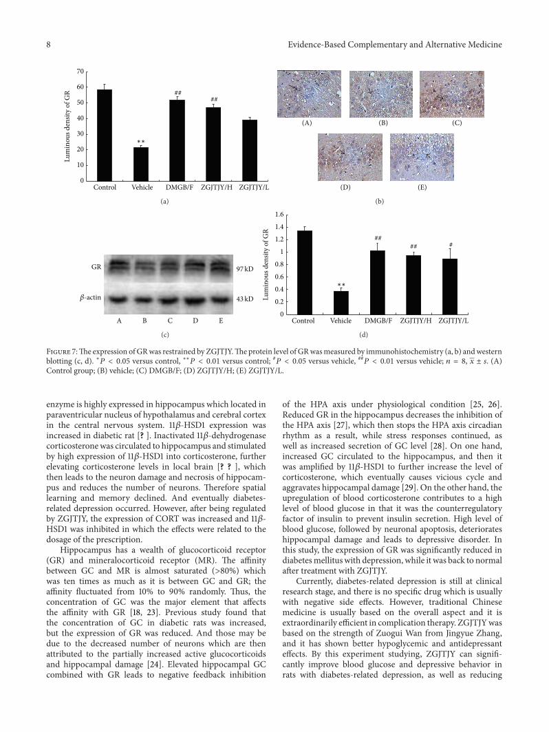
3.7. ZGJTJY Leads to an Increase of GR in Hippocampus.The protein level of GR in hippocampus was measuredby immunohistochemistry and western blotting. The resultsindicate that the vehicle (Figure 7(b)) group witnessed asignificant decrease in GR-like positive immunoreactivityneuron when compared with control group (𝑃 < 0.05)that means the expression of GR was restrained by diabetes-related depression. The content of GR was increased inDMGB/F and ZGJTJY groups. Noticeably, there is a positivecorrelation between the upregulation of GR and the dose ofZGJTJY. A remarkable increase was seen in the high doseof ZGJTJY (Figure 7(a)). When it comes to western blotting(Figures 7(c) and 7(d)), the trend of GR was almost thesame as the result of immunohistochemistry where there isa positive relationship between the increase of GR and thedosage of ZGJTJY as well.
4. Discussion
In this study, we found that ZGJTJY can effectively lowerblood glucose level in diabetes-related depression rats(Figure 3) and improve their depressive behavior (Figures 1and 2). CORT can not only regulate blood glucose variation,
Evidence-Based Complementary and Alternative Medicine 7
0
10
20
30
40
50
60
70
80
Control Vehicle DMGB/F ZGJTJY/H ZGJTJY/L
###
∗∗
Lum
inou
s den
sity
of11𝛽
-HSD
1
(a)
(A) (B) (C)
(D) (E)
(b)
A B C D E
45kD
43kD
11𝛽-HSD1
𝛽-actin
(c)
0
1
2
3
4
5
6
7
Control Vehicle DMGB/F ZGJTJY/H ZGJTJY/L
# #
∗∗
Lum
inou
s den
sity
of11𝛽
-HSD
1
(d)
Figure 6: Inhibition of 11𝛽-HSD1 was caused by ZGJTJY. The protein level was measured by immunohistochemistry (a, b) and westernblotting (c, d). ∗𝑃 < 0.05 compared with control, ∗∗𝑃 < 0.01 compared with control; #𝑃 < 0.05 compared with vehicle; ##𝑃 < 0.01 comparedwith vehicle; 𝑛 = 8, 𝑥 ± 𝑠. (A) Control group; (B) vehicle; (C) DMGB/F; (D) ZGJTJY/H; (E) ZGJTJY/L.
but also damage the hippocampus and lead to depressivebehavior because of its high concentration. Our study foundthat the disordered blood glucose and behavior attributed tothe abnormal increase of CORT in diabetes-related depres-sion rats (Figure 3). Although a lot of studies have examinedthe relationship between CORT and blood glucose, few haveobserved the association between CORT and hippocampus.In this study, we speculated that the deregulated expression of11𝛽-HSD1 in hippocampus (Figure 6) can lead to increasedCORT activity, while a decreased expression of glucocorti-coid receptor (Figure 7) leads to a situation that CORT isunable to correct the HPA axis disorders through negativefeedback and then causes CORT accumulation and damagesthe neurons.Thosemight be the important reasons to explainthe hippocampal damage in diabetes-related depression.ZGJTJY could relieve hippocampal damage by improvingCORT regulation through inhibiting the expression of 11𝛽-HSD1 and increasing GR.
The methods of establishing the diabetes-related depres-sion in the study consist of high fat diet, STZ injection,and 28 days of unpredictable chronic mild stress, whichare reliable and stable. The chronic mild stress utilized inthis research not only decreases the stimulus intensity, butbetter simulates the living pressure during the daily life as
well. Consequently, physiological changes may occur in therats after a series of stimulations; moreover, the randomlyapplied chronicmild stressmade rats difficult to adopt, whichis conducive to establishing the model. Although there aresome other methods to establish a depression model, most ofthose approaches are inappropriate to build a diabetes-relateddepression. For instance, brain injury models includingolfactory bulb resection model contribute to critical damageswhich are difficult for diabetes rats to bear. Force swim andtail suspension test with a short time model maintenance areinstable in this study, which made the results not credible.
An increased HPA axis activity was found in diabetes,which then leads to secretion of GC in adrenal glandswith an increase of CORT in blood circulation and localbrain tissue. The elevated cortisol levels in the hippocam-pal neurons cause neurotoxicity and affect the cognitivefunction, which then results in depression. GC relied onthe activation of 11𝛽-HSD1 to pose effects on target tissues.11𝛽-HSD1, a NADP(H)-dependent with a low affinity dehy-drogenation/oxidoreductase enzyme, was a stimulator thattransforms the nonactive GC, namely, cortisone (human)and 11𝛽-dehydrogenase corticosterone (rodents), into activecortisol (human) and corticosterone (rodents), respectively,and then it amplifies the local effect of GC [21, 22]. The
8 Evidence-Based Complementary and Alternative Medicine
0
10
20
30
40
50
60
70
Control Vehicle DMGB/F ZGJTJY/H ZGJTJY/L
Lum
inou
s den
sity
of G
R ####
∗∗
(a)
(A) (B) (C)
(D) (E)
(b)
A
GR
B C D E
97kD
43kD𝛽-actin
(c)
0
0.2
0.4
0.6
0.8
1
1.2
1.4
1.6
Control Vehicle DMGB/F ZGJTJY/H ZGJTJY/L
Lum
inou
s den
sity
of G
R
#### #
∗∗
(d)
Figure 7:The expression ofGRwas restrained by ZGJTJY.The protein level of GRwasmeasured by immunohistochemistry (a, b) andwesternblotting (c, d). ∗𝑃 < 0.05 versus control, ∗∗𝑃 < 0.01 versus control; #𝑃 < 0.05 versus vehicle, ##𝑃 < 0.01 versus vehicle; 𝑛 = 8, 𝑥 ± 𝑠. (A)Control group; (B) vehicle; (C) DMGB/F; (D) ZGJTJY/H; (E) ZGJTJY/L.
enzyme is highly expressed in hippocampus which located inparaventricular nucleus of hypothalamus and cerebral cortexin the central nervous system. 11𝛽-HSD1 expression wasincreased in diabetic rat [? ]. Inactivated 11𝛽-dehydrogenasecorticosteronewas circulated to hippocampus and stimulatedby high expression of 11𝛽-HSD1 into corticosterone, furtherelevating corticosterone levels in local brain [? ? ], whichthen leads to the neuron damage and necrosis of hippocam-pus and reduces the number of neurons. Therefore spatiallearning and memory declined. And eventually diabetes-related depression occurred. However, after being regulatedby ZGJTJY, the expression of CORT was increased and 11𝛽-HSD1 was inhibited in which the effects were related to thedosage of the prescription.
Hippocampus has a wealth of glucocorticoid receptor(GR) and mineralocorticoid receptor (MR). The affinitybetween GC and MR is almost saturated (>80%) whichwas ten times as much as it is between GC and GR; theaffinity fluctuated from 10% to 90% randomly. Thus, theconcentration of GC was the major element that affectsthe affinity with GR [18, 23]. Previous study found thatthe concentration of GC in diabetic rats was increased,but the expression of GR was reduced. And those may bedue to the decreased number of neurons which are thenattributed to the partially increased active glucocorticoidsand hippocampal damage [24]. Elevated hippocampal GCcombined with GR leads to negative feedback inhibition
of the HPA axis under physiological condition [25, 26].Reduced GR in the hippocampus decreases the inhibition ofthe HPA axis [27], which then stops the HPA axis circadianrhythm as a result, while stress responses continued, aswell as increased secretion of GC level [28]. On one hand,increased GC circulated to the hippocampus, and then itwas amplified by 11𝛽-HSD1 to further increase the level ofcorticosterone, which eventually causes vicious cycle andaggravates hippocampal damage [29]. On the other hand, theupregulation of blood corticosterone contributes to a highlevel of blood glucose in that it was the counterregulatoryfactor of insulin to prevent insulin secretion. High level ofblood glucose, followed by neuronal apoptosis, deteriorateshippocampal damage and leads to depressive disorder. Inthis study, the expression of GR was significantly reduced indiabetesmellitus with depression, while it was back to normalafter treatment with ZGJTJY.
Currently, diabetes-related depression is still at clinicalresearch stage, and there is no specific drug which is usuallywith negative side effects. However, traditional Chinesemedicine is usually based on the overall aspect and it isextraordinarily efficient in complication therapy. ZGJTJYwasbased on the strength of Zuogui Wan from Jingyue Zhang,and it has shown better hypoglycemic and antidepressanteffects. By this experiment studying, ZGJTJY can signifi-cantly improve blood glucose and depressive behavior inrats with diabetes-related depression, as well as reducing
Evidence-Based Complementary and Alternative Medicine 9
corticosterone levels, and inhibit the expression of 11𝛽-HSD1in the hippocampus and increase the expression of GR.In order to have a further insight of ZGJTJY, our groupintends to perform further study on the effect of ZGJTJYon cortisol regulation of diabetes-related depression rats inhippocampus.
Conflict of Interests
The authors declare that there is no conflict of interestsregarding the publication of this paper.
Acknowledgments
The project was supported by the National Natural ScienceFund Project (81373578, 81403379), the Natural Science FundProject in Hunan Province (13JJ5030), and Innovation Plat-form Open-End Fund in Colleges and Universities of HunanProvince (13K074).
References
[1] International Diabetes Federation, (1950–2014), 2014,http://www.idf.org/about-diabetes.
[2] T.Wu, G. Xie, Y. Ni et al., “Serummetabolite signatures of type 2diabetes mellitus complications,” Journal of Proteome Research.In press.
[3] D. Selvarajah and S. Tesfaye, “Central nevous system involve-ment in diabetes mellitus,” Current Diabetes Reports, vol. 6, no.6, pp. 431–438, 2006.
[4] N. Sartorius and L. Cimino, “The co-occurrence of diabetesand depression: an example of the worldwide epidemic ofcomorbidity of mental and physical illness,” Annals of theAcademy ofMedicine Singapore, vol. 41, no. 10, pp. 430–431, 2012.
[5] L. E. Egede and C. Ellis, “Diabetes and depression: globalperspectives,”Diabetes Research andClinical Practice, vol. 87, no.3, pp. 302–312, 2010.
[6] Y.-H. Wang, L.-T. Yin, H. Yang, X.-L. Li, and K.-G. Wu, “Hypo-glycemic and anti-depressant effects of Zuogui Jiangtang Jieyuformulation in a model of unpredictable chronic mild stressin rats with diabetes mellitus,” Experimental and TherapeuticMedicine, vol. 8, no. 1, pp. 281–285, 2014.
[7] S. M. Gold, I. Dziobek, V. Sweat et al., “Hippocampal damageandmemory impairments as possible early brain complicationsof type 2 diabetes,”Diabetologia, vol. 50, no. 4, pp. 711–719, 2007.
[8] Y. I. Sheline, “Depression and the hippocampus: cause oreffect?,” Biological Psychiatry, vol. 70, no. 4, pp. 308–309, 2011.
[9] K. Agyemang, L. Han, E. Liu, Y. Zhang, T. Wang, andX. Gao, “Recent advances in astragalus membranaceus anti-diabetic research: pharmacological effects of its phytochemicalconstituents,” Evidence-Based Complementary and AlternativeMedicine, vol. 2013, Article ID 654643, 9 pages, 2013.
[10] C. H. Park, J. S. Noh, T. Tanaka, S. S. Roh, J. C. Lee,and T. Yokozawa, “Polyphenol isolated from Corni Fructus,7-O-galloyl-d-sedoheptulose, modulates advanced glycationendproduct-related pathway in type 2 diabetic db/db mice,”Archives of Pharmacal Research, 2014.
[11] F. Z. Talukder, K. A. Khan, R. Uddin, N. Jahan, and M. A.Alam, “In vitro free radical scavenging and anti-hyperglycemicactivities of Achyranthes aspera extract in alloxan-induced
diabetic mice,” Drug Discoveries & Therapeutics, vol. 6, no. 6,pp. 298–305, 2012.
[12] L. Zhang, J. Luo, M. Zhang,W. Yao, X. Ma, and S. Y. Yu, “Effectsof curcumin on chronic, unpredictable, mild, stress-induceddepressive-like behaviour and structural plasticity in the lateralamygdala of rats,” International Journal of Neuropsychopharma-cology, vol. 17, no. 5, pp. 793–806, 2014.
[13] J. Tian, F. Zhang, J. Cheng, S. Guo, P. Liu, and H. Wang,“Antidepressant-like activity of adhyperforin, a novel con-stituent ofHypericum perforatum L,” Scientific Report, vol. 9, no.4, article 5632, 2014.
[14] L. An, Y.-Z. Zhang, X.-M. Liu et al., “Total flavonoids extractedfrom xiaobuxin-tang on the hyperactivity of hypothalamic-pituitary-adrenal axis in chronically stressed rats,” Evidence-Based Complementary and Alternative Medicine, vol. 2011,Article ID 367619, 7 pages, 2011.
[15] B. Budziszewska, “Effect of antidepressant drugs on thehypothalamic-pituitary-adrenal axis activity and glucocorti-coid receptor function,” Polish Journal of Pharmacology, vol. 54,no. 4, pp. 343–349, 2002.
[16] V. Butterweck,M.Hegger, andH.Winterhoff, “Flavonoids of St.John’sWort reduceHPA axis function in the rat,” PlantaMedica,vol. 70, no. 10, pp. 1008–1011, 2004.
[17] P. Willner, “Chronic Mild Stress (CMS) revisited: consistencyand behavioural-neurobiological concordance in the effects ofCMS,” Neuropsychobiology, vol. 52, no. 2, pp. 90–110, 2005.
[18] R.Thieringer and A. Hermanowski-Vosatka, “Inhibition of 11𝛽-HSD1 as a novel treatment for the metabolic syndrome: doglucocorticoids play a role?,” Expert Review of CardiovascularTherapy, vol. 3, no. 5, pp. 911–924, 2005.
[19] J. R. Seckl and B. R. Walker, “Minireview: 11𝛽-hydroxysteroiddehydrogenase type 1—a tissue-specific amplifier of glucocorti-coid action,” Endocrinology, vol. 142, no. 4, pp. 1371–1376, 2001.
[20] Y. J. Liu, Y. Nakagawa, Y. Wang et al., “Increased glucocorticqidreceptor and 11𝛽-hydroxysteroid dehydrogenase type 1 expres-sion in hepatocytes may contribute to the phenotype of type 2diabetes in 𝑑𝑏/𝑑𝑏mice,”Diabetes, vol. 54, no. 1, pp. 32–40, 2005.
[21] N. Draper and P. M. Stewart, “11𝛽-hydroxysteroid dehydroge-nase and the pre-receptor regulation of corticosteroid hormoneaction,” Journal of Endocrinology, vol. 186, no. 2, pp. 251–271,2005.
[22] R. A. S. Schweizer, A. G. Atanasov, B.M. Frey, and A. Odermatt,“A rapid screening assay for inhibitors of 11𝛽-hydroxysteroiddehydrogenases (11𝛽-HSD): flavanone selectively inhibits 11𝛽-HSD1 reductase activity,”Molecular and Cellular Endocrinology,vol. 212, no. 1-2, pp. 41–49, 2003.
[23] J. L. W. Yau, T. Olsson, R. G. M. Morris, M. J. Meaney, and J. R.Seckl, “Glucocorticoids, hippocampal corticosteroid receptorgene expression and antidepressant treatment: relationshipwithspatial learning in young and aged rats,” Neuroscience, vol. 66,no. 3, pp. 571–581, 1995.
[24] J. Beauquis, P. Roig, F. Homo-Delarche, A. de Nicola, andF. Saravia, “Reduced hippocampal neurogenesis and numberof hilar neurones in streptozotocin-induced diabetic mice:reversion by antidepressant treatment,” European Journal ofNeuroscience, vol. 23, no. 6, pp. 1539–1546, 2006.
[25] W. Katon, J. Unutzer, M.-Y. Fan et al., “Cost-effectiveness andnet benefit of enhanced treatment of depression for older adultswith diabetes and depression,” Diabetes Care, vol. 29, no. 2, pp.265–270, 2006.
10 Evidence-Based Complementary and Alternative Medicine
[26] F. Thomson and M. Craighead, “Innovative approaches for thetreatment of depression: targeting theHPA axis,”NeurochemicalResearch, vol. 33, no. 4, pp. 691–707, 2008.
[27] L. An, Y.-Z. Zhang, X.-M. Liu et al., “Total flavonoids extractedfrom Xiaobuxin-Tang on the hyperactivity of hypothalamic-pituitary-adrenal axis in chronically stressed rats,” Evidence-Based Complementary and Alternative Medicine, vol. 2011,Article ID 367619, 7 pages, 2011.
[28] P. J. Lustman and R. E. Clouse, “Treatment of depression indiabetes: impact on mood and medical outcome,” Journal ofPsychosomatic Research, vol. 53, no. 4, pp. 917–924, 2002.
[29] J. C. Fournier, R. J. DeRubeis, S. D.Hollon et al., “Antidepressantdrug effects and depression severity: a patient-level meta-analysis,” The Journal of the American Medical Association, vol.303, no. 1, pp. 47–53, 2010.

Submit your manuscripts athttp://www.hindawi.com
Stem CellsInternational
Hindawi Publishing Corporationhttp://www.hindawi.com Volume 2014
Hindawi Publishing Corporationhttp://www.hindawi.com Volume 2014
MEDIATORSINFLAMMATION
of
Hindawi Publishing Corporationhttp://www.hindawi.com Volume 2014
Behavioural Neurology
EndocrinologyInternational Journal of
Hindawi Publishing Corporationhttp://www.hindawi.com Volume 2014
Hindawi Publishing Corporationhttp://www.hindawi.com Volume 2014
Disease Markers
Hindawi Publishing Corporationhttp://www.hindawi.com Volume 2014
BioMed Research International
OncologyJournal of
Hindawi Publishing Corporationhttp://www.hindawi.com Volume 2014
Hindawi Publishing Corporationhttp://www.hindawi.com Volume 2014
Oxidative Medicine and Cellular Longevity
Hindawi Publishing Corporationhttp://www.hindawi.com Volume 2014
PPAR Research
The Scientific World JournalHindawi Publishing Corporation http://www.hindawi.com Volume 2014
Immunology ResearchHindawi Publishing Corporationhttp://www.hindawi.com Volume 2014
Journal of
ObesityJournal of
Hindawi Publishing Corporationhttp://www.hindawi.com Volume 2014
Hindawi Publishing Corporationhttp://www.hindawi.com Volume 2014
Computational and Mathematical Methods in Medicine
OphthalmologyJournal of
Hindawi Publishing Corporationhttp://www.hindawi.com Volume 2014
Diabetes ResearchJournal of
Hindawi Publishing Corporationhttp://www.hindawi.com Volume 2014
Hindawi Publishing Corporationhttp://www.hindawi.com Volume 2014
Research and TreatmentAIDS
Hindawi Publishing Corporationhttp://www.hindawi.com Volume 2014
Gastroenterology Research and Practice
Hindawi Publishing Corporationhttp://www.hindawi.com Volume 2014
Parkinson’s Disease
Evidence-Based Complementary and Alternative Medicine
Volume 2014Hindawi Publishing Corporationhttp://www.hindawi.com