Embed Size (px)
Citation preview
Research ArticleThe Establishment and Characteristics of Rat Model ofAtherosclerosis Induced by Hyperuricemia
Zhen Liu,1,2,3 Tong Chen,1,2 Haitao Niu,4 Wei Ren,1,2 Xinde Li,1,2
Lingling Cui,1,2 and Changgui Li1,2
1Shandong Provincial Key Laboratory of Metabolic Disease, The Affiliated Hospital of Qingdao University, Qingdao 266003, China2Shandong Gout Clinical Medical Center, Qingdao 266003, China3Key Laboratory of Hypertension, Qingdao 266003, China4Department of Urology, The Affiliated Hospital of Qingdao University, Qingdao 266003, China
Correspondence should be addressed to Lingling Cui; [email protected] and Changgui Li; [email protected]
Received 3 June 2015; Revised 4 July 2015; Accepted 24 August 2015
Academic Editor: Yingmei Feng
Copyright © 2016 Zhen Liu et al.This is an open access article distributed under the Creative CommonsAttribution License, whichpermits unrestricted use, distribution, and reproduction in any medium, provided the original work is properly cited.
Epidemiological studies have identified hyperuricemia as an independent risk factor for cardiovascular disease. However, themechanism whereby hyperuricemia causes atherosclerosis remains unclear. The objective of the study was to establish a new ratmodel of hyperuricemia-induced atherosclerosis. Wistar-Kyoto rats were randomly allocated to either a normal diet (ND), high-fat diet (HFD), or high-adenine diet (HAD), followed by sacrifice 4, 8, or 12 weeks later. Serum uric acid and lipid levels wereanalyzed, pathologic changes in the aorta were observed by hematoxylin and eosin staining, and mRNA expression was evaluatedby quantitative real-time polymerase chain reaction. Serum uric acid and TC were significantly increased in the HAD group at 4weeks compared with the ND group, but there was no significant difference in serum uric acid between the ND and HFD groups.Aorta calcification occurred earlier and was more severe in the HAD group, compared with the HFD group. Proliferating cellnuclear antigen, monocyte chemotactic factor-1, intercellular adhesion molecule-1, and vascular cell adhesion molecule-1 mRNAlevels were increased in the HFD and HAD groups compared with the ND group. This new animal model will be a useful tool forinvestigating the mechanisms responsible for hyperuricemia-induced atherosclerosis.
1. Introduction
Uric acid is an end product of purine metabolism in humansand is excreted in the urine. Loss of uricase function meansthat humans and other primates have relatively higher levelsof serum uric acid compared with rodents, providing thebiochemical bases for the inflammatory response in goutand an increased risk of cardiovascular disease. Gertler et al.[1] initially proposed the existence of a complex interactionbetween uric acid and coronary heart disease in 1951; sincethen increasing numbers of studies have confirmed a linkbetween raised serum uric acid levels and cardiovascularevents. A recent meta-analysis of prospective studies showedthat each additional 1mg/dL of serum uric acid equatedto a 12% increase in mortality for patients with coronaryheart disease [2]. Raised serum uric acid levels are associated
with approximately 70% increase in the risk of coronaryheart disease. In addition to its direct cardiovascular effects,hyperuricemia has also been associated with increased risksfor the development of hypertension, renal damage, andmetabolic syndrome, which indirectly lead to the occurrenceof cardiovascular events or affect the prognosis and therapyof cardiovascular disease.
However, conversely, some researchers suggest that thelack of uricase is an evolutionary advantage for primates[3, 4]. Hyperuricemia could help to stabilize blood pressure,and uric acid has antioxidant activities [3, 5, 6]. Hink et al.[7] reported that uric acid could also prevent the degradationof extracellular superoxide dismutase 3, which is a keyenzyme for maintaining the functions of endothelial cellsand blood vessels. Increased serum uric acid in patients withcardiovascular disease may be an important compensatory
Hindawi Publishing CorporationStem Cells InternationalVolume 2016, Article ID 1365257, 7 pageshttp://dx.doi.org/10.1155/2016/1365257
2 Stem Cells International
Table 1: Sequences of primers.
Gene name Product size Sense primer Antisense primerRat MCP-1 120 bp TGTCCCAAAGAAGCTGTAGTATTTGT TTCTGATCTCACTTGGTTCTGGTCRat ICAM-1 103 bp GGTGGGC AAGAACCTCATCCT CTGGCGGCT CAGTGTCTCATTRat VCAM-1 110 bp CGAAAG GCCCAGTTG AAG GA GAGCACGAGAAGCTCAGGAGA AARat 𝛽-actin 120 bp TGG ACA TCC GCA AAG AC GAA AGG GTG TAA CGC AACTA
mechanism for oxidative stress during the course of the dis-ease [8]. Hyperuricemia was associated with better prognosesin patients with stroke or other neurological disorders [9].However, these observations fail to explain why higher uricacid levels are associated with a poorer prognosis in patientswith cardiovascular disease. The validity of hyperuricemiaas an independent risk factor for cardiovascular disease thusremains controversial.
In this study, we established an animal model to investi-gate the association between hyperuricemia and atheroscle-rosis risk and also examined the specific molecules involvedin this process. The results have important implicationsfor future clinical treatment strategies and for the earlyprevention of hyperuricemia and atherosclerosis.
2. Materials and Methods
2.1. Animal Model. Ninety male Wistar-Kyoto rats were pur-chased from the Animal Center of BeijingUniversity, Beijing,China. Animal experiments were performed in accordancewith the guidelines for the Principles of Laboratory AnimalCare and the Guide for Care and Use of Laboratory Animals.Rats (200–220 g) were randomly divided into three groupsfed a normal diet (ND; 𝑛 = 30), high-fat diet (HFD; 𝑛 = 30),or high-adenine diet (HAD; 𝑛 = 30), respectively. HFD ratswere administered intragastric (i.g.) vitamin D
3(60 IU/kg)
for 3 days followed by a dose of 5mL/kg high-fat emulsioncontaining pyrimidine (200 g pork, 200 g cholesterol, 20 gbile salts, and 10 g propylthiouracil, dissolved in 1 L distilledwater) twice daily, by intragastric administration. HAD ratswere fed with fodder containing 10% yeast powder andadministered adenine (50mg/kg, i.g.) and potassium oxonate(100mg/kg) subcutaneously twice a day (8 am and 4 pm). NDrats were administered an equal volume of normal saline i.g.and fed a normal diet. The rats were housed individually inspecific pathogen-free conditions at a constant temperature(20–22∘C) and humidity (45–55%) with a 12 h light-darkcycle. Rats from the three groups were sacrificed after 4, 8,and 12 weeks, respectively.
2.2. Serum Uric Acid and Lipid Measurements. At the endof the experimental periods, rats were fasted for at least8 h and then anesthetized with 10% chloral hydrate. Bloodsamples were obtained from the right carotid artery after 4 hat room temperature and centrifuged at 3500 rpm for 15minat room temperature. Serum was separated and uric acid andlipid levels were determined using an autoanalyzer (Toshiba,Japan).
2.3. Tissue Processing, Histology, and Immunohistochemistry.At the end of the experiment, the thoracic aorta (from thearch to the diaphragm) was harvested, cut in half, and eitherfixed in buffered formalin or snap frozen. Aorta rings wereembedded in paraffin and sections were cut at 4 𝜇m andprepared for hematoxylin and eosin (HE) staining. Immuno-histochemical detection of proliferating cell nuclear antigen(PCNA) was performed using an anti-PCNA primary anti-body (1 : 500) and a horseradish peroxidase-conjugated goatanti-rat Ig secondary antibody (1 : 100) (Invitrogen, Basel,Switzerland).
2.4. Quantitative Real-Time Polymerase Chain Reaction.Total aorta RNA was extracted using Trizol reagent (Invitro-gen). For each sample, cDNA was synthesized using a Pri-meScript RT Reagent Kit (Takara, China) according to themanufacturer’s instructions. SYBR Green-based polymerasechain reaction (PCR) was performed in an automated ther-mal cycler (Bio-Rad) in a final volume of 25 𝜇L, contain-ing 2𝜇L cDNA solution, 12.5 𝜇L of SYBR Premix Ex Taq(Takara), 1 𝜇L of each primer (10 𝜇mol/L), and 8.5 𝜇L ofddH2O. The cycling reaction was performed according to
the manufacturer’s instructions via a standard two-step PCR.Experimental Ct values were normalized to 𝛽-actin andrelativemRNA expression was calculated in comparison witha reference sample. Each sample was run and analyzed intriplicate. The structures of the primers used are listed inTable 1.
2.5. Statistical Analyses. All data are expressed as mean ±standard deviation and were analyzed by one-way analysisof variance followed by Newman-Keuls multiple comparisontest as appropriate (GraphPad Prism version 5 software). Avalue of 𝑃 < 0.05 was considered statistically significant.
3. Results
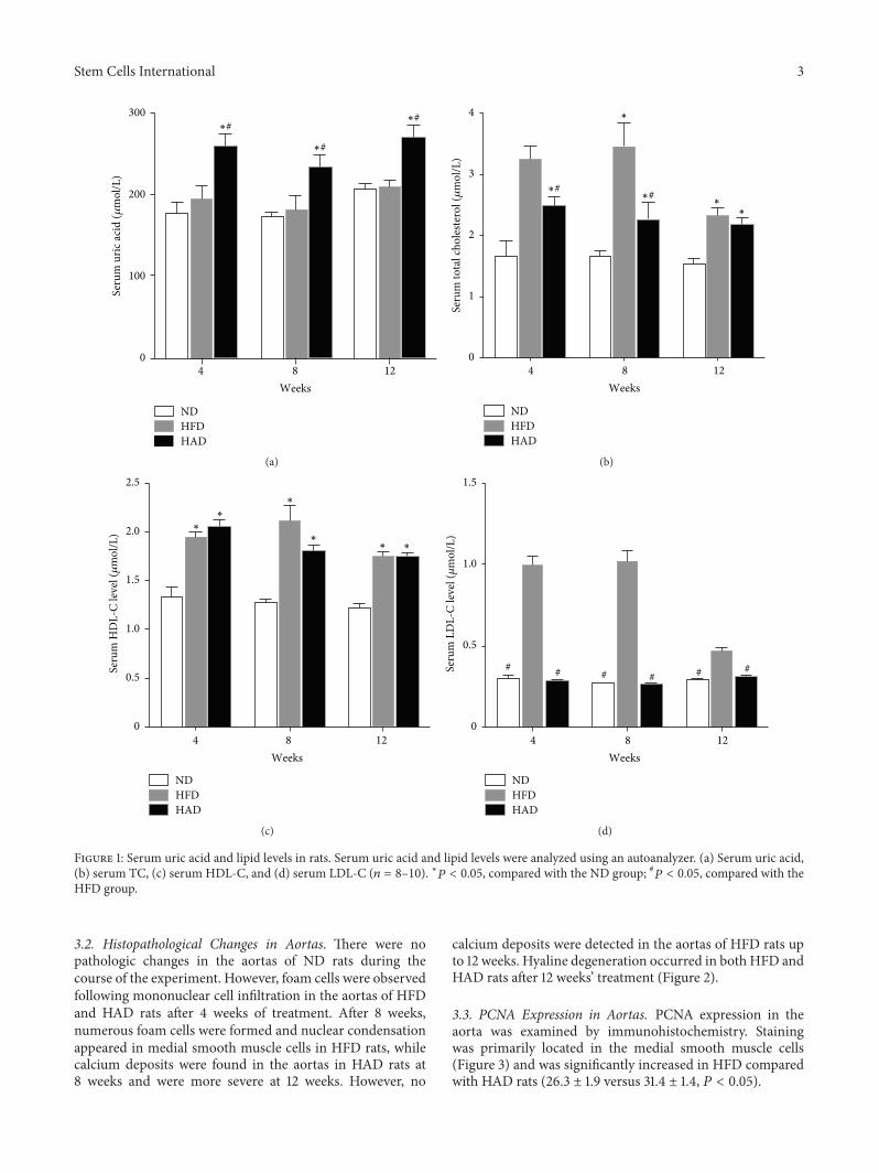
3.1. Changes in Serum Uric Acid and Lipid Levels. Serum uricacid and lipid levels were measured in all groups. Serum totalcholesterol (TC), high-density lipoprotein cholesterol (HDL-C), and low-density lipoprotein cholesterol (LDL-C) wereelevated in the HFD group compared with the ND groupafter 4, 8, and 12 weeks, respectively, while levels of UA wereunchanged. However, serum levels of UA, TC, and HDL-Cwere increased in the HAD rats. Interestingly, TC levels werelower in the HAD compared with the HFD group (Figure 1).Furthermore, mortality was increased by 30% in the HADgroup after 8weeks, comparedwith theND andHADgroups.
Stem Cells International 3
NDHFDHAD
∗#
∗#
∗#
0
100
200
300Se
rum
uric
acid
(𝜇m
ol/L
)
4 128Weeks
(a)
NDHFDHAD
Seru
m to
tal c
hole
stero
l (𝜇
mol
/L)
∗#∗#
0
1
2
3
4
1284
∗
∗
∗
Weeks
(b)
NDHFDHAD
∗
∗
∗
∗∗ ∗
0
0.5
1.0
1.5
2.0
2.5
Seru
m H
DL-
C le
vel (𝜇
mol
/L)
1284Weeks
(c)
NDHFDHAD
# # # # # #
0
0.5
1.0
1.5
Seru
m L
DL-
C le
vel (𝜇
mol
/L)
4 128Weeks
(d)
Figure 1: Serum uric acid and lipid levels in rats. Serum uric acid and lipid levels were analyzed using an autoanalyzer. (a) Serum uric acid,(b) serum TC, (c) serum HDL-C, and (d) serum LDL-C (𝑛 = 8–10). ∗𝑃 < 0.05, compared with the ND group; #𝑃 < 0.05, compared with theHFD group.
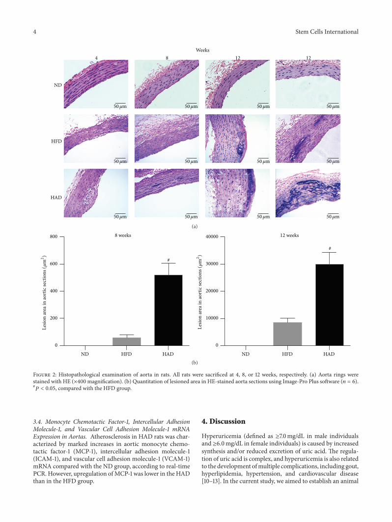
3.2. Histopathological Changes in Aortas. There were nopathologic changes in the aortas of ND rats during thecourse of the experiment. However, foam cells were observedfollowing mononuclear cell infiltration in the aortas of HFDand HAD rats after 4 weeks of treatment. After 8 weeks,numerous foam cells were formed and nuclear condensationappeared in medial smooth muscle cells in HFD rats, whilecalcium deposits were found in the aortas in HAD rats at8 weeks and were more severe at 12 weeks. However, no
calcium deposits were detected in the aortas of HFD rats upto 12 weeks. Hyaline degeneration occurred in both HFD andHAD rats after 12 weeks’ treatment (Figure 2).
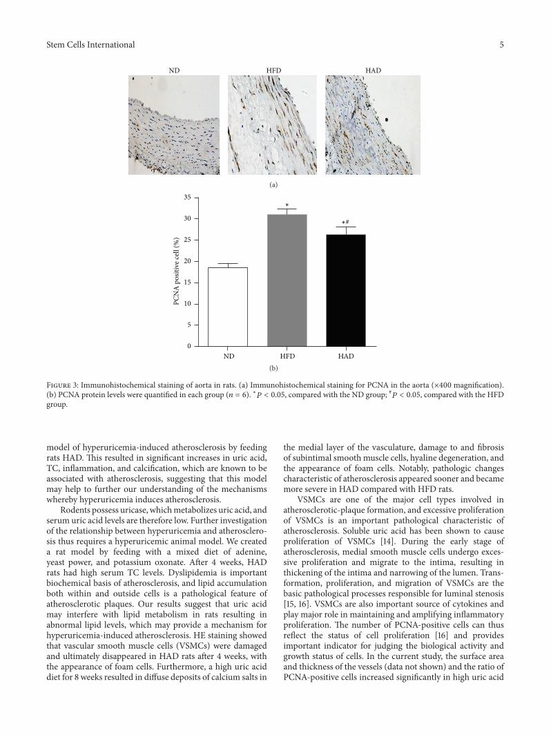
3.3. PCNA Expression in Aortas. PCNA expression in theaorta was examined by immunohistochemistry. Stainingwas primarily located in the medial smooth muscle cells(Figure 3) and was significantly increased in HFD comparedwith HAD rats (26.3 ± 1.9 versus 31.4 ± 1.4, 𝑃 < 0.05).
4 Stem Cells International
ND
HFD
HAD
4 8 12 12Weeks
50𝜇m 50𝜇m 50𝜇m 50𝜇m
50𝜇m 50𝜇m 50𝜇m 50𝜇m
50𝜇m 50𝜇m 50𝜇m 50𝜇m
(a)
8 weeks
ND HFD HAD
#
0
200
400
600
Lesio
n ar
ea in
aort
ic se
ctio
ns (𝜇
m2)
800 12 weeks
ND HFD HAD
#
0
10000
20000
30000
40000
Lesio
n ar
ea in
aort
ic se
ctio
ns (𝜇
m2)
(b)
Figure 2: Histopathological examination of aorta in rats. All rats were sacrificed at 4, 8, or 12 weeks, respectively. (a) Aorta rings werestained with HE (×400 magnification). (b) Quantitation of lesioned area in HE-stained aorta sections using Image-Pro Plus software (𝑛 = 6).#𝑃 < 0.05, compared with the HFD group.
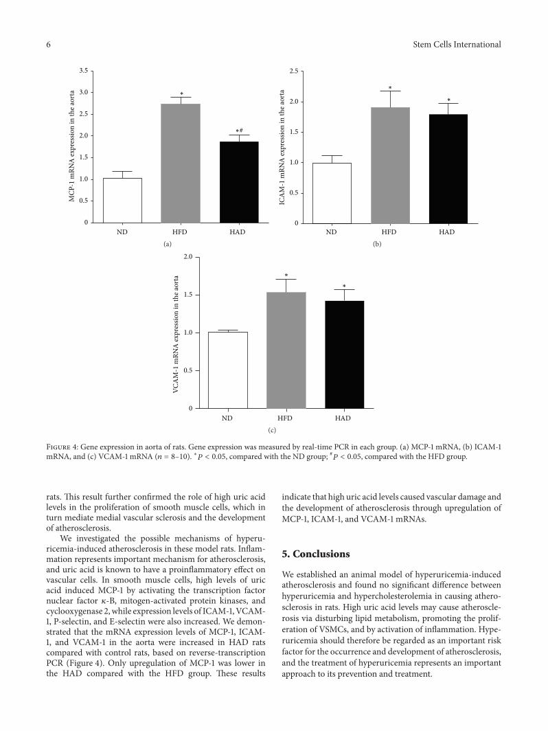
3.4. Monocyte Chemotactic Factor-1, Intercellular AdhesionMolecule-1, and Vascular Cell Adhesion Molecule-1 mRNAExpression in Aortas. Atherosclerosis in HAD rats was char-acterized by marked increases in aortic monocyte chemo-tactic factor-1 (MCP-1), intercellular adhesion molecule-1(ICAM-1), and vascular cell adhesion molecule-1 (VCAM-1)mRNA compared with the ND group, according to real-timePCR. However, upregulation ofMCP-1 was lower in the HADthan in the HFD group.
4. Discussion
Hyperuricemia (defined as ≥7.0mg/dL in male individualsand ≥6.0mg/dL in female individuals) is caused by increasedsynthesis and/or reduced excretion of uric acid. The regula-tion of uric acid is complex, and hyperuricemia is also relatedto the development ofmultiple complications, including gout,hyperlipidemia, hypertension, and cardiovascular disease[10–13]. In the current study, we aimed to establish an animal
Stem Cells International 5
ND HFD HAD
(a)
HAD0
5
10
15
20
25
30
35
PCN
A p
ositi
ve ce
ll (%
)
ND HFD
∗#
∗
(b)
Figure 3: Immunohistochemical staining of aorta in rats. (a) Immunohistochemical staining for PCNA in the aorta (×400 magnification).(b) PCNA protein levels were quantified in each group (𝑛 = 6). ∗𝑃 < 0.05, compared with the ND group; #𝑃 < 0.05, compared with the HFDgroup.
model of hyperuricemia-induced atherosclerosis by feedingrats HAD. This resulted in significant increases in uric acid,TC, inflammation, and calcification, which are known to beassociated with atherosclerosis, suggesting that this modelmay help to further our understanding of the mechanismswhereby hyperuricemia induces atherosclerosis.
Rodents possess uricase, whichmetabolizes uric acid, andserum uric acid levels are therefore low. Further investigationof the relationship between hyperuricemia and atherosclero-sis thus requires a hyperuricemic animal model. We createda rat model by feeding with a mixed diet of adenine,yeast power, and potassium oxonate. After 4 weeks, HADrats had high serum TC levels. Dyslipidemia is importantbiochemical basis of atherosclerosis, and lipid accumulationboth within and outside cells is a pathological feature ofatherosclerotic plaques. Our results suggest that uric acidmay interfere with lipid metabolism in rats resulting inabnormal lipid levels, which may provide a mechanism forhyperuricemia-induced atherosclerosis. HE staining showedthat vascular smooth muscle cells (VSMCs) were damagedand ultimately disappeared in HAD rats after 4 weeks, withthe appearance of foam cells. Furthermore, a high uric aciddiet for 8 weeks resulted in diffuse deposits of calcium salts in
the medial layer of the vasculature, damage to and fibrosisof subintimal smooth muscle cells, hyaline degeneration, andthe appearance of foam cells. Notably, pathologic changescharacteristic of atherosclerosis appeared sooner and becamemore severe in HAD compared with HFD rats.
VSMCs are one of the major cell types involved inatherosclerotic-plaque formation, and excessive proliferationof VSMCs is an important pathological characteristic ofatherosclerosis. Soluble uric acid has been shown to causeproliferation of VSMCs [14]. During the early stage ofatherosclerosis, medial smooth muscle cells undergo exces-sive proliferation and migrate to the intima, resulting inthickening of the intima and narrowing of the lumen. Trans-formation, proliferation, and migration of VSMCs are thebasic pathological processes responsible for luminal stenosis[15, 16]. VSMCs are also important source of cytokines andplay major role in maintaining and amplifying inflammatoryproliferation. The number of PCNA-positive cells can thusreflect the status of cell proliferation [16] and providesimportant indicator for judging the biological activity andgrowth status of cells. In the current study, the surface areaand thickness of the vessels (data not shown) and the ratio ofPCNA-positive cells increased significantly in high uric acid
6 Stem Cells International
ND HFD HAD
MCP
-1 m
RNA
expr
essio
n in
the a
orta
0
0.5
1.0
1.5
2.0
2.5
3.0
3.5
∗
∗#
(a)ND HFD HAD
ICA
M-1
mRN
A ex
pres
sion
in th
e aor
ta
0
0.5
1.0
1.5
2.0
2.5
∗
∗
(b)
ND HAD
VCA
M-1
mRN
A ex
pres
sion
in th
e aor
ta
2.0
1.5
1.0
0.5
0HFD
∗
∗
(c)
Figure 4: Gene expression in aorta of rats. Gene expression was measured by real-time PCR in each group. (a) MCP-1 mRNA, (b) ICAM-1mRNA, and (c) VCAM-1 mRNA (𝑛 = 8–10). ∗𝑃 < 0.05, compared with the ND group; #𝑃 < 0.05, compared with the HFD group.
rats. This result further confirmed the role of high uric acidlevels in the proliferation of smooth muscle cells, which inturn mediate medial vascular sclerosis and the developmentof atherosclerosis.
We investigated the possible mechanisms of hyperu-ricemia-induced atherosclerosis in these model rats. Inflam-mation represents important mechanism for atherosclerosis,and uric acid is known to have a proinflammatory effect onvascular cells. In smooth muscle cells, high levels of uricacid induced MCP-1 by activating the transcription factornuclear factor 𝜅-B, mitogen-activated protein kinases, andcyclooxygenase 2,while expression levels of ICAM-1,VCAM-1, P-selectin, and E-selectin were also increased. We demon-strated that the mRNA expression levels of MCP-1, ICAM-1, and VCAM-1 in the aorta were increased in HAD ratscompared with control rats, based on reverse-transcriptionPCR (Figure 4). Only upregulation of MCP-1 was lower inthe HAD compared with the HFD group. These results
indicate that high uric acid levels caused vascular damage andthe development of atherosclerosis through upregulation ofMCP-1, ICAM-1, and VCAM-1 mRNAs.
5. Conclusions
We established an animal model of hyperuricemia-inducedatherosclerosis and found no significant difference betweenhyperuricemia and hypercholesterolemia in causing athero-sclerosis in rats. High uric acid levels may cause atheroscle-rosis via disturbing lipid metabolism, promoting the prolif-eration of VSMCs, and by activation of inflammation. Hype-ruricemia should therefore be regarded as an important riskfactor for the occurrence and development of atherosclerosis,and the treatment of hyperuricemia represents an importantapproach to its prevention and treatment.
Stem Cells International 7
Conflict of Interests
The authors declare that there is no conflict of interestsregarding the publication of this paper.
Authors’ Contribution
Zhen Liu, Tong Chen, and Haitao Niu contributed equally tothis work. All authors approved the final paper as submittedand agree to be accountable for all aspects of the work.
Acknowledgments
This work was supported by the National Science Foundationof China (81500346, 81520108007, 31371272, and 31471195),Shandong Province Science and Technology DevelopmentPlan Item (2014GSF118013), the Natural Science Foundationof Shandong Province (ZR2010HZ001), and the Basic Appli-cation Research Plan of Qingdao (14-2-4-73-jch).
References
[1] M.M. Gertler, S. M. Garn, and S. A. Levine, “Serum uric acid inrelation to age and physique in health and in coronary heartdisease,” Annals of Internal Medicine, vol. 34, no. 6, pp. 1421–1431, 1951.
[2] Y. S. Kim, J. P. Guevara, K.M.Kim,H. K. Choi, D. F.Heitjan, andD. A. Albert, “Hyperuricemia and coronary heart disease: a sys-tematic review and meta-analysis,” Arthritis Care and Research,vol. 62, no. 2, pp. 170–180, 2010.
[3] B. N. Ames, R. Cathcart, E. Schwiers, and P. Hochstein, “Uricacid provides an antioxidant defense in humans against oxi-dant- and radical-caused aging and cancer: a hypothesis,” Pro-ceedings of the National Academy of Sciences of the United Statesof America, vol. 78, no. 11, pp. 6858–6862, 1981.
[4] S. Watanabe, D.-H. Kang, L. Feng et al., “Uric acid, hominoidevolution, and the pathogenesis of salt-sensitivity,” Hyperten-sion, vol. 40, no. 3, pp. 355–360, 2002.
[5] X. Wu, D. M. Muzny, C. C. Lee, and C. T. Caskey, “Two inde-pendent mutational events in the loss of urate oxidase duringhominoid evolution,” Journal ofMolecular Evolution, vol. 34, no.1, pp. 78–84, 1992.
[6] M. Tomita, S.Mizuno,H. Yamanaka et al., “Does hyperuricemiaaeffect mortality? A prospective cohort study of japanese maleworkers,” Journal of Epidemiology, vol. 10, no. 6, pp. 403–409,2000.
[7] H. U. Hink, N. Santanam, S. Dikalov et al., “Peroxidase proper-ties of extracellular superoxide dismutase: role of uric acid inmodulating in vivo activity,” Arteriosclerosis, Thrombosis, andVascular Biology, vol. 22, no. 9, pp. 1402–1408, 2002.
[8] F. J. Nieto, C. Iribarren, M. D. Gross, G.W. Comstock, and R. G.Cutler, “Uric acid and serum antioxidant capacity: a reaction toatherosclerosis?” Atherosclerosis, vol. 148, no. 1, pp. 131–139,2000.
[9] A. Chamorro, V. Obach, A. Cervera, M. Revilla, R. Deulofeu,and J. H. Aponte, “Prognostic significance of uric acid serumconcentration in patients with acute ischemic stroke,” Stroke,vol. 33, no. 4, pp. 1048–1052, 2002.
[10] T. R. Merriman and N. Dalbeth, “The genetic basis of hyper-uricaemia and gout,” Joint Bone Spine, vol. 78, no. 1, pp. 35–40,2011.
[11] J. F. Baker, E. Krishnan, L. Chen, andH. R. Schumacher, “Serumuric acid and cardiovascular disease: recent developments, andwhere do they leave us?”TheAmerican Journal of Medicine, vol.118, no. 8, pp. 816–826, 2005.
[12] Y. Li, C. Xu, C. Yu, L. Xu, and M. Miao, “Association of serumuric acid level with non-alcoholic fatty liver disease: a cross-sectional study,” Journal of Hepatology, vol. 50, no. 5, pp. 1029–1034, 2009.
[13] P. Boffetta, C. Nordenvall, O. Nyren, and W. Ye, “A prospectivestudy of gout and cancer,” European Journal of Cancer Preven-tion, vol. 18, no. 2, pp. 127–132, 2009.
[14] M. Horiuchi, T.-X. Cui, Z. Li, J.-M. Li, H. Nakagami, and M.Iwai, “Fluvastatin enhances the inhibitory effects of a selectiveangiotensin II type 1 receptor blocker, valsartan, on vascularneointimal formation,” Circulation, vol. 107, no. 1, pp. 106–112,2003.
[15] R. Candido, T. J. Allen, M. Lassila et al., “Irbesartan but notamlodipine suppresses diabetes-associated atherosclerosis,”Circulation, vol. 109, no. 12, pp. 1536–1542, 2004.
[16] T. Aizawa, S. Kokubun, and Y. Tanaka, “Apoptosis and prolif-eration of growth plate chondrocytes in rabbits,”The Journal ofBone & Joint Surgery—British Volume, vol. 79, no. 3, pp. 483–486, 1997.

Submit your manuscripts athttp://www.hindawi.com
Hindawi Publishing Corporationhttp://www.hindawi.com Volume 2014
Anatomy Research International
PeptidesInternational Journal of
Hindawi Publishing Corporationhttp://www.hindawi.com Volume 2014
Hindawi Publishing Corporation http://www.hindawi.com
International Journal of
Volume 2014
Zoology
Hindawi Publishing Corporationhttp://www.hindawi.com Volume 2014
Molecular Biology International
GenomicsInternational Journal of
Hindawi Publishing Corporationhttp://www.hindawi.com Volume 2014
The Scientific World JournalHindawi Publishing Corporation http://www.hindawi.com Volume 2014
Hindawi Publishing Corporationhttp://www.hindawi.com Volume 2014
BioinformaticsAdvances in
Marine BiologyJournal of
Hindawi Publishing Corporationhttp://www.hindawi.com Volume 2014
Hindawi Publishing Corporationhttp://www.hindawi.com Volume 2014
Signal TransductionJournal of
Hindawi Publishing Corporationhttp://www.hindawi.com Volume 2014
BioMed Research International
Evolutionary BiologyInternational Journal of
Hindawi Publishing Corporationhttp://www.hindawi.com Volume 2014
Hindawi Publishing Corporationhttp://www.hindawi.com Volume 2014
Biochemistry Research International
ArchaeaHindawi Publishing Corporationhttp://www.hindawi.com Volume 2014
Hindawi Publishing Corporationhttp://www.hindawi.com Volume 2014
Genetics Research International
Hindawi Publishing Corporationhttp://www.hindawi.com Volume 2014
Advances in
Virolog y
Hindawi Publishing Corporationhttp://www.hindawi.com
Nucleic AcidsJournal of
Volume 2014
Stem CellsInternational
Hindawi Publishing Corporationhttp://www.hindawi.com Volume 2014
Hindawi Publishing Corporationhttp://www.hindawi.com Volume 2014
Enzyme Research
Hindawi Publishing Corporationhttp://www.hindawi.com Volume 2014
International Journal of
Microbiology