Embed Size (px)
Citation preview
Hindawi Publishing CorporationOxidative Medicine and Cellular LongevityVolume 2013 Article ID 320823 15 pageshttpdxdoiorg1011552013320823
Research ArticleThe Cell Wall Sensors Mtl1 Wsc1 and Mid2 Are Required forStress-Induced Nuclear to Cytoplasmic Translocation of Cyclin Cand Programmed Cell Death in Yeast
Chunyan Jin1 Andrey V Parshin2 Ira Daly3 Randy Strich1 and Katrina F Cooper4
1 Department of Molecular Biology Graduate School of Biomedical Sciences Rowan University Stratford NJ 08055 USA2Department of Cell Biology Graduate School of Biomedical Sciences Rowan University Stratford NJ 08055 USA3Quintiles Transnational Corp Morris Plains NI 07950 USA4Department of Molecular Biology Graduate School of Biomedical Sciences Rowan University Two Medical Center DriveStratford NJ 08084 USA
Correspondence should be addressed to Katrina F Cooper cooperkarowanedu
Received 11 June 2013 Revised 14 August 2013 Accepted 16 August 2013
Academic Editor Cristina Mazzoni
Copyright copy 2013 Chunyan Jin et al This is an open access article distributed under the Creative Commons Attribution Licensewhich permits unrestricted use distribution and reproduction in any medium provided the original work is properly cited
Mtl1 is amember of a cell wall sensor family thatmonitors cell wall integrity in budding yeast In response to cell wall stressMtl1 acti-vates the cell wall integrity (CWI) MAP kinase pathway which transmits this signal to the nucleus to effect changes in gene expres-sion One target of the CWIMAP kinase is cyclin C a negative regulator of stress response genes CWI activation results in cyclin Crelocalization from the nucleus to the cytoplasmwhere it stimulates programmed cell death (PCD) before it is destroyedThis reportdemonstrates that under low oxidative stress conditions a combination of membrane sensors Mtl1 and either Wsc1 or Mid2 arerequired jointly to transmit the oxidative stress signal to initiate cyclin C destruction However when exposed to elevated oxidativestress additional pathways independent of these three sensor proteins are activated to destroy cyclin C In additionN-glycosylationis important forMtl1 function asmutating the receptor residue (Asn42) or an enzyme required for synthesis ofN-acetylglucosamine(Gfa1) reduces sensor activity Finally combining gfa1-1 with the cyclin C null allele induces a severe synthetic growth defect Thissurprising result reveals a previously unknown genetic interaction between cyclin C and plasma membrane integrity
1 Introduction
The yeast cell wall is the first level of defense against environ-mental stress Embedded in this cell wall are sensors thatdetect damage and transduce this signal to the nucleus tochange gene expression In addition this stress signal mustthen be disseminated throughout the cell to elicit changes inorganelle function and morphology For example exposureto oxidative stress induces the transcription of protein chap-erones and antioxidant enzymes alters ribosome assemblyin the nucleolus and triggers extensive fragmentation of themitochondria [1] All three examples help inform the cell ondeciding whether to arrest cell growth and repair the damageor execute the programmed cell death pathway
In yeast the cell wall integrity pathway (CWI) respondsto a variety of stresses including oxidative stress [2] heat
shock [3] and any other stress that alters the cell wallintegrity In brief the CWI pathway senses the stress via afamily of cell-surface sensors (Wsc1 Mid2 and Mtl1 [4])These sensors transmit the stress to a small G protein Rho1which in turn activates protein kinase C (Pkc1) The acti-vated Pkc1 transmits the intracellular signals to the MAPKSlt2Mpk1 via the MAPK module (see Figure 1(a)) Finallythe transcription factors (Rlm1 Swi4Swi6) regulating thecorresponding gene expression act as the output of theCWI pathway (Figure 1(a) and reviewed in [5]) In additionoxidative stress activation of this pathway triggers the Slt2-dependent cytoplasmic translocation and consequent degra-dation of cyclin C [6 7]
The cell wall sensor proteins are members of eitherthe Mid2 or Wsc subfamily [4] The Wsc sensor subfamilyincludes Wsc1 the mainWsc senor [8] Wsc2 andWsc3 [9]
2 Oxidative Medicine and Cellular Longevity
Slt2
Pkc1
Rho1
ROS
Mtl1
Mkk12
Bck1
Cyclin C translocation tocytoplasm and destruction
(MAPK)
(MEK)
(MEKK)
Mid2 Wsc1
S S I S R V T H
TCT TCT ATC TCT CGT GTC ACC CAC - GFA1
TCT TTC ATC C-- --- -TC ACC CAC - gfa1-1S S I F T H
485
[CNC1-
[CNC1-
URA3]
URA3]
[CNC1-TRP1]
[GFA1-LAP4] [LAP4] [GFA1]
[LAP4-GFA1] Vector
(a)
(b)
(c) (d)
(e)
cnc1Δ cnc1Δgfa1-1 gfa1-1 [CNC1-URA3]5-FOA5-FOA
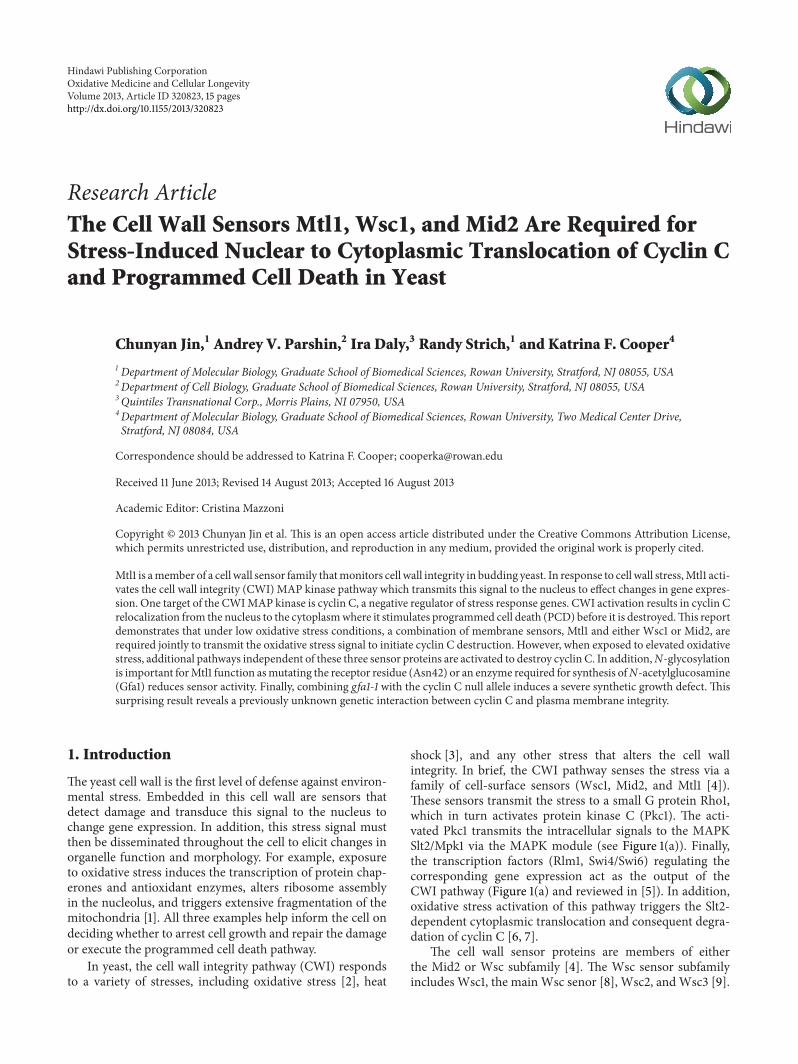
Figure 1 Negative synthetic growth defect in a cnc1Δ gfa1-1 mutant (a) Model of the cell wall signaling pathway that regulates cyclin Ccytoplasmic translocation and consequent degradation Adapted from [5] and data presented in [6 7 25] and this report (b) An isolatefollowing EMS mutagenesis harboring either a URA3 (left half) or TRP1 (right half) marked CNC1 expression plasmid was streaked onmedium containing 5-FOA (c) and (d)The gfa1-1 cnc1Δ double mutant strain was transformed with the indicated plasmids then streaked on5-FOAmedium All plates were incubated for 3 days at 30∘ before the image was taken (e) Sequence analysis of the gfa1-1 allele is shownThesix nucleotide deletion is indicated by the hash marks The predicted amino acid sequences for the wild type and gfa1-1 encoded proteins areindicated in single amino acid code
The Mid2 subfamily contains Mid2 and Mtl1 that shares 50homology with Mid2 [10] Although these cell wall sensorsshare structural similarity their sequences are not conservedThe Wsc subfamily contains an N-terminal cysteine-richregion termed theCRDdomainwhich is not present inMid2or Mtl1 The CRD domain transiently interacts with the glu-can chains in the cell wall while the transmembrane domainanchors the sensor in the plasma membrane In responseto cell wall stress the glucan chains are stretched exertinga force on the nanospring-like SerThr-rich domain Thisresults in a conformational change within the cytoplasmicdomain which triggers the interaction with Rom2 and acti-vates the downstream signaling cascade (reviewed in [11])Intermolecular interactions of the CRD domains promotesensor clustering with a concomitant increase of the down-stream signalingThis local accumulation enhances the stresssignal and the cellular response [12]
Posttranslational modification is required for properfunction of these cell wall sensors The N-glycosylation ofAsn35 is required for Mid2-dependent Slt2 activation [13]However this modification is not necessary for deposition ofMid2 to the plasma membrane suggesting that glycosylationis important for the protein to initiate the stress signalThe production of UDP-N-acetyl-D-glucosamine (UDP-GlcNAc) a building block forN- andO-linked glycosylationas well as the formation of GPI-anchors and chitin (reviewedin [14]) is controlled by both extra- and intracellular cues Forexample GFA1 which encodes glutamine fructose-6-phos-phate aminotransferase an enzyme that catalyzes the first andrate-limiting step inUDP-GlcNAc production is regulated in
response to mating pheromones [15] cell cycle progression[16 17] and cell wall damage [18] Defects in this pathwayresult in hypersensitivity to cell wall damage induced byheat shock [19] or spore wall morphogenesis [20] Defects inglycosylation also result in cell death [21 22] via Kex1 a pro-tease involved in programmed cell death induced by aceticacid or chronological aging [23]These results suggest an inti-mate relationship between glycosylation stress signaling andthe execution of cell death programs in yeast
The mechanisms underlying transcription factor activa-tion by signal transduction pathways have garnered muchof the attention when studying how exogenous cues are con-verted into changes in the gene expression program [24]Theother side of the coin that is removal of repressor proteinsis not as well understood In budding yeast repression ofmany stress response genes (SRG) including the HSP70member SSA1 [25] CIT1 and DDR2 [6] is mediated by thecyclin C-cyclin-dependent kinase 8 (Cdk8) module [26 27]This complex associates with the mediator component of theRNA polymerase holoenzyme and plays both a positive andnegative roles in transcription depending on the specific locus[28ndash30] Unlike cyclins that regulate the cell cycle cyclin Clevels do not vary significantly during the cell cycle in yeastor human cells [25 31] To relieve cyclin C-Cdk8-depend-ent repression in yeast cyclin C is destroyed by a Not4-dependent process in cultures subjected to a variety of stres-sors [6] Before it is destroyed cyclin C (but not Cdk8) trans-locates to the cytoplasm [6] where it is required for stress-induced mitochondrial hyperfission (unpublished dataK F Cooper S Khakhina S K Kim and R Strich)
Oxidative Medicine and Cellular Longevity 3
Table 1 Yeast strains used in this study
Strain Genotype SourceRSY10a 119872119860119879a ade2 ade6 can1-100 his3-11 15 leu2-3 112 trp1-1 ura3-1 [41]W303-1Bb
119872119860119879120572 ade2 can1-100 his3-11 15 leu2-3 112 trp1-1 ura3-1 [42]RSY391a cnc1LEU2 [25]RSY397 b cnc1LEU2 This studyRSY1543 b cnc1LEU2 gfa1-1 This studyRSY1539a chs3KANMX4 This studyRSY1538b mid2HIS3 This studyRSY1547a wsc1KANMX4 mid2HIS3 This studyRSY1659a MTL1-3HAHIS3 This studyRSY1608a cnc1LEU2 mtl1TRP1 This studyRSY1660a mtl1HIS3 This studyRSY1661b cnc1LEU2 gfa1-1 MTL1-3HATRP1 This studyRSY1707b wsc1KANMX4 mid2HIS3 mtl1TRP1 This studyRSY1844b wsc1KANMX4 mid2HIS3 pGAL-MTL1 This studyaGenotype119872119860119879a ade2 ade6 can1-100 his3-11 15 leu2-3 112 trp1-1 ura3-1bGenotype119872119860119879120572 ade2 can1-100 his3-11 15 leu2-3 112 trp1-1 ura3-1
Mitochondrial hyper-fission is a conserved hallmark ofthe stress response in higher eukaryotes [32ndash34] as wellas yeast [35ndash37] (see [38] for review) In many examplesmitochondrial fission is an early event in the PCD pathway[39 40] Thus the resistance to ROS-induced programmedcell death (PCD) exhibited by cyclin C null cells [6 7] is likelydue to a defect in the extensive mitochondrial fragmentationassociated with cellular damage These results indicate thatthe normal cellular response to oxidative stress requires theproper function of cell wall sensors that transduce the signalto the nucleus to mediate translocation of cyclin C to thecytoplasm This study connects a complex sensor systemrequiring proper N-glycosylation through Gfa1 function tocyclin C relocalization destruction and programmed celldeath
2 Materials and Methods
21 Yeast Strains Plasmids and Cell Culturing ConditionsThe strains used in this study are listed in Table 1 andmost arederived from W303-related strains RSY10 (MATa ade2 ade6can1-100 his3-11 15 leu2-3 112 trp1-1 ura3-1) [41] or W303-1B [42] In accordance with the Mediator nomenclatureunification effort [43] cyclin C (SSN8UME3SRB11) andCdk8 (SSN3UME5SRB10) we will use CNC1 and CDK8gene designations respectively KANMX4 deletion strainswere constructed by integrating the PCR amplifiedKANMX4deleted alleles obtained from the Research Genetics deletionstrain collection Deletion alleles using other auxotrophicmarkers or the Mtl1-3HA and pGAL-MTL1 strains wereconstructed using gene replacement methodology [44] Thecnc1Δ strains have been previously described [25] All primersused are listed in Table 2 Details about the plasmids usedin this study can be found in Table 3 The CNC1 ORF wastagged with one copy of the myc epitope placed immediatelydownstream of initiator methionine and creates a functionalprotein [25] The functional YFP-cyclin C fusion expression
plasmid construct (pBK37) was made by replacing the GFPallele in the functional GFP-cyclin C construct pBK1 [6]with PCR amplified YFP pID301 was created in two stepsFirst a 25 kbp EcoR1 fragment containing theCNC1minimalsubclone [25] was cloned into EcoR1 digested pRS316 [45]to form pKC311 Second ADE2 was inserted into pKC311 bycloning the BamHI fragment from pAZ11 [46] into BglII cutpKC311 to form pID301 pJB323 was made by cloning theCNC1minimal subclone that contains its own promotor andterminator sequences into ECOR1 cut pRS314 [45] Furtherdetails are available upon request The 3HA-tagged MTL1plasmids (pCJ3 and pCJ4) were made by PCR amplificationusing Phusion Taq (Denville Scientific) of the chromosomallytagged MTL1 ORF promotor and terminator from RSY1659The PCR fragment was cut with SacII and EcoR1 and theresulting fragment cloned into pRS423 or pRS426 [45]respectively digested with the same enzymes TheMTL1N42A
andMTL1N547A constructs were generated using site-directedmutagenesis on pCJ3 or pCJ4 according to themanufacturersdirection (Stratagene) with oligonucleotides listed in Table 2The remaining plasmids were gifts pYO964 containinghyperactive RHO1 allele (G19V) was provided by Y OhyaBCK1-20 containing the constitutively active BCK1 allele andthe SLT2-HA expression plasmids were from D Levin Cellswere grown in either rich nonselective medium (YPDA)or synthetic minimal medium (SC) to allow for plasmidselection as previously described [25] Galactose induciblegene expression was achieved by adding galactose (2 finalconcentration) to cultures grown in SC with raffinose as acarbon source
22 Synthetic Lethality Screen The colony color-sectoringmethod was used as previously described [47] Strain(RSY397 ade2 cnc1LEU2 ura3) harboring the CNC1 geneon a plasmid with the ADE2 and URA3 selectable markers(pID301) was grown to midlog phase (8 times 106) mutag-enized with ethylmethane sulfonate (EMS) as previously
4 Oxidative Medicine and Cellular Longevity
Table 2 Primers used in this study
Name Mutation created OligonucleotideMID2 F1 mid2Δ 51015840 TCGTTGAAGATTGGACATATAAAATACGCAAATCATAGTCGGATCCCCGGGTTAATTAA 31015840
MID2 R1 mid2Δ 51015840 AGGAATGAAAAGTAGCCATAAGCACTAAATGATATCAATGAATTCGAGCTCGTTTAAAC 31015840
WSC1 F wsc1Δ 51015840 GTA AAC TCG ACC AGG CAC 31015840
WSC1 R wsc1Δ 51015840 TATATCGTCTTTCAACGCGG 31015840
MTL1 F1 mtl1Δ 51015840 TTAACTTACTCCCAGTTAGTATAATATAAGTAGTTAAGGTCGGATCCCCGGGTTAATTAA 31015840
MTL1 R1 mtl1Δ 51015840 TTAAGAAGAAAAGTTATGGCAAAGCTGCTTTCGCTATGATGAATTCGAGCTCGTTTAAAC 31015840
MTL1 F2 MTL1-3HA 51015840 TATTACACGAAACCAAACAACGGCTTAAATATCACGAACTATCGGATCCCCGGGTTAATTAA 31015840
MTL1 F4 PGAL-MTL1 51015840 TTTAAACACTTCTAGTTCATTTCGGGTTGGTTCGATCTTGGAATTCGAGCTCGTTTAAAC 31015840
MTL1 R2 PGAL-MTL1 51015840 TTGAAGCAGAGCTCTTCTTCCTGGTCGGATTGCAGCTTGCCATTTTGAGATCCGGGTTTT 31015840
N42A-F Mtl1N42A 51015840 GCAGGAGTTGGTTCCAGCTGCTAGCACAACATCGAGCAC 31015840
N42A-R Mtl1N42A 51015840 GTGCTCGATGTTGTGCTAGCAGCTGGAACCAACTCCTGC 31015840
N547A-F Mtl1N547A 51015840 GAAACCAAACAACGGCTTAGCTATCACGAACTATTAAATC 31015840
N547A-R Mtl1N547A 51015840 GATTTAATAGTTCGTGATAGCTAAGCCGTTGTTTGGTTTC 31015840
KCO1234 Amplify gfa1-1 51015840 CGCTTACAAGAAAGCATTC 31015840
KCO1235 Amplify gfa1-1 51015840 GTCTAATTTAGGGCTGCAAC 31015840
Oligonucleotides used in this study with their accompanying mutation identified
Table 3 Plasmids used in this study
Plasmid name Gene Epitope tag Marker Promotor 2120583 or CEN ReferencepYO964 RHO1G19V No URA3 ADH1 CEN [66]pKC311 CNC1 No URA3 CNC1 CEN This studypKC800 GFA1 and LAP4 No TRP1 GFA1 and LAP4 CEN This studypKC811 LAP4 No TRP1 LAP4 CEN This studypKC812 GFA1 No TRP1 GFA1 CEN This studypID301 CNC1 No URA3 and ADE2 CNC1 CEN This studypBK37 CNC1 YFP TRP1 ADH1 CEN This studypLR106 BCK1-20 No HIS3 ADH1 CEN [7]pLR337 CNC1 1 myc TRP1 ADH1 CEN [25]MPK1-HA SLT2 HA LEU2 MPK1 2120583 [67]pCJ3 MTL1 3HA HIS3 ADH1 CEN This studypCJ4 MTL1 3HA URA3 ADH1 CEN This studypCJ11 MTL1N42A 3HA URA3 ADH1 CEN This studypCJ12 MTL1N42A 3HA HIS3 ADH1 CEN This studypCJ13 MTL1N547A 3HA HIS3 ADH1 CEN This study
described [41] and then plated on solid medium with lim-iting adenine (6mgmL) Colonies that were unable to losepID301 (as demonstrated by an unsectored colony) wereselected as candidates for further study To further screenthese candidates the colonies were transferred to solidmedium containing 5-fluoroorotic acid (5-FOA) an analogin the uracil biosynthetic pathway Colonies able to losethe CNC1 plasmid with the URA3 gene would continueto grow We identified thirteen colonies unable to lose theCNC1 expression plasmid suggesting that CNC1 was nowrequired for growth From this double screen one individual(RSY1543) was chosen for further study as described in thetext due to strength of the slow growth phenotype Verifica-tion of the requirement for cyclinCwas achieved by swappingpID301 for pJB323 and plating these cells on limiting adenineand 5-FOA medium The mutant allele responsible for this
synthetic phenotype was isolated by transforming RSY1543harboring pID301 with the pRS200 (TRP1-marked) genomiclibrary (ATCC77164) and identifying cells that could growon5-FOA medium Plasmid DNA (pKC800) was recovered byE coli transformation from 5-FOA resistant cells that formedwhite colonies and retransformed back into RSY1543 to verifythe complementation phenotype pKC800 was sequencedand determined to contain two ORFS GFA1 and LAP4pKC811 (LAP4 ORF promotor and terminator) and pKC812(GFA1 ORF promotor and terminator) were created frompKC800
23 Identification of the gfa1-1 Mutation The gfa1-1mutationwas identified by sequencing a PCR fragment generated fromthe chromosomal copy of themutant allele in strain RSY1543In brief yeast DNA from RSY1543 was made as previously
Oxidative Medicine and Cellular Longevity 5
described [25] and amplified using KCO1234 and KCO1235which map 200 bp upstream and 200 bp downstream ofthe GFA1 ORF respectively This PCR fragment was thensequenced (Eurofins MWG Operon) and the results werealigned with wild type GFA1 ORF Further details of primersused for sequencing are available upon request
24 Survival and Stress Assays For all stress assays cells weregrown to midlog phase (6 times 106 cellsmL) and then treatedwith H
2O2or acetic acid at the concentrations described in
the text Clonogenic viability studies were conducted withmidlog phase (6 times 106 cellsmL) treated with 100mM aceticacid for 200min and then serially diluted (1 10) and plated onminimal completemediumwith or without plasmid selectionas indicated in the text Caspase assays were conductedwith three independent cultures as described [48] exceptthat the cells were incubated with the caspase substrate(CaspSCREEN BioVision Inc) at 37∘ for 24 h in the dark Atleast 20000 cells were counted per sample Statistical analysiswas performed using the unpaired Studentrsquos t-test with 119875values lt005 being considered significant
25 Western Blot Analysis Extracts prepared for analyzingmyc-cyclin C levels were prepared from midlog cultures(6 times 106 cellsmL) as described previously [25] except thatthe lysis buffer used was 150mM NaCl 50mM Tris-HClpH 80 1 NP-40 015 deoxycholic acid sodium salt1 120583gmL pepstatin 1120583gmL leupeptin and 02 proteaseinhibitor cocktail (Sigma) In brief 500120583g of soluble extractwas immunoprecipitated using either anti-myc or anti-HAantibodies (Roche) collected on agarose A beads and thenanalyzed byWestern blot For monitoringMtl1 glycosylationa gradient acrylamide gel was used (5ndash10) to allow resolu-tion of both modified and unmodified signals on the samegel Western blot signals were detected using goat 120572-mousesecondary antibodies conjugated to alkaline phosphatase(Sigma) and the CDP-Star chemiluminescence kit (Tropix)Signals were quantitated by phosphorimaging (Kodak Inc)Half-life determinations were calculated by linear regressionanalysis with curves possessing 119903 values gt09 Relative cyclinC concentrations were standardized internally to Tub1 levelsbefore comparing to other values Slt2-HA phosphorylationwas detected using 120572-phospho-p4442 antibodies (Cell Sig-naling) as previously described [7] Tub1 was visualized using120572-tubulin antibodies (12G10) were obtained from the Devel-opmental Studies Hybridoma Bank (University of Iowa)
26 N-Glycosylation Assay Mtl1 N-glycosylation was moni-tored as described previously [13] Briefly crude membraneextracts were prepared from 250mL midlog cultures har-boring the Mtl1-3HA plasmid indicated (5 times 106 cellsmL inminimal medium) An equal volume of glass beads to the cellpellet was added and the cells lysed with 500120583L lysis buffer(150mM NaCl 50mM Tris-HCl pH 80 1 NP-40 015deoxycholic acid sodium salt 1 120583gmL pepstatin 1120583gmLleupeptin and 02 protease inhibitor cocktail (Sigma))by vortexing four times for 1min (with 1min intervals onice) Cell debris was removed by centrifugation for 5min at
3500timesg at 4∘C Crude membranes were collected from thesupernatant by centrifugation for 30min at 18000timesg at 4∘C(Beckman TLA-55 rotor) and resuspended in 100120583L lysisbuffer The crude membranes were digested with 2500 unitsof Endo H (New England Biolabs) for 2 h at 37∘C Mockincubationswere carried out without EndoH Reactionswerestopped by adding 3X SDS sample buffer and analyzed byWestern blot
27 Immunofluorescence Microscopy Localization studies ofchimeric fusion proteins were performed on cells fixed in37 paraformaldehyde and stained with 410158406-diamidino-2-phenylindole (DAPI) For all experiments the cells weregrown to midlog (5 times 106 cellsmL) treated with the H
2O2
concentrations and timepoints indicated in the figures andthen analyzed by fluorescence microscopy Images wereobtained using a Nikon microscope (model E800) with a60X objective (Plan Fluor Oil NA 13) and a CCD camera(RETIGA Exi) Data were collected using NIS software andprocessed using Image Pro software All images of individualcells were optically sectioned (02120583 slices at 06120583 spacing) anddeconvolved
3 Results
31 Gfa1 and Cyclin C Display a Negative Genetic InteractionCyclin C is a target of the cell wall integrity pathwayand required for programmed cell death [7] To identifyadditional components of this regulatory network a syntheticlethality screen was undertaken (see Section 2 for details)These studies identified a mutant that was unable to growin the presence of the counterselection drug 5-FOA whencyclin C was expressed from a plasmid containing the URA3selectablemarker (Figure 1(b) left panel) However introduc-tion of CNC1 on a plasmid with the TRP1 selectable markerwas able to lose theURA3 based plasmid indicating the cyclinC expression was required for normal cell growth (Figure1(b) right panel) Continued incubation of these plates didpermit limited growth of the double mutant strain (data notshown) This indicates that the phenotype observed was notdue to synthetic lethality but rather a severe growth defect
To identify the gene corresponding to the mutant alleleresponsible for this synthetic phenotype a genomic librarywas introduced into this strain and transformants wereidentified that were now able to grow in the absence of cyclinC One transformant was identified that contained a genomiccontig with two intact genes GFA1 and APE1LAP4 (Figure1(c) left panel) To determine which of these genes possessedthe complementation activity plasmids were introduced intothe mutant strain expressing either GFA1 or APE1 Thisexperiment revealed that GFA1 complemented the syntheticgrowth phenotype (Figure 1(d)) Since GFA1 is an essentialgene our allele (gfa1-1) must be hypomorphic but stillpossessing sufficient activity for survival To determine thenature of this allele DNA sequence analysis was performedon PCR products specific to the GFA1 coding region gen-erated from the gfa1-1 strain This analysis revealed a sixnucleotide deletion which changed S486 to a phenylalanine
6 Oxidative Medicine and Cellular Longevity
YFP-cylin C MergeDAPINomh+H2O2
0
2
0
2
GFA1
GFA1
gfa1-1
gfa1-1
gfa1-1
Cyclin C
Cyclin C
Tub1 p
Tub1
Cyclin C
Tub1
Cyclin C
Tub1
WT
chs3Δ
0 1 2 4
h + H2O2
VV
Glutamine + fructose-6-phosphate
Gfa1
glucosamine-6-phosphate
UDP-GlcNAc
N- and O-linkedglycosylation
Chitin GPI anchors
(a)
(b)
(c)
(d)
RHO1G
19V
RHO1G
19V
BCK1
-20
BCK1
-20
Figure 2 Gfa1 is required for cyclin C relocalization and destruction (a) Diagram ofGfa1 functions in the cell (b) Gfa1 is required for cyclin Cdestruction Wild type (RSY10) gfa1-1 cnc1Δ (RSY1543) and chs3Δ (RSY1539) midlog phase cultures expressing myc-cyclin C (pLR337) weretreated with 04mMH
2
O2
for the indicated times in hours Cyclin C levels were determined byWestern blot analysis of immunoprecipitatesTub1 levels were used as a loading control (c) YFP-cyclin C subcellular localization was monitored in a wild type and gfa1-1 cnc1Δ strainsharboring pBK37 before and following H
2
O2
treatment DAPI staining was used to identify the nucleus (d) Gfa1 functions upstream of theCWI pathway Cyclin C levels were determined byWestern blot analysis of immunoprecipitates in a midlog phase wild type (RSY10) or gfa1-1cnc1Δ (RSY1543) cultures expressing myc-cyclin C (pLR337) and either a vector control (v) BCK1-20 or RHO1G19V Tub1 levels were used asa loading control
and deleted R487 and V488 (Figure 1(e)) The remainder ofthe protein appears unaltered This small deletion resides inone of two sugar isomerase (SIS) domains found in manysugar binding proteins [49]This result suggests that the smalldeletion in Gfa1 reduces but does not eliminate Gfa1 activity
32 Gfa1 Is Required for Cyclin C Destruction in Responseto Oxidative Stress GFA1 encodes an essential glutamine-fructose-6-phosphate aminotransferase that catalyzes thefirst step in GlcNAc biosynthesis [15] GlcNAc is involved inseveral biological processes including GPI anchor formationchitin biosynthesis and the substrate for N- and O-linkedglycosylation (Figure 2(a)) Given the established role ofGfa1 in cell wall maintenance and our previous findingsthat cell wall stressors induce cyclin C destruction we nextdetermined if Gfa1 is required for oxidative stress-induceddegradation of cyclin C A CNC1myc tagged allele under the
control of the ADH1 promoter was placed on a single copyplasmid and introduced into the gfa1-1 mutant Myc-cyclinC levels were monitored by Western blot analysis followingexposure to hydrogen peroxide (04mM) Compared to wildtype myc-cyclin C levels were not reduced in the gfa1-1 strain (Figure 2(b)) These results indicate that Gfa1 isrequired for normal cyclin C destruction However chitinsynthase (CHS3) is not required for cyclin C destruction(Figure 2(b)) Although other chitin synthases are present inthe cell chs3 mutants display several phenotypes similar togfa1mutants including spore wall assembly and temperaturesensitive growth [50] Taken together these results suggestthat Gfa1 functions other than stimulating chitin formationare involved in regulating cyclin C destruction
Our previous report indicated that cyclin C relocalizationfrom the nucleus to the cytoplasm was required forboth cyclin C destruction and programmed cell death
Oxidative Medicine and Cellular Longevity 7
execution [6] Therefore we next asked whether Gfa1 wasalso required for cyclin C relocalization A wild type andgfa1-1 strain was transformed with a plasmid expressing YFP-cyclin C We have previously demonstrated that this reporterproteinwas functional and recapitulated normal cyclin C reg-ulation [6] In response to H
2O2treatment YFP-cyclin C foci
were observed in the cytoplasm in the wild-type strain (Fig-ure 2(c)) However YFP-cyclin C remained predominantlynuclear in the gfa1-1 strain These results indicate that Gfa1 isrequired for ROS-induced nuclear to cytoplasmic transloca-tion of cyclin C
The results just described indicate that Gfa1 is requiredfor the oxidative stress signal to induce cyclin C relocalizationand destruction We have reported that the cell wall integrityMAP kinase pathway is necessary for this process (see Figure1(a) and [7]) To determine if Gfa1 functions upstream ordownstream of this signaling pathway epistasis experimentswere conducted Plasmids expressing either constitutivelyactive alleles of RHO1 (RHO1G19V) or BCK1 (BCK1-20) wereintroduced into a wild type strain and the gfa1-1 mutantCyclin C levels were monitored in these cultures in theabsence of stress As previously reported [7] constitutiveactivation of Bck1 or Rho1 (this study) can induce cyclinC destruction in the absence of stress (Figure 2(d)) Asimilar result was obtained in the gfa1-1 strain The low levelretention of cyclin C in the gfa1-1 culture expressing Rho1G19Vwas reproducible suggesting that Gfa1 function is partiallyrequired for Rho1G19V-induced cyclin C destruction Theseresults indicate that Gfa1 mediates stress-induced cyclin Crelocalization and destructionThese results suggest that Gfa1may regulate cyclin C destruction through glycosylation of aCWI pathway component
33 Mtl1 Mid2 andWsc1 Are Required for Cyclin C Degrada-tion in Response to Moderate Oxidative Stress N-Glycosyla-tion is required for the proper function of Mid2 a cell wallsensor that is required to transduce the cell wall stress signal[13] To determine which cell wall sensor (or sensors) isrequired to transmit the stress signal to cyclinCwe examinedcyclin C levels in sensor mutants following exposure to H
2O2
(04mM) Cyclin C was protected from destruction in themtl1Δ strain indicating that Mtl1 is required for this process(Figure 3(a) quantitated in Figure 3(c)) However deletingMID2 did not significantly alter cyclin C degradation kinetics(Figure 3(b)) suggesting that a functional specialization existsbetween these two members of the N-glucosamine modifiedfamily Previous studies have shown that Mid2 and Wsc1have redundant functions as oxidative stress sensors [2]Consistent with this finding cyclin C was not destroyedfollowing H
2O2stress in mid2Δ wsc1Δ cells (Figure 2(b))
indicating that these proteins perform overlapping functionscontrolling cyclin C destruction Interestingly the prestresscyclin C levels were lower in the double mutant comparedto the wild type This observation may reflect the activationof another stress pathway that recognizes instability in thecell wall due to the loss of these sensors (reviewed in[11]) resulting in partial cyclin C destruction A similarreduction in cyclin C levels was observed in unstressed
slt2Δmutants [7] Taken together these data suggest that twosensor groups Mid2Wsc1 and Mtl1 mediate H
2O2-induced
cyclin C destructionPrevious studies indicated that the CWI MAPK Slt2
is required for H2O2induced cyclin C destruction [7]
To determine if the Mid2Wsc1 and Mtl1 sensor groupssignal cyclin C destruction through Slt2 its activation wasmonitored by Thr and Tyr T-loop phosphorylation [51] T-loop phosphorylation specific antibodies were used to probeWestern blots of Slt2 immunoprecipitated from extracts pre-pared from wild type mtl1Δ or mid2Δ wsc1Δ strains beforeand after exposure to 04mM H
2O2 As previously reported
[7] a transient elevation in phosphorylated Slt2 was detectedin wild-type cells (Figure 3(d)) A similar result was obtainedin the mtl1Δ strain while the mid2Δ wsc1Δ double mutantdisplayed a reduction in total Slt2 activation To determine ifboth sensor groups contributed to Slt2 activation the exper-iment was repeated in the mid2Δ wsc1Δ mtl1Δ triple mutantThis analysis revealed no detectable Slt2 phosphorylationunder these conditions (Figure 3(e)) These results suggestthat both Mid2Wsc1 and Mtl1 groups are required fornormal Slt2 activation in response to low-level H
2O2expo-
sure
34 Mtl1 Is N-Glycosylated on Asn42 in a Gfa1-DependentManner Our results indicating that both Mtl1 and Gfa1 arerequired for ROS-induced cyclin C destruction promptedfurther experiments to determine if these proteins werefunctionally related Given the role of Gfa1 in productionof GlcNAc we tested whether Mtl1 was N-glycosylatedThe MTL1 gene was tagged in the chromosome with threeHA epitopes (3HA) Mtl1-3HA was immunoprecipitated andthis immunoprecipitate was split and one half was treatedwith Endo H to remove glycosylation Western blot analysisrevealed that most of the Mtl1-3HA signal migrated at anapparent molecular weight of 56 kDa close to its predictedsize (arrowhead Figure 4(a)) A significantly slower specieswas also observed (open arrow) that shifted to a faster mobil-ity upon Endo H treatment (solid arrow) The remaininghigh molecular weight Mtl1 species was most likely dueto O-mannosylation These results indicate that as recentlyreported [52] Mtl1 is N-glycosylated Inspection of the Mtl1sequence for consensus N-glycosylation recognition sites(NxST x =P) revealed twomotifs at Asn42 andAsn547 Site-directedmutagenesis was used to substitute alanines for theseAsn residues and the experiments were repeated Unlike theN547A mutant Mtl1N47A did not display the characteristicshift in mobility upon EndoH treatment (Figure 4(b))Theseresults indicate that Mtl1 is modified on N42
Finally Mtl1 modification was examined in the gfa1-1mutant In these experiments two differences were observedcompared to the wild-type control First Mtl1 mobility wasenhanced following PAGE in the untreated mutant extractcompared to wild type (Figure 4(c) compare open and darkgrey arrows) Treatment of these samples with Endo Hresulted in furthermobility enhancement (compare solid andlight grey arrows) although to a lesser extent These resultssuggest that Mtl1 is still N-glycosylated to some extent mostlikely due to residual Gfa1-1 activity The second result was
8 Oxidative Medicine and Cellular Longevity
0 1 2 4
Cyclin C
Tub1
0 1 2 4
h + 04mM H2O2
WT mtl1Δ
(a)
Cyclin C
Tub1
0 1 2 4 0 1 2 4 0 1 2 4
h + 04mM H2O2
mid2Δ wsc1Δmtl1Δ
mid2Δ mid2Δ wsc1Δ
(b)
0
08
12
16
20
log
initi
al cy
clin
C (
)
3 41 2h + 04mM H2O2
WTmtl1Δ mid2Δ wsc1Δ mtl1Δ
mid2Δ wsc1Δ
mid2Δ
(c)
0 1 2 2 0 01 1 2
h + 04mM H2O2
WT
Slt2p-HA
Slt2p- P
mid2Δ wsc1Δmtl1Δ
(d)
0 1 2 0 1 2
h + 04mM H2O2
WT
Slt2p-HA
Slt2p- P
mid2Δ wsc1Δmtl1Δ
(e)
Figure 3 Mtl1 andMid2Wsc1 membrane sensors are required for cyclin C destruction following 04mMH2
O2
stress (a)Wild type (RSY10)and mtl1Δ (RSY1660) expressing myc-cyclin C (pLR337) were grown to midlog phase (0 h) and then treated with 04mM H
2
O2
for theindicated times in hours Cyclin C levels were determined byWestern blot analysis of immunoprecipitates Tub1 levels were used as a loadingcontrol (b) Cyclin C levels were monitored in mid2Δ (RSY1538) mid2Δ wsc1Δ (RSY1547) and mid2Δ wsc1Δ mtl1Δ (RSY1707) strains asdescribed in (a) (c) Quantification of the results obtained in (a) and (b) (d) T-loop phosphorylation (top panel) and immunoprecipitations(bottom panel) of Slt2-HA in wild type mtl1Δ mid2Δ wsc1Δ and (e) mid2Δ wsc1Δ mtl1Δ following 04mM H
2
O2
treatment for the timesindicated (h)
that the total amount of modified Mtl1 was reduced in thegfa1-1 mutant with a corresponding increase in the unmodi-fied form compared to wild-type extracts In this experimenttwice the amount of the gfa1-1 extract was required tovisualize modified Mtl1 These results indicate that Mtl1 isN-glycosylated on Asn42 and that this modification requiresGfa1 In addition the loss of Gfa1 activity reduces the steadystate levels of modified Mtl1
35Mtl1 N-Glycosylation Is Required for Normal ROS-InducedPCD and Cyclin C Degradation Previous studies have foundthat mutants lacking Mtl1 are hypersensitive to H
2O2treat-
ment [53] To determine whether this loss in viability was dueto PCD or necrosis an mtl1Δ strain was transformed withplasmids expressing wild-type MTL1 or the vector controlThree independent transformants were grown to midlogphase and subjected to 04mMH
2O2for 4 hThese cells were
Oxidative Medicine and Cellular Longevity 9
MTL1
(kD
a)minus + Endo H
250
150
100
50
WT
(a)
mtl1Δ
MTL1minus + minus + minus + Endo H
250
150
100
50
N42A N547A
(kD
a)
(b)
MTL1minus + minus + Endo H
250
150
100
50
WT gfa-1
(kD
a)
(c)
Figure 4 Mtl1 is glycosylated on Asn42 Immunoprecipitates from extracts prepared from a wild-type strain expressing Mtl1-3HA from itsnormal promoter were either subjected (+) or not (minus) to Endo H treatment Molecular weight standards (kDa) are given on the left Thearrowhead indicates the migration of the unmodified Mtl1-3HA Open and closed arrows indicate modified Mtl1-3HA before and after EndoH treatment respectively (b) Site directed mutagenesis was used to substitute alanine for asparagine at position 42 or 547 The experimentsdescribed in (a) were repeated with the modified forms of Mtl1 (c) The experiment described in (a) was repeated with a wild type and gfa1-1mutant Open and dark grey arrows indicated fully modified form in wild type and gfa1-1mutant respectively Closed and light grey arrowsindicate the Endo H treated samples in wild type and the gfa1-1mutant respectively
analyzed for caspase activity using a fluorescent substrateand flow cytometry (see Section 2 for details) These studiesrevealed a significant increase in caspase active cells in thecells lackingMTL1 expression (Figure 5(a)) indicating that atleast a portion of the enhanced cell death observed in mtl1Δmutants is due to PCD To determine if N-glycosylation wasrequired for this activity the experiment was repeated withthe mtl1Δ strain harboring a plasmid expressing mtl1N42AThis study revealed an intermediate elevation in PCD com-pared to the wild type and the null strain valuesThese resultssuggest that loss of N-glycosylation reduces but does noteliminate Mtl1 function with respect to H
2O2-induced PCD
executionWe next determined whether this intermediate pheno-
type was observed under two additional stress conditionsknown to induce PCD namely higher H
2O2concentrations
or exposure to acetic acid Surprisingly exposing thesecultures to higher H
2O2levels did not significantly change
caspase activation between the three cultures Converselythe acetic acid treated mtl1N42A expressing strain exhibitedsimilar levels of caspase activation compared to the vectorcontrolThis hypersensitivity to acetic acid was also observedin plating assays (Figure 5(b)) indicating thatN-glycosylationis important forMtl1 function following acetic acid treatmentThese results indicate that Gfa1-dependent N-glycosylationof Mtl1 is required for the normal cellular response to lowlevels of H
2O2and acetic acid However this activity is
dispensable in cells exposed to higher concentrations ofperoxide These results suggest that the cell can modulate thesignal transduction configuration based on the extent andtype of damage encountered
36 Increasing H2O2Concentrations Elevates CWI Pathway
Activation The results just described indicated that Mtl1 isimportant for transducing a low-level ROS signal but not that
of a higher concentration To examine this question furtherwe investigated the role of the Mtl1 and Mid2-Wsc1 sensorgroups in transducing high-level H
2O2exposure Western
blot analysis was used to monitor cyclin C levels before andfollowing the addition of 08mM H
2O2to wild type mtl1Δ
mid2Δwsc1Δ andmtl1Δmid2Δwsc1Δmidlog-phase culturesUnlike the results obtained with lower H
2O2concentration
loss of either the Mtl1 or Mid2Wsc1 groups did not protectcyclin C from destruction (Figure 6(a)) Consistent with thisfinding Slt2 activation was readily observed in both mtl1Δand mid2Δ wsc1Δ mutants (Figure 6(b)) However eliminat-ing both groups (mtl1Δmid2Δ wsc1Δ) stabilized cyclin C andsuppressed Slt2 activation These results suggest that as ROSexposure increases either Mtl1 or Mid2Wsc1 can initiatea signal sufficient to trigger cyclin C destruction Howevercyclin C destruction still occurs in the triple sensor mutantwhen the H
2O2concentration is raised to 12mM (Figure
6(a)) These findings suggest that an additional pathway(s)is now enabled that can initiate cyclin C destruction undersevere stress exposure (see Section 4)
The above results suggest a model in which H2O2-
mediated cyclin C destruction is regulated differentlydepending on the ROS dose Twomodels seemmost likely todescribe these results First theremay be a linear relationshipbetween the amount of ROS applied and the effective ratefor cyclin C destruction Alternatively there may be separatepathways whose activation occurs under specified stressconditions To begin testing these possibilities cyclin C levelswere monitored in a wild-type strain exposed to increasingconcentrations of H
2O2 For these experiments a single
poststress timepoint of one hour was chosen as it representsan intermediate time so that changes in destruction kineticscan be identified In wild-type cells the addition of H
2O2
up to 04mM does not induce an appreciable decline incyclin C levels after 1 h (Figure 6(c) quantitated Figure 6(d))
10 Oxidative Medicine and Cellular Longevity
05
10
10
15
20
0
25
30
04 01M
Fold
casp
ase+
[H2O2] mM
Vec
acetic acid
PlasmidMTL1
lowastlowastlowast
lowast
mtl1N42A
(a)
Control 01M acetic acid
Dilutionmtl1Δ [MTL1]mtl1Δ [mtl1N42A]
(b)
Figure 5 Mtl1 N-glycosylation is required for efficient stress-induced PCD execution (a) The mtl1Δ strain (RSY1660) trans-formed with plasmids expressing MTL1 (pCJ4) MTL1N42A (pCJ10)or the vector control was subjected to H
2
O2
or acetic acid atthe indicated concentrations for 240 200 and 200m respectivelyCaspase assays were performed on three independent cultures andthe mean plusmn SEM is indicated lowast119875 lt 005 lowastlowast119875 lt 001 (b) Strainsdescribed in (a) were grown to midlog phase and then either treatedor not with 100mM acetic acid for 200min The cells were seriallydiluted (1 10) and then spotted onto growth medium The imageswere obtained following 2 days incubation at 30∘C
However a significant reduction in cyclin C levels is observedat 06mM H
2O2 A continued reduction in cyclin C levels
is observed at 08mM but they remain relatively constantthrough 12mM These results are more consistent withthe second model that additional pathways able to triggercyclin C destruction are enabled in the 08ndash12mM H
2O2
concentrations To further test this model the experimentwas repeated with the mtl1Δ mutant Interestingly followinga delay in cyclin C destruction until 06mM a very similarprofile was observed except that the 08 to 12mM plateauoccurred around 50 of cyclin C remaining versus approxi-mately 15 for the wild-type strainThese results suggest thatMtl1 contributes to the oxidative stress signal regardless ofthe level of ROS and that additional pathways are activated todestroy cyclin C as ROS exposure increases (see Section 4)
37 Sensor Concentration Regulates the Cyclin C Destruc-tion Signal The results just described suggest a model thatincreased oxidative stress is able to induce more rapiddestruction of cyclin C To determine if this difference indestruction kinetics could be explained by elevated Slt2 acti-vation the extent of T-loop phosphorylation was monitoredin cultures exposed to low (04mM) or high (08mM) H
2O2
concentrations These concentrations were chosen as theyrepresent the two steps observed with cyclin C destruction(see Figure 6(d)) These experiments revealed that Slt2 acti-vation was elevated in cultures subjected to 08mM H
2O2
compared to 04mM (Figure 7(a)) In addition cyclin Cdestruction kinetics are enhanced in response to high ROSconditions (compare cyclin C degradation kinetics betweenFigures 3(a) and 6(a)) Taken together these results suggestthat activation ofmultiple stress pathways leads to heightenedSlt2 activation and more rapid cyclin C destruction
Since elevated H2O2concentrations are able to increase
Slt2 stimulation we next asked if the different stress sensorsgenerated a specific signal or Slt2 was simply responding tooverall signal intensity To test these possibilities a plasmidcontainingMTL1 under the control of the galactose induciblepromoter (GAL1) was introduced into amid2Δ wsc1Δ doublemutant In the presence of the repressing carbon sourceglucose cyclin C was not destroyed in response to low H
2O2
levels in the double mutant (Figure 7(b)) as we observedpreviously (see Figure 3(b)) Growing this culture in galac-tose medium which induced Mtl1 overexpression allowednormal cyclin C destruction to occur These results indicatethat overexpression of Mtl1 can compensate for the loss oftheMid2Wsc1 signalThis finding is consistent with amodelthat overall sensor activity and not a sensor-specific signalmediates cyclin C destruction Taken together these resultsindicate that elevated sensor signaling either by increasingthe sensor or H
2O2concentrations can more efficiently
induce cyclin C destruction
4 Discussion
ROS generated by oxidative phosphorylation in the mito-chondria are normally neutralized by the intrinsic anti-oxidant systemThe addition of exogenous prooxidants (per-oxides heavy metals) triggers the oxidative stress responseLow-level oxidative stress induces cell cycle arrest untilthe damage has been repaired Conversely extensive ROS-induced damage activates the programmed cell death path-way Our previous studies found that oxidative stress inducesthe nuclear to cytoplasmic relocalization and destruction ofthe stress response gene transcriptional repressor cyclin CIn addition cyclin C translocation is required for efficientPCD execution making the signaling pathway that transmitsthe ROS signal critical for the decision to enter PCD Inthis report we describe the sensor array that transmits thecyclin C destruction signal following H
2O2treatment Using
cyclin C relocalization and destruction as readouts this studyrevealed that the cell wall sensors Mtl1 and either Mid2 orWsc1 combinations are required for low-level ROS signalingto cyclin C However our data suggest that the cell is sensingthe total signal not a specific activity derived from each
Oxidative Medicine and Cellular Longevity 11
Cyclin C
Tub1
Cyclin C
Tub1
WT
mtl1Δ
0 02 1506 08 10 1204
[H2O2] mM
20
40
60
80
0
100
WTmtl1Δ
02 08 10 12 140604[H2O2] mM
Initi
al cy
clin
C (
)
0
Cyclin C
Tub1
0 1 2 4 0 1 2 4 0 1 2 4 0 1 2 4 0 1 2 4
h + 08mM H2O2 h + 12mM H2O2
mid2Δ wsc1Δmtl1Δ
mid2Δ wsc1Δmid2Δ wsc1Δmtl1Δ
WT mtl1Δ
(a)
0 1 2 0 1 2 0 1 2 0 1 2 0 1 2
h + 08mM H2O2
mid2Δ wsc1Δmtl1Δ
mid2Δ wsc1ΔWT WTmtl1Δ
Slt2-HA
Slt2- P
(b)
(c) (d)
Figure 6Membrane sensors are not required for cyclinC destruction following 08mMH2
O2
stress (a)Wild type (RSY10)mtl1Δ (RSY1660)mids2Δ wsc1Δ (RSY1547) and mid2Δ wsc1Δ mtl1Δ (RSY1707) strains expressing myc-cyclin C (pLR337) were grown to midlog phase (0 hr)and then treated with 08mM or 12mM H
2
O2
for the indicated times Cyclin C levels were determined by Western blot analysis ofimmunoprecipitates Tub1 levels were used as a loading control (b) Phosphorylation (top panels) and immunoprecipitation (bottom panels)of Slt2-HA in wild type mtl1Δ mids2Δ wsc1Δ and mid2Δ wsc1Δ mtl1Δ strains following 08mM H
2
O2
treatment (c) Kinetics of cyclin Cdegradation are dependent upon the H
2
O2
dose Wild type (RSY10) and mtl1Δ (RSY1600) strains expressing myc-cyclin C (pLR337) weregrown to midlog phase (0 hr) and then treated with increasing amounts of H
2
O2
for 1 hour Cyclin C levels were determined byWestern blotanalysis of immunoprecipitates Tub1 levels were used as a loading control (d) Quantification of cyclin C levels in (c)
of these sensors In addition Mtl1 signaling requires N-glycosylated via a Gfa1-dependent process In response tohigher concentrations of prooxidant additional pathways areactivated that do not require these sensors These findingsindicate that the cell is able to respond to differing levelsof oxidative stress through activation of multiple signalingpathways
This study was initiated by the finding that a muta-tion in GFA1 caused cells deleted for cyclin C to growvery slowly This synthetic growth defect suggested thatthese factors somehow functioned in similar or redundant
pathways However our data indicate that Gfa1 is requiredfor the ROS-induced destruction of cyclin C indicatingopposing functions Thus why are cnc1Δ gfa1-1 mutantssynthetically sick One possibility takes into considerationthat cyclin C is part of the Cdk8 module that represses over100 genes [55 56] while Gfa1 controls the modification ofproteins through the production of GlcNAc andGPI anchorsTherefore there are multiple opportunities for these twomutations to interact For example cyclin C-Cdk8 repressesthe transcription of GIP2 [55] an activator of the proteinphosphatase Glc7 [57] Hyperactivation of Glc7 displays a
12 Oxidative Medicine and Cellular Longevity
0 1 2 4 0 1 2 4
Slt2-HA
Slt2- P
h + 04mM H2O2 h + 08mM H2O2
(a)
GalactoseGlucose
Cyclin C
Tub1
0 1 0 1
pGAL-MTL1
h + H2O2
mid2Δ wsc1Δ
(b)
CW
PM
Low ROS stress
Cyclin C relocalization slow
High ROS stress Extreme ROS stress
Cyclin C relocalization fast Cyclin C relocalization faster
Mtl1 Wsc1Mid2
(c)
Figure 7 Mtl1 overexpression suppressesmid2Δ wsc1Δ signaling defect (a) Phosphorylation (top panels) and immunoprecipitation (bottompanels) of Slt2-HA in wild-type strain (RSY10) following 04mM and 08mM H
2
O2
treatment as indicated (b) A mid2Δ wsc1Δ mutant(RSY1844) harboring an integrated copy ofMTL1 under the control of the GAL1 promoter and cyclin C-myc (pLR337) was grown to midlogin raffinose The culture was split and treated with either 2 galactose or glucose for 2 h before the addition of 04mMH
2
O2
Cyclin C levelswere monitored byWestern blot analysis of immunoprecipitates Tub1 levels were used as a loading control (c) Model of combinatorial signaltransduction pathway control for cyclin C regulation Sensors associated with the plasma membrane (PM) and cell wall (CW) are indicatedSensor activity in response to low high and extreme ROS exposure is indicated Under low stress conditions the combined signals fromboth Mtl1 and Mid2Wsc1 are required to trigger cyclin C relocalization and destruction High stress conditions generate more signal fromeach group perhaps through sensor clustering as proposed for Wsc1 [12 54] Extreme stress environment proposes the existence of anotherpathway represented here by the question mark that contributes to the overall stress The location of this pathway at the plasma membranewas done for convenience and does not imply another cell wall sensor system per se
growth defect in combination with mutations in the CWIpathway [58] which requires glycosylated Mlt1 for normalactivity Therefore although the interactions may not bedirect the proper function of both cyclin C and Gfa1 asregulators of the stress response is important for normal cellgrowth
An important question is how the cell responds to differ-ences in stress signal intensities The Wsc group is proposedto function as a mechanosensor in which the transmembranedomain anchors the protein in the plasma membrane whilethe head groups provide a dynamic interaction with the cellwall [24 54 59] Here we have identified specific sensorcombinations that are required for cyclin C destruction Acurrent model in the field suggests that sensor concentrationis an important factor for relaying the intensity of the damagesignal [54 59] Our results can also be viewed in this overallframework At low ROS levels the combined activity of theMtl1 and Wsc1Mid2 sensor array is necessary to generate asignal sufficient to induce cyclin C destruction (Figure 7(c))In response to elevated oxidative stress each sensor group
generates a signal on its own sufficient to induce cyclin Ctranslocation to the cytoplasm This ability may be partiallyexplained by a clustering mechanism just described At highstress levels inactivating either Mtl1 or Mid2Wsc1 stillpermits the generation of a signal sufficient to induce cyclinC relocalization and destruction Our finding that simplyincreasing the levels of Mtl1 can suppress the requirement forMid2 and Wsc1 suggests that overall signal intensity not aspecific signal generated by each senor type is being read bythe cell By sensing the increased overall signal the cell couldrespond by a more intense and sustained Slt2 activationwhich in turn promotes a more rapid translocation of cyclinC to the cytoplasm Thus the rate of cyclin C appearance inthe cytoplasm may represent a measure for the extent of thecellular damage encountered
Since cyclin C is still destroyed in the absence of Mtl1 andMid2Wsc1 at extreme stress levels this model would requirethat an additional yet undefined pathway (or pathways)activated is able to contribute cyclin C translocation Onepossible pathway would involve the additional Wsc family
Oxidative Medicine and Cellular Longevity 13
members Wsc2 and Wsc3 Wsc2 overexpression is able tosuppress the requirement for Mid2 [10] suggesting that afunctional overlap exists between these sensors An alterna-tive but not mutually exclusive possibility is that as exoge-nously added H
2O2increases lipids protein and finally
DNA are oxidized [60] Oxidized proteins are selectivelydegraded by the proteasome [61] while DNA damage issensed by several well-characterized pathways [62] Main-tenance of the redox state in the ER is critical for properdisulfide bond formation by Ero1 during nascent peptidefolding (see [63] for review) An oxidized ER environmentrestricts Ero1 function resulting in misfolded proteins andultimately ER stress In addition a product of Ero1 functionis H2O2 which can add to the oxidative stress load especially
under condition of elevated protein synthesis andor defectsin the protein folding system For exampleN-glycan additionis important for the folding of secretory proteins Inactivationof this N-glycosylation step through the use of tunicamycincan initiate ER stress which involves an ER to nuclear translo-cation of a transcription factor [64] Interestingly cyclinC is partially destroyed in cells treated with tunicamycin[65] although Slt2 was not activated These observationssuggest that ER stress induced by protein folding deficienciescan also trigger cyclin C destruction Therefore dependingon the level of ROS exposure the signaling repertoire ofthe cell is altered to reflect detected damage at differentcellular compartments By having these pathways conflatingtheir respective signals at cyclin C the cell could detectand respond to significant damage even if one pathway isdefective
Acknowledgments
The authors thank David Levin and Yoshikazu Ohya for plas-mids This work was supported by Grants from the NationalInstitutes of Health (CA099003 GM086788) to Randy Strichand the WW Smith Charitable Trust (noCO604) to KatrinaF Cooper
References
[1] K A Morano C M Grant and W S Moye-Rowley ldquoTheresponse to heat shock and oxidative stress in Saccharomycescerevisiaerdquo Genetics vol 190 no 4 pp 1157ndash1195 2012
[2] F Vilella E Herrero J Torres and M A de la Torre-RuizldquoPkc1 and the upstream elements of the cell integrity pathwayin Saccharomyces cerevisiae Rom2 and Mtl1 are required forcellular responses to oxidative stressrdquo Journal of BiologicalChemistry vol 280 no 10 pp 9149ndash9159 2005
[3] Y Kamada U S Jung J Piotrowski and D E Levin ldquoThe pro-tein kinase C-activated MAP kinase pathway of Saccharomycescerevisiae mediates a novel aspect of the heat shock responserdquoGenes and Development vol 9 no 13 pp 1559ndash1571 1995
[4] R Rodicio and J J Heinisch ldquoTogether we are strongmdashcell wallintegrity sensors in yeastsrdquoYeast vol 27 no 8 pp 531ndash540 2010
[5] D E Levin ldquoRegulation of cell wall biogenesis in Saccharomycescerevisiae the cell wall integrity signaling pathwayrdquo Geneticsvol 189 no 4 pp 1145ndash1175 2011
[6] K F Cooper M S Scarnati E Krasley et al ldquoOxidative-stress-induced nuclear to cytoplasmic relocalization is required forNot4-dependent cyclin C destructionrdquo Journal of Cell Sciencevol 125 pp 1015ndash1026 2012
[7] E Krasley K F Cooper M J Mallory R Dunbrack and RStrich ldquoRegulation of the oxidative stress response throughSlt2p-dependent destruction of cyclin C in Saccharomycescerevisiaerdquo Genetics vol 172 no 3 pp 1477ndash1486 2006
[8] J V Gray J P Ogas Y Kamada M Stone D E Levin andI Herskowitz ldquoA role for the Pkc1 MAP kinase pathway ofSaccharomyces cerevisiae in bud emergence and identificationof a putative upstream regulatorrdquo The EMBO Journal vol 16no 16 pp 4924ndash4937 1997
[9] J Verna A Lodder K Lee A Vagts and R Ballester ldquoA familyof genes required for maintenance of cell wall integrity and forthe stress response in Saccharomyces cerevisiaerdquo Proceedings ofthe National Academy of Sciences of the United States of Americavol 94 no 25 pp 13804ndash13809 1997
[10] M Rajavel B Philip B M Buehrer B Errede and D ELevin ldquoMid2 is a putative sensor for cell integrity signaling inSaccharomyces cerevisiaerdquo Molecular and Cellular Biology vol19 no 6 pp 3969ndash3976 1999
[11] A Jendretzki J Wittland S Wilk A Straede and J J HeinischldquoHow do I begin Sensing extracellular stress to maintain yeastcell wall integrityrdquo European Journal of Cell Biology vol 90 no9 pp 740ndash744 2011
[12] J J Heinisch V Dupres D Alsteens and Y F DufreneldquoMeasurement of the mechanical behavior of yeast membranesensors using single-molecule atomic forcemicroscopyrdquoNatureProtocols vol 5 no 4 pp 670ndash677 2010
[13] F Hutzler R Gerstl M Lommel and S Strahl ldquoProtein N-glycosylation determines functionality of the Saccharomycescerevisiae cell wall integrity sensor Mid2prdquo Molecular Microbi-ology vol 68 no 6 pp 1438ndash1449 2008
[14] T R Gemmill and R B Trimble ldquoOverview of N- andO-linkedoligosaccharide structures found in various yeast speciesrdquoBiochimica et Biophysica Acta vol 1426 no 2 pp 227ndash237 1999
[15] G Watzele and W Tanner ldquoCloning of the glutaminefructose-6-phosphate amidotransferase gene from yeast Pheromonalregulation of its transcriptionrdquo Journal of Biological Chemistryvol 264 no 15 pp 8753ndash8758 1989
[16] D A Bulik M Olczak H A Lucero B C Osmond P WRobbins and C A Specht ldquoChitin synthesis in Saccharomycescerevisiae in response to supplementation of growth mediumwith glucosamine and cell wall stressrdquo Eukaryotic Cell vol 2no 5 pp 886ndash900 2003
[17] M-H Kuo and E Grayhack ldquoA library of yeast genomicMCM1binding sites contains genes involved in cell cycle control cellwall and membrane structure and metabolismrdquoMolecular andCellular Biology vol 14 no 1 pp 348ndash359 1994
[18] A Boorsma H de Nobel B ter Riet et al ldquoCharacterization ofthe transcriptional response to cell wall stress in Saccharomycescerevisiaerdquo Yeast vol 21 no 5 pp 413ndash427 2004
[19] L Ballou J R Grove R J Roon J Wiggs and C E Bal-lou ldquoTemperature-sensitive glucosamine auxotroph of Saccha-romyces cerevisiaerdquoMolecular and Cellular Biology vol 1 no 1pp 9ndash12 1981
[20] W L Whelan and C E Ballou ldquoSporulation in D glucosamineauxotrophs of Saccharomyces cerevisiae meiosis with defectiveascospore wall formationrdquo Journal of Bacteriology vol 124 no3 pp 1545ndash1557 1975
14 Oxidative Medicine and Cellular Longevity
[21] P Hauptmann C Riel L A Kunz-Schughart K-U FrohlichF Madeo and L Lehle ldquoDefects in N-glycosylation induceapoptosis in yeastrdquo Molecular Microbiology vol 59 no 3 pp765ndash778 2006
[22] M Sugiura and H Takagi ldquoYeast cell death caused by mutationof the OST2 gene encoding the 120576-subunit of Saccharomycescerevisiae oligosaccharyltransferaserdquo Bioscience Biotechnologyand Biochemistry vol 70 no 5 pp 1234ndash1241 2006
[23] P Hauptmann and L Lehle ldquoKex1 protease is involved in yeastcell death induced by defective N-glycosylation acetic acid andchronological agingrdquo Journal of Biological Chemistry vol 283no 27 pp 19151ndash19163 2008
[24] D E Levin ldquoCell wall integrity signaling in SaccharomycescerevisiaerdquoMicrobiology andMolecular Biology Reviews vol 69no 2 pp 262ndash291 2005
[25] K F Cooper M J Mallory J B Smith and R Strich ldquoStressand developmental regulation of the yeast C-type cyclinUme3p(Srb11pSsn8p)rdquo The EMBO Journal vol 16 no 15 pp 4665ndash4675 1997
[26] S-M Liao J Zhang D A Jeffery et al ldquoA kinase-cyclin pair inthe RNA polymerase II holoenzymerdquoNature vol 374 no 6518pp 193ndash196 1995
[27] K F Cooper and R Strich ldquoFunctional analysis of theUme3pSrb11p-RNA polymerase II holoenzyme interactionrdquoGene Expression vol 8 no 1 pp 43ndash57 1999
[28] M Hirst M S Kobor N Kuriakose J Greenblatt andI Sadowski ldquoGAL4 is regulated by the RNA polymeraseII holoenzyme-associated cyclin-dependent protein kinaseSRB10CDK8rdquoMolecular Cell vol 3 no 5 pp 673ndash678 1999
[29] O Vincent S Kuchin S-P Hong R Townley V K Vyas andM Carlson ldquoInteraction of the Srb10 kinase with Sip4 a tran-scriptional activator of gluconeogenic genes in Saccharomycescerevisiaerdquo Molecular and Cellular Biology vol 21 no 17 pp5790ndash5796 2001
[30] Y Chi M J Huddleston X Zhang et al ldquoNegative regulationof Gcn4 and Msn2 transcription factors by Srb10 cyclin-dependent kinaserdquo Genes and Development vol 15 no 9 pp1078ndash1092 2001
[31] D J Lew V Dulic and S I Reed ldquoIsolation of three novelhuman cyclins by rescue of G1 cyclin (Cln) function in yeastrdquoCell vol 66 no 6 pp 1197ndash1206 1991
[32] T Igaki H Kanuka N Inohara et al ldquoDrob-1 a Drosophilamember of the Bcl-2CED-9 family that promotes cell deathrdquoProceedings of the National Academy of Sciences of the UnitedStates of America vol 97 no 2 pp 662ndash667 2000
[33] M Karbowski Y-J Lee B Gaume et al ldquoSpatial and temporalassociation of Bax with mitochondrial fission sites Drp1 andMfn2 during apoptosisrdquo Journal of Cell Biology vol 159 no 6pp 931ndash938 2002
[34] H L A Vieira P Boya I Cohen et al ldquoCell permeable BH3-peptides overcome the cytoprotective effect of Bcl-2 and Bcl-XLrdquo Oncogene vol 21 no 13 pp 1963ndash1977 2002
[35] A Bink G Govaert I E J A Francois et al ldquoA fungici-dal piperazine-1-carboxamidine induces mitochondrial fission-dependent apoptosis in yeastrdquo FEMS Yeast Research vol 10 no7 pp 812ndash818 2010
[36] C Q Scheckhuber N Erjavec A Tinazli A Hamann TNystrom andH D Osiewacz ldquoReducingmitochondrial fissionresults in increased life span and fitness of two fungal ageingmodelsrdquo Nature Cell Biology vol 9 no 1 pp 99ndash105 2007
[37] I Ivanovska and J M Hardwick ldquoViruses activate a geneticallyconserved cell death pathway in a unicellular organismrdquo Journalof Cell Biology vol 170 no 3 pp 391ndash399 2005
[38] N Guaragnella M Zdralevic L Antonacci S Passarella EMarra and S Giannattasio ldquoThe role of mitochondria in yeastprogrammed cell deathrdquo Frontiers in Oncology vol 2 article 702012
[39] D G Breckenridge M Stojanovic R C Marcellus and G CShore ldquoCaspase cleavage product of BAP31 induces mitochon-drial fission through endoplasmic reticulum calcium signalsenhancing cytochrome c release to the cytosolrdquo Journal of CellBiology vol 160 no 7 pp 1115ndash1127 2003
[40] S Frank B Gaume E S Bergmann-Leitner et al ldquoThe role ofdynamin-related protein 1 a mediator of mitochondrial fissionin apoptosisrdquoDevelopmental Cell vol 1 no 4 pp 515ndash525 2001
[41] R Strich M R Slater and R E Esposito ldquoIdentification ofnegative regulatory genes that govern the expression of earlymeiotic genes in yeastrdquo Proceedings of the National Academyof Sciences of the United States of America vol 86 no 24 pp10018ndash10022 1989
[42] B JThomas and R Rothstein ldquoElevated recombination rates intranscriptionally active DNArdquo Cell vol 56 no 4 pp 619ndash6301989
[43] H-M Bourbon ldquoComparative genomics supports a deepevolutionary origin for the large four-module transcriptionalmediator complexrdquo Nucleic Acids Research vol 36 no 12 pp3993ndash4008 2008
[44] M S Longtine A McKenzie D J Demarini et al ldquoAdditionalmodules for versatile and economical PCR-based gene deletionandmodification in Saccharomyces cerevisiaerdquo Yeast vol 14 pp953ndash961 1998
[45] T W Christianson R S Sikorski M Dante J H Sheroand P Hieter ldquoMultifunctional yeast high-copy-number shuttlevectorsrdquo Gene vol 110 no 1 pp 119ndash122 1992
[46] A Stotz and P Linder ldquoThe ADE2 gene from Saccharomycescerevisiae sequence and new vectorsrdquo Gene vol 95 no 1 pp91ndash98 1990
[47] A Bender and J R Pringle ldquoUse of a screen for syntheticlethal and multicopy suppressee mutants to identify two newgenes involved in morphogenesis in Saccharomyces cerevisiaerdquoMolecular and Cellular Biology vol 11 no 3 pp 1295ndash1305 1991
[48] L Vachova and Z Palkova ldquoPhysiological regulation of yeastcell death in multicellular colonies is triggered by ammoniardquoJournal of Cell Biology vol 169 no 5 pp 711ndash717 2005
[49] A Bateman ldquoThe SIS domain a phosphosugar-bindingdomainrdquo Trends in Biochemical Sciences vol 24 no 3 pp 94ndash95 1999
[50] J A Shaw P C Mol B Bowers et al ldquoThe function of chitinsynthases 2 and 3 in the Saccharomyces cerevisiae cell cyclerdquoJournal of Cell Biology vol 114 no 1 pp 111ndash123 1991
[51] J J Heinisch A Lorberg H-P Schmitz and J J Jacoby ldquoTheprotein kinaseC-mediatedMAPkinase pathway involved in themaintenance of cellular integrity in Saccharomyces cerevisiaerdquoMolecular Microbiology vol 32 no 4 pp 671ndash680 1999
[52] M I Petkova N Pujol-Carrion and M A de la Torre-RuizldquoMtl1 O-mannosylation mediated by both Pmt1 and Pmt2 isimportant for cell survival under oxidative conditions and TORblockaderdquo Fungal Genetics and Biology vol 49 no 11 pp 903ndash914 2012
[53] M I Petkova N Pujol-Carrion J Arroyo J Garcıa-Cantalejoand M A de la Torre-Ruiz ldquoMtl1 is required to activate general
Oxidative Medicine and Cellular Longevity 15
stress response through TOR1 and RAS2 inhibition underconditions of glucose starvation and oxidative stressrdquo Journalof Biological Chemistry vol 285 no 25 pp 19521ndash19531 2010
[54] J J Heinisch V Dupres S Wilk A Jendretzki and Y FDufrene ldquoSingle-molecule atomic force microscopy revealsclustering of the yeast plasma-membrane sensor Wsc1rdquo PLoSOne vol 5 no 6 Article ID e11104 2010
[55] F C P Holstege E G Jennings J J Wyrick et al ldquoDissectingthe regulatory circuitry of a eukaryotic genomerdquo Cell vol 95no 5 pp 717ndash728 1998
[56] J van de Peppel N Kettelarij H van Bakel T T J P Kock-elkorn D van Leenen and F C P Holstege ldquoMediator expres-sion profiling epistasis reveals a signal transduction pathwaywith antagonistic submodules and highly specific downstreamtargetsrdquoMolecular Cell vol 19 no 4 pp 511ndash522 2005
[57] J P Bharucha J R Larson L Gao L K Daves and K TatchellldquoYpi1 a positive regulator of nuclear protein phosphatase type1 activity in Saccharomyces cerevisiaerdquo Molecular Biology of theCell vol 19 no 3 pp 1032ndash1045 2008
[58] M A Garcıa-Gimeno I Munoz J Arino and P Sanz ldquoMolec-ular characterization of Ypi1 a novel Saccharomyces cerevisiaetype 1 protein phosphatase inhibitorrdquo Journal of BiologicalChemistry vol 278 no 48 pp 47744ndash47752 2003
[59] V Dupres D Alsteens S Wilk B Hansen J J Heinisch andY F Dufrene ldquoThe yeast Wsc1 cell surface sensor behaves like ananospring in vivordquo Nature Chemical Biology vol 5 no 11 pp857ndash862 2009
[60] P Moradas-Ferreira V Costa P Piper and W Mager ldquoThemolecular defences against reactive oxygen species in yeastrdquoMolecular Microbiology vol 19 no 4 pp 651ndash658 1996
[61] D Poppek andT Grune ldquoProteasomal defense of oxidative pro-teinmodificationsrdquoAntioxidants and Redox Signaling vol 8 no1-2 pp 173ndash184 2006
[62] S Boiteux and S Jinks-Robertson ldquoDNA repair mechanismsand the bypass of DNA damage in Saccharomyces cerevisiaerdquoGenetics vol 193 pp 1025ndash1064 2013
[63] C S Sevier and C A Kaiser ldquoEro1 and redox homeostasis inthe endoplasmic reticulumrdquo Biochimica et Biophysica Acta vol1783 no 4 pp 549ndash556 2008
[64] L W Ruddock and M Molinari ldquoN-glycan processing in ERquality controlrdquo Journal of Cell Science vol 119 no 21 pp 4373ndash4380 2006
[65] T J CohenM JMallory R Strich and T-P Yao ldquoHos2pSet3pdeacetylase complex signals secretory stress through theMpk1pcell integrity pathwayrdquoEukaryotic Cell vol 7 no 7 pp 1191ndash11992008
[66] M Sekiya-Kawasaki M Abe A Saka et al ldquoDissection ofupstream regulatory components of the Rho1p effector 13-120573-glucan synthase in Saccharomyces cerevisiaerdquoGenetics vol 162no 2 pp 663ndash676 2002
[67] K Irie M Takase K S Lee et al ldquoMKK1 and MKK2 whichencode Saccharomyces cerevisiae mitogen-activated proteinkinase-kinase homologs function in the pathway mediated byprotein kinase CrdquoMolecular and Cellular Biology vol 13 no 5pp 3076ndash3083 1993
Submit your manuscripts athttpwwwhindawicom
Stem CellsInternational
Hindawi Publishing Corporationhttpwwwhindawicom Volume 2014
Hindawi Publishing Corporationhttpwwwhindawicom Volume 2014
MEDIATORSINFLAMMATION
of
Hindawi Publishing Corporationhttpwwwhindawicom Volume 2014
Behavioural Neurology
EndocrinologyInternational Journal of
Hindawi Publishing Corporationhttpwwwhindawicom Volume 2014
Hindawi Publishing Corporationhttpwwwhindawicom Volume 2014
Disease Markers
Hindawi Publishing Corporationhttpwwwhindawicom Volume 2014
BioMed Research International
OncologyJournal of
Hindawi Publishing Corporationhttpwwwhindawicom Volume 2014
Hindawi Publishing Corporationhttpwwwhindawicom Volume 2014
Oxidative Medicine and Cellular Longevity
Hindawi Publishing Corporationhttpwwwhindawicom Volume 2014
PPAR Research
The Scientific World JournalHindawi Publishing Corporation httpwwwhindawicom Volume 2014
Immunology ResearchHindawi Publishing Corporationhttpwwwhindawicom Volume 2014
Journal of
ObesityJournal of
Hindawi Publishing Corporationhttpwwwhindawicom Volume 2014
Hindawi Publishing Corporationhttpwwwhindawicom Volume 2014
Computational and Mathematical Methods in Medicine
OphthalmologyJournal of
Hindawi Publishing Corporationhttpwwwhindawicom Volume 2014
Diabetes ResearchJournal of
Hindawi Publishing Corporationhttpwwwhindawicom Volume 2014
Hindawi Publishing Corporationhttpwwwhindawicom Volume 2014
Research and TreatmentAIDS
Hindawi Publishing Corporationhttpwwwhindawicom Volume 2014
Gastroenterology Research and Practice
Hindawi Publishing Corporationhttpwwwhindawicom Volume 2014
Parkinsonrsquos Disease
Evidence-Based Complementary and Alternative Medicine
Volume 2014Hindawi Publishing Corporationhttpwwwhindawicom
2 Oxidative Medicine and Cellular Longevity
Slt2
Pkc1
Rho1
ROS
Mtl1
Mkk12
Bck1
Cyclin C translocation tocytoplasm and destruction
(MAPK)
(MEK)
(MEKK)
Mid2 Wsc1
S S I S R V T H
TCT TCT ATC TCT CGT GTC ACC CAC - GFA1
TCT TTC ATC C-- --- -TC ACC CAC - gfa1-1S S I F T H
485
[CNC1-
[CNC1-
URA3]
URA3]
[CNC1-TRP1]
[GFA1-LAP4] [LAP4] [GFA1]
[LAP4-GFA1] Vector
(a)
(b)
(c) (d)
(e)
cnc1Δ cnc1Δgfa1-1 gfa1-1 [CNC1-URA3]5-FOA5-FOA
Figure 1 Negative synthetic growth defect in a cnc1Δ gfa1-1 mutant (a) Model of the cell wall signaling pathway that regulates cyclin Ccytoplasmic translocation and consequent degradation Adapted from [5] and data presented in [6 7 25] and this report (b) An isolatefollowing EMS mutagenesis harboring either a URA3 (left half) or TRP1 (right half) marked CNC1 expression plasmid was streaked onmedium containing 5-FOA (c) and (d)The gfa1-1 cnc1Δ double mutant strain was transformed with the indicated plasmids then streaked on5-FOAmedium All plates were incubated for 3 days at 30∘ before the image was taken (e) Sequence analysis of the gfa1-1 allele is shownThesix nucleotide deletion is indicated by the hash marks The predicted amino acid sequences for the wild type and gfa1-1 encoded proteins areindicated in single amino acid code
The Mid2 subfamily contains Mid2 and Mtl1 that shares 50homology with Mid2 [10] Although these cell wall sensorsshare structural similarity their sequences are not conservedThe Wsc subfamily contains an N-terminal cysteine-richregion termed theCRDdomainwhich is not present inMid2or Mtl1 The CRD domain transiently interacts with the glu-can chains in the cell wall while the transmembrane domainanchors the sensor in the plasma membrane In responseto cell wall stress the glucan chains are stretched exertinga force on the nanospring-like SerThr-rich domain Thisresults in a conformational change within the cytoplasmicdomain which triggers the interaction with Rom2 and acti-vates the downstream signaling cascade (reviewed in [11])Intermolecular interactions of the CRD domains promotesensor clustering with a concomitant increase of the down-stream signalingThis local accumulation enhances the stresssignal and the cellular response [12]
Posttranslational modification is required for properfunction of these cell wall sensors The N-glycosylation ofAsn35 is required for Mid2-dependent Slt2 activation [13]However this modification is not necessary for deposition ofMid2 to the plasma membrane suggesting that glycosylationis important for the protein to initiate the stress signalThe production of UDP-N-acetyl-D-glucosamine (UDP-GlcNAc) a building block forN- andO-linked glycosylationas well as the formation of GPI-anchors and chitin (reviewedin [14]) is controlled by both extra- and intracellular cues Forexample GFA1 which encodes glutamine fructose-6-phos-phate aminotransferase an enzyme that catalyzes the first andrate-limiting step inUDP-GlcNAc production is regulated in
response to mating pheromones [15] cell cycle progression[16 17] and cell wall damage [18] Defects in this pathwayresult in hypersensitivity to cell wall damage induced byheat shock [19] or spore wall morphogenesis [20] Defects inglycosylation also result in cell death [21 22] via Kex1 a pro-tease involved in programmed cell death induced by aceticacid or chronological aging [23]These results suggest an inti-mate relationship between glycosylation stress signaling andthe execution of cell death programs in yeast
The mechanisms underlying transcription factor activa-tion by signal transduction pathways have garnered muchof the attention when studying how exogenous cues are con-verted into changes in the gene expression program [24]Theother side of the coin that is removal of repressor proteinsis not as well understood In budding yeast repression ofmany stress response genes (SRG) including the HSP70member SSA1 [25] CIT1 and DDR2 [6] is mediated by thecyclin C-cyclin-dependent kinase 8 (Cdk8) module [26 27]This complex associates with the mediator component of theRNA polymerase holoenzyme and plays both a positive andnegative roles in transcription depending on the specific locus[28ndash30] Unlike cyclins that regulate the cell cycle cyclin Clevels do not vary significantly during the cell cycle in yeastor human cells [25 31] To relieve cyclin C-Cdk8-depend-ent repression in yeast cyclin C is destroyed by a Not4-dependent process in cultures subjected to a variety of stres-sors [6] Before it is destroyed cyclin C (but not Cdk8) trans-locates to the cytoplasm [6] where it is required for stress-induced mitochondrial hyperfission (unpublished dataK F Cooper S Khakhina S K Kim and R Strich)
Oxidative Medicine and Cellular Longevity 3
Table 1 Yeast strains used in this study
Strain Genotype SourceRSY10a 119872119860119879a ade2 ade6 can1-100 his3-11 15 leu2-3 112 trp1-1 ura3-1 [41]W303-1Bb
119872119860119879120572 ade2 can1-100 his3-11 15 leu2-3 112 trp1-1 ura3-1 [42]RSY391a cnc1LEU2 [25]RSY397 b cnc1LEU2 This studyRSY1543 b cnc1LEU2 gfa1-1 This studyRSY1539a chs3KANMX4 This studyRSY1538b mid2HIS3 This studyRSY1547a wsc1KANMX4 mid2HIS3 This studyRSY1659a MTL1-3HAHIS3 This studyRSY1608a cnc1LEU2 mtl1TRP1 This studyRSY1660a mtl1HIS3 This studyRSY1661b cnc1LEU2 gfa1-1 MTL1-3HATRP1 This studyRSY1707b wsc1KANMX4 mid2HIS3 mtl1TRP1 This studyRSY1844b wsc1KANMX4 mid2HIS3 pGAL-MTL1 This studyaGenotype119872119860119879a ade2 ade6 can1-100 his3-11 15 leu2-3 112 trp1-1 ura3-1bGenotype119872119860119879120572 ade2 can1-100 his3-11 15 leu2-3 112 trp1-1 ura3-1
Mitochondrial hyper-fission is a conserved hallmark ofthe stress response in higher eukaryotes [32ndash34] as wellas yeast [35ndash37] (see [38] for review) In many examplesmitochondrial fission is an early event in the PCD pathway[39 40] Thus the resistance to ROS-induced programmedcell death (PCD) exhibited by cyclin C null cells [6 7] is likelydue to a defect in the extensive mitochondrial fragmentationassociated with cellular damage These results indicate thatthe normal cellular response to oxidative stress requires theproper function of cell wall sensors that transduce the signalto the nucleus to mediate translocation of cyclin C to thecytoplasm This study connects a complex sensor systemrequiring proper N-glycosylation through Gfa1 function tocyclin C relocalization destruction and programmed celldeath
2 Materials and Methods
21 Yeast Strains Plasmids and Cell Culturing ConditionsThe strains used in this study are listed in Table 1 andmost arederived from W303-related strains RSY10 (MATa ade2 ade6can1-100 his3-11 15 leu2-3 112 trp1-1 ura3-1) [41] or W303-1B [42] In accordance with the Mediator nomenclatureunification effort [43] cyclin C (SSN8UME3SRB11) andCdk8 (SSN3UME5SRB10) we will use CNC1 and CDK8gene designations respectively KANMX4 deletion strainswere constructed by integrating the PCR amplifiedKANMX4deleted alleles obtained from the Research Genetics deletionstrain collection Deletion alleles using other auxotrophicmarkers or the Mtl1-3HA and pGAL-MTL1 strains wereconstructed using gene replacement methodology [44] Thecnc1Δ strains have been previously described [25] All primersused are listed in Table 2 Details about the plasmids usedin this study can be found in Table 3 The CNC1 ORF wastagged with one copy of the myc epitope placed immediatelydownstream of initiator methionine and creates a functionalprotein [25] The functional YFP-cyclin C fusion expression
plasmid construct (pBK37) was made by replacing the GFPallele in the functional GFP-cyclin C construct pBK1 [6]with PCR amplified YFP pID301 was created in two stepsFirst a 25 kbp EcoR1 fragment containing theCNC1minimalsubclone [25] was cloned into EcoR1 digested pRS316 [45]to form pKC311 Second ADE2 was inserted into pKC311 bycloning the BamHI fragment from pAZ11 [46] into BglII cutpKC311 to form pID301 pJB323 was made by cloning theCNC1minimal subclone that contains its own promotor andterminator sequences into ECOR1 cut pRS314 [45] Furtherdetails are available upon request The 3HA-tagged MTL1plasmids (pCJ3 and pCJ4) were made by PCR amplificationusing Phusion Taq (Denville Scientific) of the chromosomallytagged MTL1 ORF promotor and terminator from RSY1659The PCR fragment was cut with SacII and EcoR1 and theresulting fragment cloned into pRS423 or pRS426 [45]respectively digested with the same enzymes TheMTL1N42A
andMTL1N547A constructs were generated using site-directedmutagenesis on pCJ3 or pCJ4 according to themanufacturersdirection (Stratagene) with oligonucleotides listed in Table 2The remaining plasmids were gifts pYO964 containinghyperactive RHO1 allele (G19V) was provided by Y OhyaBCK1-20 containing the constitutively active BCK1 allele andthe SLT2-HA expression plasmids were from D Levin Cellswere grown in either rich nonselective medium (YPDA)or synthetic minimal medium (SC) to allow for plasmidselection as previously described [25] Galactose induciblegene expression was achieved by adding galactose (2 finalconcentration) to cultures grown in SC with raffinose as acarbon source
22 Synthetic Lethality Screen The colony color-sectoringmethod was used as previously described [47] Strain(RSY397 ade2 cnc1LEU2 ura3) harboring the CNC1 geneon a plasmid with the ADE2 and URA3 selectable markers(pID301) was grown to midlog phase (8 times 106) mutag-enized with ethylmethane sulfonate (EMS) as previously
4 Oxidative Medicine and Cellular Longevity
Table 2 Primers used in this study
Name Mutation created OligonucleotideMID2 F1 mid2Δ 51015840 TCGTTGAAGATTGGACATATAAAATACGCAAATCATAGTCGGATCCCCGGGTTAATTAA 31015840
MID2 R1 mid2Δ 51015840 AGGAATGAAAAGTAGCCATAAGCACTAAATGATATCAATGAATTCGAGCTCGTTTAAAC 31015840
WSC1 F wsc1Δ 51015840 GTA AAC TCG ACC AGG CAC 31015840
WSC1 R wsc1Δ 51015840 TATATCGTCTTTCAACGCGG 31015840
MTL1 F1 mtl1Δ 51015840 TTAACTTACTCCCAGTTAGTATAATATAAGTAGTTAAGGTCGGATCCCCGGGTTAATTAA 31015840
MTL1 R1 mtl1Δ 51015840 TTAAGAAGAAAAGTTATGGCAAAGCTGCTTTCGCTATGATGAATTCGAGCTCGTTTAAAC 31015840
MTL1 F2 MTL1-3HA 51015840 TATTACACGAAACCAAACAACGGCTTAAATATCACGAACTATCGGATCCCCGGGTTAATTAA 31015840
MTL1 F4 PGAL-MTL1 51015840 TTTAAACACTTCTAGTTCATTTCGGGTTGGTTCGATCTTGGAATTCGAGCTCGTTTAAAC 31015840
MTL1 R2 PGAL-MTL1 51015840 TTGAAGCAGAGCTCTTCTTCCTGGTCGGATTGCAGCTTGCCATTTTGAGATCCGGGTTTT 31015840
N42A-F Mtl1N42A 51015840 GCAGGAGTTGGTTCCAGCTGCTAGCACAACATCGAGCAC 31015840
N42A-R Mtl1N42A 51015840 GTGCTCGATGTTGTGCTAGCAGCTGGAACCAACTCCTGC 31015840
N547A-F Mtl1N547A 51015840 GAAACCAAACAACGGCTTAGCTATCACGAACTATTAAATC 31015840
N547A-R Mtl1N547A 51015840 GATTTAATAGTTCGTGATAGCTAAGCCGTTGTTTGGTTTC 31015840
KCO1234 Amplify gfa1-1 51015840 CGCTTACAAGAAAGCATTC 31015840
KCO1235 Amplify gfa1-1 51015840 GTCTAATTTAGGGCTGCAAC 31015840
Oligonucleotides used in this study with their accompanying mutation identified
Table 3 Plasmids used in this study
Plasmid name Gene Epitope tag Marker Promotor 2120583 or CEN ReferencepYO964 RHO1G19V No URA3 ADH1 CEN [66]pKC311 CNC1 No URA3 CNC1 CEN This studypKC800 GFA1 and LAP4 No TRP1 GFA1 and LAP4 CEN This studypKC811 LAP4 No TRP1 LAP4 CEN This studypKC812 GFA1 No TRP1 GFA1 CEN This studypID301 CNC1 No URA3 and ADE2 CNC1 CEN This studypBK37 CNC1 YFP TRP1 ADH1 CEN This studypLR106 BCK1-20 No HIS3 ADH1 CEN [7]pLR337 CNC1 1 myc TRP1 ADH1 CEN [25]MPK1-HA SLT2 HA LEU2 MPK1 2120583 [67]pCJ3 MTL1 3HA HIS3 ADH1 CEN This studypCJ4 MTL1 3HA URA3 ADH1 CEN This studypCJ11 MTL1N42A 3HA URA3 ADH1 CEN This studypCJ12 MTL1N42A 3HA HIS3 ADH1 CEN This studypCJ13 MTL1N547A 3HA HIS3 ADH1 CEN This study
described [41] and then plated on solid medium with lim-iting adenine (6mgmL) Colonies that were unable to losepID301 (as demonstrated by an unsectored colony) wereselected as candidates for further study To further screenthese candidates the colonies were transferred to solidmedium containing 5-fluoroorotic acid (5-FOA) an analogin the uracil biosynthetic pathway Colonies able to losethe CNC1 plasmid with the URA3 gene would continueto grow We identified thirteen colonies unable to lose theCNC1 expression plasmid suggesting that CNC1 was nowrequired for growth From this double screen one individual(RSY1543) was chosen for further study as described in thetext due to strength of the slow growth phenotype Verifica-tion of the requirement for cyclinCwas achieved by swappingpID301 for pJB323 and plating these cells on limiting adenineand 5-FOA medium The mutant allele responsible for this
synthetic phenotype was isolated by transforming RSY1543harboring pID301 with the pRS200 (TRP1-marked) genomiclibrary (ATCC77164) and identifying cells that could growon5-FOA medium Plasmid DNA (pKC800) was recovered byE coli transformation from 5-FOA resistant cells that formedwhite colonies and retransformed back into RSY1543 to verifythe complementation phenotype pKC800 was sequencedand determined to contain two ORFS GFA1 and LAP4pKC811 (LAP4 ORF promotor and terminator) and pKC812(GFA1 ORF promotor and terminator) were created frompKC800
23 Identification of the gfa1-1 Mutation The gfa1-1mutationwas identified by sequencing a PCR fragment generated fromthe chromosomal copy of themutant allele in strain RSY1543In brief yeast DNA from RSY1543 was made as previously
Oxidative Medicine and Cellular Longevity 5
described [25] and amplified using KCO1234 and KCO1235which map 200 bp upstream and 200 bp downstream ofthe GFA1 ORF respectively This PCR fragment was thensequenced (Eurofins MWG Operon) and the results werealigned with wild type GFA1 ORF Further details of primersused for sequencing are available upon request
24 Survival and Stress Assays For all stress assays cells weregrown to midlog phase (6 times 106 cellsmL) and then treatedwith H
2O2or acetic acid at the concentrations described in
the text Clonogenic viability studies were conducted withmidlog phase (6 times 106 cellsmL) treated with 100mM aceticacid for 200min and then serially diluted (1 10) and plated onminimal completemediumwith or without plasmid selectionas indicated in the text Caspase assays were conductedwith three independent cultures as described [48] exceptthat the cells were incubated with the caspase substrate(CaspSCREEN BioVision Inc) at 37∘ for 24 h in the dark Atleast 20000 cells were counted per sample Statistical analysiswas performed using the unpaired Studentrsquos t-test with 119875values lt005 being considered significant
25 Western Blot Analysis Extracts prepared for analyzingmyc-cyclin C levels were prepared from midlog cultures(6 times 106 cellsmL) as described previously [25] except thatthe lysis buffer used was 150mM NaCl 50mM Tris-HClpH 80 1 NP-40 015 deoxycholic acid sodium salt1 120583gmL pepstatin 1120583gmL leupeptin and 02 proteaseinhibitor cocktail (Sigma) In brief 500120583g of soluble extractwas immunoprecipitated using either anti-myc or anti-HAantibodies (Roche) collected on agarose A beads and thenanalyzed byWestern blot For monitoringMtl1 glycosylationa gradient acrylamide gel was used (5ndash10) to allow resolu-tion of both modified and unmodified signals on the samegel Western blot signals were detected using goat 120572-mousesecondary antibodies conjugated to alkaline phosphatase(Sigma) and the CDP-Star chemiluminescence kit (Tropix)Signals were quantitated by phosphorimaging (Kodak Inc)Half-life determinations were calculated by linear regressionanalysis with curves possessing 119903 values gt09 Relative cyclinC concentrations were standardized internally to Tub1 levelsbefore comparing to other values Slt2-HA phosphorylationwas detected using 120572-phospho-p4442 antibodies (Cell Sig-naling) as previously described [7] Tub1 was visualized using120572-tubulin antibodies (12G10) were obtained from the Devel-opmental Studies Hybridoma Bank (University of Iowa)
26 N-Glycosylation Assay Mtl1 N-glycosylation was moni-tored as described previously [13] Briefly crude membraneextracts were prepared from 250mL midlog cultures har-boring the Mtl1-3HA plasmid indicated (5 times 106 cellsmL inminimal medium) An equal volume of glass beads to the cellpellet was added and the cells lysed with 500120583L lysis buffer(150mM NaCl 50mM Tris-HCl pH 80 1 NP-40 015deoxycholic acid sodium salt 1 120583gmL pepstatin 1120583gmLleupeptin and 02 protease inhibitor cocktail (Sigma))by vortexing four times for 1min (with 1min intervals onice) Cell debris was removed by centrifugation for 5min at
3500timesg at 4∘C Crude membranes were collected from thesupernatant by centrifugation for 30min at 18000timesg at 4∘C(Beckman TLA-55 rotor) and resuspended in 100120583L lysisbuffer The crude membranes were digested with 2500 unitsof Endo H (New England Biolabs) for 2 h at 37∘C Mockincubationswere carried out without EndoH Reactionswerestopped by adding 3X SDS sample buffer and analyzed byWestern blot
27 Immunofluorescence Microscopy Localization studies ofchimeric fusion proteins were performed on cells fixed in37 paraformaldehyde and stained with 410158406-diamidino-2-phenylindole (DAPI) For all experiments the cells weregrown to midlog (5 times 106 cellsmL) treated with the H
2O2
concentrations and timepoints indicated in the figures andthen analyzed by fluorescence microscopy Images wereobtained using a Nikon microscope (model E800) with a60X objective (Plan Fluor Oil NA 13) and a CCD camera(RETIGA Exi) Data were collected using NIS software andprocessed using Image Pro software All images of individualcells were optically sectioned (02120583 slices at 06120583 spacing) anddeconvolved
3 Results
31 Gfa1 and Cyclin C Display a Negative Genetic InteractionCyclin C is a target of the cell wall integrity pathwayand required for programmed cell death [7] To identifyadditional components of this regulatory network a syntheticlethality screen was undertaken (see Section 2 for details)These studies identified a mutant that was unable to growin the presence of the counterselection drug 5-FOA whencyclin C was expressed from a plasmid containing the URA3selectablemarker (Figure 1(b) left panel) However introduc-tion of CNC1 on a plasmid with the TRP1 selectable markerwas able to lose theURA3 based plasmid indicating the cyclinC expression was required for normal cell growth (Figure1(b) right panel) Continued incubation of these plates didpermit limited growth of the double mutant strain (data notshown) This indicates that the phenotype observed was notdue to synthetic lethality but rather a severe growth defect
To identify the gene corresponding to the mutant alleleresponsible for this synthetic phenotype a genomic librarywas introduced into this strain and transformants wereidentified that were now able to grow in the absence of cyclinC One transformant was identified that contained a genomiccontig with two intact genes GFA1 and APE1LAP4 (Figure1(c) left panel) To determine which of these genes possessedthe complementation activity plasmids were introduced intothe mutant strain expressing either GFA1 or APE1 Thisexperiment revealed that GFA1 complemented the syntheticgrowth phenotype (Figure 1(d)) Since GFA1 is an essentialgene our allele (gfa1-1) must be hypomorphic but stillpossessing sufficient activity for survival To determine thenature of this allele DNA sequence analysis was performedon PCR products specific to the GFA1 coding region gen-erated from the gfa1-1 strain This analysis revealed a sixnucleotide deletion which changed S486 to a phenylalanine
6 Oxidative Medicine and Cellular Longevity
YFP-cylin C MergeDAPINomh+H2O2
0
2
0
2
GFA1
GFA1
gfa1-1
gfa1-1
gfa1-1
Cyclin C
Cyclin C
Tub1 p
Tub1
Cyclin C
Tub1
Cyclin C
Tub1
WT
chs3Δ
0 1 2 4
h + H2O2
VV
Glutamine + fructose-6-phosphate
Gfa1
glucosamine-6-phosphate
UDP-GlcNAc
N- and O-linkedglycosylation
Chitin GPI anchors
(a)
(b)
(c)
(d)
RHO1G
19V
RHO1G
19V
BCK1
-20
BCK1
-20
Figure 2 Gfa1 is required for cyclin C relocalization and destruction (a) Diagram ofGfa1 functions in the cell (b) Gfa1 is required for cyclin Cdestruction Wild type (RSY10) gfa1-1 cnc1Δ (RSY1543) and chs3Δ (RSY1539) midlog phase cultures expressing myc-cyclin C (pLR337) weretreated with 04mMH
2
O2
for the indicated times in hours Cyclin C levels were determined byWestern blot analysis of immunoprecipitatesTub1 levels were used as a loading control (c) YFP-cyclin C subcellular localization was monitored in a wild type and gfa1-1 cnc1Δ strainsharboring pBK37 before and following H
2
O2
treatment DAPI staining was used to identify the nucleus (d) Gfa1 functions upstream of theCWI pathway Cyclin C levels were determined byWestern blot analysis of immunoprecipitates in a midlog phase wild type (RSY10) or gfa1-1cnc1Δ (RSY1543) cultures expressing myc-cyclin C (pLR337) and either a vector control (v) BCK1-20 or RHO1G19V Tub1 levels were used asa loading control
and deleted R487 and V488 (Figure 1(e)) The remainder ofthe protein appears unaltered This small deletion resides inone of two sugar isomerase (SIS) domains found in manysugar binding proteins [49]This result suggests that the smalldeletion in Gfa1 reduces but does not eliminate Gfa1 activity
32 Gfa1 Is Required for Cyclin C Destruction in Responseto Oxidative Stress GFA1 encodes an essential glutamine-fructose-6-phosphate aminotransferase that catalyzes thefirst step in GlcNAc biosynthesis [15] GlcNAc is involved inseveral biological processes including GPI anchor formationchitin biosynthesis and the substrate for N- and O-linkedglycosylation (Figure 2(a)) Given the established role ofGfa1 in cell wall maintenance and our previous findingsthat cell wall stressors induce cyclin C destruction we nextdetermined if Gfa1 is required for oxidative stress-induceddegradation of cyclin C A CNC1myc tagged allele under the
control of the ADH1 promoter was placed on a single copyplasmid and introduced into the gfa1-1 mutant Myc-cyclinC levels were monitored by Western blot analysis followingexposure to hydrogen peroxide (04mM) Compared to wildtype myc-cyclin C levels were not reduced in the gfa1-1 strain (Figure 2(b)) These results indicate that Gfa1 isrequired for normal cyclin C destruction However chitinsynthase (CHS3) is not required for cyclin C destruction(Figure 2(b)) Although other chitin synthases are present inthe cell chs3 mutants display several phenotypes similar togfa1mutants including spore wall assembly and temperaturesensitive growth [50] Taken together these results suggestthat Gfa1 functions other than stimulating chitin formationare involved in regulating cyclin C destruction
Our previous report indicated that cyclin C relocalizationfrom the nucleus to the cytoplasm was required forboth cyclin C destruction and programmed cell death
Oxidative Medicine and Cellular Longevity 7
execution [6] Therefore we next asked whether Gfa1 wasalso required for cyclin C relocalization A wild type andgfa1-1 strain was transformed with a plasmid expressing YFP-cyclin C We have previously demonstrated that this reporterproteinwas functional and recapitulated normal cyclin C reg-ulation [6] In response to H
2O2treatment YFP-cyclin C foci
were observed in the cytoplasm in the wild-type strain (Fig-ure 2(c)) However YFP-cyclin C remained predominantlynuclear in the gfa1-1 strain These results indicate that Gfa1 isrequired for ROS-induced nuclear to cytoplasmic transloca-tion of cyclin C
The results just described indicate that Gfa1 is requiredfor the oxidative stress signal to induce cyclin C relocalizationand destruction We have reported that the cell wall integrityMAP kinase pathway is necessary for this process (see Figure1(a) and [7]) To determine if Gfa1 functions upstream ordownstream of this signaling pathway epistasis experimentswere conducted Plasmids expressing either constitutivelyactive alleles of RHO1 (RHO1G19V) or BCK1 (BCK1-20) wereintroduced into a wild type strain and the gfa1-1 mutantCyclin C levels were monitored in these cultures in theabsence of stress As previously reported [7] constitutiveactivation of Bck1 or Rho1 (this study) can induce cyclinC destruction in the absence of stress (Figure 2(d)) Asimilar result was obtained in the gfa1-1 strain The low levelretention of cyclin C in the gfa1-1 culture expressing Rho1G19Vwas reproducible suggesting that Gfa1 function is partiallyrequired for Rho1G19V-induced cyclin C destruction Theseresults indicate that Gfa1 mediates stress-induced cyclin Crelocalization and destructionThese results suggest that Gfa1may regulate cyclin C destruction through glycosylation of aCWI pathway component
33 Mtl1 Mid2 andWsc1 Are Required for Cyclin C Degrada-tion in Response to Moderate Oxidative Stress N-Glycosyla-tion is required for the proper function of Mid2 a cell wallsensor that is required to transduce the cell wall stress signal[13] To determine which cell wall sensor (or sensors) isrequired to transmit the stress signal to cyclinCwe examinedcyclin C levels in sensor mutants following exposure to H
2O2
(04mM) Cyclin C was protected from destruction in themtl1Δ strain indicating that Mtl1 is required for this process(Figure 3(a) quantitated in Figure 3(c)) However deletingMID2 did not significantly alter cyclin C degradation kinetics(Figure 3(b)) suggesting that a functional specialization existsbetween these two members of the N-glucosamine modifiedfamily Previous studies have shown that Mid2 and Wsc1have redundant functions as oxidative stress sensors [2]Consistent with this finding cyclin C was not destroyedfollowing H
2O2stress in mid2Δ wsc1Δ cells (Figure 2(b))
indicating that these proteins perform overlapping functionscontrolling cyclin C destruction Interestingly the prestresscyclin C levels were lower in the double mutant comparedto the wild type This observation may reflect the activationof another stress pathway that recognizes instability in thecell wall due to the loss of these sensors (reviewed in[11]) resulting in partial cyclin C destruction A similarreduction in cyclin C levels was observed in unstressed
slt2Δmutants [7] Taken together these data suggest that twosensor groups Mid2Wsc1 and Mtl1 mediate H
2O2-induced
cyclin C destructionPrevious studies indicated that the CWI MAPK Slt2
is required for H2O2induced cyclin C destruction [7]
To determine if the Mid2Wsc1 and Mtl1 sensor groupssignal cyclin C destruction through Slt2 its activation wasmonitored by Thr and Tyr T-loop phosphorylation [51] T-loop phosphorylation specific antibodies were used to probeWestern blots of Slt2 immunoprecipitated from extracts pre-pared from wild type mtl1Δ or mid2Δ wsc1Δ strains beforeand after exposure to 04mM H
2O2 As previously reported
[7] a transient elevation in phosphorylated Slt2 was detectedin wild-type cells (Figure 3(d)) A similar result was obtainedin the mtl1Δ strain while the mid2Δ wsc1Δ double mutantdisplayed a reduction in total Slt2 activation To determine ifboth sensor groups contributed to Slt2 activation the exper-iment was repeated in the mid2Δ wsc1Δ mtl1Δ triple mutantThis analysis revealed no detectable Slt2 phosphorylationunder these conditions (Figure 3(e)) These results suggestthat both Mid2Wsc1 and Mtl1 groups are required fornormal Slt2 activation in response to low-level H
2O2expo-
sure
34 Mtl1 Is N-Glycosylated on Asn42 in a Gfa1-DependentManner Our results indicating that both Mtl1 and Gfa1 arerequired for ROS-induced cyclin C destruction promptedfurther experiments to determine if these proteins werefunctionally related Given the role of Gfa1 in productionof GlcNAc we tested whether Mtl1 was N-glycosylatedThe MTL1 gene was tagged in the chromosome with threeHA epitopes (3HA) Mtl1-3HA was immunoprecipitated andthis immunoprecipitate was split and one half was treatedwith Endo H to remove glycosylation Western blot analysisrevealed that most of the Mtl1-3HA signal migrated at anapparent molecular weight of 56 kDa close to its predictedsize (arrowhead Figure 4(a)) A significantly slower specieswas also observed (open arrow) that shifted to a faster mobil-ity upon Endo H treatment (solid arrow) The remaininghigh molecular weight Mtl1 species was most likely dueto O-mannosylation These results indicate that as recentlyreported [52] Mtl1 is N-glycosylated Inspection of the Mtl1sequence for consensus N-glycosylation recognition sites(NxST x =P) revealed twomotifs at Asn42 andAsn547 Site-directedmutagenesis was used to substitute alanines for theseAsn residues and the experiments were repeated Unlike theN547A mutant Mtl1N47A did not display the characteristicshift in mobility upon EndoH treatment (Figure 4(b))Theseresults indicate that Mtl1 is modified on N42
Finally Mtl1 modification was examined in the gfa1-1mutant In these experiments two differences were observedcompared to the wild-type control First Mtl1 mobility wasenhanced following PAGE in the untreated mutant extractcompared to wild type (Figure 4(c) compare open and darkgrey arrows) Treatment of these samples with Endo Hresulted in furthermobility enhancement (compare solid andlight grey arrows) although to a lesser extent These resultssuggest that Mtl1 is still N-glycosylated to some extent mostlikely due to residual Gfa1-1 activity The second result was
8 Oxidative Medicine and Cellular Longevity
0 1 2 4
Cyclin C
Tub1
0 1 2 4
h + 04mM H2O2
WT mtl1Δ
(a)
Cyclin C
Tub1
0 1 2 4 0 1 2 4 0 1 2 4
h + 04mM H2O2
mid2Δ wsc1Δmtl1Δ
mid2Δ mid2Δ wsc1Δ
(b)
0
08
12
16
20
log
initi
al cy
clin
C (
)
3 41 2h + 04mM H2O2
WTmtl1Δ mid2Δ wsc1Δ mtl1Δ
mid2Δ wsc1Δ
mid2Δ
(c)
0 1 2 2 0 01 1 2
h + 04mM H2O2
WT
Slt2p-HA
Slt2p- P
mid2Δ wsc1Δmtl1Δ
(d)
0 1 2 0 1 2
h + 04mM H2O2
WT
Slt2p-HA
Slt2p- P
mid2Δ wsc1Δmtl1Δ
(e)
Figure 3 Mtl1 andMid2Wsc1 membrane sensors are required for cyclin C destruction following 04mMH2
O2
stress (a)Wild type (RSY10)and mtl1Δ (RSY1660) expressing myc-cyclin C (pLR337) were grown to midlog phase (0 h) and then treated with 04mM H
2
O2
for theindicated times in hours Cyclin C levels were determined byWestern blot analysis of immunoprecipitates Tub1 levels were used as a loadingcontrol (b) Cyclin C levels were monitored in mid2Δ (RSY1538) mid2Δ wsc1Δ (RSY1547) and mid2Δ wsc1Δ mtl1Δ (RSY1707) strains asdescribed in (a) (c) Quantification of the results obtained in (a) and (b) (d) T-loop phosphorylation (top panel) and immunoprecipitations(bottom panel) of Slt2-HA in wild type mtl1Δ mid2Δ wsc1Δ and (e) mid2Δ wsc1Δ mtl1Δ following 04mM H
2
O2
treatment for the timesindicated (h)
that the total amount of modified Mtl1 was reduced in thegfa1-1 mutant with a corresponding increase in the unmodi-fied form compared to wild-type extracts In this experimenttwice the amount of the gfa1-1 extract was required tovisualize modified Mtl1 These results indicate that Mtl1 isN-glycosylated on Asn42 and that this modification requiresGfa1 In addition the loss of Gfa1 activity reduces the steadystate levels of modified Mtl1
35Mtl1 N-Glycosylation Is Required for Normal ROS-InducedPCD and Cyclin C Degradation Previous studies have foundthat mutants lacking Mtl1 are hypersensitive to H
2O2treat-
ment [53] To determine whether this loss in viability was dueto PCD or necrosis an mtl1Δ strain was transformed withplasmids expressing wild-type MTL1 or the vector controlThree independent transformants were grown to midlogphase and subjected to 04mMH
2O2for 4 hThese cells were
Oxidative Medicine and Cellular Longevity 9
MTL1
(kD
a)minus + Endo H
250
150
100
50
WT
(a)
mtl1Δ
MTL1minus + minus + minus + Endo H
250
150
100
50
N42A N547A
(kD
a)
(b)
MTL1minus + minus + Endo H
250
150
100
50
WT gfa-1
(kD
a)
(c)
Figure 4 Mtl1 is glycosylated on Asn42 Immunoprecipitates from extracts prepared from a wild-type strain expressing Mtl1-3HA from itsnormal promoter were either subjected (+) or not (minus) to Endo H treatment Molecular weight standards (kDa) are given on the left Thearrowhead indicates the migration of the unmodified Mtl1-3HA Open and closed arrows indicate modified Mtl1-3HA before and after EndoH treatment respectively (b) Site directed mutagenesis was used to substitute alanine for asparagine at position 42 or 547 The experimentsdescribed in (a) were repeated with the modified forms of Mtl1 (c) The experiment described in (a) was repeated with a wild type and gfa1-1mutant Open and dark grey arrows indicated fully modified form in wild type and gfa1-1mutant respectively Closed and light grey arrowsindicate the Endo H treated samples in wild type and the gfa1-1mutant respectively
analyzed for caspase activity using a fluorescent substrateand flow cytometry (see Section 2 for details) These studiesrevealed a significant increase in caspase active cells in thecells lackingMTL1 expression (Figure 5(a)) indicating that atleast a portion of the enhanced cell death observed in mtl1Δmutants is due to PCD To determine if N-glycosylation wasrequired for this activity the experiment was repeated withthe mtl1Δ strain harboring a plasmid expressing mtl1N42AThis study revealed an intermediate elevation in PCD com-pared to the wild type and the null strain valuesThese resultssuggest that loss of N-glycosylation reduces but does noteliminate Mtl1 function with respect to H
2O2-induced PCD
executionWe next determined whether this intermediate pheno-
type was observed under two additional stress conditionsknown to induce PCD namely higher H
2O2concentrations
or exposure to acetic acid Surprisingly exposing thesecultures to higher H
2O2levels did not significantly change
caspase activation between the three cultures Converselythe acetic acid treated mtl1N42A expressing strain exhibitedsimilar levels of caspase activation compared to the vectorcontrolThis hypersensitivity to acetic acid was also observedin plating assays (Figure 5(b)) indicating thatN-glycosylationis important forMtl1 function following acetic acid treatmentThese results indicate that Gfa1-dependent N-glycosylationof Mtl1 is required for the normal cellular response to lowlevels of H
2O2and acetic acid However this activity is
dispensable in cells exposed to higher concentrations ofperoxide These results suggest that the cell can modulate thesignal transduction configuration based on the extent andtype of damage encountered
36 Increasing H2O2Concentrations Elevates CWI Pathway
Activation The results just described indicated that Mtl1 isimportant for transducing a low-level ROS signal but not that
of a higher concentration To examine this question furtherwe investigated the role of the Mtl1 and Mid2-Wsc1 sensorgroups in transducing high-level H
2O2exposure Western
blot analysis was used to monitor cyclin C levels before andfollowing the addition of 08mM H
2O2to wild type mtl1Δ
mid2Δwsc1Δ andmtl1Δmid2Δwsc1Δmidlog-phase culturesUnlike the results obtained with lower H
2O2concentration
loss of either the Mtl1 or Mid2Wsc1 groups did not protectcyclin C from destruction (Figure 6(a)) Consistent with thisfinding Slt2 activation was readily observed in both mtl1Δand mid2Δ wsc1Δ mutants (Figure 6(b)) However eliminat-ing both groups (mtl1Δmid2Δ wsc1Δ) stabilized cyclin C andsuppressed Slt2 activation These results suggest that as ROSexposure increases either Mtl1 or Mid2Wsc1 can initiatea signal sufficient to trigger cyclin C destruction Howevercyclin C destruction still occurs in the triple sensor mutantwhen the H
2O2concentration is raised to 12mM (Figure
6(a)) These findings suggest that an additional pathway(s)is now enabled that can initiate cyclin C destruction undersevere stress exposure (see Section 4)
The above results suggest a model in which H2O2-
mediated cyclin C destruction is regulated differentlydepending on the ROS dose Twomodels seemmost likely todescribe these results First theremay be a linear relationshipbetween the amount of ROS applied and the effective ratefor cyclin C destruction Alternatively there may be separatepathways whose activation occurs under specified stressconditions To begin testing these possibilities cyclin C levelswere monitored in a wild-type strain exposed to increasingconcentrations of H
2O2 For these experiments a single
poststress timepoint of one hour was chosen as it representsan intermediate time so that changes in destruction kineticscan be identified In wild-type cells the addition of H
2O2
up to 04mM does not induce an appreciable decline incyclin C levels after 1 h (Figure 6(c) quantitated Figure 6(d))
10 Oxidative Medicine and Cellular Longevity
05
10
10
15
20
0
25
30
04 01M
Fold
casp
ase+
[H2O2] mM
Vec
acetic acid
PlasmidMTL1
lowastlowastlowast
lowast
mtl1N42A
(a)
Control 01M acetic acid
Dilutionmtl1Δ [MTL1]mtl1Δ [mtl1N42A]
(b)
Figure 5 Mtl1 N-glycosylation is required for efficient stress-induced PCD execution (a) The mtl1Δ strain (RSY1660) trans-formed with plasmids expressing MTL1 (pCJ4) MTL1N42A (pCJ10)or the vector control was subjected to H
2
O2
or acetic acid atthe indicated concentrations for 240 200 and 200m respectivelyCaspase assays were performed on three independent cultures andthe mean plusmn SEM is indicated lowast119875 lt 005 lowastlowast119875 lt 001 (b) Strainsdescribed in (a) were grown to midlog phase and then either treatedor not with 100mM acetic acid for 200min The cells were seriallydiluted (1 10) and then spotted onto growth medium The imageswere obtained following 2 days incubation at 30∘C
However a significant reduction in cyclin C levels is observedat 06mM H
2O2 A continued reduction in cyclin C levels
is observed at 08mM but they remain relatively constantthrough 12mM These results are more consistent withthe second model that additional pathways able to triggercyclin C destruction are enabled in the 08ndash12mM H
2O2
concentrations To further test this model the experimentwas repeated with the mtl1Δ mutant Interestingly followinga delay in cyclin C destruction until 06mM a very similarprofile was observed except that the 08 to 12mM plateauoccurred around 50 of cyclin C remaining versus approxi-mately 15 for the wild-type strainThese results suggest thatMtl1 contributes to the oxidative stress signal regardless ofthe level of ROS and that additional pathways are activated todestroy cyclin C as ROS exposure increases (see Section 4)
37 Sensor Concentration Regulates the Cyclin C Destruc-tion Signal The results just described suggest a model thatincreased oxidative stress is able to induce more rapiddestruction of cyclin C To determine if this difference indestruction kinetics could be explained by elevated Slt2 acti-vation the extent of T-loop phosphorylation was monitoredin cultures exposed to low (04mM) or high (08mM) H
2O2
concentrations These concentrations were chosen as theyrepresent the two steps observed with cyclin C destruction(see Figure 6(d)) These experiments revealed that Slt2 acti-vation was elevated in cultures subjected to 08mM H
2O2
compared to 04mM (Figure 7(a)) In addition cyclin Cdestruction kinetics are enhanced in response to high ROSconditions (compare cyclin C degradation kinetics betweenFigures 3(a) and 6(a)) Taken together these results suggestthat activation ofmultiple stress pathways leads to heightenedSlt2 activation and more rapid cyclin C destruction
Since elevated H2O2concentrations are able to increase
Slt2 stimulation we next asked if the different stress sensorsgenerated a specific signal or Slt2 was simply responding tooverall signal intensity To test these possibilities a plasmidcontainingMTL1 under the control of the galactose induciblepromoter (GAL1) was introduced into amid2Δ wsc1Δ doublemutant In the presence of the repressing carbon sourceglucose cyclin C was not destroyed in response to low H
2O2
levels in the double mutant (Figure 7(b)) as we observedpreviously (see Figure 3(b)) Growing this culture in galac-tose medium which induced Mtl1 overexpression allowednormal cyclin C destruction to occur These results indicatethat overexpression of Mtl1 can compensate for the loss oftheMid2Wsc1 signalThis finding is consistent with amodelthat overall sensor activity and not a sensor-specific signalmediates cyclin C destruction Taken together these resultsindicate that elevated sensor signaling either by increasingthe sensor or H
2O2concentrations can more efficiently
induce cyclin C destruction
4 Discussion
ROS generated by oxidative phosphorylation in the mito-chondria are normally neutralized by the intrinsic anti-oxidant systemThe addition of exogenous prooxidants (per-oxides heavy metals) triggers the oxidative stress responseLow-level oxidative stress induces cell cycle arrest untilthe damage has been repaired Conversely extensive ROS-induced damage activates the programmed cell death path-way Our previous studies found that oxidative stress inducesthe nuclear to cytoplasmic relocalization and destruction ofthe stress response gene transcriptional repressor cyclin CIn addition cyclin C translocation is required for efficientPCD execution making the signaling pathway that transmitsthe ROS signal critical for the decision to enter PCD Inthis report we describe the sensor array that transmits thecyclin C destruction signal following H
2O2treatment Using
cyclin C relocalization and destruction as readouts this studyrevealed that the cell wall sensors Mtl1 and either Mid2 orWsc1 combinations are required for low-level ROS signalingto cyclin C However our data suggest that the cell is sensingthe total signal not a specific activity derived from each
Oxidative Medicine and Cellular Longevity 11
Cyclin C
Tub1
Cyclin C
Tub1
WT
mtl1Δ
0 02 1506 08 10 1204
[H2O2] mM
20
40
60
80
0
100
WTmtl1Δ
02 08 10 12 140604[H2O2] mM
Initi
al cy
clin
C (
)
0
Cyclin C
Tub1
0 1 2 4 0 1 2 4 0 1 2 4 0 1 2 4 0 1 2 4
h + 08mM H2O2 h + 12mM H2O2
mid2Δ wsc1Δmtl1Δ
mid2Δ wsc1Δmid2Δ wsc1Δmtl1Δ
WT mtl1Δ
(a)
0 1 2 0 1 2 0 1 2 0 1 2 0 1 2
h + 08mM H2O2
mid2Δ wsc1Δmtl1Δ
mid2Δ wsc1ΔWT WTmtl1Δ
Slt2-HA
Slt2- P
(b)
(c) (d)
Figure 6Membrane sensors are not required for cyclinC destruction following 08mMH2
O2
stress (a)Wild type (RSY10)mtl1Δ (RSY1660)mids2Δ wsc1Δ (RSY1547) and mid2Δ wsc1Δ mtl1Δ (RSY1707) strains expressing myc-cyclin C (pLR337) were grown to midlog phase (0 hr)and then treated with 08mM or 12mM H
2
O2
for the indicated times Cyclin C levels were determined by Western blot analysis ofimmunoprecipitates Tub1 levels were used as a loading control (b) Phosphorylation (top panels) and immunoprecipitation (bottom panels)of Slt2-HA in wild type mtl1Δ mids2Δ wsc1Δ and mid2Δ wsc1Δ mtl1Δ strains following 08mM H
2
O2
treatment (c) Kinetics of cyclin Cdegradation are dependent upon the H
2
O2
dose Wild type (RSY10) and mtl1Δ (RSY1600) strains expressing myc-cyclin C (pLR337) weregrown to midlog phase (0 hr) and then treated with increasing amounts of H
2
O2
for 1 hour Cyclin C levels were determined byWestern blotanalysis of immunoprecipitates Tub1 levels were used as a loading control (d) Quantification of cyclin C levels in (c)
of these sensors In addition Mtl1 signaling requires N-glycosylated via a Gfa1-dependent process In response tohigher concentrations of prooxidant additional pathways areactivated that do not require these sensors These findingsindicate that the cell is able to respond to differing levelsof oxidative stress through activation of multiple signalingpathways
This study was initiated by the finding that a muta-tion in GFA1 caused cells deleted for cyclin C to growvery slowly This synthetic growth defect suggested thatthese factors somehow functioned in similar or redundant
pathways However our data indicate that Gfa1 is requiredfor the ROS-induced destruction of cyclin C indicatingopposing functions Thus why are cnc1Δ gfa1-1 mutantssynthetically sick One possibility takes into considerationthat cyclin C is part of the Cdk8 module that represses over100 genes [55 56] while Gfa1 controls the modification ofproteins through the production of GlcNAc andGPI anchorsTherefore there are multiple opportunities for these twomutations to interact For example cyclin C-Cdk8 repressesthe transcription of GIP2 [55] an activator of the proteinphosphatase Glc7 [57] Hyperactivation of Glc7 displays a
12 Oxidative Medicine and Cellular Longevity
0 1 2 4 0 1 2 4
Slt2-HA
Slt2- P
h + 04mM H2O2 h + 08mM H2O2
(a)
GalactoseGlucose
Cyclin C
Tub1
0 1 0 1
pGAL-MTL1
h + H2O2
mid2Δ wsc1Δ
(b)
CW
PM
Low ROS stress
Cyclin C relocalization slow
High ROS stress Extreme ROS stress
Cyclin C relocalization fast Cyclin C relocalization faster
Mtl1 Wsc1Mid2
(c)
Figure 7 Mtl1 overexpression suppressesmid2Δ wsc1Δ signaling defect (a) Phosphorylation (top panels) and immunoprecipitation (bottompanels) of Slt2-HA in wild-type strain (RSY10) following 04mM and 08mM H
2
O2
treatment as indicated (b) A mid2Δ wsc1Δ mutant(RSY1844) harboring an integrated copy ofMTL1 under the control of the GAL1 promoter and cyclin C-myc (pLR337) was grown to midlogin raffinose The culture was split and treated with either 2 galactose or glucose for 2 h before the addition of 04mMH
2
O2
Cyclin C levelswere monitored byWestern blot analysis of immunoprecipitates Tub1 levels were used as a loading control (c) Model of combinatorial signaltransduction pathway control for cyclin C regulation Sensors associated with the plasma membrane (PM) and cell wall (CW) are indicatedSensor activity in response to low high and extreme ROS exposure is indicated Under low stress conditions the combined signals fromboth Mtl1 and Mid2Wsc1 are required to trigger cyclin C relocalization and destruction High stress conditions generate more signal fromeach group perhaps through sensor clustering as proposed for Wsc1 [12 54] Extreme stress environment proposes the existence of anotherpathway represented here by the question mark that contributes to the overall stress The location of this pathway at the plasma membranewas done for convenience and does not imply another cell wall sensor system per se
growth defect in combination with mutations in the CWIpathway [58] which requires glycosylated Mlt1 for normalactivity Therefore although the interactions may not bedirect the proper function of both cyclin C and Gfa1 asregulators of the stress response is important for normal cellgrowth
An important question is how the cell responds to differ-ences in stress signal intensities The Wsc group is proposedto function as a mechanosensor in which the transmembranedomain anchors the protein in the plasma membrane whilethe head groups provide a dynamic interaction with the cellwall [24 54 59] Here we have identified specific sensorcombinations that are required for cyclin C destruction Acurrent model in the field suggests that sensor concentrationis an important factor for relaying the intensity of the damagesignal [54 59] Our results can also be viewed in this overallframework At low ROS levels the combined activity of theMtl1 and Wsc1Mid2 sensor array is necessary to generate asignal sufficient to induce cyclin C destruction (Figure 7(c))In response to elevated oxidative stress each sensor group
generates a signal on its own sufficient to induce cyclin Ctranslocation to the cytoplasm This ability may be partiallyexplained by a clustering mechanism just described At highstress levels inactivating either Mtl1 or Mid2Wsc1 stillpermits the generation of a signal sufficient to induce cyclinC relocalization and destruction Our finding that simplyincreasing the levels of Mtl1 can suppress the requirement forMid2 and Wsc1 suggests that overall signal intensity not aspecific signal generated by each senor type is being read bythe cell By sensing the increased overall signal the cell couldrespond by a more intense and sustained Slt2 activationwhich in turn promotes a more rapid translocation of cyclinC to the cytoplasm Thus the rate of cyclin C appearance inthe cytoplasm may represent a measure for the extent of thecellular damage encountered
Since cyclin C is still destroyed in the absence of Mtl1 andMid2Wsc1 at extreme stress levels this model would requirethat an additional yet undefined pathway (or pathways)activated is able to contribute cyclin C translocation Onepossible pathway would involve the additional Wsc family
Oxidative Medicine and Cellular Longevity 13
members Wsc2 and Wsc3 Wsc2 overexpression is able tosuppress the requirement for Mid2 [10] suggesting that afunctional overlap exists between these sensors An alterna-tive but not mutually exclusive possibility is that as exoge-nously added H
2O2increases lipids protein and finally
DNA are oxidized [60] Oxidized proteins are selectivelydegraded by the proteasome [61] while DNA damage issensed by several well-characterized pathways [62] Main-tenance of the redox state in the ER is critical for properdisulfide bond formation by Ero1 during nascent peptidefolding (see [63] for review) An oxidized ER environmentrestricts Ero1 function resulting in misfolded proteins andultimately ER stress In addition a product of Ero1 functionis H2O2 which can add to the oxidative stress load especially
under condition of elevated protein synthesis andor defectsin the protein folding system For exampleN-glycan additionis important for the folding of secretory proteins Inactivationof this N-glycosylation step through the use of tunicamycincan initiate ER stress which involves an ER to nuclear translo-cation of a transcription factor [64] Interestingly cyclinC is partially destroyed in cells treated with tunicamycin[65] although Slt2 was not activated These observationssuggest that ER stress induced by protein folding deficienciescan also trigger cyclin C destruction Therefore dependingon the level of ROS exposure the signaling repertoire ofthe cell is altered to reflect detected damage at differentcellular compartments By having these pathways conflatingtheir respective signals at cyclin C the cell could detectand respond to significant damage even if one pathway isdefective
Acknowledgments
The authors thank David Levin and Yoshikazu Ohya for plas-mids This work was supported by Grants from the NationalInstitutes of Health (CA099003 GM086788) to Randy Strichand the WW Smith Charitable Trust (noCO604) to KatrinaF Cooper
References
[1] K A Morano C M Grant and W S Moye-Rowley ldquoTheresponse to heat shock and oxidative stress in Saccharomycescerevisiaerdquo Genetics vol 190 no 4 pp 1157ndash1195 2012
[2] F Vilella E Herrero J Torres and M A de la Torre-RuizldquoPkc1 and the upstream elements of the cell integrity pathwayin Saccharomyces cerevisiae Rom2 and Mtl1 are required forcellular responses to oxidative stressrdquo Journal of BiologicalChemistry vol 280 no 10 pp 9149ndash9159 2005
[3] Y Kamada U S Jung J Piotrowski and D E Levin ldquoThe pro-tein kinase C-activated MAP kinase pathway of Saccharomycescerevisiae mediates a novel aspect of the heat shock responserdquoGenes and Development vol 9 no 13 pp 1559ndash1571 1995
[4] R Rodicio and J J Heinisch ldquoTogether we are strongmdashcell wallintegrity sensors in yeastsrdquoYeast vol 27 no 8 pp 531ndash540 2010
[5] D E Levin ldquoRegulation of cell wall biogenesis in Saccharomycescerevisiae the cell wall integrity signaling pathwayrdquo Geneticsvol 189 no 4 pp 1145ndash1175 2011
[6] K F Cooper M S Scarnati E Krasley et al ldquoOxidative-stress-induced nuclear to cytoplasmic relocalization is required forNot4-dependent cyclin C destructionrdquo Journal of Cell Sciencevol 125 pp 1015ndash1026 2012
[7] E Krasley K F Cooper M J Mallory R Dunbrack and RStrich ldquoRegulation of the oxidative stress response throughSlt2p-dependent destruction of cyclin C in Saccharomycescerevisiaerdquo Genetics vol 172 no 3 pp 1477ndash1486 2006
[8] J V Gray J P Ogas Y Kamada M Stone D E Levin andI Herskowitz ldquoA role for the Pkc1 MAP kinase pathway ofSaccharomyces cerevisiae in bud emergence and identificationof a putative upstream regulatorrdquo The EMBO Journal vol 16no 16 pp 4924ndash4937 1997
[9] J Verna A Lodder K Lee A Vagts and R Ballester ldquoA familyof genes required for maintenance of cell wall integrity and forthe stress response in Saccharomyces cerevisiaerdquo Proceedings ofthe National Academy of Sciences of the United States of Americavol 94 no 25 pp 13804ndash13809 1997
[10] M Rajavel B Philip B M Buehrer B Errede and D ELevin ldquoMid2 is a putative sensor for cell integrity signaling inSaccharomyces cerevisiaerdquo Molecular and Cellular Biology vol19 no 6 pp 3969ndash3976 1999
[11] A Jendretzki J Wittland S Wilk A Straede and J J HeinischldquoHow do I begin Sensing extracellular stress to maintain yeastcell wall integrityrdquo European Journal of Cell Biology vol 90 no9 pp 740ndash744 2011
[12] J J Heinisch V Dupres D Alsteens and Y F DufreneldquoMeasurement of the mechanical behavior of yeast membranesensors using single-molecule atomic forcemicroscopyrdquoNatureProtocols vol 5 no 4 pp 670ndash677 2010
[13] F Hutzler R Gerstl M Lommel and S Strahl ldquoProtein N-glycosylation determines functionality of the Saccharomycescerevisiae cell wall integrity sensor Mid2prdquo Molecular Microbi-ology vol 68 no 6 pp 1438ndash1449 2008
[14] T R Gemmill and R B Trimble ldquoOverview of N- andO-linkedoligosaccharide structures found in various yeast speciesrdquoBiochimica et Biophysica Acta vol 1426 no 2 pp 227ndash237 1999
[15] G Watzele and W Tanner ldquoCloning of the glutaminefructose-6-phosphate amidotransferase gene from yeast Pheromonalregulation of its transcriptionrdquo Journal of Biological Chemistryvol 264 no 15 pp 8753ndash8758 1989
[16] D A Bulik M Olczak H A Lucero B C Osmond P WRobbins and C A Specht ldquoChitin synthesis in Saccharomycescerevisiae in response to supplementation of growth mediumwith glucosamine and cell wall stressrdquo Eukaryotic Cell vol 2no 5 pp 886ndash900 2003
[17] M-H Kuo and E Grayhack ldquoA library of yeast genomicMCM1binding sites contains genes involved in cell cycle control cellwall and membrane structure and metabolismrdquoMolecular andCellular Biology vol 14 no 1 pp 348ndash359 1994
[18] A Boorsma H de Nobel B ter Riet et al ldquoCharacterization ofthe transcriptional response to cell wall stress in Saccharomycescerevisiaerdquo Yeast vol 21 no 5 pp 413ndash427 2004
[19] L Ballou J R Grove R J Roon J Wiggs and C E Bal-lou ldquoTemperature-sensitive glucosamine auxotroph of Saccha-romyces cerevisiaerdquoMolecular and Cellular Biology vol 1 no 1pp 9ndash12 1981
[20] W L Whelan and C E Ballou ldquoSporulation in D glucosamineauxotrophs of Saccharomyces cerevisiae meiosis with defectiveascospore wall formationrdquo Journal of Bacteriology vol 124 no3 pp 1545ndash1557 1975
14 Oxidative Medicine and Cellular Longevity
[21] P Hauptmann C Riel L A Kunz-Schughart K-U FrohlichF Madeo and L Lehle ldquoDefects in N-glycosylation induceapoptosis in yeastrdquo Molecular Microbiology vol 59 no 3 pp765ndash778 2006
[22] M Sugiura and H Takagi ldquoYeast cell death caused by mutationof the OST2 gene encoding the 120576-subunit of Saccharomycescerevisiae oligosaccharyltransferaserdquo Bioscience Biotechnologyand Biochemistry vol 70 no 5 pp 1234ndash1241 2006
[23] P Hauptmann and L Lehle ldquoKex1 protease is involved in yeastcell death induced by defective N-glycosylation acetic acid andchronological agingrdquo Journal of Biological Chemistry vol 283no 27 pp 19151ndash19163 2008
[24] D E Levin ldquoCell wall integrity signaling in SaccharomycescerevisiaerdquoMicrobiology andMolecular Biology Reviews vol 69no 2 pp 262ndash291 2005
[25] K F Cooper M J Mallory J B Smith and R Strich ldquoStressand developmental regulation of the yeast C-type cyclinUme3p(Srb11pSsn8p)rdquo The EMBO Journal vol 16 no 15 pp 4665ndash4675 1997
[26] S-M Liao J Zhang D A Jeffery et al ldquoA kinase-cyclin pair inthe RNA polymerase II holoenzymerdquoNature vol 374 no 6518pp 193ndash196 1995
[27] K F Cooper and R Strich ldquoFunctional analysis of theUme3pSrb11p-RNA polymerase II holoenzyme interactionrdquoGene Expression vol 8 no 1 pp 43ndash57 1999
[28] M Hirst M S Kobor N Kuriakose J Greenblatt andI Sadowski ldquoGAL4 is regulated by the RNA polymeraseII holoenzyme-associated cyclin-dependent protein kinaseSRB10CDK8rdquoMolecular Cell vol 3 no 5 pp 673ndash678 1999
[29] O Vincent S Kuchin S-P Hong R Townley V K Vyas andM Carlson ldquoInteraction of the Srb10 kinase with Sip4 a tran-scriptional activator of gluconeogenic genes in Saccharomycescerevisiaerdquo Molecular and Cellular Biology vol 21 no 17 pp5790ndash5796 2001
[30] Y Chi M J Huddleston X Zhang et al ldquoNegative regulationof Gcn4 and Msn2 transcription factors by Srb10 cyclin-dependent kinaserdquo Genes and Development vol 15 no 9 pp1078ndash1092 2001
[31] D J Lew V Dulic and S I Reed ldquoIsolation of three novelhuman cyclins by rescue of G1 cyclin (Cln) function in yeastrdquoCell vol 66 no 6 pp 1197ndash1206 1991
[32] T Igaki H Kanuka N Inohara et al ldquoDrob-1 a Drosophilamember of the Bcl-2CED-9 family that promotes cell deathrdquoProceedings of the National Academy of Sciences of the UnitedStates of America vol 97 no 2 pp 662ndash667 2000
[33] M Karbowski Y-J Lee B Gaume et al ldquoSpatial and temporalassociation of Bax with mitochondrial fission sites Drp1 andMfn2 during apoptosisrdquo Journal of Cell Biology vol 159 no 6pp 931ndash938 2002
[34] H L A Vieira P Boya I Cohen et al ldquoCell permeable BH3-peptides overcome the cytoprotective effect of Bcl-2 and Bcl-XLrdquo Oncogene vol 21 no 13 pp 1963ndash1977 2002
[35] A Bink G Govaert I E J A Francois et al ldquoA fungici-dal piperazine-1-carboxamidine induces mitochondrial fission-dependent apoptosis in yeastrdquo FEMS Yeast Research vol 10 no7 pp 812ndash818 2010
[36] C Q Scheckhuber N Erjavec A Tinazli A Hamann TNystrom andH D Osiewacz ldquoReducingmitochondrial fissionresults in increased life span and fitness of two fungal ageingmodelsrdquo Nature Cell Biology vol 9 no 1 pp 99ndash105 2007
[37] I Ivanovska and J M Hardwick ldquoViruses activate a geneticallyconserved cell death pathway in a unicellular organismrdquo Journalof Cell Biology vol 170 no 3 pp 391ndash399 2005
[38] N Guaragnella M Zdralevic L Antonacci S Passarella EMarra and S Giannattasio ldquoThe role of mitochondria in yeastprogrammed cell deathrdquo Frontiers in Oncology vol 2 article 702012
[39] D G Breckenridge M Stojanovic R C Marcellus and G CShore ldquoCaspase cleavage product of BAP31 induces mitochon-drial fission through endoplasmic reticulum calcium signalsenhancing cytochrome c release to the cytosolrdquo Journal of CellBiology vol 160 no 7 pp 1115ndash1127 2003
[40] S Frank B Gaume E S Bergmann-Leitner et al ldquoThe role ofdynamin-related protein 1 a mediator of mitochondrial fissionin apoptosisrdquoDevelopmental Cell vol 1 no 4 pp 515ndash525 2001
[41] R Strich M R Slater and R E Esposito ldquoIdentification ofnegative regulatory genes that govern the expression of earlymeiotic genes in yeastrdquo Proceedings of the National Academyof Sciences of the United States of America vol 86 no 24 pp10018ndash10022 1989
[42] B JThomas and R Rothstein ldquoElevated recombination rates intranscriptionally active DNArdquo Cell vol 56 no 4 pp 619ndash6301989
[43] H-M Bourbon ldquoComparative genomics supports a deepevolutionary origin for the large four-module transcriptionalmediator complexrdquo Nucleic Acids Research vol 36 no 12 pp3993ndash4008 2008
[44] M S Longtine A McKenzie D J Demarini et al ldquoAdditionalmodules for versatile and economical PCR-based gene deletionandmodification in Saccharomyces cerevisiaerdquo Yeast vol 14 pp953ndash961 1998
[45] T W Christianson R S Sikorski M Dante J H Sheroand P Hieter ldquoMultifunctional yeast high-copy-number shuttlevectorsrdquo Gene vol 110 no 1 pp 119ndash122 1992
[46] A Stotz and P Linder ldquoThe ADE2 gene from Saccharomycescerevisiae sequence and new vectorsrdquo Gene vol 95 no 1 pp91ndash98 1990
[47] A Bender and J R Pringle ldquoUse of a screen for syntheticlethal and multicopy suppressee mutants to identify two newgenes involved in morphogenesis in Saccharomyces cerevisiaerdquoMolecular and Cellular Biology vol 11 no 3 pp 1295ndash1305 1991
[48] L Vachova and Z Palkova ldquoPhysiological regulation of yeastcell death in multicellular colonies is triggered by ammoniardquoJournal of Cell Biology vol 169 no 5 pp 711ndash717 2005
[49] A Bateman ldquoThe SIS domain a phosphosugar-bindingdomainrdquo Trends in Biochemical Sciences vol 24 no 3 pp 94ndash95 1999
[50] J A Shaw P C Mol B Bowers et al ldquoThe function of chitinsynthases 2 and 3 in the Saccharomyces cerevisiae cell cyclerdquoJournal of Cell Biology vol 114 no 1 pp 111ndash123 1991
[51] J J Heinisch A Lorberg H-P Schmitz and J J Jacoby ldquoTheprotein kinaseC-mediatedMAPkinase pathway involved in themaintenance of cellular integrity in Saccharomyces cerevisiaerdquoMolecular Microbiology vol 32 no 4 pp 671ndash680 1999
[52] M I Petkova N Pujol-Carrion and M A de la Torre-RuizldquoMtl1 O-mannosylation mediated by both Pmt1 and Pmt2 isimportant for cell survival under oxidative conditions and TORblockaderdquo Fungal Genetics and Biology vol 49 no 11 pp 903ndash914 2012
[53] M I Petkova N Pujol-Carrion J Arroyo J Garcıa-Cantalejoand M A de la Torre-Ruiz ldquoMtl1 is required to activate general
Oxidative Medicine and Cellular Longevity 15
stress response through TOR1 and RAS2 inhibition underconditions of glucose starvation and oxidative stressrdquo Journalof Biological Chemistry vol 285 no 25 pp 19521ndash19531 2010
[54] J J Heinisch V Dupres S Wilk A Jendretzki and Y FDufrene ldquoSingle-molecule atomic force microscopy revealsclustering of the yeast plasma-membrane sensor Wsc1rdquo PLoSOne vol 5 no 6 Article ID e11104 2010
[55] F C P Holstege E G Jennings J J Wyrick et al ldquoDissectingthe regulatory circuitry of a eukaryotic genomerdquo Cell vol 95no 5 pp 717ndash728 1998
[56] J van de Peppel N Kettelarij H van Bakel T T J P Kock-elkorn D van Leenen and F C P Holstege ldquoMediator expres-sion profiling epistasis reveals a signal transduction pathwaywith antagonistic submodules and highly specific downstreamtargetsrdquoMolecular Cell vol 19 no 4 pp 511ndash522 2005
[57] J P Bharucha J R Larson L Gao L K Daves and K TatchellldquoYpi1 a positive regulator of nuclear protein phosphatase type1 activity in Saccharomyces cerevisiaerdquo Molecular Biology of theCell vol 19 no 3 pp 1032ndash1045 2008
[58] M A Garcıa-Gimeno I Munoz J Arino and P Sanz ldquoMolec-ular characterization of Ypi1 a novel Saccharomyces cerevisiaetype 1 protein phosphatase inhibitorrdquo Journal of BiologicalChemistry vol 278 no 48 pp 47744ndash47752 2003
[59] V Dupres D Alsteens S Wilk B Hansen J J Heinisch andY F Dufrene ldquoThe yeast Wsc1 cell surface sensor behaves like ananospring in vivordquo Nature Chemical Biology vol 5 no 11 pp857ndash862 2009
[60] P Moradas-Ferreira V Costa P Piper and W Mager ldquoThemolecular defences against reactive oxygen species in yeastrdquoMolecular Microbiology vol 19 no 4 pp 651ndash658 1996
[61] D Poppek andT Grune ldquoProteasomal defense of oxidative pro-teinmodificationsrdquoAntioxidants and Redox Signaling vol 8 no1-2 pp 173ndash184 2006
[62] S Boiteux and S Jinks-Robertson ldquoDNA repair mechanismsand the bypass of DNA damage in Saccharomyces cerevisiaerdquoGenetics vol 193 pp 1025ndash1064 2013
[63] C S Sevier and C A Kaiser ldquoEro1 and redox homeostasis inthe endoplasmic reticulumrdquo Biochimica et Biophysica Acta vol1783 no 4 pp 549ndash556 2008
[64] L W Ruddock and M Molinari ldquoN-glycan processing in ERquality controlrdquo Journal of Cell Science vol 119 no 21 pp 4373ndash4380 2006
[65] T J CohenM JMallory R Strich and T-P Yao ldquoHos2pSet3pdeacetylase complex signals secretory stress through theMpk1pcell integrity pathwayrdquoEukaryotic Cell vol 7 no 7 pp 1191ndash11992008
[66] M Sekiya-Kawasaki M Abe A Saka et al ldquoDissection ofupstream regulatory components of the Rho1p effector 13-120573-glucan synthase in Saccharomyces cerevisiaerdquoGenetics vol 162no 2 pp 663ndash676 2002
[67] K Irie M Takase K S Lee et al ldquoMKK1 and MKK2 whichencode Saccharomyces cerevisiae mitogen-activated proteinkinase-kinase homologs function in the pathway mediated byprotein kinase CrdquoMolecular and Cellular Biology vol 13 no 5pp 3076ndash3083 1993
Submit your manuscripts athttpwwwhindawicom
Stem CellsInternational
Hindawi Publishing Corporationhttpwwwhindawicom Volume 2014
Hindawi Publishing Corporationhttpwwwhindawicom Volume 2014
MEDIATORSINFLAMMATION
of
Hindawi Publishing Corporationhttpwwwhindawicom Volume 2014
Behavioural Neurology
EndocrinologyInternational Journal of
Hindawi Publishing Corporationhttpwwwhindawicom Volume 2014
Hindawi Publishing Corporationhttpwwwhindawicom Volume 2014
Disease Markers
Hindawi Publishing Corporationhttpwwwhindawicom Volume 2014
BioMed Research International
OncologyJournal of
Hindawi Publishing Corporationhttpwwwhindawicom Volume 2014
Hindawi Publishing Corporationhttpwwwhindawicom Volume 2014
Oxidative Medicine and Cellular Longevity
Hindawi Publishing Corporationhttpwwwhindawicom Volume 2014
PPAR Research
The Scientific World JournalHindawi Publishing Corporation httpwwwhindawicom Volume 2014
Immunology ResearchHindawi Publishing Corporationhttpwwwhindawicom Volume 2014
Journal of
ObesityJournal of
Hindawi Publishing Corporationhttpwwwhindawicom Volume 2014
Hindawi Publishing Corporationhttpwwwhindawicom Volume 2014
Computational and Mathematical Methods in Medicine
OphthalmologyJournal of
Hindawi Publishing Corporationhttpwwwhindawicom Volume 2014
Diabetes ResearchJournal of
Hindawi Publishing Corporationhttpwwwhindawicom Volume 2014
Hindawi Publishing Corporationhttpwwwhindawicom Volume 2014
Research and TreatmentAIDS
Hindawi Publishing Corporationhttpwwwhindawicom Volume 2014
Gastroenterology Research and Practice
Hindawi Publishing Corporationhttpwwwhindawicom Volume 2014
Parkinsonrsquos Disease
Evidence-Based Complementary and Alternative Medicine
Volume 2014Hindawi Publishing Corporationhttpwwwhindawicom
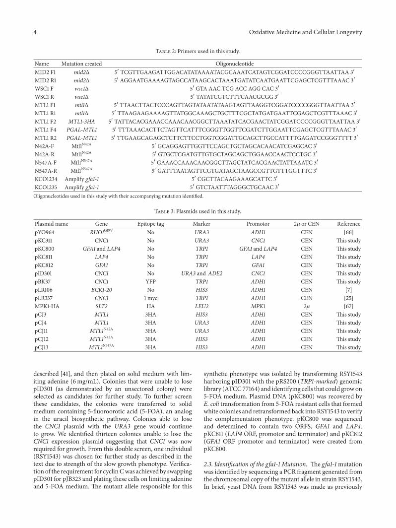
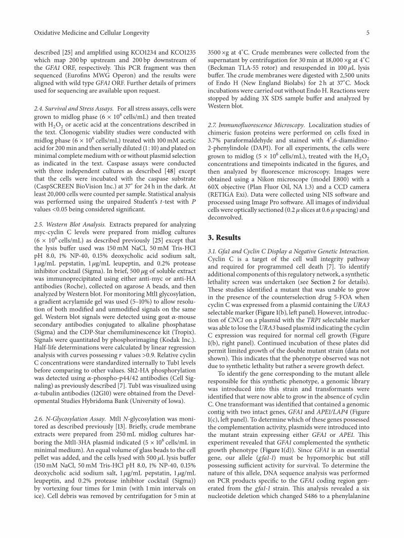
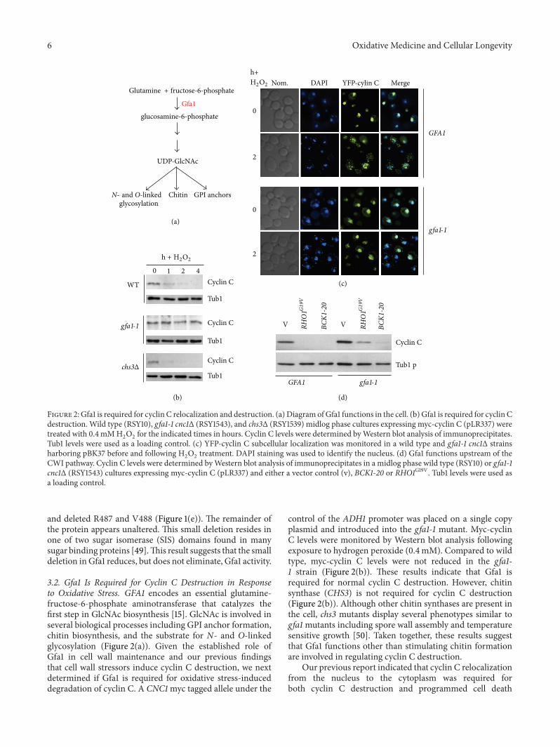
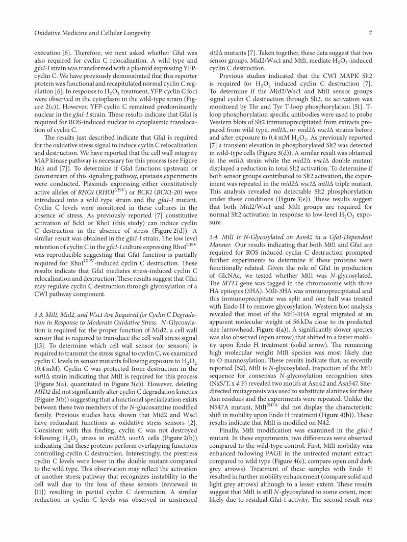
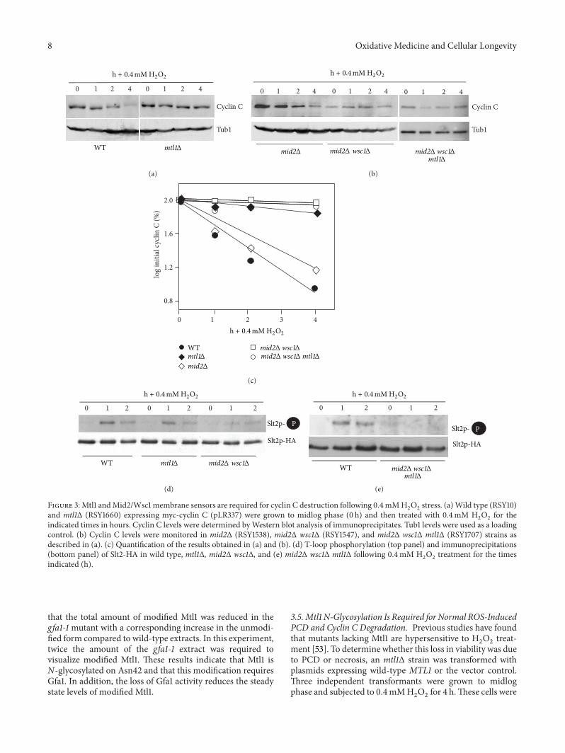
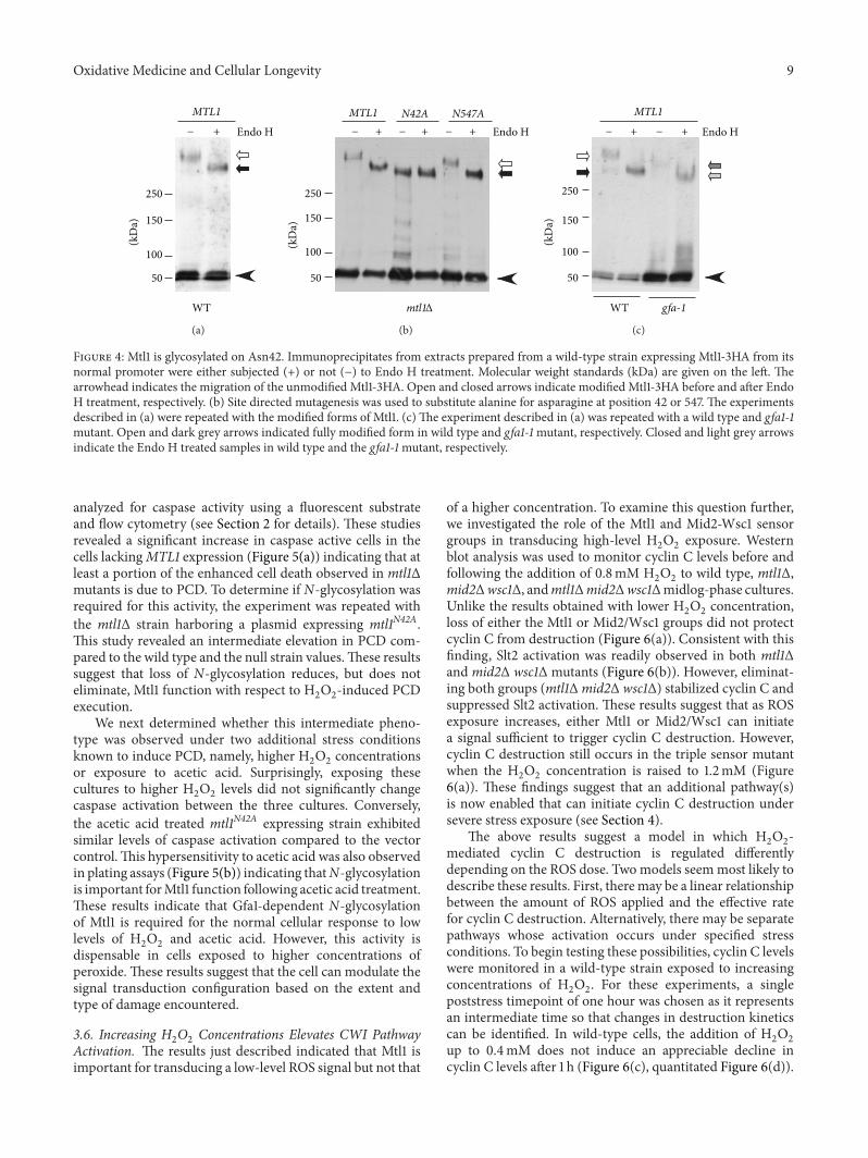
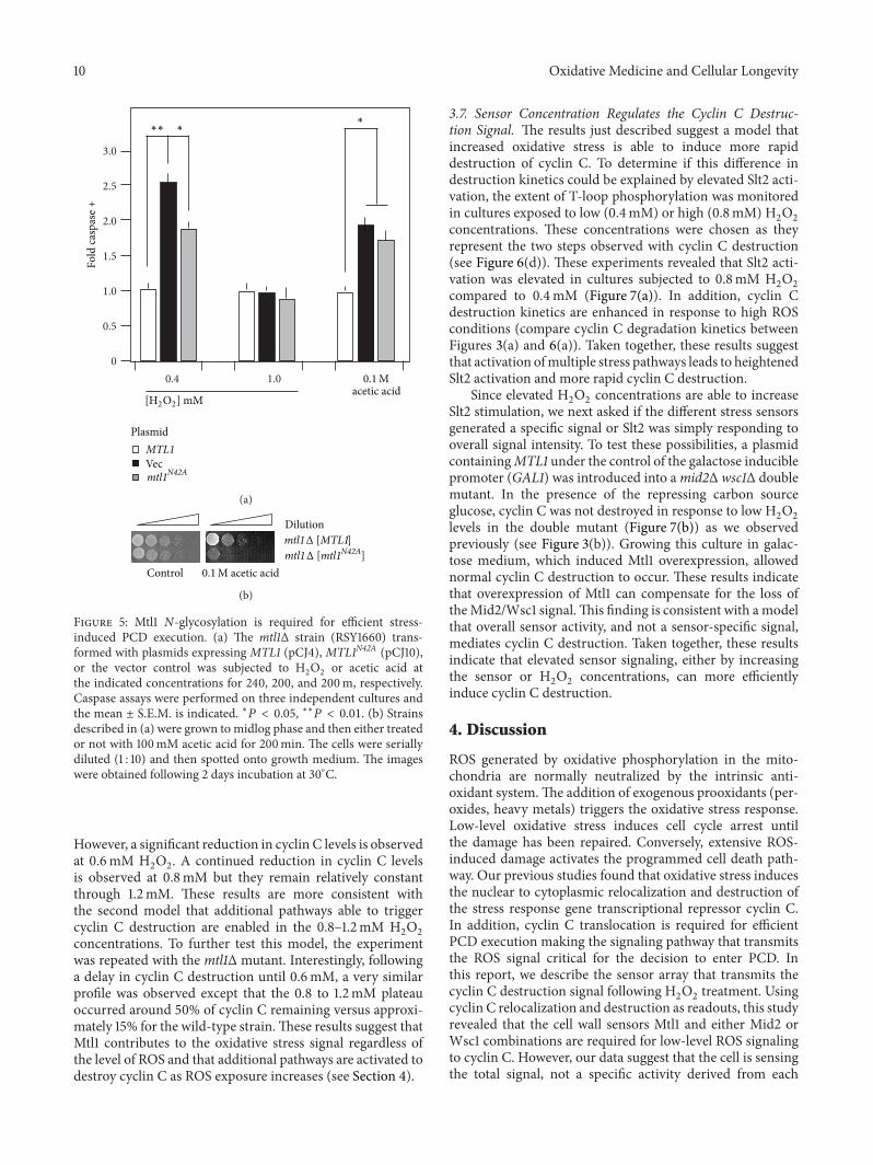
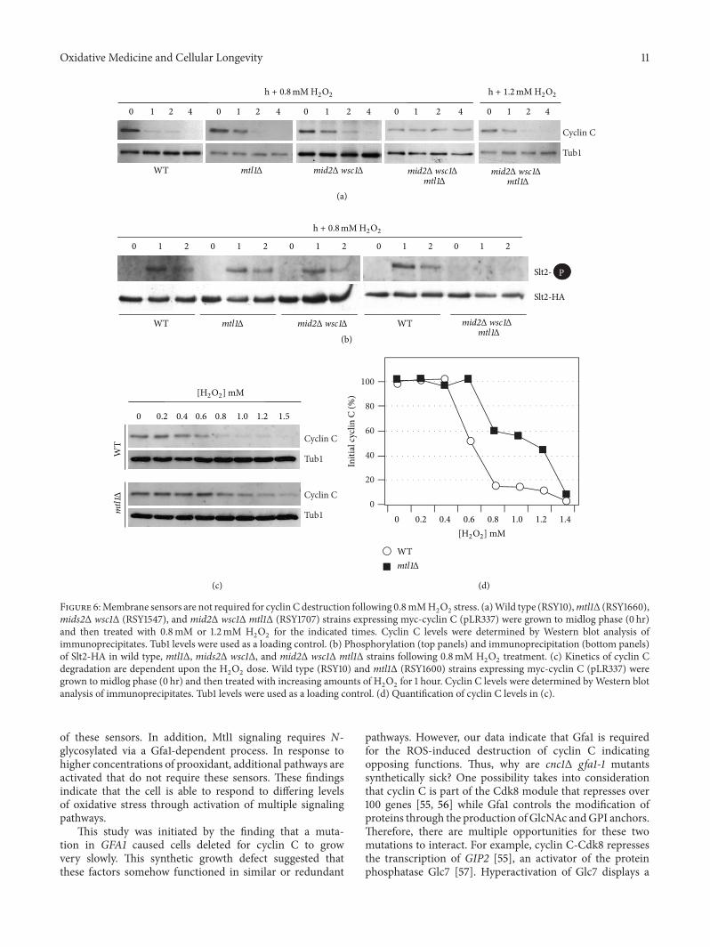
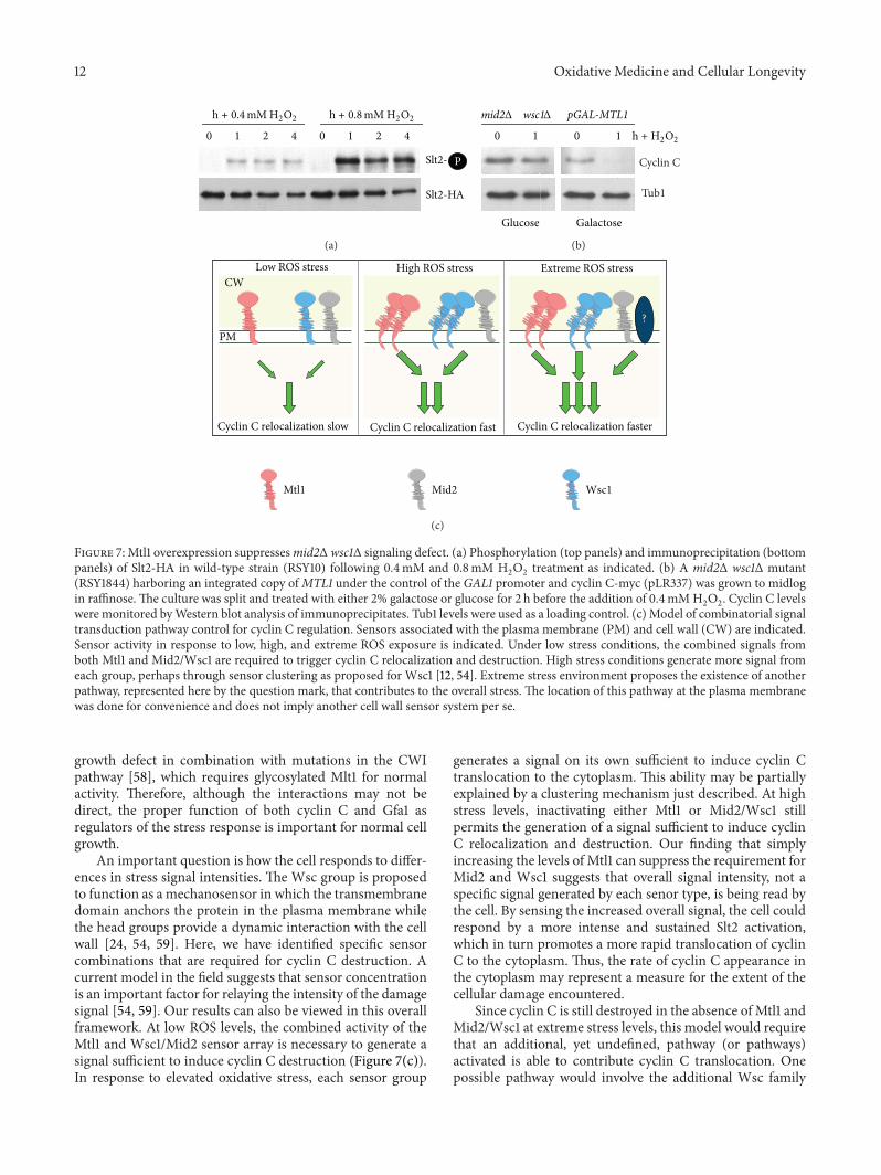
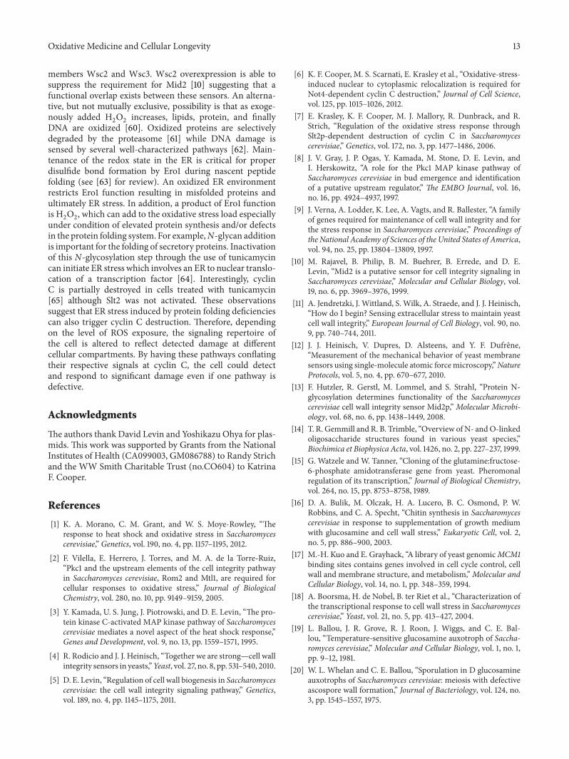
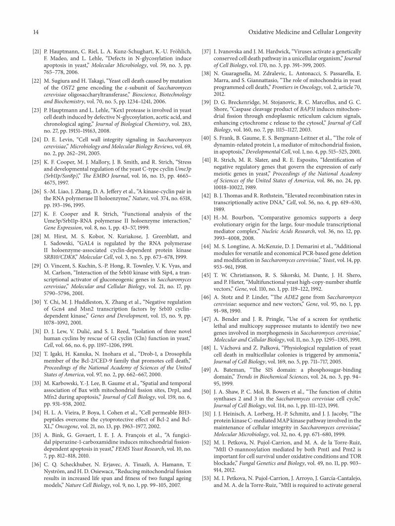


![DAA MID2 [UandiStar.org]](https://img.pdfslide.us/doc/110x75/545a7372af7959755d8b5b4b/daa-mid2-uandistarorg.jpg)