Embed Size (px)
Citation preview
Research ArticleThe Effect of tert-Butyl Hydroperoxide-Induced Oxidative Stresson Lean and Steatotic Rat Hepatocytes In Vitro
Otto KuIera1 Reneacute Endlicher2 Tomaacuteš Roušar1 Halka Lotkovaacute1
Tomaacuteš Garnol1 Zdenjk Drahota1 and Zuzana Hervinkovaacute1
1 Department of Physiology Charles University in Prague-Faculty of Medicine in Hradec Kralove Simkova 870500 38 Hradec Kralove Czech Republic
2 Department of Anatomy Charles University in Prague-Faculty of Medicine in Hradec Kralove Simkova 870500 38 Hradec Kralove Czech Republic
Correspondence should be addressed to Otto Kucera kuceraolfhkcunicz
Received 23 January 2014 Accepted 22 February 2014 Published 31 March 2014
Academic Editor Jakub Rohlena
Copyright copy 2014 Otto Kucera et al This is an open access article distributed under the Creative Commons Attribution Licensewhich permits unrestricted use distribution and reproduction in any medium provided the original work is properly cited
Oxidative stress and mitochondrial dysfunction play an important role in the pathogenesis of nonalcoholic fatty liver diseaseand toxic liver injury The present study was designed to evaluate the effect of exogenous inducer of oxidative stress (tert-butylhydroperoxide tBHP) on nonfatty and steatotic hepatocytes isolated from the liver of rats fed by standard and high-fat dietrespectively In control steatotic hepatocytes we found higher generation of ROS increased lipoperoxidation an altered redox stateof glutathione and decreased ADP-stimulated respiration using NADH-linked substrates as compared to intact lean hepatocytesFatty hepatocytes exposed to tBHP exert more severe damage lower reduced glutathione to total glutathione ratio and higherformation of ROS and production of malondialdehyde and are more susceptible to tBHP-induced decrease in mitochondrialmembrane potential Respiratory control ratio of complex Iwas significantly reduced by tBHP in both lean and steatotic hepatocytesbut reduction in NADH-dependent state 3 respiration was more severe in fatty cells In summary our results collectively indicatethat steatotic rat hepatocytes occur under conditions of enhanced oxidative stress and are more sensitive to the exogenous sourceof oxidative injury This confirms the hypothesis of steatosis being the first hit sensitizing hepatocytes to further damage
1 Introduction
Nonalcoholic fatty liver disease (NAFLD) is the most com-mon chronic liver disease in the USA and theWestern worldand the prevalence of NAFLD has recently dramaticallyincreased and it seems that this trend will continue [1 2]There is accumulating evidence that hepatic mitochondrialdysfunction and oxidative stress play an important role in thepathogenesis of NAFLD [3] There are several mitochondrialabnormalities associated with NAFLD including ultrastruc-tural lesions depletion of mitochondrial DNA decreasedactivity of respiratory chain complexes and impaired mito-chondrial 120573-oxidation [4ndash6] Oxidative stress and resultingaltered redox balance seem to be crucial in the pathogenesisof steatosis steatohepatitis and fibrosis [7] Mitochondrialdysfunction and increased oxidative stress are closely related
Mitochondrial dysfunction can directly lead to the excessof reactive oxygen species (ROS) production [8] On theother hand oxidative stress may be a cause of mitochondrialdysfunction [9] Altered hepatic redox status was proofed notonly in the advanced forms of NAFLD such as nonalcoholicsteatohepatitis (NASH) but also in simple steatosis [10]Enhanced oxidative stress is thought to belong to secondhits that participate in the progression of simple steatosisto steatohepatitis [11] Increased generation of free radicalsand other highly reactive substances results from fat accu-mulation in the liver particularly due to direct toxicity offatty acids increased peroxisomal and microsomal oxidationof fatty acids and mitochondrial dysfunction [12 13] Othersources of oxidative stress such as xenobiotic-induced hep-atotoxicity may very likely potentiate progression of simplesteatosis to steatohepatitis In previous experiments we have
Hindawi Publishing CorporationOxidative Medicine and Cellular LongevityVolume 2014 Article ID 752506 12 pageshttpdxdoiorg1011552014752506
2 Oxidative Medicine and Cellular Longevity
found in accordance to others that rat steatotic hepatocytesexert higher sensitivity to the acute injury caused by hepa-totoxins in vivo [14ndash16] and in vitro [17] NAFLD includingsimple steatosis predisposes the liver to the increased risk ofhepatotoxicity [18]
Oxidative stress is one of the general mechanismsinvolved in hepatotoxicity Free radicals initiate lipid perox-idation of polyunsaturated fatty acids in membranes whichresults in membrane disruption formation of reactive alde-hydes and depletion of cellular storage of reduced glu-tathione (GSH) tert-Butyl hydroperoxide (t-BHP) is com-monly used as a model substance for evaluation of mecha-nisms of cellular alterations resulting from oxidative stress incells and tissues There are two pathways by which tBHP ismetabolized both of them induce oxidative stress The firstprovided by cytochrome P450 leads to production of peroxyland alkoxyl radicals [19]These radicals initiate lipoperoxida-tion of membrane phospholipids with subsequent alterationsto membrane fluidity and permeability The other pathwayemploys glutathione peroxidase tBHP is detoxified to tert-butanol and GSH is depleted by oxidation to its disulphideform (GSSG) [20] Lipoperoxidation depletion of GSH andthe onset of mitochondrial permeability transition (MPT)are general mechanisms involved in cell injury caused byoxidative stressThus usage of exogenous inducer of oxidativestress such as tBHP may simulate situation of augmentedoxidative stress in fatty hepatocytes and helps us to under-stand particular mechanisms in the pathogenesis of NAFLD
The present study was designed to determine and tocompare tBHP-induced oxidative stress in isolated lean andsteatotic rat hepatocytes in primary culture For our model ofcell injury we used tBHP a short-chain organic hydroperox-ide which is an analogue of the products of lipoperoxidationformed during oxidative stress and may mimic oxidativestress in human diseases
2 Material and Methods
21 Chemicals Medium Williamrsquos E (without phenol red)foetal bovine serum penicillin streptomycin and glutaminewere purchased from PAN BIOTECH GmbH (AidenbachGermany) Collagenase (Collagenase NB 4 Standard Gradefrom Clostridium histolyticum) was obtained from SERVAElectrophoresis GmbH (Heidelberg Germany) insulin andglucagon (Actrapid Novo Nordisk AS Bagsvaerd Den-mark) and prednisolon (Merck KGaA Darmstadt Germany)were from the suppliers mentioned in brackets Type Icollagen trypan blue tert-butyl hydroperoxide solution (Catnumber 458139) and all other chemicals were purchasedfrom Sigma-Aldrich (St Louis MO)
22 Animals Male albino Wistar rats (BioTest KonaroviceCzech Republic) were housed at 23 plusmn 1∘C 55 plusmn 10 relativehumidity air exchange of 12ndash14 timesh and 12 h light-darkcycle periods (600 h to 1800 h) Rats used for isolation ofnonsteatotic hepatocytes (NH)were fed ad libitum a standardpelleted diet (ST-1 diet Velaz Prague Czech Republic 10energy fat 30 energy proteins and 60 energy saccharides)
Table 1 Scheme of protocol with 5 120583M TFP pretreatment
Group of lean and steatotichepatocytesTime of incubation 01015840ndash301015840 301015840ndash601015840
Control 0 0TFP 5120583M TFP 5120583M TFPtBHP 0 tBHPtBHP + TFP 5120583M TFP 5120583M TFP + tBHP0 Williamrsquos E medium without TFP or tBHP TFP trifluoperazine tBHPtert-butyl hydroperoxide at tested concentration
for 6 weeks For isolation of fatty hepatocytes (SH) animalswere fed high-fat gelled diet (71 energy fat 18 energyproteins and 11 energy saccharides) for 6 weeks [21] Theanimals had free access to tap water All animals receivedcare according to the guidelines set by the Animal-WelfareBody of the Charles University Prague Czech Republic andthe International Guiding Principles for Biomedical ResearchInvolving Animals All animal experiments were approvedby the committee mentioned above and by the Ministryof Education Youth and Sports (authorisation referencenumber 1315-285722012-30)
23 Hepatocyte Isolation Cultivation and Treatment Underether anaesthesia hepatocytes were isolated by two-stepcollagenase perfusion from rat liver [22] with viability higherthan 90 (trypan blue exclusion test) Isolated hepatocyteswere suspended in supplemented Williamrsquos E medium with6 foetal bovine serum [17] and plated in collagen-coatedPetri dishes (60mm 2 times 106 cellsdish) or 12- (4 times 105cellswell) and 96-well (3 times 104 cellswell) plates Hepato-cytes were allowed to establish monolayer in a humidifiedatmosphere containing 95 air and 5 CO
2at 37∘C for 2 h
Then the medium was replaced with a fresh supplementedmedium without foetal bovine serum according to theexperimental protocol Nonfatty and steatotic hepatocytes inprimary cultures were incubated with tBHP (001ndash1mmolL)for up to 60min The other portion of hepatocytes waspreincubated in Williamrsquos E medium with or without 5 120583Mtrifluoperazine (TFP) for 30min and then exposed to tBHP(025 0375 05mmolL) with or without 5120583M TFP forfurther 30min (Table 1) After incubation the medium wascollected and cells were harvested for the required assays Sus-pension of digitonin-permeabilized hepatocytes pretreatedwith 025mM tBHP for 5min was used for evaluation ofmitochondrial respiration
24 Cytotoxicity Assays Plasma membrane integrity of cul-tured hepatocytes was determined by lactate dehydrogenase(LDH) activity in the medium using a commercial kitfrom DiaSys (Holzheim Germany) Cell viability was alsoevaluated by measurement of the activity of cellular dehy-drogenases using Cell Proliferation Reagent WST-1 (RocheDiagnostics Mannheim Germany) [23]
25 Estimation of GSH to Total Glutathione Ratio and Pro-duction of Reactive Oxygen Species (ROS) and Malondialde-hyde (MDA) For assessment of intracellular GSH to total
Oxidative Medicine and Cellular Longevity 3
glutathione ratio [GSH(2 timesGSSG +GSH)] cells were firstlylysed and harvested and then GSH and GSSG were measuredby spectrofluorometric assay based on the reaction betweenGSH and o-phthalaldehyde (120582 (exc) = 340 nm pH 60) [24]
The production of ROS was evaluated using 5- and6-chloromethyl-2101584071015840-dichlorodihydrofluorescein diacetate(CM-H2DCFDA Molecular Probes Eugene OR) Afterincubation the cells were washed in Williamrsquos E mediumand loaded by 1 120583M CM-H2DCFDA for 45min and thenrinsed again in nonsupplemented Williamrsquos E mediumThenfluorescence intensity was measured for 40min (TECANInfinite M200 Tecan Austria GmbH Grodig Austriaexcitation and emission wavelength of 485 and 535 nmresp) Results are expressed in percent where controlnonsteatotic hepatocytes are 100 of fluorescence intensitydifference (at 401015840 minus at 01015840)
Secondary end product of lipoperoxidation MDA inculture medium was determined by the assessment of thio-barbituric acid reactive substances [25]
26 Production of Albumin Functional capacity of culturedhepatocytes was evaluated by the amount of albumin secretedinto the culture medium using Rat Albumin ELISAQuantifi-cation Kit (Bethyl Laboratories Montgomery TX)
27 Visualization of Mitochondrial Membrane Potential(MMP) MMPwas depicted using hepatocyte uptake of JC-1(Molecular Probes Inc Oregon USA) a cationic carbocya-nine dye that accumulates in mitochondria according to itsmembrane potential At low membrane potential JC-1 exertsa green fluorescence (120582em 525 nm) At higher potentialsJC-1 forms red-fluorescent ldquoJ-aggregatesrdquo (120582em 590 nm)Hepatocytes were incubated with 10 120583M JC-1 (dissolved inWilliamrsquos E medium) in humidified atmosphere containing95 air and 5 CO
2at 37∘C for 30min then the cells were
washed twice with fresh media MMP was visualized usingfluorescence microscope Olympus IX51 (Olympus Japan)equipped with the digital camera Olympus E600 (OlympusJapan) Results are expressed as percentage of cells containingmitochondria with high membrane potential
28 Measurement of Oxygen Uptake by Isolated HepatocytesOxygen consumption was measured using a High ResolutionOxygraph 2K (OROBOROS INSTRUMENTS GmbH Inns-bruck Austria) Digitonin-permeabilized (10120583gmL) hepato-cytes (125000mL) were incubated in 2mL of K+-medium[26] at 30∘C State 4 respiration (10mM glutamate + 25mMmalate) and state 3 respiration (10mM glutamate + 25mMmalate + 15mM ADP) were measured and respiratorycontrol ratio (RCR ratio of O
2consumption rate in state 3 to
state 4) was calculated Oxygen uptake at state 3 and state 4 isexpressed as pmoles oxygen per second per million cells Forevaluation of oxygen uptake OROBOROS software (DatLab31 OROBOROS INSTRUMENTS GmbH) was used
29 Statistical Analysis Experiments were performed atleast three times using different isolations of hepatocytesDue to high interexperiment variability the data analyses
were conducted by experiment The results are expressed asmeans plusmn SD of a single representative experiment After test-ing the normality statistical analysis was performed by one-way analysis of variance (GraphPad Prism 601 GraphPadSoftware Inc La Jolla CA)When significance was detectedTukey-Kramerrsquos post hoc test was used for comparisonsbetween the different groups 119875 lt 005 was considered to bestatistically significant
3 Results
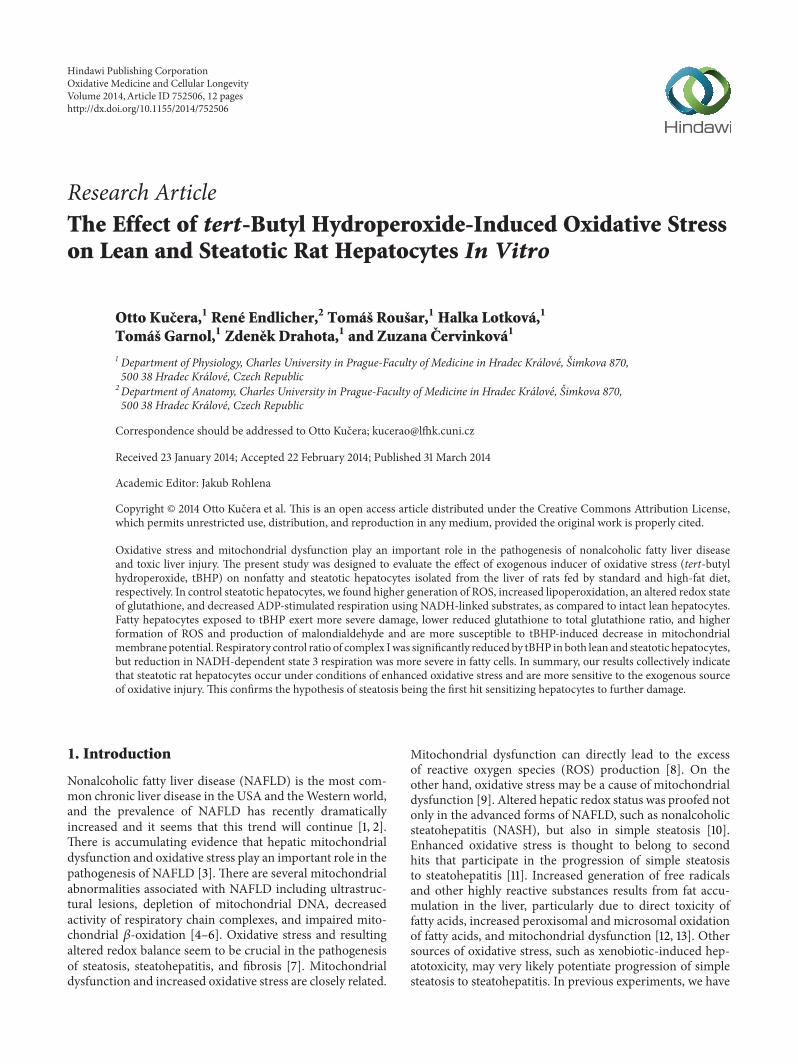
31 Characteristics of Control Lean and Fatty HepatocytesSteatotic hepatocytes compared to nonfatty cells exert sig-nificantly lower activity of cellular dehydrogenases (WST-1test Figure 1(a)) and almost 2-fold higher activity of LDHin culture medium (Figure 1(b)) Control fatty hepatocytesare also significantly more affected by oxidative stress asdocumented by higher production of ROS (Figures 1(c) and3(a)) and MDA in culture medium (Figure 3(b)) and lowerintracellular ratio of GSH to total glutathione (Figure 3(c))There are no differences between control lean and steatotichepatocytes in albumin production (Figure 2(c)) percentageof hepatocytes with energized mitochondria (Figures 4(a)4(b) and 5) and oxygen consumption at state 4 respirationwhereas oxygen consumption at state 3 respiration wassignificantly reduced in steatotic cells (Table 2)
32 Effect of tBHP on Nonfatty and Steatotic Hepato-cytes WST-1 test showed (Figure 1(a)) that lean hepatocytesexposed to tBHP for a period of 60min are significantlyaffected from the concentration of 025mmolL whereasin fatty hepatocytes WST-1 test was already decreased attBHP concentration of 01mmolL Activity of LDH inculture medium did not exert any increase of LDH activ-ity in nonsteatotic cells treated with 025mM tBHP upto 60min (Figure 1(b)) In fatty hepatocytes LDH activitywas significantly elevated even in 15min after exposureto 025mM tBHP tBHP (025 and 0375mmolL) inducedmore pronounced depression of albumin production in fattyhepatocytes as compared to lean cells (Figure 2(c))
Incubation with tBHP for 30min revealed higher sus-ceptibility of steatotic hepatocytes to oxidative stress Gen-eration of ROS after exposure to tBHP exerts dose andtime dependent manner and is more pronounced in fattyhepatocytes (Figures 1(c) and 3(a)) Concentration of MDAin culture medium of fatty cells incubated with 025mMtBHP was almost 2-fold higher than in lean hepatocytes(Figure 3(b)) tBHP at concentration of 025mmolL did notcause significant change in GSH to total glutathione ratio innonsteatotic cells and incubation with 0375mM tBHP leadsto onlymild decrease by 5 in the ratio In contrast reductionin this ratio to 66 and 33was observed in fatty hepatocytesexposed to tBHP at concentrations of 025 and 0375mmolLrespectively (Figure 3(c))
Figures 4(c) 4(d) 4(g) 4(h) and 5 show that steatotichepatocytes exert higher susceptibility to tBHP-induceddecrease in MMP In lean cells tBHP from concentration of0375mmolL leads to reduction of percentage of hepatocytes
4 Oxidative Medicine and Cellular Longevity
120
100
80
60
40
20
0
WST-1 test(incubation with tBHP for 60 min)
lowastlowastlowast
lowastlowastlowastlowastlowastlowast
lowastlowastlowast
xxx
xxx
xxx
NH
NH+
tBH
P00
1
NH+
tBH
P01
NH+
tBH
P02
5N
H+
tBH
P05
NH+
tBH
P1 SHSH
+tB
HP0
01
SH+
tBH
P01
SH+
tBH
P02
5SH
+tB
HP0
5SH
+tB
HP1
NH
( o
f con
trol)
(a)
2000
1800
1600
1400
1200
1000
800
600
400
200
0
NS SH
(IU
L)
Time (min)
Activity of LDH in culture medium
lowastlowastlowastlowastlowastlowast lowastlowastlowast
lowastlowastlowast
NS + tBHP025 SH + tBHP025
xxxxxx
xxxxxx
15998400 30998400 45998400 60998400
(b)
1200
1000
800
600
400
200
0
CtBHP 15
tBHP
tBHPtBHP
Production of ROS
NH+
tBH
P02
5
NH+
tBH
P03
75
SH+
tBH
P02
5
SH+
tBH
P03
75
NH+
tBH
P05
SH+
tBH
P05
NH
( o
f con
trol)
998400
30998400
45998400
60998400
(c)
Figure 1 (a) WST-1 test of nonsteatotic (NH) and steatotic rat hepatocytes (SH) in primary culture treated with 001ndash1mM tBHP (NH +tBHP SH + tBHP) for 60min The values are means plusmn SD (119899 = 8) Results are expressed in percent where 100 is the activity of cellulardehydrogenases in control NH lowastlowastlowast119875 lt 0001 versus control NH
119875 lt 0001 versus control SH xxx119875 lt 0001 versus corresponding
NH + tBHP (b) Time course of LDH activity (IUL) in media of lean (NH) and steatotic rat hepatocytes (SH) in primary cultures treatedwith 025mM tBHP (NH + tBHP SH + tBHP) for up to 60min The values are means plusmn SD (119899 = 6) lowastlowastlowast119875 lt 0001 versus control NH119875 lt 001 and
119875 lt 0001 versus control SH at corresponding time xxx119875 lt 0001 versus NH + tBHP at corresponding time (c) Time and
concentration course of ROS generation (CM-H2DCFDA) in nonfatty (NH) and steatotic rat hepatocytes (SH) in primary culture treatedwith 025ndash05mM tBHP (NH + tBHP SH + tBHP) for up to 60min The values are means plusmn SD (119899 = 8) Results are expressed in percentwhere 100 is production of ROS by control NH for each concentration of tBHP 119875 values are not shown
with energized mitochondria whereas in fatty cells thereduction was more expressed and was found from tBHPconcentration of 025mmolL
Exposure to 025mM tBHP for 5min resulted in anonsignificant increase in state 4 respiration by 24 and 26 inlean and fatty cells respectively (compared with control non-steatotic and steatotic hepatocytes resp) RCR of complex Iand oxygen consumption at state 3 were reduced by tBHP inboth lean and steatotic cells NADH-dependent respiration at
state 3 was significantly lower in fatty hepatocytes than in leancontrols (Table 2)
33 Effect of TFP on Control Lean and Fatty HepatocytesIncubation of both lean and steatotic hepatocytes with 5 120583MTFP for a period of 60min (301015840 + 301015840) did not lead tosignificant changes in LDH activity in culture medium(Figure 2(a)) TFP did not reduce activity of cellular dehydro-genases in nonsteatotic and fatty hepatocytes when compared
Oxidative Medicine and Cellular Longevity 5
Activity of LDH in culture medium400
350
300
250
200
150
100
50
0
(IU
L)
lowastlowastlowastN
H
NH+
TFP
NH+
TFP+
tBH
P02
5
NH+
tBH
P02
5
SH
SH+
TFP
SH+
tBH
P02
5
SH+
TFP+
tBH
P02
5
xxx
xx$$$
(a)
180
160
140
120
100
80
60
40
20
0
WST-1 test
xxx
xx
NH
NH+
TFP
NH+
TFP+
tBH
P02
5
NH+
tBH
P02
5
SH
SH+
TFP
SH+
tBH
P02
5
SH+
TFP+
tBH
P02
5
xxx
NH
( o
f con
trol)
(b)
25
20
15
10
5
0
Albumin production
lowast
lowastlowastlowast
lowastlowastlowastlowastlowastlowastlowastlowastlowast
(120583g
L)
xxx xxx
NH
NH+
TFP
NH+
TFP+
tBH
P02
5
NH+
tBH
P02
5
NH+
TFP+
tBH
P03
75
NH+
tBH
P03
75 SH
SH+
TFP
SH+
tBH
P02
5
SH+
TFP+
tBH
P02
5
SH+
tBH
P03
75
SH+
TFP+
tBH
P03
75
ampampamp
(c)
Figure 2 (a) LDH activity (IUL) in medium of lean (NH) and steatotic rat hepatocytes (SH) in primary culture treated with 5120583M TFP(NH + TFP SH + TFP) with 025mM tBHP (NH + tBHP SH + tBHP) or with 5120583M TFP and 025mM tBHP (NH + TFP + tBHP SH +TFP + tBHP) Cells were exposed to tBHP for a period of 30min The values are means plusmn SD (119899 = 6) lowastlowastlowast119875 lt 0001 versus control NH119875 lt 0001 versus control SH xx
119875 lt 001 and xxx119875 lt 0001 versus corresponding group in lean hepatocytes $$$
119875 lt 0001 versus SH +tBHP (b) WST-1 test of nonsteatotic (NH) and steatotic rat hepatocytes (SH) in primary culture treated with 5120583M TFP (NH + TFP SH +TFP) with 025mM tBHP (NH + tBHP SH + tBHP) or with 5 120583M TFP and 025mM tBHP (NH + TFP + tBHP SH + TFP + tBHP) Cellswere exposed to tBHP for 30min The values are means plusmn SD (119899 = 6) Results are expressed in percent where 100 is the activity of cellulardehydrogenases in control NH
119875 lt 0001 versus control SH xx119875 lt 001 and xxx
119875 lt 0001 versus corresponding group in lean hepatocytes(c) Concentration of albumin (120583gL) in culture medium of nonsteatotic (NH) and steatotic rat hepatocytes (SH) treated with 5 120583MTFP (NH+ TFP SH + TFP) with 0250375mM tBHP (NH + tBHP SH + tBHP) or with 5120583M TFP and 0250375mM tBHP (NH + TFP + tBHPSH + TFP + tBHP) Cells were exposed to tBHP for 30min The values are means plusmn SD (119899 = 5) lowast119875 lt 005 and lowastlowastlowast119875 lt 0001 versus controlNH ampampamp
119875 lt 0001 versus NH + tBHP025 119875 lt 001 and
119875 lt 0001 versus control SH xxx119875 lt 0001 versus corresponding group in lean
hepatocytes
with corresponding controls (Figure 2(b)) Exposure to TFPleads to the elevation of intracellular GSH to total glutathioneratio in control nonsteatotic hepatocytes and a similar non-significant trend was also found in fatty cells (Figure 3(c))In contrast to these beneficial effects TFP alone significantlyreduced production of albumin in both lean and steatotic cells(Figure 2(c))
34 Potential Beneficial Effect of TFP on tBHP-Induced InjuryWecompared two protocols of cell incubationwith 5120583MTFPFirstly hepatocytes were coincubated with tBHP and TFP for30min Secondly cells were at the earliest preincubated withTFP for a period of 30min and subsequently coincubatedwith tBHP and TFP for additional 30min We found thatTFPwas able to partially reduce tBHP-induced damage (LDH
6 Oxidative Medicine and Cellular Longevity
400
350
300
250
200
150
100
50
0
Production of ROS
lowastlowastlowastlowastlowastlowast
lowastlowastlowast
NH
NH+
TFP
NH+
TFP+
tBH
P02
5
NH+
tBH
P02
5
SH
SH+
TFP
SH+
tBH
P02
5
SH+
TFP+
tBH
P02
5
xxx
xxx$$$
ampamp
NH
fluo
resc
ence
( o
f con
trol)
(a)
3
25
2
15
1
05
0
Concentration of MDA in culture media
lowastlowastlowastlowast
xxx
xxx
NH
NH+
TFP+
tBH
P02
5
NH+
tBH
P02
5
SH
SH+
tBH
P02
5
SH+
TFP+
tBH
P02
5
(120583m
olL
)
(b)
1009080706050403020100
Intracellular GSH to total glutathione ratiolowast
lowast lowastlowast xxx
xxx
NH
NH+
TFP
NH+
TFP+
tBH
P02
5
NH+
tBH
P02
5
NH+
TFP+
tBH
P03
75
NH+
tBH
P03
75 SH
SH+
TFP
SH+
tBH
P02
5
SH+
TFP+
tBH
P02
5
SH+
tBH
P03
75
SH+
TFP+
tBH
P03
75
ampampamp
GSH
(G
SG+
2times
GSS
G)
()
(c)
Figure 3 (a) Production of ROS (CM-H2DCFDA) in nonsteatotic (NH) and steatotic rat hepatocytes (SH) treated with 5 120583M TFP (NH +TFP SH + TFP) with 025mM tBHP (NH + tBHP SH + tBHP) or with 5 120583M TFP and 025mM tBHP (NH + TFP + tBHP SH + TFP +tBHP) Cells were exposed to tBHP for 30minThe values are means plusmn SD (119899 = 8) Results are expressed in percent where 100 is productionof ROS by control NH lowastlowastlowast119875 lt 0001 versus control NH ampamp
119875 lt 001 versus NH + tBHP 119875 lt 0001 versus control SH xxx
119875 lt 0001
versus corresponding group in lean hepatocytes $$$119875 lt 0001 versus SH + tBHP (b) Concentration of MDA (120583molL) in culture media of
nonsteatotic (NH) and steatotic rat hepatocytes (SH) treated with 025mM tBHP (NH + tBHP SH + tBHP) or with 5120583MTFP and 025mMtBHP (NH + TFP + tBHP SH + TFP + tBHP) Cells were exposed to tBHP for 30min The values are means plusmn SD (119899 = 6) lowast119875 lt 005 andlowastlowastlowast
119875 lt 0001 versus control NH 119875 lt 001 versus control SH xxx
119875 lt 0001 versus corresponding group in lean hepatocytes (c) IntracellularGSH to total glutathione ratio in nonsteatotic (NH) and steatotic rat hepatocytes (SH) treated with 5 120583M TFP (NH + TFP SH + TFP) with025mM tBHP (NH + tBHP SH + tBHP) or with 5 120583MTFP and 025mM tBHP (NH + TFP + tBHP SH + TFP + tBHP) Cells were exposedto tBHP for 30min The values are means plusmn SD (119899 = 5) Results are expressed in percent of GSH from total glutathione lowast119875 lt 005 andlowastlowast
119875 lt 001 versus control NH ampampamp119875 lt 0001 versus NH + tBHP0375
119875 lt 005 and 119875 lt 0001 versus control SH xxx
119875 lt 0001 versuscorresponding group in lean hepatocytes
activity andROSproduction data not shown) onlywhen cellswere firstly preincubated with TFP prior to tBHP exposureTherefore we only present results with preincubation fol-lowed by coincubation
TFPwas able to partially prevent tBHP-induced elevationin LDH activity in culture medium of steatotic hepato-cytes (Figure 2(a)) increase in production of ROS in both
lean and fatty cells (Figure 3(a)) decrease in GSHtotalglutathione ratio in nonfatty hepatocytes exposed to 0375tBHP (Figure 3(c)) and reduction in the percentage of cellswith energized mitochondria in nonsteatotic hepatocytesexposed to 0375 and 05mM tBHP and in steatotic cellsincubated with 025 and 0375mM tBHP (Figures 4 and 5)We did not observe any beneficial effect of TFP on activity
Oxidative Medicine and Cellular Longevity 7
(a) (b) (c)
(d) (e) (f)
(g) (h) (i)
(j)
Figure 4 Visualization of changes in mitochondrial membrane potential using mitochondria specific fluorescent probe JC-1 Mitochondriawith intact membrane potential concentrates JC-1 into aggregates (J-aggregates red fluorescence at 590 nm) whereas deenergizedmitochondria cannot concentrate JC-1 (green fluorescence at 530 nm) Microphotographs of nonsteatotic and fatty rat hepatocytes culturedinWilliamrsquos E medium (control (a) and (b) resp) with tBHP at concentration of 025mmolL ((c) and (d) resp) and 0375mmolL ((g) and(h) resp) or with 5 120583MTFP and 025mM ((e) and (f) resp) or 5 120583MTFP and 0375mM tBHP ((i) and (j) resp) Cells were exposed to tBHPfor a period of 30min Magnification 400x
8 Oxidative Medicine and Cellular Longevity
Table 2 Respiration of digitonin-permeabilized (10 120583gmL) hepatocytes (125000mL) in K+-medium at a temperature of 30∘C State 4respiration (10mM glutamate + 25mM malate) and state 3 respiration (10mM glutamate + 25mM malate + 15mM ADP) were measuredand respiratory control ratio (RCR state 3state 4) was calculated in control nonsteatotic (NH) and steatotic rat hepatocytes (SH) and in leanand fatty hepatocytes preincubated with 025mM tBHP for 5min (NH + tBHP SH + tBHP) Oxygen uptake at state 3 and state 4 is expressedas pmoles oxygen per second per million cells lowastlowast119875 lt 001 and lowastlowastlowast119875 lt 0001 versus NH 119875 lt 0001 versus SH $$119875 lt 001 versus NH +tBHP
State 3 State 4 RCR of complex I(pmolO2s10
6 cells) (pmolO2s106 cells)
NH (119899 = 11) 10155 plusmn 1451 1885 plusmn 445 54 plusmn 12
NH + tBHP (119899 = 9) 7610 plusmn 1456 2330 plusmn 616 33 plusmn 07
lowast lowast lowast ns versus NH lowast lowast lowast
SH (119899 = 10) 8197 plusmn 1125 1724 plusmn 425 48 plusmn 08
lowastlowast ns versus NH ns versus NH
SH + tBHP (119899 = 8)5204 plusmn 982 2176 plusmn 760 24 plusmn 06
ns versus SH $$ ns versus NH + tBHP ns versus NH + tBHP
100
90
80
70
60
50
40
30
20
10
0
()
lowastlowastlowast lowastlowastlowast
lowastlowastlowast
xxx
xxx
xxx
$$$
$$$
Hepatocytes with energized mitochondria ()
NH
NH+
TFP
NH+
TFP+
tBH
P02
5N
H+
tBH
P02
5
NH+
TFP+
tBH
P05
NH+
tBH
P05
NH+
TFP+
tBH
P03
75N
H+
tBH
P03
75 SHSH
+TF
PSH
+tB
HP0
25
SH+
TFP+
tBH
P02
5
SH+
tBH
P05
SH+
TFP+
tBH
P05
SH+
tBH
P03
75SH
+TF
P+
tBH
P03
75
ampampamp
ampampamp
Figure 5 Percentage of nonsteatotic (NH) and steatotic rat hepato-cytes (SH)with energizedmitochondria treatedwith 5120583MTFP (NH+ TFP SH + TFP) with 025 0375 and 05mM tBHP respectively(NH + tBHP SH + tBHP) or with 5120583M TFP and 025 0375 and05mM tBHP respectively (NH + TFP + tBHP SH + TFP + tBHP)Cells were exposed to tBHP for a period of 30min The values aremeans plusmn SD (119899 = 16) lowastlowastlowast119875 lt 0001 versus control NH ampampamp
119875 lt
0001 versus corresponding NH + tBHP group 119875 lt 0001 versus
control SH xxx119875 lt 0001 versus corresponding NH + tBHP group
$$$119875 lt 0001 versus corresponding SH + tBHP group
of cellular dehydrogenases (Figure 2(b)) production of albu-min (Figure 2(c)) and production of MDA (Figure 3(b)) intBHP-treated nonfatty and steatotic hepatocytes
4 Discussion
In this experiment we studied oxidative stress-inducedchanges in hepatocytes isolated from nonfatty and steatoticrat liver For evaluation of peroxidative damage hepatocytes
were exposed to tBHP a prooxidant compound frequentlyused for assessment of mechanisms involving in oxidativestress in biological systems Oxidative stress plays commonlya key role in the pathogenesis of both xenobioticdrug-induced hepatotoxicity [27] and NAFLD [28] We analysedthe time course and the dose dependence of the perox-idative injury to hepatocytes induced by tBHP and wecorrelated changes of cell viability markers of oxidativestress (production of ROS lipoperoxidation and intracellularGSH content) the mitochondrial membrane potential func-tional capacity of hepatocytes and respiration of digitonin-permeabilized hepatocytes
tBHP is known to cause peroxidation of membrane lipids[19] and deplete cellular GSH [20] We and others havepreviously reported lower amounts of GSH in steatotic liverin vivo in patients [29] and in experimental models [21]and in vitro in mouse hepatocyte line (AML12 cells) treatedwith free fatty acids [30] or in rat hepatocytes isolated fromfatty liver [17] In contrast induction of steatosis in thehuman liver cell line (HepG2C3A) leads to elevation ofcellular GSH [31] Similarly Grattagliano et al [32] observedan early increase of liver GSH followed by its progressivedecrease in a rat model of steatosis This transient incrementof GSH seems to be only a cellular adaptive antioxidantresponse to increased oxidative stress induced by excess offat Glutathione depletion is considered a potential biomarkerof drug-induced hepatotoxicity Moreover tBHP is partiallymetabolized via glutathione peroxidase [20] thus our obser-vation of altered balance of intracellular glutathione redoxstate in steatotic cells predisposes to its toxicity and tosusceptibility to oxidative stress in general Decreased GSHto total glutathione ratio in control fatty hepatocytes is ina good concordance with findings of about twofold higherproduction of ROS and increased MDA production in thesecells tBHP-induced generation of ROS in lean and steatotichepatocytes correlates well with the time of incubation andthe dose of tBHP and is significantly more pronounced infatty cells Our study clearly showed that steatotic hepatocytesare more susceptible to oxidative injury caused by tBHP in
Oxidative Medicine and Cellular Longevity 9
primary culture than lean hepatocytes as documented bythe activity of cellular dehydrogenases and LDH In leanhepatocytes we did not observe any damage to plasmamembrane after incubation with 025mM tBHP for up to60min In contrast LDH activity in the culture mediumof fatty hepatocytes was significantly elevated even aftercultivation with 025mM tBHP for 15min Despite the sameviability of lean and steatotic hepatocytes at beginning of theexperiment (data not shown) plasma membrane integrity ofcontrol fatty cells was more disrupted than that of controllean cells Altered redox balance and S-thiolation of crucialcellular components may be responsible for the inhibitionof protein synthesis during the oxidative stress [33] Weshowed that albumin production was reduced to 63 and23 of control values in lean and steatotic hepatocytesrespectively after 30min incubation with 025mM tBHPThus proteosynthetic function of hepatocytes seems to bemore sensitive to oxidative stress in fatty hepatocytes
Mitochondrial functions are often altered in the liveraffected by NAFLD [5] Thus induction of ROS productionin the terrain of NAFLD leads to further progression ofmitochondrial dysfunction with all consequences Electronflowdisruption at any point of the respiratory chain augmentsgeneration of ROS via transfer of electrons to molecularoxygen [8] Besides other known mechanisms ROS exerttheir toxicity also through the induction ofMPT [34] tBHP isknown to induceMPT in isolated hepatocytes [35] Althoughthe onset of MPT was not directly measured in this studywe believe that JC-1-visualized changes of mitochondrialmembrane potential (MMP) result from MPT To supportthis statement we treated hepatocytes with a MPT inhibitorSince cyclosporinA is weakly effective in prevention of tBHP-induced cytotoxicity in hepatocytes we used trifluoperazinewhich is known to specifically reduce MPT-mediated injury[35] Broekemeier and Pfeiffer [36] suggested that TFP isable to increase the gating potential of the MPT pore andtherefore block the MPT TFP was capable of attenuatingtBHP-induced decrease in MMP in both lean and steatotichepatocytes Thus changes of MMP are at least partiallycaused by MPT Moreover TFP reduced partially plasmamembrane damage in steatotic hepatocytes ROS productionin both nonsteatotic and fatty cells and altered GSH statusin nonfatty hepatocytes exposed to tBHP Similarly Shenet al [37] also showed that MPT inhibitors may reducesuperoxide mediated cytochrome c release and mitochon-drial depolarization and subsequently inhibit apoptosis inHepG
2cells Nevertheless TFP was not significantly effective
in prevention of reduced activity of cellular dehydrogenasesdecreased albumin synthesis enhanced lipoperoxidationand surprisingly altered mitochondrial complex I activity(data not shown)
In steatotic liver there is a higher offer of fatty acids to beperoxidized which together with an insufficient antioxidantcapacity of the liver [29] leads to augmented lipoperoxida-tion Here we proved that production ofMDA in control fattycells is twice as much that in lean cells Additional exposureto external inducer of oxidative stress caused higher increasein MDA production in steatotic cells Peroxidized lipids [38]together with ROS (ie superoxide anion) [39] belong to the
activators of phospholipases A2 Since the involvement of freefatty acids products of phospholipase A2 activity in the trig-gering of MPT was shown [40] lipoperoxidation productsand superoxide anion are thought to play an important rolein this event in the environment of enhanced oxidative stressTFP is also known to inhibit phospholipase A2 activity whichmay explain more effective prevention of tBHP-inducedcytotoxicity in hepatocytes in comparison with cyclosporineA Bohm et al showed that feeding rats corn oil containingperoxidized fatty acids may trigger the development of hep-atic inflammation [41] Thus lipoperoxidation participatesconsiderably in the pathogenesis of NAFLD and mediatesprogression from simple steatosis to advanced forms ofNAFLD [42] with further increasing of oxidative stress
Mitochondria as the main energy provision systemis a crucial site of action of many hepatotoxic substances[12] In the liver with accumulated fat decreased activitiesof mitochondrial complexes I II IV and V [21 43] andelevated formation of ROS [3 7 10 44] were detected Mito-chondrial dysfunction is characterized by permeabilizationof mitochondrial outer membrane and resulting release ofproteins from intermembrane space into the cytosol caspaseactivation disruption of themitochondrial respiratory chainloss of MMP and augmented free-radical production [45]Mitochondrial dysfunction is thought to represent a centralabnormality responsible for progression from simple fattyliver to steatohepatitis [46] Herein we show respirationof digitonin-permeabilized rat hepatocytes When observingcomplex I respiration we found significant reduction in state3 oxygen consumption and a trend of lower RCR in controlfatty cells In contrast to changes of complex I Cardoso etal reported trends of increased state 3 respiration and RCRof complex II (succinate as a substrate) in mitochondriaisolated from liver of high-fat diet fed mice [44] This isin agreement with our previous results showing that ADP-stimulated respiration using succinate together with NADH-linked substrates was not affected in steatotic permeabilizedhepatocytes thus flavoprotein-dependent substrates mightcompensate decreased activity of complex I [47] Oxidativestress induced by tBHP reduces mitochondrial function inisolated hepatocytes [48] We have previously proved thatmitochondrial complex I is more sensitive to peroxidativedamage of tBHP than complex II in nonfatty hepatocytes[26 48] Palmitoyl carnitine oxidation is strongly depressedby very low concentration of tBHP [49] thus even mildoxidative stress leads to reduction of fatty acid oxidation bymitochondria and worsening of hepatocyte steatosis Supplyof succinate (a substrate of complex II) and inhibition ofMPT by cyclosporine A may restore tBHP-induced decreasein MMP [26] Our previous observations [47] and presentedresults show that respiratory complex I in permeabilizedsteatotic hepatocytes is even more susceptible to the effectof tBHP than in lean cells Decreased activity of complexI substantially contributes to mitochondrial dysfunction byreducing the electron transport and the proton-motive forceIn addition to reduced generation of ATP dysfunction ofrespiratory complex I results in augmented production ofsuperoxide anion [50] Besides activation of apoptosis lossof cytochrome c from intermembranous space leads to
10 Oxidative Medicine and Cellular Longevity
a dramatic increase in ROS generation and inhibition ofrespiration in mitochondria oxidising complex I substrates[50] In our study we observed a nonsignificant elevation ofstate 4 activity of complex I after exposure to tBHP in bothlean and steatotic hepatocytes Higher state 4 respirationmayindicate tBHP-induced damage to the inner mitochondrialmembrane and is proportionate to the rate of proton leakageacross the inner membrane [51] RCR was also considerablyaffected by the action of tBHP in both lean and fatty cellsThe significant decrease of RCR induced by tBHP is causedby both the inhibition of ADP-dependent respiration and theelevation of state 4 respiration Evenmild inhibition (by 20)of complex I activity in contrast to inactivation of complexIII results in considerable increase in ROS production inmitochondria [52] In addition to damage of complex Ilipoperoxidation further aggravates mitochondrial functionByproducts of lipid peroxidation such as MDA and 4-hydroxynonenal are able to form adducts with cytochrome coxidase and reduce its activity [53] Oxidative stress inducesmitochondrial dysfunction which causes an increase in ROSproduction and further injury to mitochondria
5 Conclusion
In summary we demonstrated that there are higher produc-tion of ROS increased lipid peroxidation lower redox stateof glutathione and decreased ADP-stimulated respirationusing NADH-linked substrates in control fatty hepatocytesas compared to control lean hepatocytes We provided evi-dence that steatotic rat hepatocytes isolated from fatty liverare more susceptible to oxidative injury caused by tBHPin primary culture According to the partial effect of TFPin the prevention of tBHP induced injury MPT seems toparticipate in the toxicity of tBHP in lean and steatotichepatocytes and the onset of MPT appears to be causedby lower concentration of tBHP in fatty cells In additionthe present study confirmed the significance of inhibitionof complex I activity induced by tBHP in both lean andsteatotic cells Our results collectively indicate that steatoticrat hepatocytes in primary culture are under conditions ofenhanced oxidative stress Moreover these fatty hepatocytesaremore sensitive to the exogenous source of oxidative injuryFree radicals are not only a cause but also a consequence ofhuman pathologies such as NAFLD Mitochondria play anessential role in the generation of ROS and at the same timemitochondria are an important target for toxic action of freeradicals [54] Our results confirmwidely accepted hypothesisthat steatosis is the first hit that sensitizes hepatocytes tofurther damage
Conflict of Interests
There is no conflict of interests regarding the publication ofthis paper
Acknowledgments
This study was supported by the Programme PRVOUKP3702 The authors are grateful to Mr Remus AnthraperMD for linguistic revision of the paper
References
[1] G Vernon A Baranova and Z M Younossi ldquoSystematicreview the epidemiology and natural history of non-alcoholicfatty liver disease and non-alcoholic steatohepatitis in adultsrdquoAlimentary Pharmacology and Therapeutics vol 34 no 3 pp274ndash285 2011
[2] R Williams ldquoGlobal challenges in liver diseaserdquo Hepatologyvol 44 no 3 pp 521ndash526 2006
[3] A P Rolo J S Teodoro and C M Palmeira ldquoRole of oxidativestress in the pathogenesis of nonalcoholic steatohepatitisrdquo FreeRadical Biology and Medicine vol 52 no 1 pp 59ndash69 2012
[4] D Pessayre and B Fromenty ldquoNASH a mitochondrial diseaserdquoJournal of Hepatology vol 42 no 6 pp 928ndash940 2005
[5] Y Wei R S Rector J P Thyfault and J A Ibdah ldquoNonalco-holic fatty liver disease and mitochondrial dysfunctionrdquoWorldJournal of Gastroenterology vol 14 no 2 pp 193ndash199 2008
[6] G Serviddio J Sastre F Bellanti J Vina G Vendemialeand E Altomare ldquoMitochondrial involvement in non-alcoholicsteatohepatitisrdquo Molecular Aspects of Medicine vol 29 no 1-2pp 22ndash35 2008
[7] R Gambino G Musso andM Cassader ldquoRedox balance in thepathogenesis of nonalcoholic fatty liver disease mechanismsand therapeutic opportunitiesrdquo Antioxidants and Redox Signal-ing vol 15 no 5 pp 1325ndash1365 2011
[8] C Garcia-Ruiz A Colell A Morales N Kaplowitz and J CFernandez-Checa ldquoRole of oxidative stress generated from themitochondrial electron transport chain and mitochondrial glu-tathione status in loss of mitochondrial function and activationof transcription factor nuclear factor-120581B studies with isolatedmitochondria and rat hepatocytesrdquo Molecular Pharmacologyvol 48 no 5 pp 825ndash834 1995
[9] Z Cervinkova H Lotkova P Krivakova et al ldquoEvaluation ofmitochondrial function in isolated rat hepatocytes and mito-chondria during oxidative stressrdquo Alternatives to LaboratoryAnimals vol 35 no 3 pp 353ndash361 2007
[10] D Gao C Wei L Chen J Huang S Yang and A M DiehlldquoOxidative DNA damage and DNA repair enzyme expressionare inversely related in murine models of fatty liver diseaserdquoAmerican Journal of Physiology-Gastrointestinal and Liver Phys-iology vol 287 no 5 pp G1070ndashG1077 2004
[11] C P Day ldquoFrom fat to inflammationrdquoGastroenterology vol 130no 1 pp 207ndash210 2006
[12] H Jaeschke G J Gores A I Cederbaum J A Hinson DPessayre and J J Lemasters ldquoMechanisms of hepatotoxicityrdquoToxicological Sciences vol 65 no 2 pp 166ndash176 2002
[13] Z Li M Berk T M McIntyre G J Gores and A E FeldsteinldquoThe lysosomal-mitochondrial axis in free fatty acid-inducedhepatic lipotoxicityrdquo Hepatology vol 47 no 5 pp 1495ndash15032008
[14] O Kucera H Lotkova P Stankova et al ldquoIs rat liver affectedby non-alcoholic steatosis more susceptible to the acute toxiceffect of thioacetamiderdquo International Journal of ExperimentalPathology vol 92 no 4 pp 281ndash289 2011
Oxidative Medicine and Cellular Longevity 11
[15] O Kucera T Rousar P Stankova et al ldquoSusceptibility ofrat non-alcoholic fatty liver to the acute toxic effect ofacetaminophenrdquo Journal of Gastroenterology and Hepatologyvol 27 no 2 pp 323ndash330 2012
[16] S Chatterjee R Rana J Corbett M B Kadiiska J GoldsteinandR PMason ldquoP2X7 receptor-NADPHoxidase axismediatesprotein radical formation and Kupffer cell activation in carbontetrachloride-mediated steatohepatitis in obesemicerdquo Free Rad-ical Biology and Medicine vol 52 no 9 pp 1666ndash1679 2012
[17] O Kucera S Al-Dury H Lotkova T Rousar D RychtrmocandZ Cervinkova ldquoSteatotic rat hepatocytes in primary culturearemore susceptible to the acute toxic effect of acetaminophenrdquoPhysiological Research vol 61 supplement 2 pp S93ndashS101 2012
[18] G Tarantino P Conca V Basile et al ldquoA prospective study ofacute drug-induced liver injury in patients suffering from non-alcoholic fatty liver diseaserdquo Hepatology Research vol 37 no 6pp 410ndash415 2007
[19] M J Davies ldquoDetection of peroxyl and alkoxyl radicals pro-duced by reaction of hydroperoxides with rat liver microsomalfractionsrdquoBiochemical Journal vol 257 no 2 pp 603ndash606 1989
[20] D Crane D Haussinger P Graf and H Sies ldquoDecreased fluxthrough pyruvate dehydrogenase by thiol oxidation during t-butyl hydroperoxide metabolism in perfused rat liverrdquo Hoppe-Seylerrsquos Zeitschrift fur Physiologische Chemie vol 364 no 8 pp977ndash987 1983
[21] O Kucera T Garnol H Lotkova et al ldquoThe effect of rat straindiet composition and feeding period on the development of anutritional model of non-alcoholic fatty liver disease in ratsrdquoPhysiological Research vol 60 no 2 pp 317ndash328 2011
[22] M N Berry ldquoChapter 2 High-yield preparation of isolatedhepatocytes from rat liverrdquo in Laboratory Techniques in Bio-chemistry and Molecular Biology R H Burdon and P H VKnippenberg Eds pp 15ndash58 Elsevier New York NY USA1991
[23] H Lotkova O Kucera T Rousar et al ldquoEffect of s-adenosylmethionine on acetaminophen-induced toxic injury ofrat hepatocytes in vitrordquoActa Veterinaria Brno vol 78 no 4 pp603ndash613 2009
[24] T Rousar O Kucera H Lotkova and Z Cervinkova ldquoAssess-ment of reduced glutathione comparison of an optimized flu-orometric assay with enzymatic recycling methodrdquo AnalyticalBiochemistry vol 423 no 2 pp 236ndash240 2012
[25] H Ohkawa N Ohishi and K Yagi ldquoAssay for lipid peroxidesin animal tissues by thiobarbituric acid reactionrdquo AnalyticalBiochemistry vol 95 no 2 pp 351ndash358 1979
[26] Z Cervinkova P Krivakova A Labajova et al ldquoMechanismsparticipating in oxidative damage of isolated rat hepatocytesrdquoArchives of Toxicology vol 83 no 4 pp 363ndash372 2009
[27] H Jaeschke M R McGill and A Ramachandran ldquoOxidantstress mitochondria and cell death mechanisms in drug-induced liver injury lessons learned from acetaminophenhepatotoxicityrdquoDrugMetabolism Reviews vol 44 no 1 pp 88ndash106 2012
[28] G Serviddio F Bellanti and G Vendemiale ldquoFree radicalbiology for medicine learning from nonalcoholic fatty liverdiseaserdquo Free Radical Biology andMedicine vol 65 pp 952ndash9682013
[29] L A Videla R Rodrigo M Orellana et al ldquoOxidative stress-related parameters in the liver of non-alcoholic fatty liverdisease patientsrdquo Clinical Science vol 106 no 3 pp 261ndash2682004
[30] S Anavi N B Harmelin Z Madar and O Tirosh ldquoOxida-tive stress impairs HIF1120572 activation a novel mechanism forincreased vulnerability of steatotic hepatocytes to hypoxicstressrdquo Free Radical Biology andMedicine vol 52 no 9 pp 1531ndash1542 2012
[31] M C Garcia M Amankwa-Sakyi and T J Flynn ldquoCellularglutathione in fatty liver in vitro modelsrdquo Toxicology in Vitrovol 25 no 7 pp 1501ndash1506 2011
[32] I Grattagliano P Caraceni G Calamita et al ldquoSevere liversteatosis correlates with nitrosative and oxidative stress in ratsrdquoEuropean Journal of Clinical Investigation vol 38 no 7 pp 523ndash530 2008
[33] I Latour E De Ros J Denef and P Buc Calderon ldquoProteinS-thiolation can mediate the inhibition of protein synthesisinduced by tert-butyl hydroperoxide in isolated rat hepato-cytesrdquo Toxicology and Applied Pharmacology vol 160 no 1 pp1ndash9 1999
[34] A P Halestrap G P McStay and S J Clarke ldquoThe permeabilitytransition pore complex another viewrdquo Biochimie vol 84 no2-3 pp 153ndash166 2002
[35] R Imberti A-L Nieminen B Herman and J J LemastersldquoMitochondrial and glycolytic dysfunction in lethal injury tohepatocytes by t-butylhydroperoxide protection by fructosecyclosporin A and trifluoperazinerdquo Journal of Pharmacologyand Experimental Therapeutics vol 265 no 1 pp 392ndash4001993
[36] K M Broekemeier and D R Pfeiffer ldquoInhibition of themitochondrial permeability transition by cyclosporin A dur-ing long time frame experiments relationship between poreopening and the activity of mitochondrial phospholipasesrdquoBiochemistry vol 34 no 50 pp 16440ndash16449 1995
[37] H M Shen C F Yang W X Ding J Liu and C N OngldquoSuperoxide radical-initiated apoptotic signalling pathway inselenite-treated HepG2 cells mitochondria serve as the maintargetrdquo Free Radical Biology and Medicine vol 30 no 1 pp 9ndash21 2001
[38] J Rashba-Step A Tatoyan R Duncan D Ann T R Pushpa-Rehka and A Sevanian ldquoPhospholipid peroxidation inducescytosolic phospholipase A2 activity membrane effects versusenzyme phosphorylationrdquo Archives of Biochemistry and Bio-physics vol 343 no 1 pp 44ndash54 1997
[39] M Madesh and K A Balasubramanian ldquoActivation of livermitochondrial phospholipase A2 by superoxiderdquo Archives ofBiochemistry and Biophysics vol 346 no 2 pp 187ndash192 1997
[40] M E Gadd K M Broekemeier E D Crouser J KumarG Graff and D R Pfeiffer ldquoMitochondrial iPLA2 activitymodulates the release of cytochrome c from mitochondria andinfluences the permeability transitionrdquo Journal of BiologicalChemistry vol 281 no 11 pp 6931ndash6939 2006
[41] T Bohm H Berger M Nejabat et al ldquoFood-derived perox-idized fatty acids may trigger hepatic inflammation a novelhypothesis to explain steatohepatitisrdquo Journal ofHepatology vol59 no 3 pp 563ndash570 2013
[42] R Singh Y Wang J M Schattenberg Y Xiang and M JCzaja ldquoChronic oxidative stress sensitizes hepatocytes to deathfrom 4-hydroxynonenal by JNKc-Jun overactivationrdquo Ameri-can Journal of Physiology-Gastrointestinal and Liver Physiologyvol 297 no 5 pp G907ndashG917 2009
[43] M Perez-Carreras P Del Hoyo M A Martın et al ldquoDefectivehepatic mitochondrial respiratory chain in patients with nonal-coholic steatohepatitisrdquoHepatology vol 38 no 4 pp 999ndash10072003
12 Oxidative Medicine and Cellular Longevity
[44] A R Cardoso P A Kakimoto and A J Kowaltowski ldquoDiet-sensitive sources of reactive oxygen species in liver mitochon-dria role of very long chain acyl-CoA dehydrogenasesrdquo PLoSONE vol 8 no 10 Article ID e77088 2013
[45] J-E Ricci N Waterhouse and D R Green ldquoMitochondrialfunctions during cell death a complex (I-V) dilemmardquo CellDeath and Differentiation vol 10 no 5 pp 488ndash492 2003
[46] J D Browning and J D Horton ldquoMolecular mediators of hep-atic steatosis and liver injuryrdquo Journal of Clinical Investigationvol 114 no 2 pp 147ndash152 2004
[47] T Garnol R Endlicher O Kucera Z Drahota and ZCervinkova ldquoImpairment of mitochondrial function of rathepatocytes by high fat diet and oxidative stressrdquo PhysiologicalResearch 2014
[48] Z Drahota P Krivakova Z Cervinkova et al ldquoTert-butylhydroperoxide selectively inhibits mitochondrial respiratory-chain enzymes in isolated rat hepatocytesrdquo PhysiologicalResearch vol 54 no 1 pp 67ndash72 2005
[49] R Endlicher P Krivakova H Rauchova H Nuskova ZCervinkova and Z Drahota ldquoPeroxidative damage of mito-chondrial respiration is substrate-dependentrdquo PhysiologicalResearch vol 58 no 5 pp 685ndash692 2009
[50] Y Kushnareva A N Murphy and A Andreyev ldquoComplexI-mediated reactive oxygen species generation modulationby cytochrome c and NAD(P)+ oxidation-reduction staterdquoBiochemical Journal vol 368 no 2 pp 545ndash553 2002
[51] D F Rolfe A J Hulbert and M D Brand ldquoCharacteristicsof mitochondrial proton leak and control of oxidative phos-phorylation in the major oxygen-consuming tissues of the ratrdquoBiochimica et Biophysica Acta-Bioenergetics vol 1188 no 3 pp405ndash416 1994
[52] I Sipos L Tretter and V Adam-Vizi ldquoThe production ofreactive oxygen species in intact isolated nerve terminals isindependent of the mitochondrial membrane potentialrdquo Neu-rochemical Research vol 28 no 10 pp 1575ndash1581 2003
[53] J Chen D R Petersen S Schenker and G I Henderson ldquoFor-mation of malondialdehyde adducts in livers of rats exposedto ethanol role in ethanol mediated inhibition of cytochromec oxidaserdquo Alcoholism Clinical and Experimental Research vol24 no 4 pp 544ndash552 2000
[54] J F Turrens ldquoMitochondrial formation of reactive oxygenspeciesrdquo Journal of Physiology vol 552 no 2 pp 335ndash344 2003
Submit your manuscripts athttpwwwhindawicom
Stem CellsInternational
Hindawi Publishing Corporationhttpwwwhindawicom Volume 2014
Hindawi Publishing Corporationhttpwwwhindawicom Volume 2014
MEDIATORSINFLAMMATION
of
Hindawi Publishing Corporationhttpwwwhindawicom Volume 2014
Behavioural Neurology
EndocrinologyInternational Journal of
Hindawi Publishing Corporationhttpwwwhindawicom Volume 2014
Hindawi Publishing Corporationhttpwwwhindawicom Volume 2014
Disease Markers
Hindawi Publishing Corporationhttpwwwhindawicom Volume 2014
BioMed Research International
OncologyJournal of
Hindawi Publishing Corporationhttpwwwhindawicom Volume 2014
Hindawi Publishing Corporationhttpwwwhindawicom Volume 2014
Oxidative Medicine and Cellular Longevity
Hindawi Publishing Corporationhttpwwwhindawicom Volume 2014
PPAR Research
The Scientific World JournalHindawi Publishing Corporation httpwwwhindawicom Volume 2014
Immunology ResearchHindawi Publishing Corporationhttpwwwhindawicom Volume 2014
Journal of
ObesityJournal of
Hindawi Publishing Corporationhttpwwwhindawicom Volume 2014
Hindawi Publishing Corporationhttpwwwhindawicom Volume 2014
Computational and Mathematical Methods in Medicine
OphthalmologyJournal of
Hindawi Publishing Corporationhttpwwwhindawicom Volume 2014
Diabetes ResearchJournal of
Hindawi Publishing Corporationhttpwwwhindawicom Volume 2014
Hindawi Publishing Corporationhttpwwwhindawicom Volume 2014
Research and TreatmentAIDS
Hindawi Publishing Corporationhttpwwwhindawicom Volume 2014
Gastroenterology Research and Practice
Hindawi Publishing Corporationhttpwwwhindawicom Volume 2014
Parkinsonrsquos Disease
Evidence-Based Complementary and Alternative Medicine
Volume 2014Hindawi Publishing Corporationhttpwwwhindawicom

2 Oxidative Medicine and Cellular Longevity
found in accordance to others that rat steatotic hepatocytesexert higher sensitivity to the acute injury caused by hepa-totoxins in vivo [14ndash16] and in vitro [17] NAFLD includingsimple steatosis predisposes the liver to the increased risk ofhepatotoxicity [18]
Oxidative stress is one of the general mechanismsinvolved in hepatotoxicity Free radicals initiate lipid perox-idation of polyunsaturated fatty acids in membranes whichresults in membrane disruption formation of reactive alde-hydes and depletion of cellular storage of reduced glu-tathione (GSH) tert-Butyl hydroperoxide (t-BHP) is com-monly used as a model substance for evaluation of mecha-nisms of cellular alterations resulting from oxidative stress incells and tissues There are two pathways by which tBHP ismetabolized both of them induce oxidative stress The firstprovided by cytochrome P450 leads to production of peroxyland alkoxyl radicals [19]These radicals initiate lipoperoxida-tion of membrane phospholipids with subsequent alterationsto membrane fluidity and permeability The other pathwayemploys glutathione peroxidase tBHP is detoxified to tert-butanol and GSH is depleted by oxidation to its disulphideform (GSSG) [20] Lipoperoxidation depletion of GSH andthe onset of mitochondrial permeability transition (MPT)are general mechanisms involved in cell injury caused byoxidative stressThus usage of exogenous inducer of oxidativestress such as tBHP may simulate situation of augmentedoxidative stress in fatty hepatocytes and helps us to under-stand particular mechanisms in the pathogenesis of NAFLD
The present study was designed to determine and tocompare tBHP-induced oxidative stress in isolated lean andsteatotic rat hepatocytes in primary culture For our model ofcell injury we used tBHP a short-chain organic hydroperox-ide which is an analogue of the products of lipoperoxidationformed during oxidative stress and may mimic oxidativestress in human diseases
2 Material and Methods
21 Chemicals Medium Williamrsquos E (without phenol red)foetal bovine serum penicillin streptomycin and glutaminewere purchased from PAN BIOTECH GmbH (AidenbachGermany) Collagenase (Collagenase NB 4 Standard Gradefrom Clostridium histolyticum) was obtained from SERVAElectrophoresis GmbH (Heidelberg Germany) insulin andglucagon (Actrapid Novo Nordisk AS Bagsvaerd Den-mark) and prednisolon (Merck KGaA Darmstadt Germany)were from the suppliers mentioned in brackets Type Icollagen trypan blue tert-butyl hydroperoxide solution (Catnumber 458139) and all other chemicals were purchasedfrom Sigma-Aldrich (St Louis MO)
22 Animals Male albino Wistar rats (BioTest KonaroviceCzech Republic) were housed at 23 plusmn 1∘C 55 plusmn 10 relativehumidity air exchange of 12ndash14 timesh and 12 h light-darkcycle periods (600 h to 1800 h) Rats used for isolation ofnonsteatotic hepatocytes (NH)were fed ad libitum a standardpelleted diet (ST-1 diet Velaz Prague Czech Republic 10energy fat 30 energy proteins and 60 energy saccharides)
Table 1 Scheme of protocol with 5 120583M TFP pretreatment
Group of lean and steatotichepatocytesTime of incubation 01015840ndash301015840 301015840ndash601015840
Control 0 0TFP 5120583M TFP 5120583M TFPtBHP 0 tBHPtBHP + TFP 5120583M TFP 5120583M TFP + tBHP0 Williamrsquos E medium without TFP or tBHP TFP trifluoperazine tBHPtert-butyl hydroperoxide at tested concentration
for 6 weeks For isolation of fatty hepatocytes (SH) animalswere fed high-fat gelled diet (71 energy fat 18 energyproteins and 11 energy saccharides) for 6 weeks [21] Theanimals had free access to tap water All animals receivedcare according to the guidelines set by the Animal-WelfareBody of the Charles University Prague Czech Republic andthe International Guiding Principles for Biomedical ResearchInvolving Animals All animal experiments were approvedby the committee mentioned above and by the Ministryof Education Youth and Sports (authorisation referencenumber 1315-285722012-30)
23 Hepatocyte Isolation Cultivation and Treatment Underether anaesthesia hepatocytes were isolated by two-stepcollagenase perfusion from rat liver [22] with viability higherthan 90 (trypan blue exclusion test) Isolated hepatocyteswere suspended in supplemented Williamrsquos E medium with6 foetal bovine serum [17] and plated in collagen-coatedPetri dishes (60mm 2 times 106 cellsdish) or 12- (4 times 105cellswell) and 96-well (3 times 104 cellswell) plates Hepato-cytes were allowed to establish monolayer in a humidifiedatmosphere containing 95 air and 5 CO
2at 37∘C for 2 h
Then the medium was replaced with a fresh supplementedmedium without foetal bovine serum according to theexperimental protocol Nonfatty and steatotic hepatocytes inprimary cultures were incubated with tBHP (001ndash1mmolL)for up to 60min The other portion of hepatocytes waspreincubated in Williamrsquos E medium with or without 5 120583Mtrifluoperazine (TFP) for 30min and then exposed to tBHP(025 0375 05mmolL) with or without 5120583M TFP forfurther 30min (Table 1) After incubation the medium wascollected and cells were harvested for the required assays Sus-pension of digitonin-permeabilized hepatocytes pretreatedwith 025mM tBHP for 5min was used for evaluation ofmitochondrial respiration
24 Cytotoxicity Assays Plasma membrane integrity of cul-tured hepatocytes was determined by lactate dehydrogenase(LDH) activity in the medium using a commercial kitfrom DiaSys (Holzheim Germany) Cell viability was alsoevaluated by measurement of the activity of cellular dehy-drogenases using Cell Proliferation Reagent WST-1 (RocheDiagnostics Mannheim Germany) [23]
25 Estimation of GSH to Total Glutathione Ratio and Pro-duction of Reactive Oxygen Species (ROS) and Malondialde-hyde (MDA) For assessment of intracellular GSH to total
Oxidative Medicine and Cellular Longevity 3
glutathione ratio [GSH(2 timesGSSG +GSH)] cells were firstlylysed and harvested and then GSH and GSSG were measuredby spectrofluorometric assay based on the reaction betweenGSH and o-phthalaldehyde (120582 (exc) = 340 nm pH 60) [24]
The production of ROS was evaluated using 5- and6-chloromethyl-2101584071015840-dichlorodihydrofluorescein diacetate(CM-H2DCFDA Molecular Probes Eugene OR) Afterincubation the cells were washed in Williamrsquos E mediumand loaded by 1 120583M CM-H2DCFDA for 45min and thenrinsed again in nonsupplemented Williamrsquos E mediumThenfluorescence intensity was measured for 40min (TECANInfinite M200 Tecan Austria GmbH Grodig Austriaexcitation and emission wavelength of 485 and 535 nmresp) Results are expressed in percent where controlnonsteatotic hepatocytes are 100 of fluorescence intensitydifference (at 401015840 minus at 01015840)
Secondary end product of lipoperoxidation MDA inculture medium was determined by the assessment of thio-barbituric acid reactive substances [25]
26 Production of Albumin Functional capacity of culturedhepatocytes was evaluated by the amount of albumin secretedinto the culture medium using Rat Albumin ELISAQuantifi-cation Kit (Bethyl Laboratories Montgomery TX)
27 Visualization of Mitochondrial Membrane Potential(MMP) MMPwas depicted using hepatocyte uptake of JC-1(Molecular Probes Inc Oregon USA) a cationic carbocya-nine dye that accumulates in mitochondria according to itsmembrane potential At low membrane potential JC-1 exertsa green fluorescence (120582em 525 nm) At higher potentialsJC-1 forms red-fluorescent ldquoJ-aggregatesrdquo (120582em 590 nm)Hepatocytes were incubated with 10 120583M JC-1 (dissolved inWilliamrsquos E medium) in humidified atmosphere containing95 air and 5 CO
2at 37∘C for 30min then the cells were
washed twice with fresh media MMP was visualized usingfluorescence microscope Olympus IX51 (Olympus Japan)equipped with the digital camera Olympus E600 (OlympusJapan) Results are expressed as percentage of cells containingmitochondria with high membrane potential
28 Measurement of Oxygen Uptake by Isolated HepatocytesOxygen consumption was measured using a High ResolutionOxygraph 2K (OROBOROS INSTRUMENTS GmbH Inns-bruck Austria) Digitonin-permeabilized (10120583gmL) hepato-cytes (125000mL) were incubated in 2mL of K+-medium[26] at 30∘C State 4 respiration (10mM glutamate + 25mMmalate) and state 3 respiration (10mM glutamate + 25mMmalate + 15mM ADP) were measured and respiratorycontrol ratio (RCR ratio of O
2consumption rate in state 3 to
state 4) was calculated Oxygen uptake at state 3 and state 4 isexpressed as pmoles oxygen per second per million cells Forevaluation of oxygen uptake OROBOROS software (DatLab31 OROBOROS INSTRUMENTS GmbH) was used
29 Statistical Analysis Experiments were performed atleast three times using different isolations of hepatocytesDue to high interexperiment variability the data analyses
were conducted by experiment The results are expressed asmeans plusmn SD of a single representative experiment After test-ing the normality statistical analysis was performed by one-way analysis of variance (GraphPad Prism 601 GraphPadSoftware Inc La Jolla CA)When significance was detectedTukey-Kramerrsquos post hoc test was used for comparisonsbetween the different groups 119875 lt 005 was considered to bestatistically significant
3 Results
31 Characteristics of Control Lean and Fatty HepatocytesSteatotic hepatocytes compared to nonfatty cells exert sig-nificantly lower activity of cellular dehydrogenases (WST-1test Figure 1(a)) and almost 2-fold higher activity of LDHin culture medium (Figure 1(b)) Control fatty hepatocytesare also significantly more affected by oxidative stress asdocumented by higher production of ROS (Figures 1(c) and3(a)) and MDA in culture medium (Figure 3(b)) and lowerintracellular ratio of GSH to total glutathione (Figure 3(c))There are no differences between control lean and steatotichepatocytes in albumin production (Figure 2(c)) percentageof hepatocytes with energized mitochondria (Figures 4(a)4(b) and 5) and oxygen consumption at state 4 respirationwhereas oxygen consumption at state 3 respiration wassignificantly reduced in steatotic cells (Table 2)
32 Effect of tBHP on Nonfatty and Steatotic Hepato-cytes WST-1 test showed (Figure 1(a)) that lean hepatocytesexposed to tBHP for a period of 60min are significantlyaffected from the concentration of 025mmolL whereasin fatty hepatocytes WST-1 test was already decreased attBHP concentration of 01mmolL Activity of LDH inculture medium did not exert any increase of LDH activ-ity in nonsteatotic cells treated with 025mM tBHP upto 60min (Figure 1(b)) In fatty hepatocytes LDH activitywas significantly elevated even in 15min after exposureto 025mM tBHP tBHP (025 and 0375mmolL) inducedmore pronounced depression of albumin production in fattyhepatocytes as compared to lean cells (Figure 2(c))
Incubation with tBHP for 30min revealed higher sus-ceptibility of steatotic hepatocytes to oxidative stress Gen-eration of ROS after exposure to tBHP exerts dose andtime dependent manner and is more pronounced in fattyhepatocytes (Figures 1(c) and 3(a)) Concentration of MDAin culture medium of fatty cells incubated with 025mMtBHP was almost 2-fold higher than in lean hepatocytes(Figure 3(b)) tBHP at concentration of 025mmolL did notcause significant change in GSH to total glutathione ratio innonsteatotic cells and incubation with 0375mM tBHP leadsto onlymild decrease by 5 in the ratio In contrast reductionin this ratio to 66 and 33was observed in fatty hepatocytesexposed to tBHP at concentrations of 025 and 0375mmolLrespectively (Figure 3(c))
Figures 4(c) 4(d) 4(g) 4(h) and 5 show that steatotichepatocytes exert higher susceptibility to tBHP-induceddecrease in MMP In lean cells tBHP from concentration of0375mmolL leads to reduction of percentage of hepatocytes
4 Oxidative Medicine and Cellular Longevity
120
100
80
60
40
20
0
WST-1 test(incubation with tBHP for 60 min)
lowastlowastlowast
lowastlowastlowastlowastlowastlowast
lowastlowastlowast
xxx
xxx
xxx
NH
NH+
tBH
P00
1
NH+
tBH
P01
NH+
tBH
P02
5N
H+
tBH
P05
NH+
tBH
P1 SHSH
+tB
HP0
01
SH+
tBH
P01
SH+
tBH
P02
5SH
+tB
HP0
5SH
+tB
HP1
NH
( o
f con
trol)
(a)
2000
1800
1600
1400
1200
1000
800
600
400
200
0
NS SH
(IU
L)
Time (min)
Activity of LDH in culture medium
lowastlowastlowastlowastlowastlowast lowastlowastlowast
lowastlowastlowast
NS + tBHP025 SH + tBHP025
xxxxxx
xxxxxx
15998400 30998400 45998400 60998400
(b)
1200
1000
800
600
400
200
0
CtBHP 15
tBHP
tBHPtBHP
Production of ROS
NH+
tBH
P02
5
NH+
tBH
P03
75
SH+
tBH
P02
5
SH+
tBH
P03
75
NH+
tBH
P05
SH+
tBH
P05
NH
( o
f con
trol)
998400
30998400
45998400
60998400
(c)
Figure 1 (a) WST-1 test of nonsteatotic (NH) and steatotic rat hepatocytes (SH) in primary culture treated with 001ndash1mM tBHP (NH +tBHP SH + tBHP) for 60min The values are means plusmn SD (119899 = 8) Results are expressed in percent where 100 is the activity of cellulardehydrogenases in control NH lowastlowastlowast119875 lt 0001 versus control NH
119875 lt 0001 versus control SH xxx119875 lt 0001 versus corresponding
NH + tBHP (b) Time course of LDH activity (IUL) in media of lean (NH) and steatotic rat hepatocytes (SH) in primary cultures treatedwith 025mM tBHP (NH + tBHP SH + tBHP) for up to 60min The values are means plusmn SD (119899 = 6) lowastlowastlowast119875 lt 0001 versus control NH119875 lt 001 and
119875 lt 0001 versus control SH at corresponding time xxx119875 lt 0001 versus NH + tBHP at corresponding time (c) Time and
concentration course of ROS generation (CM-H2DCFDA) in nonfatty (NH) and steatotic rat hepatocytes (SH) in primary culture treatedwith 025ndash05mM tBHP (NH + tBHP SH + tBHP) for up to 60min The values are means plusmn SD (119899 = 8) Results are expressed in percentwhere 100 is production of ROS by control NH for each concentration of tBHP 119875 values are not shown
with energized mitochondria whereas in fatty cells thereduction was more expressed and was found from tBHPconcentration of 025mmolL
Exposure to 025mM tBHP for 5min resulted in anonsignificant increase in state 4 respiration by 24 and 26 inlean and fatty cells respectively (compared with control non-steatotic and steatotic hepatocytes resp) RCR of complex Iand oxygen consumption at state 3 were reduced by tBHP inboth lean and steatotic cells NADH-dependent respiration at
state 3 was significantly lower in fatty hepatocytes than in leancontrols (Table 2)
33 Effect of TFP on Control Lean and Fatty HepatocytesIncubation of both lean and steatotic hepatocytes with 5 120583MTFP for a period of 60min (301015840 + 301015840) did not lead tosignificant changes in LDH activity in culture medium(Figure 2(a)) TFP did not reduce activity of cellular dehydro-genases in nonsteatotic and fatty hepatocytes when compared
Oxidative Medicine and Cellular Longevity 5
Activity of LDH in culture medium400
350
300
250
200
150
100
50
0
(IU
L)
lowastlowastlowastN
H
NH+
TFP
NH+
TFP+
tBH
P02
5
NH+
tBH
P02
5
SH
SH+
TFP
SH+
tBH
P02
5
SH+
TFP+
tBH
P02
5
xxx
xx$$$
(a)
180
160
140
120
100
80
60
40
20
0
WST-1 test
xxx
xx
NH
NH+
TFP
NH+
TFP+
tBH
P02
5
NH+
tBH
P02
5
SH
SH+
TFP
SH+
tBH
P02
5
SH+
TFP+
tBH
P02
5
xxx
NH
( o
f con
trol)
(b)
25
20
15
10
5
0
Albumin production
lowast
lowastlowastlowast
lowastlowastlowastlowastlowastlowastlowastlowastlowast
(120583g
L)
xxx xxx
NH
NH+
TFP
NH+
TFP+
tBH
P02
5
NH+
tBH
P02
5
NH+
TFP+
tBH
P03
75
NH+
tBH
P03
75 SH
SH+
TFP
SH+
tBH
P02
5
SH+
TFP+
tBH
P02
5
SH+
tBH
P03
75
SH+
TFP+
tBH
P03
75
ampampamp
(c)
Figure 2 (a) LDH activity (IUL) in medium of lean (NH) and steatotic rat hepatocytes (SH) in primary culture treated with 5120583M TFP(NH + TFP SH + TFP) with 025mM tBHP (NH + tBHP SH + tBHP) or with 5120583M TFP and 025mM tBHP (NH + TFP + tBHP SH +TFP + tBHP) Cells were exposed to tBHP for a period of 30min The values are means plusmn SD (119899 = 6) lowastlowastlowast119875 lt 0001 versus control NH119875 lt 0001 versus control SH xx
119875 lt 001 and xxx119875 lt 0001 versus corresponding group in lean hepatocytes $$$
119875 lt 0001 versus SH +tBHP (b) WST-1 test of nonsteatotic (NH) and steatotic rat hepatocytes (SH) in primary culture treated with 5120583M TFP (NH + TFP SH +TFP) with 025mM tBHP (NH + tBHP SH + tBHP) or with 5 120583M TFP and 025mM tBHP (NH + TFP + tBHP SH + TFP + tBHP) Cellswere exposed to tBHP for 30min The values are means plusmn SD (119899 = 6) Results are expressed in percent where 100 is the activity of cellulardehydrogenases in control NH
119875 lt 0001 versus control SH xx119875 lt 001 and xxx
119875 lt 0001 versus corresponding group in lean hepatocytes(c) Concentration of albumin (120583gL) in culture medium of nonsteatotic (NH) and steatotic rat hepatocytes (SH) treated with 5 120583MTFP (NH+ TFP SH + TFP) with 0250375mM tBHP (NH + tBHP SH + tBHP) or with 5120583M TFP and 0250375mM tBHP (NH + TFP + tBHPSH + TFP + tBHP) Cells were exposed to tBHP for 30min The values are means plusmn SD (119899 = 5) lowast119875 lt 005 and lowastlowastlowast119875 lt 0001 versus controlNH ampampamp
119875 lt 0001 versus NH + tBHP025 119875 lt 001 and
119875 lt 0001 versus control SH xxx119875 lt 0001 versus corresponding group in lean
hepatocytes
with corresponding controls (Figure 2(b)) Exposure to TFPleads to the elevation of intracellular GSH to total glutathioneratio in control nonsteatotic hepatocytes and a similar non-significant trend was also found in fatty cells (Figure 3(c))In contrast to these beneficial effects TFP alone significantlyreduced production of albumin in both lean and steatotic cells(Figure 2(c))
34 Potential Beneficial Effect of TFP on tBHP-Induced InjuryWecompared two protocols of cell incubationwith 5120583MTFPFirstly hepatocytes were coincubated with tBHP and TFP for30min Secondly cells were at the earliest preincubated withTFP for a period of 30min and subsequently coincubatedwith tBHP and TFP for additional 30min We found thatTFPwas able to partially reduce tBHP-induced damage (LDH
6 Oxidative Medicine and Cellular Longevity
400
350
300
250
200
150
100
50
0
Production of ROS
lowastlowastlowastlowastlowastlowast
lowastlowastlowast
NH
NH+
TFP
NH+
TFP+
tBH
P02
5
NH+
tBH
P02
5
SH
SH+
TFP
SH+
tBH
P02
5
SH+
TFP+
tBH
P02
5
xxx
xxx$$$
ampamp
NH
fluo
resc
ence
( o
f con
trol)
(a)
3
25
2
15
1
05
0
Concentration of MDA in culture media
lowastlowastlowastlowast
xxx
xxx
NH
NH+
TFP+
tBH
P02
5
NH+
tBH
P02
5
SH
SH+
tBH
P02
5
SH+
TFP+
tBH
P02
5
(120583m
olL
)
(b)
1009080706050403020100
Intracellular GSH to total glutathione ratiolowast
lowast lowastlowast xxx
xxx
NH
NH+
TFP
NH+
TFP+
tBH
P02
5
NH+
tBH
P02
5
NH+
TFP+
tBH
P03
75
NH+
tBH
P03
75 SH
SH+
TFP
SH+
tBH
P02
5
SH+
TFP+
tBH
P02
5
SH+
tBH
P03
75
SH+
TFP+
tBH
P03
75
ampampamp
GSH
(G
SG+
2times
GSS
G)
()
(c)
Figure 3 (a) Production of ROS (CM-H2DCFDA) in nonsteatotic (NH) and steatotic rat hepatocytes (SH) treated with 5 120583M TFP (NH +TFP SH + TFP) with 025mM tBHP (NH + tBHP SH + tBHP) or with 5 120583M TFP and 025mM tBHP (NH + TFP + tBHP SH + TFP +tBHP) Cells were exposed to tBHP for 30minThe values are means plusmn SD (119899 = 8) Results are expressed in percent where 100 is productionof ROS by control NH lowastlowastlowast119875 lt 0001 versus control NH ampamp
119875 lt 001 versus NH + tBHP 119875 lt 0001 versus control SH xxx
119875 lt 0001
versus corresponding group in lean hepatocytes $$$119875 lt 0001 versus SH + tBHP (b) Concentration of MDA (120583molL) in culture media of
nonsteatotic (NH) and steatotic rat hepatocytes (SH) treated with 025mM tBHP (NH + tBHP SH + tBHP) or with 5120583MTFP and 025mMtBHP (NH + TFP + tBHP SH + TFP + tBHP) Cells were exposed to tBHP for 30min The values are means plusmn SD (119899 = 6) lowast119875 lt 005 andlowastlowastlowast
119875 lt 0001 versus control NH 119875 lt 001 versus control SH xxx
119875 lt 0001 versus corresponding group in lean hepatocytes (c) IntracellularGSH to total glutathione ratio in nonsteatotic (NH) and steatotic rat hepatocytes (SH) treated with 5 120583M TFP (NH + TFP SH + TFP) with025mM tBHP (NH + tBHP SH + tBHP) or with 5 120583MTFP and 025mM tBHP (NH + TFP + tBHP SH + TFP + tBHP) Cells were exposedto tBHP for 30min The values are means plusmn SD (119899 = 5) Results are expressed in percent of GSH from total glutathione lowast119875 lt 005 andlowastlowast
119875 lt 001 versus control NH ampampamp119875 lt 0001 versus NH + tBHP0375
119875 lt 005 and 119875 lt 0001 versus control SH xxx
119875 lt 0001 versuscorresponding group in lean hepatocytes
activity andROSproduction data not shown) onlywhen cellswere firstly preincubated with TFP prior to tBHP exposureTherefore we only present results with preincubation fol-lowed by coincubation
TFPwas able to partially prevent tBHP-induced elevationin LDH activity in culture medium of steatotic hepato-cytes (Figure 2(a)) increase in production of ROS in both
lean and fatty cells (Figure 3(a)) decrease in GSHtotalglutathione ratio in nonfatty hepatocytes exposed to 0375tBHP (Figure 3(c)) and reduction in the percentage of cellswith energized mitochondria in nonsteatotic hepatocytesexposed to 0375 and 05mM tBHP and in steatotic cellsincubated with 025 and 0375mM tBHP (Figures 4 and 5)We did not observe any beneficial effect of TFP on activity
Oxidative Medicine and Cellular Longevity 7
(a) (b) (c)
(d) (e) (f)
(g) (h) (i)
(j)
Figure 4 Visualization of changes in mitochondrial membrane potential using mitochondria specific fluorescent probe JC-1 Mitochondriawith intact membrane potential concentrates JC-1 into aggregates (J-aggregates red fluorescence at 590 nm) whereas deenergizedmitochondria cannot concentrate JC-1 (green fluorescence at 530 nm) Microphotographs of nonsteatotic and fatty rat hepatocytes culturedinWilliamrsquos E medium (control (a) and (b) resp) with tBHP at concentration of 025mmolL ((c) and (d) resp) and 0375mmolL ((g) and(h) resp) or with 5 120583MTFP and 025mM ((e) and (f) resp) or 5 120583MTFP and 0375mM tBHP ((i) and (j) resp) Cells were exposed to tBHPfor a period of 30min Magnification 400x
8 Oxidative Medicine and Cellular Longevity
Table 2 Respiration of digitonin-permeabilized (10 120583gmL) hepatocytes (125000mL) in K+-medium at a temperature of 30∘C State 4respiration (10mM glutamate + 25mM malate) and state 3 respiration (10mM glutamate + 25mM malate + 15mM ADP) were measuredand respiratory control ratio (RCR state 3state 4) was calculated in control nonsteatotic (NH) and steatotic rat hepatocytes (SH) and in leanand fatty hepatocytes preincubated with 025mM tBHP for 5min (NH + tBHP SH + tBHP) Oxygen uptake at state 3 and state 4 is expressedas pmoles oxygen per second per million cells lowastlowast119875 lt 001 and lowastlowastlowast119875 lt 0001 versus NH 119875 lt 0001 versus SH $$119875 lt 001 versus NH +tBHP
State 3 State 4 RCR of complex I(pmolO2s10
6 cells) (pmolO2s106 cells)
NH (119899 = 11) 10155 plusmn 1451 1885 plusmn 445 54 plusmn 12
NH + tBHP (119899 = 9) 7610 plusmn 1456 2330 plusmn 616 33 plusmn 07
lowast lowast lowast ns versus NH lowast lowast lowast
SH (119899 = 10) 8197 plusmn 1125 1724 plusmn 425 48 plusmn 08
lowastlowast ns versus NH ns versus NH
SH + tBHP (119899 = 8)5204 plusmn 982 2176 plusmn 760 24 plusmn 06
ns versus SH $$ ns versus NH + tBHP ns versus NH + tBHP
100
90
80
70
60
50
40
30
20
10
0
()
lowastlowastlowast lowastlowastlowast
lowastlowastlowast
xxx
xxx
xxx
$$$
$$$
Hepatocytes with energized mitochondria ()
NH
NH+
TFP
NH+
TFP+
tBH
P02
5N
H+
tBH
P02
5
NH+
TFP+
tBH
P05
NH+
tBH
P05
NH+
TFP+
tBH
P03
75N
H+
tBH
P03
75 SHSH
+TF
PSH
+tB
HP0
25
SH+
TFP+
tBH
P02
5
SH+
tBH
P05
SH+
TFP+
tBH
P05
SH+
tBH
P03
75SH
+TF
P+
tBH
P03
75
ampampamp
ampampamp
Figure 5 Percentage of nonsteatotic (NH) and steatotic rat hepato-cytes (SH)with energizedmitochondria treatedwith 5120583MTFP (NH+ TFP SH + TFP) with 025 0375 and 05mM tBHP respectively(NH + tBHP SH + tBHP) or with 5120583M TFP and 025 0375 and05mM tBHP respectively (NH + TFP + tBHP SH + TFP + tBHP)Cells were exposed to tBHP for a period of 30min The values aremeans plusmn SD (119899 = 16) lowastlowastlowast119875 lt 0001 versus control NH ampampamp
119875 lt
0001 versus corresponding NH + tBHP group 119875 lt 0001 versus
control SH xxx119875 lt 0001 versus corresponding NH + tBHP group
$$$119875 lt 0001 versus corresponding SH + tBHP group
of cellular dehydrogenases (Figure 2(b)) production of albu-min (Figure 2(c)) and production of MDA (Figure 3(b)) intBHP-treated nonfatty and steatotic hepatocytes
4 Discussion
In this experiment we studied oxidative stress-inducedchanges in hepatocytes isolated from nonfatty and steatoticrat liver For evaluation of peroxidative damage hepatocytes
were exposed to tBHP a prooxidant compound frequentlyused for assessment of mechanisms involving in oxidativestress in biological systems Oxidative stress plays commonlya key role in the pathogenesis of both xenobioticdrug-induced hepatotoxicity [27] and NAFLD [28] We analysedthe time course and the dose dependence of the perox-idative injury to hepatocytes induced by tBHP and wecorrelated changes of cell viability markers of oxidativestress (production of ROS lipoperoxidation and intracellularGSH content) the mitochondrial membrane potential func-tional capacity of hepatocytes and respiration of digitonin-permeabilized hepatocytes
tBHP is known to cause peroxidation of membrane lipids[19] and deplete cellular GSH [20] We and others havepreviously reported lower amounts of GSH in steatotic liverin vivo in patients [29] and in experimental models [21]and in vitro in mouse hepatocyte line (AML12 cells) treatedwith free fatty acids [30] or in rat hepatocytes isolated fromfatty liver [17] In contrast induction of steatosis in thehuman liver cell line (HepG2C3A) leads to elevation ofcellular GSH [31] Similarly Grattagliano et al [32] observedan early increase of liver GSH followed by its progressivedecrease in a rat model of steatosis This transient incrementof GSH seems to be only a cellular adaptive antioxidantresponse to increased oxidative stress induced by excess offat Glutathione depletion is considered a potential biomarkerof drug-induced hepatotoxicity Moreover tBHP is partiallymetabolized via glutathione peroxidase [20] thus our obser-vation of altered balance of intracellular glutathione redoxstate in steatotic cells predisposes to its toxicity and tosusceptibility to oxidative stress in general Decreased GSHto total glutathione ratio in control fatty hepatocytes is ina good concordance with findings of about twofold higherproduction of ROS and increased MDA production in thesecells tBHP-induced generation of ROS in lean and steatotichepatocytes correlates well with the time of incubation andthe dose of tBHP and is significantly more pronounced infatty cells Our study clearly showed that steatotic hepatocytesare more susceptible to oxidative injury caused by tBHP in
Oxidative Medicine and Cellular Longevity 9
primary culture than lean hepatocytes as documented bythe activity of cellular dehydrogenases and LDH In leanhepatocytes we did not observe any damage to plasmamembrane after incubation with 025mM tBHP for up to60min In contrast LDH activity in the culture mediumof fatty hepatocytes was significantly elevated even aftercultivation with 025mM tBHP for 15min Despite the sameviability of lean and steatotic hepatocytes at beginning of theexperiment (data not shown) plasma membrane integrity ofcontrol fatty cells was more disrupted than that of controllean cells Altered redox balance and S-thiolation of crucialcellular components may be responsible for the inhibitionof protein synthesis during the oxidative stress [33] Weshowed that albumin production was reduced to 63 and23 of control values in lean and steatotic hepatocytesrespectively after 30min incubation with 025mM tBHPThus proteosynthetic function of hepatocytes seems to bemore sensitive to oxidative stress in fatty hepatocytes
Mitochondrial functions are often altered in the liveraffected by NAFLD [5] Thus induction of ROS productionin the terrain of NAFLD leads to further progression ofmitochondrial dysfunction with all consequences Electronflowdisruption at any point of the respiratory chain augmentsgeneration of ROS via transfer of electrons to molecularoxygen [8] Besides other known mechanisms ROS exerttheir toxicity also through the induction ofMPT [34] tBHP isknown to induceMPT in isolated hepatocytes [35] Althoughthe onset of MPT was not directly measured in this studywe believe that JC-1-visualized changes of mitochondrialmembrane potential (MMP) result from MPT To supportthis statement we treated hepatocytes with a MPT inhibitorSince cyclosporinA is weakly effective in prevention of tBHP-induced cytotoxicity in hepatocytes we used trifluoperazinewhich is known to specifically reduce MPT-mediated injury[35] Broekemeier and Pfeiffer [36] suggested that TFP isable to increase the gating potential of the MPT pore andtherefore block the MPT TFP was capable of attenuatingtBHP-induced decrease in MMP in both lean and steatotichepatocytes Thus changes of MMP are at least partiallycaused by MPT Moreover TFP reduced partially plasmamembrane damage in steatotic hepatocytes ROS productionin both nonsteatotic and fatty cells and altered GSH statusin nonfatty hepatocytes exposed to tBHP Similarly Shenet al [37] also showed that MPT inhibitors may reducesuperoxide mediated cytochrome c release and mitochon-drial depolarization and subsequently inhibit apoptosis inHepG
2cells Nevertheless TFP was not significantly effective
in prevention of reduced activity of cellular dehydrogenasesdecreased albumin synthesis enhanced lipoperoxidationand surprisingly altered mitochondrial complex I activity(data not shown)
In steatotic liver there is a higher offer of fatty acids to beperoxidized which together with an insufficient antioxidantcapacity of the liver [29] leads to augmented lipoperoxida-tion Here we proved that production ofMDA in control fattycells is twice as much that in lean cells Additional exposureto external inducer of oxidative stress caused higher increasein MDA production in steatotic cells Peroxidized lipids [38]together with ROS (ie superoxide anion) [39] belong to the
activators of phospholipases A2 Since the involvement of freefatty acids products of phospholipase A2 activity in the trig-gering of MPT was shown [40] lipoperoxidation productsand superoxide anion are thought to play an important rolein this event in the environment of enhanced oxidative stressTFP is also known to inhibit phospholipase A2 activity whichmay explain more effective prevention of tBHP-inducedcytotoxicity in hepatocytes in comparison with cyclosporineA Bohm et al showed that feeding rats corn oil containingperoxidized fatty acids may trigger the development of hep-atic inflammation [41] Thus lipoperoxidation participatesconsiderably in the pathogenesis of NAFLD and mediatesprogression from simple steatosis to advanced forms ofNAFLD [42] with further increasing of oxidative stress
Mitochondria as the main energy provision systemis a crucial site of action of many hepatotoxic substances[12] In the liver with accumulated fat decreased activitiesof mitochondrial complexes I II IV and V [21 43] andelevated formation of ROS [3 7 10 44] were detected Mito-chondrial dysfunction is characterized by permeabilizationof mitochondrial outer membrane and resulting release ofproteins from intermembrane space into the cytosol caspaseactivation disruption of themitochondrial respiratory chainloss of MMP and augmented free-radical production [45]Mitochondrial dysfunction is thought to represent a centralabnormality responsible for progression from simple fattyliver to steatohepatitis [46] Herein we show respirationof digitonin-permeabilized rat hepatocytes When observingcomplex I respiration we found significant reduction in state3 oxygen consumption and a trend of lower RCR in controlfatty cells In contrast to changes of complex I Cardoso etal reported trends of increased state 3 respiration and RCRof complex II (succinate as a substrate) in mitochondriaisolated from liver of high-fat diet fed mice [44] This isin agreement with our previous results showing that ADP-stimulated respiration using succinate together with NADH-linked substrates was not affected in steatotic permeabilizedhepatocytes thus flavoprotein-dependent substrates mightcompensate decreased activity of complex I [47] Oxidativestress induced by tBHP reduces mitochondrial function inisolated hepatocytes [48] We have previously proved thatmitochondrial complex I is more sensitive to peroxidativedamage of tBHP than complex II in nonfatty hepatocytes[26 48] Palmitoyl carnitine oxidation is strongly depressedby very low concentration of tBHP [49] thus even mildoxidative stress leads to reduction of fatty acid oxidation bymitochondria and worsening of hepatocyte steatosis Supplyof succinate (a substrate of complex II) and inhibition ofMPT by cyclosporine A may restore tBHP-induced decreasein MMP [26] Our previous observations [47] and presentedresults show that respiratory complex I in permeabilizedsteatotic hepatocytes is even more susceptible to the effectof tBHP than in lean cells Decreased activity of complexI substantially contributes to mitochondrial dysfunction byreducing the electron transport and the proton-motive forceIn addition to reduced generation of ATP dysfunction ofrespiratory complex I results in augmented production ofsuperoxide anion [50] Besides activation of apoptosis lossof cytochrome c from intermembranous space leads to
10 Oxidative Medicine and Cellular Longevity
a dramatic increase in ROS generation and inhibition ofrespiration in mitochondria oxidising complex I substrates[50] In our study we observed a nonsignificant elevation ofstate 4 activity of complex I after exposure to tBHP in bothlean and steatotic hepatocytes Higher state 4 respirationmayindicate tBHP-induced damage to the inner mitochondrialmembrane and is proportionate to the rate of proton leakageacross the inner membrane [51] RCR was also considerablyaffected by the action of tBHP in both lean and fatty cellsThe significant decrease of RCR induced by tBHP is causedby both the inhibition of ADP-dependent respiration and theelevation of state 4 respiration Evenmild inhibition (by 20)of complex I activity in contrast to inactivation of complexIII results in considerable increase in ROS production inmitochondria [52] In addition to damage of complex Ilipoperoxidation further aggravates mitochondrial functionByproducts of lipid peroxidation such as MDA and 4-hydroxynonenal are able to form adducts with cytochrome coxidase and reduce its activity [53] Oxidative stress inducesmitochondrial dysfunction which causes an increase in ROSproduction and further injury to mitochondria
5 Conclusion
In summary we demonstrated that there are higher produc-tion of ROS increased lipid peroxidation lower redox stateof glutathione and decreased ADP-stimulated respirationusing NADH-linked substrates in control fatty hepatocytesas compared to control lean hepatocytes We provided evi-dence that steatotic rat hepatocytes isolated from fatty liverare more susceptible to oxidative injury caused by tBHPin primary culture According to the partial effect of TFPin the prevention of tBHP induced injury MPT seems toparticipate in the toxicity of tBHP in lean and steatotichepatocytes and the onset of MPT appears to be causedby lower concentration of tBHP in fatty cells In additionthe present study confirmed the significance of inhibitionof complex I activity induced by tBHP in both lean andsteatotic cells Our results collectively indicate that steatoticrat hepatocytes in primary culture are under conditions ofenhanced oxidative stress Moreover these fatty hepatocytesaremore sensitive to the exogenous source of oxidative injuryFree radicals are not only a cause but also a consequence ofhuman pathologies such as NAFLD Mitochondria play anessential role in the generation of ROS and at the same timemitochondria are an important target for toxic action of freeradicals [54] Our results confirmwidely accepted hypothesisthat steatosis is the first hit that sensitizes hepatocytes tofurther damage
Conflict of Interests
There is no conflict of interests regarding the publication ofthis paper
Acknowledgments
This study was supported by the Programme PRVOUKP3702 The authors are grateful to Mr Remus AnthraperMD for linguistic revision of the paper
References
[1] G Vernon A Baranova and Z M Younossi ldquoSystematicreview the epidemiology and natural history of non-alcoholicfatty liver disease and non-alcoholic steatohepatitis in adultsrdquoAlimentary Pharmacology and Therapeutics vol 34 no 3 pp274ndash285 2011
[2] R Williams ldquoGlobal challenges in liver diseaserdquo Hepatologyvol 44 no 3 pp 521ndash526 2006
[3] A P Rolo J S Teodoro and C M Palmeira ldquoRole of oxidativestress in the pathogenesis of nonalcoholic steatohepatitisrdquo FreeRadical Biology and Medicine vol 52 no 1 pp 59ndash69 2012
[4] D Pessayre and B Fromenty ldquoNASH a mitochondrial diseaserdquoJournal of Hepatology vol 42 no 6 pp 928ndash940 2005
[5] Y Wei R S Rector J P Thyfault and J A Ibdah ldquoNonalco-holic fatty liver disease and mitochondrial dysfunctionrdquoWorldJournal of Gastroenterology vol 14 no 2 pp 193ndash199 2008
[6] G Serviddio J Sastre F Bellanti J Vina G Vendemialeand E Altomare ldquoMitochondrial involvement in non-alcoholicsteatohepatitisrdquo Molecular Aspects of Medicine vol 29 no 1-2pp 22ndash35 2008
[7] R Gambino G Musso andM Cassader ldquoRedox balance in thepathogenesis of nonalcoholic fatty liver disease mechanismsand therapeutic opportunitiesrdquo Antioxidants and Redox Signal-ing vol 15 no 5 pp 1325ndash1365 2011
[8] C Garcia-Ruiz A Colell A Morales N Kaplowitz and J CFernandez-Checa ldquoRole of oxidative stress generated from themitochondrial electron transport chain and mitochondrial glu-tathione status in loss of mitochondrial function and activationof transcription factor nuclear factor-120581B studies with isolatedmitochondria and rat hepatocytesrdquo Molecular Pharmacologyvol 48 no 5 pp 825ndash834 1995
[9] Z Cervinkova H Lotkova P Krivakova et al ldquoEvaluation ofmitochondrial function in isolated rat hepatocytes and mito-chondria during oxidative stressrdquo Alternatives to LaboratoryAnimals vol 35 no 3 pp 353ndash361 2007
[10] D Gao C Wei L Chen J Huang S Yang and A M DiehlldquoOxidative DNA damage and DNA repair enzyme expressionare inversely related in murine models of fatty liver diseaserdquoAmerican Journal of Physiology-Gastrointestinal and Liver Phys-iology vol 287 no 5 pp G1070ndashG1077 2004
[11] C P Day ldquoFrom fat to inflammationrdquoGastroenterology vol 130no 1 pp 207ndash210 2006
[12] H Jaeschke G J Gores A I Cederbaum J A Hinson DPessayre and J J Lemasters ldquoMechanisms of hepatotoxicityrdquoToxicological Sciences vol 65 no 2 pp 166ndash176 2002
[13] Z Li M Berk T M McIntyre G J Gores and A E FeldsteinldquoThe lysosomal-mitochondrial axis in free fatty acid-inducedhepatic lipotoxicityrdquo Hepatology vol 47 no 5 pp 1495ndash15032008
[14] O Kucera H Lotkova P Stankova et al ldquoIs rat liver affectedby non-alcoholic steatosis more susceptible to the acute toxiceffect of thioacetamiderdquo International Journal of ExperimentalPathology vol 92 no 4 pp 281ndash289 2011
Oxidative Medicine and Cellular Longevity 11
[15] O Kucera T Rousar P Stankova et al ldquoSusceptibility ofrat non-alcoholic fatty liver to the acute toxic effect ofacetaminophenrdquo Journal of Gastroenterology and Hepatologyvol 27 no 2 pp 323ndash330 2012
[16] S Chatterjee R Rana J Corbett M B Kadiiska J GoldsteinandR PMason ldquoP2X7 receptor-NADPHoxidase axismediatesprotein radical formation and Kupffer cell activation in carbontetrachloride-mediated steatohepatitis in obesemicerdquo Free Rad-ical Biology and Medicine vol 52 no 9 pp 1666ndash1679 2012
[17] O Kucera S Al-Dury H Lotkova T Rousar D RychtrmocandZ Cervinkova ldquoSteatotic rat hepatocytes in primary culturearemore susceptible to the acute toxic effect of acetaminophenrdquoPhysiological Research vol 61 supplement 2 pp S93ndashS101 2012
[18] G Tarantino P Conca V Basile et al ldquoA prospective study ofacute drug-induced liver injury in patients suffering from non-alcoholic fatty liver diseaserdquo Hepatology Research vol 37 no 6pp 410ndash415 2007
[19] M J Davies ldquoDetection of peroxyl and alkoxyl radicals pro-duced by reaction of hydroperoxides with rat liver microsomalfractionsrdquoBiochemical Journal vol 257 no 2 pp 603ndash606 1989
[20] D Crane D Haussinger P Graf and H Sies ldquoDecreased fluxthrough pyruvate dehydrogenase by thiol oxidation during t-butyl hydroperoxide metabolism in perfused rat liverrdquo Hoppe-Seylerrsquos Zeitschrift fur Physiologische Chemie vol 364 no 8 pp977ndash987 1983
[21] O Kucera T Garnol H Lotkova et al ldquoThe effect of rat straindiet composition and feeding period on the development of anutritional model of non-alcoholic fatty liver disease in ratsrdquoPhysiological Research vol 60 no 2 pp 317ndash328 2011
[22] M N Berry ldquoChapter 2 High-yield preparation of isolatedhepatocytes from rat liverrdquo in Laboratory Techniques in Bio-chemistry and Molecular Biology R H Burdon and P H VKnippenberg Eds pp 15ndash58 Elsevier New York NY USA1991
[23] H Lotkova O Kucera T Rousar et al ldquoEffect of s-adenosylmethionine on acetaminophen-induced toxic injury ofrat hepatocytes in vitrordquoActa Veterinaria Brno vol 78 no 4 pp603ndash613 2009
[24] T Rousar O Kucera H Lotkova and Z Cervinkova ldquoAssess-ment of reduced glutathione comparison of an optimized flu-orometric assay with enzymatic recycling methodrdquo AnalyticalBiochemistry vol 423 no 2 pp 236ndash240 2012
[25] H Ohkawa N Ohishi and K Yagi ldquoAssay for lipid peroxidesin animal tissues by thiobarbituric acid reactionrdquo AnalyticalBiochemistry vol 95 no 2 pp 351ndash358 1979
[26] Z Cervinkova P Krivakova A Labajova et al ldquoMechanismsparticipating in oxidative damage of isolated rat hepatocytesrdquoArchives of Toxicology vol 83 no 4 pp 363ndash372 2009
[27] H Jaeschke M R McGill and A Ramachandran ldquoOxidantstress mitochondria and cell death mechanisms in drug-induced liver injury lessons learned from acetaminophenhepatotoxicityrdquoDrugMetabolism Reviews vol 44 no 1 pp 88ndash106 2012
[28] G Serviddio F Bellanti and G Vendemiale ldquoFree radicalbiology for medicine learning from nonalcoholic fatty liverdiseaserdquo Free Radical Biology andMedicine vol 65 pp 952ndash9682013
[29] L A Videla R Rodrigo M Orellana et al ldquoOxidative stress-related parameters in the liver of non-alcoholic fatty liverdisease patientsrdquo Clinical Science vol 106 no 3 pp 261ndash2682004
[30] S Anavi N B Harmelin Z Madar and O Tirosh ldquoOxida-tive stress impairs HIF1120572 activation a novel mechanism forincreased vulnerability of steatotic hepatocytes to hypoxicstressrdquo Free Radical Biology andMedicine vol 52 no 9 pp 1531ndash1542 2012
[31] M C Garcia M Amankwa-Sakyi and T J Flynn ldquoCellularglutathione in fatty liver in vitro modelsrdquo Toxicology in Vitrovol 25 no 7 pp 1501ndash1506 2011
[32] I Grattagliano P Caraceni G Calamita et al ldquoSevere liversteatosis correlates with nitrosative and oxidative stress in ratsrdquoEuropean Journal of Clinical Investigation vol 38 no 7 pp 523ndash530 2008
[33] I Latour E De Ros J Denef and P Buc Calderon ldquoProteinS-thiolation can mediate the inhibition of protein synthesisinduced by tert-butyl hydroperoxide in isolated rat hepato-cytesrdquo Toxicology and Applied Pharmacology vol 160 no 1 pp1ndash9 1999
[34] A P Halestrap G P McStay and S J Clarke ldquoThe permeabilitytransition pore complex another viewrdquo Biochimie vol 84 no2-3 pp 153ndash166 2002
[35] R Imberti A-L Nieminen B Herman and J J LemastersldquoMitochondrial and glycolytic dysfunction in lethal injury tohepatocytes by t-butylhydroperoxide protection by fructosecyclosporin A and trifluoperazinerdquo Journal of Pharmacologyand Experimental Therapeutics vol 265 no 1 pp 392ndash4001993
[36] K M Broekemeier and D R Pfeiffer ldquoInhibition of themitochondrial permeability transition by cyclosporin A dur-ing long time frame experiments relationship between poreopening and the activity of mitochondrial phospholipasesrdquoBiochemistry vol 34 no 50 pp 16440ndash16449 1995
[37] H M Shen C F Yang W X Ding J Liu and C N OngldquoSuperoxide radical-initiated apoptotic signalling pathway inselenite-treated HepG2 cells mitochondria serve as the maintargetrdquo Free Radical Biology and Medicine vol 30 no 1 pp 9ndash21 2001
[38] J Rashba-Step A Tatoyan R Duncan D Ann T R Pushpa-Rehka and A Sevanian ldquoPhospholipid peroxidation inducescytosolic phospholipase A2 activity membrane effects versusenzyme phosphorylationrdquo Archives of Biochemistry and Bio-physics vol 343 no 1 pp 44ndash54 1997
[39] M Madesh and K A Balasubramanian ldquoActivation of livermitochondrial phospholipase A2 by superoxiderdquo Archives ofBiochemistry and Biophysics vol 346 no 2 pp 187ndash192 1997
[40] M E Gadd K M Broekemeier E D Crouser J KumarG Graff and D R Pfeiffer ldquoMitochondrial iPLA2 activitymodulates the release of cytochrome c from mitochondria andinfluences the permeability transitionrdquo Journal of BiologicalChemistry vol 281 no 11 pp 6931ndash6939 2006
[41] T Bohm H Berger M Nejabat et al ldquoFood-derived perox-idized fatty acids may trigger hepatic inflammation a novelhypothesis to explain steatohepatitisrdquo Journal ofHepatology vol59 no 3 pp 563ndash570 2013
[42] R Singh Y Wang J M Schattenberg Y Xiang and M JCzaja ldquoChronic oxidative stress sensitizes hepatocytes to deathfrom 4-hydroxynonenal by JNKc-Jun overactivationrdquo Ameri-can Journal of Physiology-Gastrointestinal and Liver Physiologyvol 297 no 5 pp G907ndashG917 2009
[43] M Perez-Carreras P Del Hoyo M A Martın et al ldquoDefectivehepatic mitochondrial respiratory chain in patients with nonal-coholic steatohepatitisrdquoHepatology vol 38 no 4 pp 999ndash10072003
12 Oxidative Medicine and Cellular Longevity
[44] A R Cardoso P A Kakimoto and A J Kowaltowski ldquoDiet-sensitive sources of reactive oxygen species in liver mitochon-dria role of very long chain acyl-CoA dehydrogenasesrdquo PLoSONE vol 8 no 10 Article ID e77088 2013
[45] J-E Ricci N Waterhouse and D R Green ldquoMitochondrialfunctions during cell death a complex (I-V) dilemmardquo CellDeath and Differentiation vol 10 no 5 pp 488ndash492 2003
[46] J D Browning and J D Horton ldquoMolecular mediators of hep-atic steatosis and liver injuryrdquo Journal of Clinical Investigationvol 114 no 2 pp 147ndash152 2004
[47] T Garnol R Endlicher O Kucera Z Drahota and ZCervinkova ldquoImpairment of mitochondrial function of rathepatocytes by high fat diet and oxidative stressrdquo PhysiologicalResearch 2014
[48] Z Drahota P Krivakova Z Cervinkova et al ldquoTert-butylhydroperoxide selectively inhibits mitochondrial respiratory-chain enzymes in isolated rat hepatocytesrdquo PhysiologicalResearch vol 54 no 1 pp 67ndash72 2005
[49] R Endlicher P Krivakova H Rauchova H Nuskova ZCervinkova and Z Drahota ldquoPeroxidative damage of mito-chondrial respiration is substrate-dependentrdquo PhysiologicalResearch vol 58 no 5 pp 685ndash692 2009
[50] Y Kushnareva A N Murphy and A Andreyev ldquoComplexI-mediated reactive oxygen species generation modulationby cytochrome c and NAD(P)+ oxidation-reduction staterdquoBiochemical Journal vol 368 no 2 pp 545ndash553 2002
[51] D F Rolfe A J Hulbert and M D Brand ldquoCharacteristicsof mitochondrial proton leak and control of oxidative phos-phorylation in the major oxygen-consuming tissues of the ratrdquoBiochimica et Biophysica Acta-Bioenergetics vol 1188 no 3 pp405ndash416 1994
[52] I Sipos L Tretter and V Adam-Vizi ldquoThe production ofreactive oxygen species in intact isolated nerve terminals isindependent of the mitochondrial membrane potentialrdquo Neu-rochemical Research vol 28 no 10 pp 1575ndash1581 2003
[53] J Chen D R Petersen S Schenker and G I Henderson ldquoFor-mation of malondialdehyde adducts in livers of rats exposedto ethanol role in ethanol mediated inhibition of cytochromec oxidaserdquo Alcoholism Clinical and Experimental Research vol24 no 4 pp 544ndash552 2000
[54] J F Turrens ldquoMitochondrial formation of reactive oxygenspeciesrdquo Journal of Physiology vol 552 no 2 pp 335ndash344 2003
Submit your manuscripts athttpwwwhindawicom
Stem CellsInternational
Hindawi Publishing Corporationhttpwwwhindawicom Volume 2014
Hindawi Publishing Corporationhttpwwwhindawicom Volume 2014
MEDIATORSINFLAMMATION
of
Hindawi Publishing Corporationhttpwwwhindawicom Volume 2014
Behavioural Neurology
EndocrinologyInternational Journal of
Hindawi Publishing Corporationhttpwwwhindawicom Volume 2014
Hindawi Publishing Corporationhttpwwwhindawicom Volume 2014
Disease Markers
Hindawi Publishing Corporationhttpwwwhindawicom Volume 2014
BioMed Research International
OncologyJournal of
Hindawi Publishing Corporationhttpwwwhindawicom Volume 2014
Hindawi Publishing Corporationhttpwwwhindawicom Volume 2014
Oxidative Medicine and Cellular Longevity
Hindawi Publishing Corporationhttpwwwhindawicom Volume 2014
PPAR Research
The Scientific World JournalHindawi Publishing Corporation httpwwwhindawicom Volume 2014
Immunology ResearchHindawi Publishing Corporationhttpwwwhindawicom Volume 2014
Journal of
ObesityJournal of
Hindawi Publishing Corporationhttpwwwhindawicom Volume 2014
Hindawi Publishing Corporationhttpwwwhindawicom Volume 2014
Computational and Mathematical Methods in Medicine
OphthalmologyJournal of
Hindawi Publishing Corporationhttpwwwhindawicom Volume 2014
Diabetes ResearchJournal of
Hindawi Publishing Corporationhttpwwwhindawicom Volume 2014
Hindawi Publishing Corporationhttpwwwhindawicom Volume 2014
Research and TreatmentAIDS
Hindawi Publishing Corporationhttpwwwhindawicom Volume 2014
Gastroenterology Research and Practice
Hindawi Publishing Corporationhttpwwwhindawicom Volume 2014
Parkinsonrsquos Disease
Evidence-Based Complementary and Alternative Medicine
Volume 2014Hindawi Publishing Corporationhttpwwwhindawicom
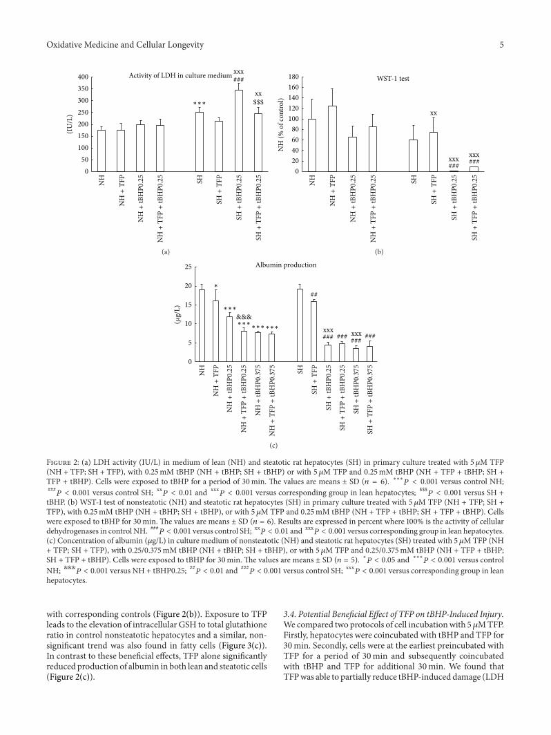